Embed Size (px)
Citation preview
1
Basic Stroke for the New Recruit
Authors
• Erin Conahan MSN, RN, ACNS-BC, CNRN, SCRN
• Julie Fussner BSN, RN, CPHQ, SCRN
• The authors have nothing to disclose.

2
Objectives
• List causes of small vessel stroke vs large vessel stroke and differences in treatment
• Describe inclusion/exclusion criteria for tPA and endovascular treatment
• List elements of acute stroke work-up to identify risk factors
Stroke Facts
• Each year 795,000 strokes occur in the United States
• Stroke is the 5th leading cause of death in the United
States
• Stroke is the leading cause of adult disability
• Up to 80% of strokes are preventable
• During a stroke ~32,000 brain cells are lost per second…
~2 million brain cells lost per minute.
• The brain ages 3.6 years for each hour untreated…
•Time is Brain
3
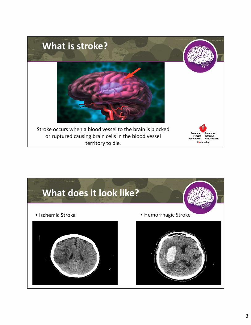
What is stroke?
Stroke occurs when a blood vessel to the brain is blocked
or ruptured causing brain cells in the blood vessel
territory to die.
What does it look like?
• Ischemic Stroke • Hemorrhagic Stroke

4
Cerebral Circulation
• Circle of Willis
• Located at the base of the skull
• Provides collateral circulation
• Anterior Circulation
• Carotid arteries
• Anterior cerebral
• Middle cerebral
• Anterior communicating
• Posterior Circulation
• Vertebral
• Basilar
• Posterior cerebral
• Posterior communicating
http://www.merckmanuals.com/professional/neurologic_disorders/stroke_cva/overview_of_stroke.html
5 Stroke Syndromes
1. Left Hemisphere
2. Right Hemisphere
3. Cerebellar
4. Brainstem
5. Hemorrhage

5
Left Hemisphere
Signs:
• Aphasia
• Right side weakness
• Right side sensory loss
• Right visual field cut
• Left gaze
Right Hemisphere
Signs:
• Neglect
• Left side weakness
• Left side sensory loss
• Left visual field cut
• Right gaze

6
Cerebellar
Signs:
• Ataxia
• Gait disturbance
• Vertigo
• Nystagmus
• Ipsilateral Findings
Retrieved on 9/25/15 from:
http://biology.clc.uc.edu/fankhauser/Labs/Anatomy_&_Physiology/A&P202/202_lecture_notes/05_Mesencephalon_Diencephalon.Jan12.
Brainstem

7
Brainstem
The 5 D’s
Dizziness
Diplopia
Dysarthria
Dysphagia
Dystaxia
Retrieved 9/25/15 from: http://www.americannursetoday.com/assets/0/434/436/440/5120/5122/5154/5156/904adb93-6d32-
4770-83d7-e6f1ad1667d2.pdf
Intracranial Hemorrhages
4 Types
Epidural (trauma)
Subdural (trauma)
Subarachnoid (traumatic or stroke)
Intracerebral hemorrhages (stroke)
Note difference between intraCRANIAL and intraCEREBRAL – both abbreviated “ICH”

8
Hemorrhage
Meninges (Outer to Inner)
Retrieved from: http://www.mdguidelines.com/subarachnoid-hemorrhage-non-traumatic. Source: Medical Disability Advisor
• Dura Mater – thick, fibrous covering of brain (and spinal cord)
• Arachnoid Mater – thin web-like membrane between dura and pia, below which flows the CSF in the subarachnoid space
• Pia Mater – delicate covering of the brain and directly adherent to tissue
SAH
Etiology
– Traumatic (fall, blow to head)
– Aneurysmal (ruptured aneurysm)
– AVM (burst of AV malformation)
Signs and symptoms – occur suddenly
– “Worst headache of my life” (thunderclap HA)
– Decrease/loss of consciousness/confusion
– N/V
– Photophobia
– Hemiparesis/hemiplegia
– Meningeal irritation

9
Intracerebral Hemorrhage
• Clinical Presentation
• SBP often >220 mmHg
• HA
• N/V
• Ataxia
• Dizziness/Vertigo
• Dysarthria
• Nuchal Rigidity
• Alterations in LOC
Stroke Assessments
• Support airway/breathing/circulation
• Vital signs
• POC Glucose
• CT
• NIHSS
• Labs, EKG, CXR
• CTA or MRA assess vessels
• LP for SAH

10
NIH Stroke Scale
• Systematic assessment tool that provides a quantitative measure of stroke used world wide
• 42 point scale
• Higher the number the worse the stroke
• NIHSS 2 or greater tPA given
• “Too good” to treat research
• www.nihss.org
NIH Stroke Scale
• Level of Consciousness Questions & Commands
• Visual Fields
• Facial Palsy
• Motor arm & leg
• Limb ataxia
• Sensory
• Best Language
• Dysarthria
• Neglect

11
What type of stroke did your patient have?
• Large vessel occlusion
• Lacunar Infarct
• Transient Ischemic Attack
• Stroke mimics
AIS: Large Artery AtherosclerosisThrombosis
• Plaque narrows the vessel lumen resulting in turbulent blood flow.
• The atherosclerotic plaque becomes unstable and ruptures, clotting factors are attracted and a thrombus forms.
• Carotids / Aorta
• Afib

12
AIS: Small Vessel Atherosclerosis
• Small-vessel ischemia occurs when plaque occludes small perforating vessels.
• Typically results in lacunar strokes which accounts for ~ 25% of ischemic strokes.
• Chronic medical conditions such as DM, HTN, HLD and smoking increases risk of small vessel disease.
TIA
• “Mini stroke” - Avoid term
• Brief episode of neurological dysfunction lasting < 1 hour
• Symptoms usually last 10-20 mins
• Old Definition: less than 24 hours
• New Definition: Evidence on MRI imaging
• Important determinant of stroke risk
• 3-10% pts stroke within 2 days
• 9-17% pts stroke within 90 days
• Within 1 year of TIA =12% will die

13
Stroke Mimics
• Hypoglycemia
• Seizures
• Migraine
• Tumor
• Abscess
• Subdural Hematoma
Imaging: CT vs MRI
• Why do they order the scan?
• CT better for seeing acute blood or skull fractures
• MRI better for ischemic stroke and tumor
• CTA/MRA looking for blood vessel abnormalities (“A” means angio)

14
Treatment options: Time is brain• Blood pressure control for all types!!
• IV tPA
• Endovascular• Pharmacologic• Mechanical
• Neurosurgery• Aneurysmal SAH Clipping and Coiling• External Ventricular Device • Decompressive Hemi craniotomy
• Research
• Neuroscience ICU / Stroke unit
**Find the cause to prevent future events!
tPA Inclusion/Exclusion Criteria
• Contraindications
• ICH, SAH, active internal bleeding
• Recent intracranial or intraspinal surgery or head trauma
• Presence of intracranial conditions that may increase risk of bleeding
• Bleeding diathesis
• Uncontrolled hypertension
• INR > 1.7 or use of NOAC
• 3-4.5 hour window
• >80 years old
• Any anticoagulant use
• History of CVA and diabetes

15
Modifiable Risk Factors
• Cardiovascular disease (CVD)
• Hypertension (HTN)
• Asymptomatic carotid stenosis
• A Fib
• Diabetes mellitus (DM)
• Dyslipidemia
• Cigarette smoking
• Drug use ( cocaine)
• Alcohol
• Obesity
• Physical inactivity
• Sickle cell disease
• Postmenopausal hormone therapy
• Hypercoagulable states
Stroke Work-Up
• Cardiac monitoring
• Angiography (CTA, MRA, angio)
• Carotid duplex
• Echocardiogram/TEE
• FLP, HgA1C, Hypercoag panels
• Patient History!!
16
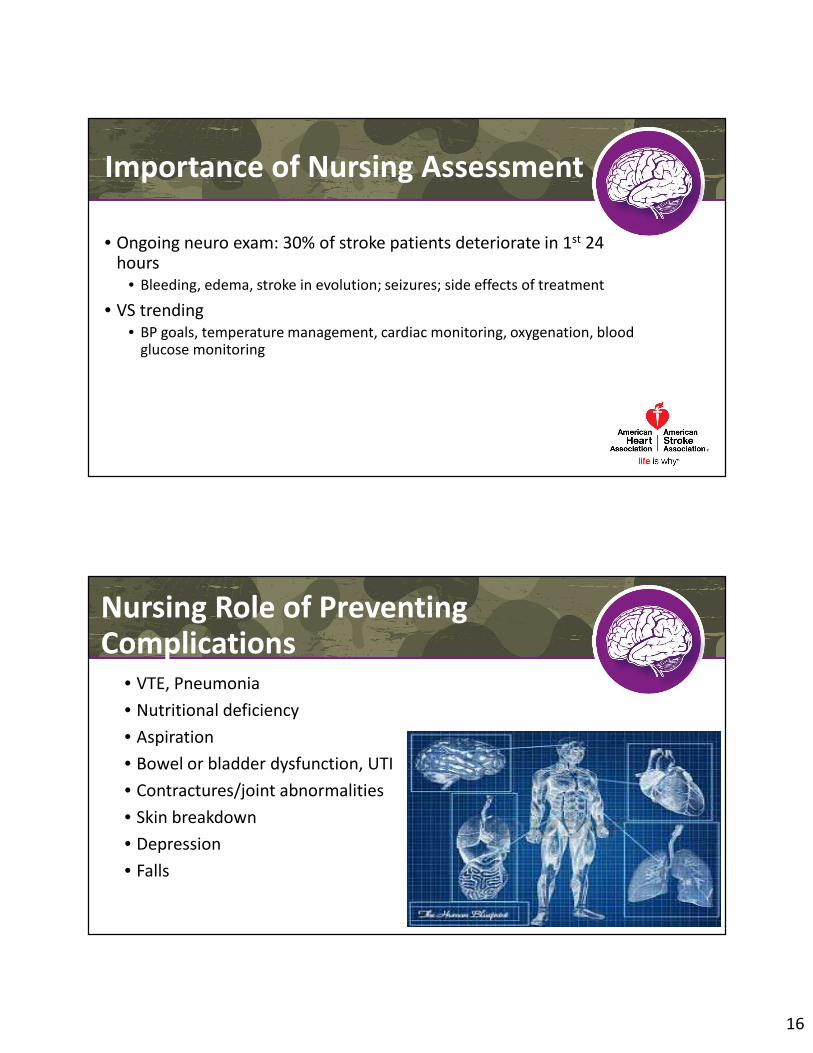
Importance of Nursing Assessment
• Ongoing neuro exam: 30% of stroke patients deteriorate in 1st 24 hours
• Bleeding, edema, stroke in evolution; seizures; side effects of treatment
• VS trending
• BP goals, temperature management, cardiac monitoring, oxygenation, blood glucose monitoring
Nursing Role of Preventing Complications
• VTE, Pneumonia
• Nutritional deficiency
• Aspiration
• Bowel or bladder dysfunction, UTI
• Contractures/joint abnormalities
• Skin breakdown
• Depression
• Falls