Embed Size (px)
Citation preview

IMMUNOLOGY: BASIC CONCEPTS IN THE IMMUNE RESPONSE (I)Doç Dr Nevriye Gönüllü

Our bodies’ defenses are similar to a military defenseThe initial defence mechanisms arebarriers: skin
acidbilemucus
These barriers inactivate and prevent entryof the foreign agents

If these barriers are compromised or theagent gains entry in another way
The local militia of innate responses (e.g., complement, natural killer cells, neutrophils, macrophages)
must quickly rally to the challenge andprevent expansion of the invasion

If this step is not effective
a major campaign must be specificallydirected against the invader by immuneresponses (antibody and T cells) knowledge of the characteristics of theenemy (antigens) through immunization,enables the body to mount a faster, moreeffective response (activation of memory B and T cells on rechallenge

The interactions of elements of theimmune response
The different elements of immune systeminteract and communicate with solublemolecules and by direct cell-to-cellinteraction.These interactions provide the mechanismsfor activation and control of the protectiveresponses.

Activators and stimulators of immunefunction
Immune cells communicate:by direct cell-to-cell interactions andby the sensing of soluble moleculesincluding:
Proteins: cytokines, chemokines, interferons and
Lipids: steroids and prostaglandines

Cytokines
Proteins produced by lymphoid and othercellsStimulate and regulate the immune response

Interferons
Low-molecular-weight proteins
produced in response to viral infectionsInterferon-α and interferon-β
on activation of the immune responseInterferon-γpromote antiviral and antitumor immuneresponses

Chemokines
Small proteins ~ 8000 DaThat are associated with inflammatory response andActivateNeutrophiles, basophils, monocytes, T cellsexpress receptors
Chemokines and other proteins (e.g., the C3a and C5a products of the complement) are chemotactic factors thatattract phagocytic and inflammatory cells to the site of infection

Cells of the immune responseImmune response are mediated by specific cellswith defined functions.The white blood cells can be distinguishedmorphologically on the basis of:
1. Histologic staining2. Immunologic functions3. Intracellular and cell surface markers; these
markers have been defined within clusters ofdifferentiation indicated by CD and numbers ( e.g.,CD4, CD8).

Intracellular and cell surface markers
APCs: antigen presenting cellsB cells, macrophages, some othersare a special class of cells that express class II major histocompatibility complex (MHC II)antigensIn addition, these and all nucleated cells expressclass I MHC (MHC I) antigens (HLA-A, HLA-B, HLA-C)
Hematopoietic cell differentiation
Differentiation of a common progenitor cell, termed the pluripotent stem cell, gives rise toall blood cells. Differentiation of these cells begins duringdevelopment of the fetus and continuesthroughout life.

Hematopoietic cell differentiation
The pluripotent stem cell differentiate intostem cells for different lineages of blood cellsincluding: the lymphoid (T and B cells)
myeloiderythrocytic, andmegakaryoblastic (source
of platelets) lineages

Hematopoietic cell differentiation
• The pluripotent stem cells reside primarily in thebone marrow but can also be isolated from thefetal blood in umbilical cords and as rare cells in adult blood.
• Differentiation of stem cells into the functionalblood cells is triggered by specific cell surfaceinteractions : with the stromal cells of the marrow
and specific cytokines produced bythese and other cells.

Hematopoietic cell differentiation
• The thymus and the bursal equivalent in the Peyer’s patches promote developmentof T cells and B cells, respectively.
• Helper T cells, macrophages, and othercells release cytokines that promotehematopoietic cell growth and terminal differenciation.

Lymphoid organsPrimary lymphoid organs
Bone marrowThymus
Secondary lymphoid organsLymph nodesSpleenMALT (mucosa-associated lymphoid tissues)
GALT (e.g., Peyer patches)BALT (e.g., tonsils, appendix)
These sites are where B and Tcells reside and respond toantigenic challenge.

The lymph nodes
• The lymph nodes are kidney-shaped organs 2 to 8 mm in diameterfilter the fluid that passes from
intercellular into lymphatic system• is constructed to optimize the
meeting of the innate (dendritic cells and macrophages) and the immune response (B and T) cells to initiate and expand specific immune responses

The lymph nodes
• consists of the following three layers:• the cortex: B cells and macrophages
arranged in clusters called follicles• the paracortex: contains dendritic cells that
bring antigens from the tissues to be presented to the T cells to initiate immune responses.
• the medulla: contain B and T cells and antibody-producing plasma cells

The spleen
• A large organ that filters antigens fromblood and removes aged cells andplatelets.
• Consists of two types of tissue:the white pulpthe red pulp

The spleen
• The white pulp consists of:arterioles surrounded by lymphoid cells, in
which the T cells surround the central arteriole.B cells are organized into primary and
secondary follicles, that have a germinal center.
• The red pulp is a storage site for blood cells andthe site of turnover of aged platelets anderythrocytes.

Polymorphonuclear Leukocytes (I)Neutrophiles
11-14 µm in diameter, lack mitochondria50%-70% of circulating blood cellsThey are short-lived cells: circulate 7-10 hours in blood, than migrate into the tissue where they live
3 days
They are a primary phagocytic defense againstbacterial infection

Polymorphonuclear Leukocytes (II)
During infection, the neutrophils in the bloodincrease in number and include precursors formsThese precursors are termed band forms, in contrast to the terminally differentiated andsegmented neutrophils. An increase in neutrophils by a blood count is sometimes termed “a left shift with an increase in bands vs segmenteds”
Ingest bacteria by phagocytosis: expose thebacteria to granules: primary (azurophilic), secondary(specific)

Polymorphonuclear Leukocytes (III)Eosinophiles: are heavily granulated cells with a bilobed nucleus that stain with the acid dye eosin
PhagocytisMotileGranulated: contain acid phosphatase, peroxidase andeosinophilic basic proteins
“Play a role in the defense against parasitic infections”Basophiles
Not phagocyticRelease the contents of their granules during allergicresponse (type 1 hypersensitivity)

Mononuclear phagocytic system (RES)
Previously called the reticuloendothelial systemAre myeloid cells and consist of:
monocytes in the blood andcells derived from monocytes, such as:
alveolar macrophages (lungs)Kupffer cells (liver)intraglomerular mesangial cells (kidney)histiocytes (connective tissue)synovial A cells (synovia)microglial cells (brain)dendritic cells

Mononuclear phagocytic system (RES)
Monocytes blood
Alveolar macrophages Kupffer cells Mesangial cells HistiocytesSynovial cells Microglial cells
Dendritic cells

Monocytes
Are 10 to 18 µm in diameterSingle-lobedKidney bean-shaped nucleus3% to 8% of peripheral blood leukocytes

Monocytes
Different cytokines promote myeloid stemcells and monocytes to differentiate into thevarious macrophages and dendritic cellsThese mature forms have differentmorphologies corresponding to their ultimatetissue location and function andmay not express all of the macrophageactivities or cell surface markers
Macrophages
Long-lived cellsPhagocyticContain lysosomes andUnlike neutrophiles, have mitochondria
Macrophages-Functions
PhagocytosisAntigen presentation (AP) to T cells to initiatespecific immune responsesSecretion of cytokines to activate andpromote innate and immune responses

Macrophages-cell surface receptors (I)
Fc portion of Immunoglobulines (IgG)
Fc-γ RIFc-γ RII,Fc-γ RIII
C3b product of thecomplement cascade
CR1CR3
Facilitate thephagocytosisof antigen, bacteria, viruses(coated withtheseproteins)

Macrophages-cell surface receptors (II)
Macrophages also express theClass II MHC antigen
which allows these cells to present antigen toCD4 helper T cells to initiate the immuneresponse

Macrophages secrete :
Interleukin-1Interleukin-6Tumor necrosis factor(TNF)Interleukin -12
stimulate immune andinflammatory responses

Macrophages are activated by :Interferon-γ
T-cell derived lymphokineActivated macrophages have:
enhanced phagocytickillingantigen-presenting capabilities
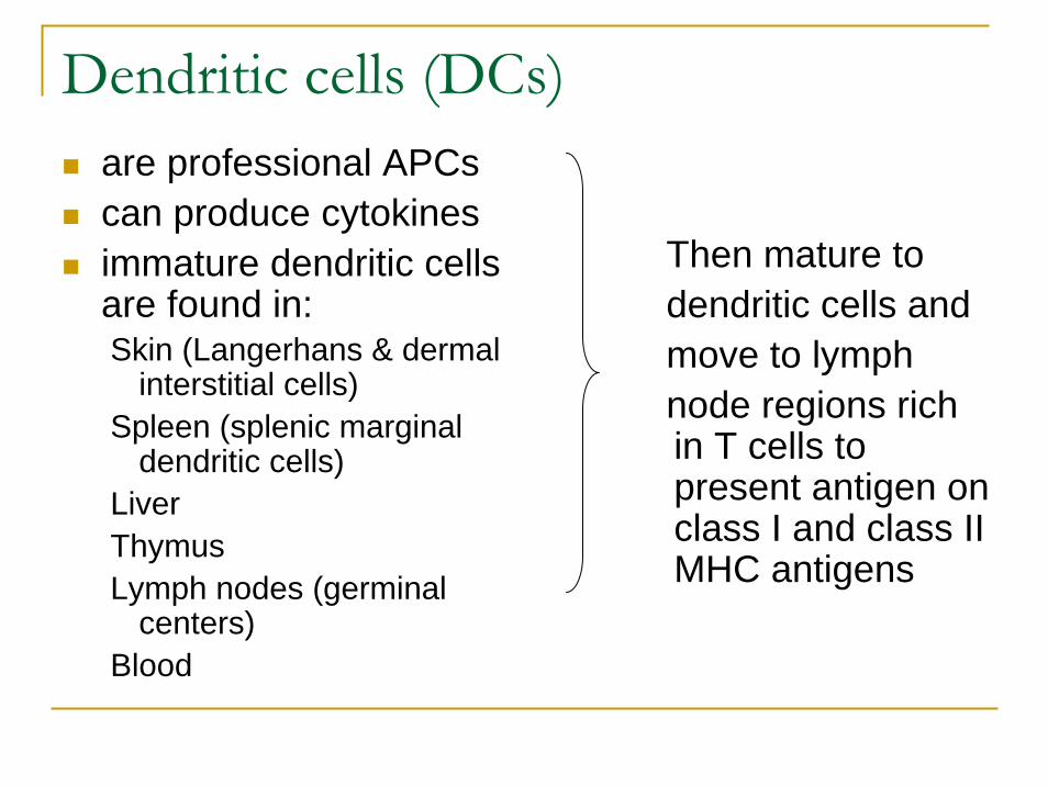
Dendritic cells (DCs)are professional APCscan produce cytokinesimmature dendritic cellsare found in: Skin (Langerhans & dermal
interstitial cells)Spleen (splenic marginal
dendritic cells)LiverThymusLymph nodes (germinal
centers)Blood
Then mature todendritic cells andmove to lymphnode regions richin T cells topresent antigen on class I and class II MHC antigens

Lymphocytes (I)Are 6-10 µm in diameter, which is smallerthan leukocytes. The two major classes are:
B cellsT cells
have a large nucleus and smaller, agranularcytoplasmB and T cells are indistinguishable by theirmorphologic features, butthey can be distinguished on the basis of function and surface markers.

Lymphocytes (II)
Lymphoid cells that are not B or T cells (non-B/non-T cells or null cells) are:
large, granular lymphocytes, and
also known as natural killer (NK) cells

Lymphocytes, B cells (I)The primary function of B cells is to makeantibodythey also internalize antigen, process theantigen, and
Present the antigen to T cellsto initiate or enhance the immune response.

Lymphocytes, B cells (II)
The B-cell name is derived from its site of differentiation in birds, bursa of Fabricius, and the bone
marrow of mammalsB-cell differentiation also takes place in thefetal liver and fetal spleenB cells can be identified by the presence of:
immunoglobulinsClass II MHC receptors for C3b and C3d of complementcascade (CR1 and CR2) on their cell surface.

Lymphocytes, B cells
Activated B cells either develop into:memory cells which express the
CD45RO cell surface marker and circulateuntil activated by specific antigen, or
plasma cells:have small nuclei and large
cytoplasmtheir job is producers of antibody

B cells (III)Activated B cells
Memory cellsCD45RO
Plasma cells activation by spesificantigen
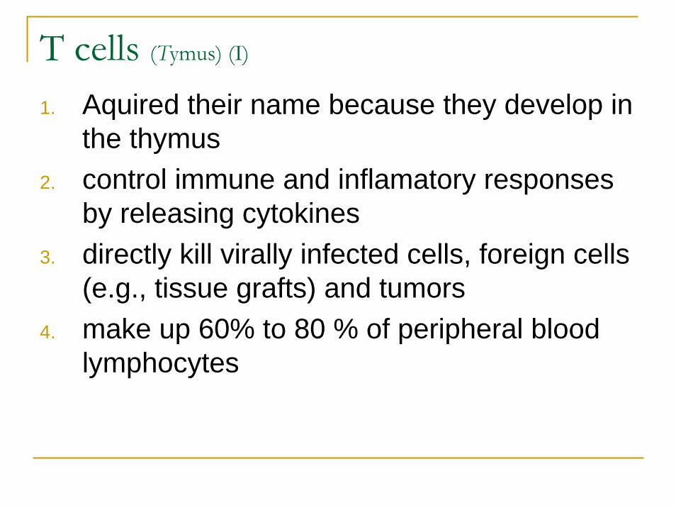
T cells (Tymus) (I)
1. Aquired their name because they develop in the thymus
2. control immune and inflamatory responsesby releasing cytokines
3. directly kill virally infected cells, foreign cells(e.g., tissue grafts) and tumors
4. make up 60% to 80 % of peripheral bloodlymphocytes

T cells (II)
T cells were initially distinguished from B cellson the basis of their ability to bind andsurround themselves (forming rosettes)All T cells express cell surface markers:
T-cell receptor (TCR): which resemblesbut differs from antibody
CD2-associated proteinsCD3-associated proteins

T cells (III)
Most lymphocytes express the αβ TCRA variable number of Tcells express theγδ TCR but do not express CD4 or CD8T cells are divided into three majorgroups on the basis of the type of TCR and the cell surface expression of twoprotein:
CD4 or CD8

CD4-expressing T cells
are cytokine-producing cells which help initiate andmature immune responses andactivate macrophages to induce delayed-typehypersensitivity responses (DTH)CD4: TH1, TH2, and TH3 subgroups according to thespectrum of cytokines they secrete:
TH1 promote: local, cellular inflammatory and DTH responses
TH2 promote : antibody productionTH3 promote: IgA production and T-cell tolerance

CD8-expressing T cells
release cytokinesrecognize and kill virally infected cells, foreigntissue transplants (non-self-grafts), andtumor cells as cytotoxic killer T cellssuppress immune response

T cells
T cells also produce memory cells thatexpress CD45RO
Terminally differenciated CD4 and CD8 express class II MHC antigen

T cells
NK cells (large granular lymphocytes)resemble CD8 T cells (in cytolytic function toward
virally infected and tumor cells, but differ in themechanisms for identifying the target cell)
also have Fc receptors, which are used in antibody-dependent killing and are also calledantibody-dependent cellular cytotoxicity (ADCC or K) cells.

The Humoral Immun Response
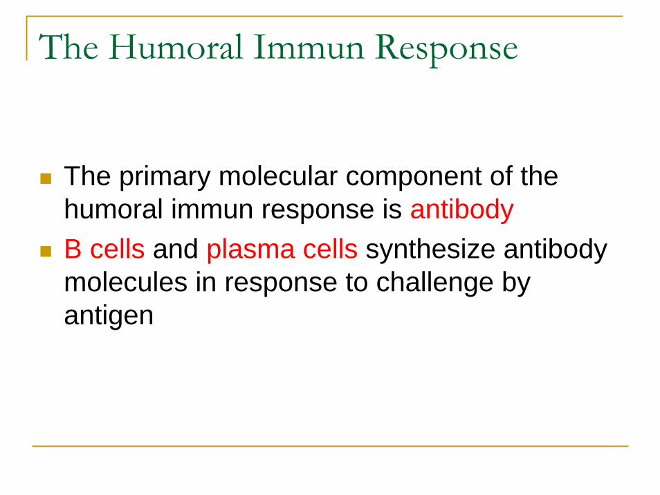
The Humoral Immun Response
The primary molecular component of thehumoral immun response is antibodyB cells and plasma cells synthesize antibodymolecules in response to challenge byantigen

Antibody-Functions
provide protection from rechallenge by an infectious agent
block spread of the agent in the bloodfacilitate elimination of the infectious agentto accomplish these tasks, a large
repertoire of antibody molecules must be available to recognize the tremendousnumber of infectious agents that challengeour bodies.
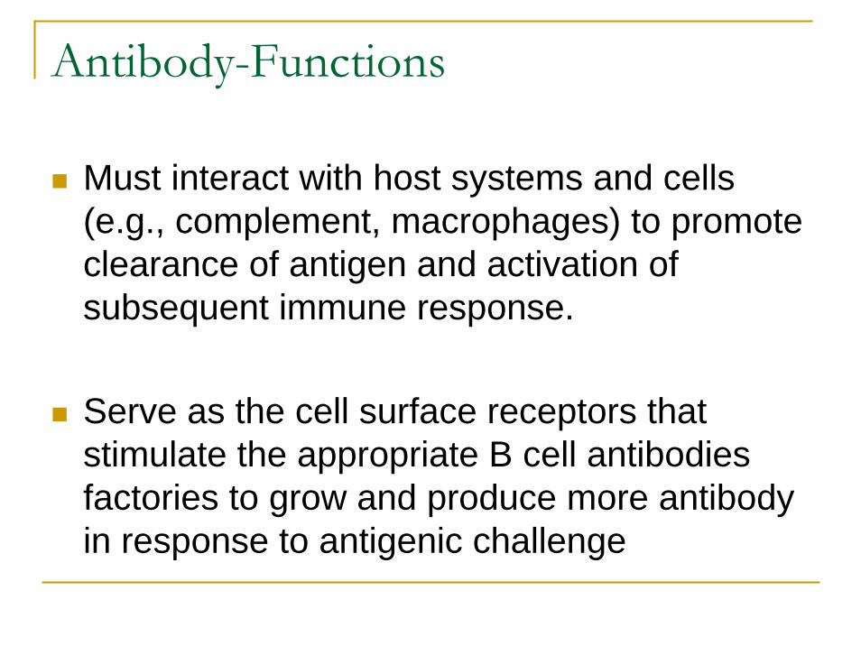
Antibody-Functions
Must interact with host systems and cells(e.g., complement, macrophages) to promoteclearance of antigen and activation of subsequent immune response.
Serve as the cell surface receptors thatstimulate the appropriate B cell antibodiesfactories to grow and produce more antibodyin response to antigenic challenge

Immunogens, Antigens and Epitopes
Almost all of the proteins and carbohydratesassociated with an infectious agent(bacterium, fungus, virus or parasite) areconsidered foreign to the human host and
have the potential to induce an immuneresponse

Definitions I
Immunogen: a substance (protein or carbohydrate) that challenges the immune system and can initiatean immune response; may contain more than oneantigen (e.g., bacteria).
Antigen: is a molecule recognized by specificantibody or T cells

Definitions (III)
Epitope (antigenic determinant): themolecular structure that interacts with a single antibody moleculeLinear epitope: whithin a protein, an epitopemay be formed by a specific sequenceConformational epitope: an epitope formedby a three-dimensional structure

Definitions (IV)
Not all molecules are immunogens. Proteinsare the best immunogens, carbohydrates areweaker immunogens and lipids are poorimmunogens.Hapten (incomplete immunogen) are oftentoo small to immunize (initiate a response) an individual but can be recognized by antibody.Haptens can be made immunogenic byattachment to a carrier molecule, such as a protein.

Definitions IIAdjuvant: substance that usually prolong thepresence of antigen in the tissue and activateor promote uptake of the immunogen bydendritic cells, macrophages andlymphocytes.
During artificial immunization (e.g.,vaccines), an adjuvant is used to enhance the responseto antigen.

Immune response
The type of immune response initiated by an immunogen depends on its molecularstructure.
A primitive but rapid antibody response can be initiated toward bacterial polysaccharides, peptidoglycan, or flagellin.

T-independent antigens
These molecules have a large, repetitivestructure, which is sufficient to activate Bcells directly to make antibody without theparticipation of T-cell help.In these cases the response is limited toproduction of IgM antibody andfails to stimulate an anamnestic (booster) response.

T-dependent antigens
antigens that must be presented to T and Bcells for antibody productionare usually proteinsthey stimulate all five classes of immunoglobulins andcan elicit an anamnestic (secondary-booster) response

Immunoglobulin types
Y-shaped moleculesFab and Fc fragmentsHeavy and light chains

Immunoglobulin types
Immunglobulins are composed of at least twoheavy chains and two light chains, a dimer of dimers. They are subdivided into classes based on the structure and antijenic distinction of theirheavy chains. IgG,IgM,IgA are the major antibody formsIgD and IgE make up less than 1% of thetotal immunoglobulin

Immunoglobulin types
The IgA and IgG classes of immunglobulinare divided further into subclasses based on differences in the Fc portion.There are four subclasses of IgG, designatedas IgG1 to IgG4, and two IgA subclasses(IgA1 and IgA2).
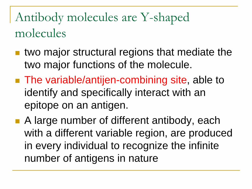
Antibody molecules are Y-shaped molecules
two major structural regions that mediate the two major functions of the molecule.The variable/antijen-combining site, able to identify and specifically interact with an epitope on an antigen.A large number of different antibody, eachwith a different variable region, are producedin every individual to recognize the infinitenumber of antigens in nature

Antibody-the Fc portion
Stem of the antibody (Y), interacts with hostsystems and cells to promote activation of subsequent immune responsesis responsible for fixation of complement andbinding of the molecule to cell surface immunoglobulin receptors (FcR) on macrophages, natural killer cells, and T cells.

Proteolytic digestion of immunoglobulin
IgG and IgA have a flexible hinge region rich in proline and susceptible to cleavage by proteolytic enzymes.Digestion of molecules with papain yields two Fab fragments and one Fc fragment. Each Fab fragment has one antigen-binding site.Pepsin cleaves the molecule, producing an F(ab’)2 fragment with two antigen-binding sites and a pFc’ fragment.

Immunglobulin D
IgD has a molecular mass of 185 kDa
Accounts for less than 1% of serum immunglobulins
Activate B cell growth.

Immunglobulin M (I)
The first antibody produced in response to antigenic challengeCan be produced in a T-cell-independent manner.IgM makes up 5% to 10% of the total immunglobulins in adults.

Immunglobulin M
It is a pentameric molecule with five immunoglobulin units joined by disulfide bounds and J chain, with a total mass of 900 kDa. It is the most efficient immunoglobulin for fixing (binding) complement. A single IgMpentamer can activate the classical complement pathway

Immunglobulin G
Comprises approximately 85% of the immunglobulins in adults. It has a molecular mass of 154 kDa, based on two L chains of 22000 Da each and two H chains of 55000 each.The four subclasses of IgG differ in structure, relative concentration, and function.Production of IgG requires T-cell help.

Immunglobulin G
IgG has the longest half-life (23 days) of the five immunoglobulin classes, crosses the placenta.Is the principal antibody in the anamnestic or booster response.Shows high avidity (binding capacity) for antigens, fixes complement, stimulates chemotaxis, and acts as an opsonin to facilitate phagocytosis.

Immunglobulin A
Comprises 5% to 15% of the serum immunglobulins and has a half-life of 6 days.It has a molecular mass of 160 kDa and a basic four-chain monomeric structure.It can occur as monomers, dimmers, trimers, and multimers combined by the J chain (similar to IgM). In addition to the serum IgA, a secretory IgAappears in body secretions and provides localized immunity.

Immunglobulin A
IgA production requires specialized T-cell help and mucosal stimulation.An adult secretes approximately 2 g of IgAper day.Secretory IgA appears in colostrums, intestinal and respiratory secretions, saliva, tears and other secretions.IgA-deficient individuals have an increased incidence of respiratory tract infections.

Immunglobulin E
Accounts for less than 1% of the total immunglobulins and has a half-life of approximately 2.5 days.Most IgE is bound to Fc receptors on mast cells.

Immunglobulin E
When sufficient antigen binds to the IgE on the mast cell, the mast cell releaseshistamine, prostaglandin, platelet-activatingfactor, and cytokines.
Important against parasitic infection andresponsible for anaphylactic hypersensitivity(type 1) (rapid allergic reactions)

Complement cascade
The complement system is an alarm and a weaponagainst infection, especially bacterial infection.The complement system is activated:
directly by bacteria and bacterial products(alternate or properdin pathway).
by lectin binding to sugars on the bacterialcell surface (mannose-binding protein), or
by complexes of antibody and antigen(classical pathway).

Complement activation
Activation by either pathway iniatiates a cascade of proteolytic events thatproduce chemotactic factors to attractphagocytic and inflammatory cells to the site,increase vascular permeability to allowaccess to the site of infection,bind to the agent to promote theirphagocytosis (opsonization) and elimination, and directly kill the infecting agent.

Complement
The three activation pathways of complementcaolesce at a common junction point, theactivation of the C3 component.

Alternate pathway
The alternate pathway is activated directly bybacterial cell components (e.g., endotoksin, microbial polysaccarides).This pathway can be activated before theestablishment of an immune responsebecause
it does not depend on antibody anddoes not involve the early complement
components (C1, C2 and C4).

Alternate pathway
Activation is mediated by properdin factor B binding to C3b and than properdin factor D, which splits factor B in Bb active fragmentthat remains linked to C3b (activation unit)C3b sticks to the cell surface and inactive Bbleading to the activation of many C3 moleculesThe complement cascade continues in a manner analougous to the classical pathway.

Classical pathway
The classical complement cascade is initiatedby binding to the Fc portion of antibody that is bound to cell surface antigens, or in an immune complex with soluble antigens.
The first complement component, designatedC1, consists of a complex of three separateproteins designated C1q, C1r and C1s.
Classical pathway
The first complement component, C1, consistof a complex of three separate protein: C1q, C1r, and C1sOne molecule each of C1q and C1s with twomolecules of C1r comprises the C1 complexor recognition unit.Binding of C1q activates C1r and C1s andthan cleaves C4 to C4a and C4b and
C2 to C2a and C2b

Classical pathway
Split of numerous C2 and C4 represents an amplification mechanism in the complementcascade.The union of C4b and C2a produces C4b2a, which is known as C3 convertase.This complex bind to the cell membrane andcleaves C3 into C3a and C3b

Classical pathway
The interaction of C3b with C4b2a bound tothe cell membrane produces C4b3b2a, whichis termed C5 convertase
This activation unit splits C5 into C5a andC5b fragments and represents yet anotheramplification step

Lectin pathway
The lectin pathway is also a bacterial andfungal defense mechanism.
Manose-binding protein is a large serum protein that binds to nonreduced mannose, fucose and glucosamine on bacterial andother cell surface.

Lectin pathway
This protein resembles and replaces the C1q component and activates the cleavage of manose-binding protein-associated serine protease.
This cleaves the C4 and C2 components toproduce the C3 convertase, the junction pointof complement cascade.