Embed Size (px)
Citation preview
Basic Cellular Physiology and Anatomy
The Breakdown of Organic Molecules Takes the Place in Sequences of
Enzyme-catalized Reactions
• While your textbook is in a stable state, it could burst into flames and exist in a more stable state. It must have something that provides energy of activation to move it from its current state to a more stable state.
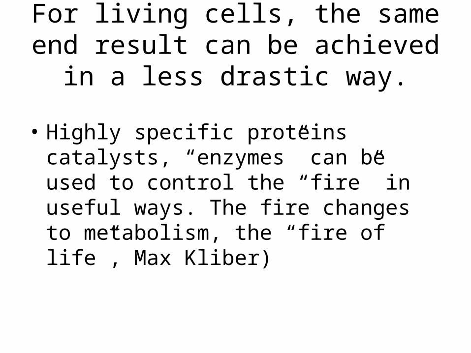
For living cells, the same end result can be achieved in a less drastic way.
• Highly specific proteins catalysts, “enzymes” can be used to control the “fire” in useful ways. The fire changes to metabolism, the “fire of life”, Max Kliber)
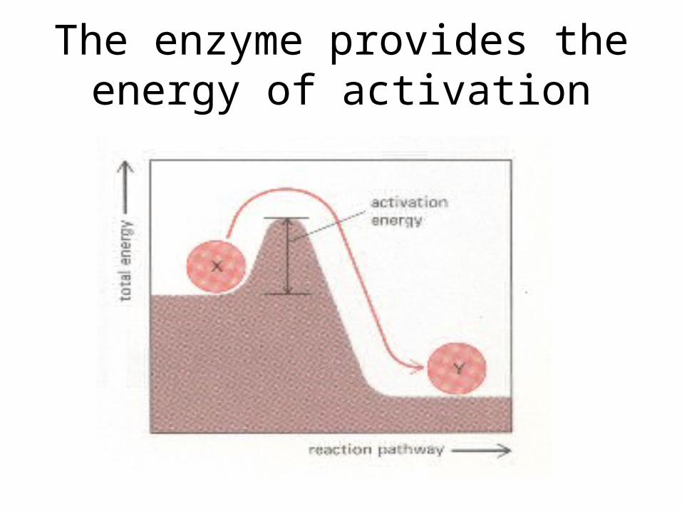
The enzyme provides the energy of activation
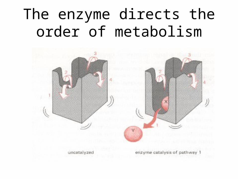
The enzyme directs the order of metabolism
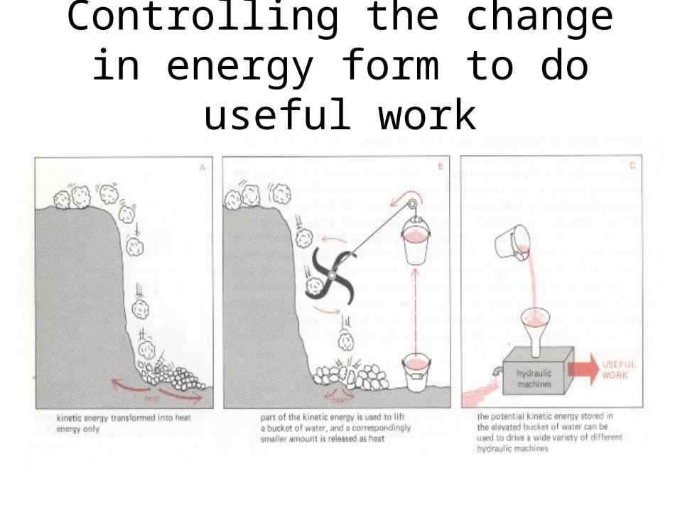
Controlling the change in energy form to do useful work
• Enzymes allow for useful work by controlling the path and flow of energy change.
• Enzymes allow for the conversion of energy from the sun to be converted into energy that can be used to drive biological processes (ATP adenosinetriphosphate - ATP). Used in the manufacture of all proteins and enzymes.
• The majority of this ATP production by a non-photosynthetic aerobic eukaryote cell takes place in the mitochondria, which can make up nearly 25% of the total volume of a typical cell.
• It is the high energy bond of ATP that raises the substrate plus the enzyme to a higher level of activation allowing the metabolic process to go.

Three distinct membrane systems
• These three structure can be visualized with an electron microscope.
• The membranes are made of different proteins
• Serve separate functions within the cell
• This is usually called ultrastructure.

The major membrane system
• The major membrane system extends from the nucleus to the external surface of the plasma membrane.
• Includes:– Nuclear envelope– Endoplasmic reticulum: tubes, fibers, channels– The Golgi apparatus– Secretory granules– Endosomes– The plasma membrane that surrounds the cell
Lysosomes
• The second major membrane system that breaks down proteins and lipids.

Mitochondria
• The third system is mitochondria (see below)

I Major membrane system
• Extends from the nuclear membrane to the plasma membrane: contains 6 different parts
(1) Nuclear envelope
• Surrounds the nucleus of the neuron
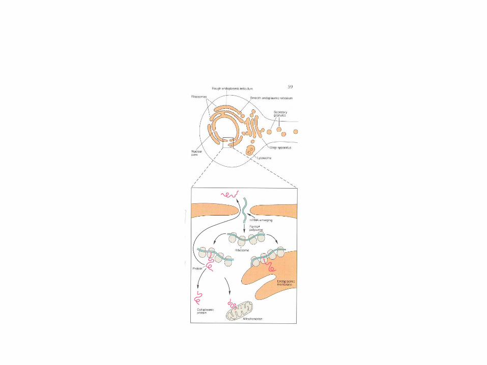
(2) Endoplasmic reticulum
• A continuation of the nuclear membrane as a set of extensive channels.
• In ultrastructure (electron microscopy) appears in both a smooth membrane and a rough membrane
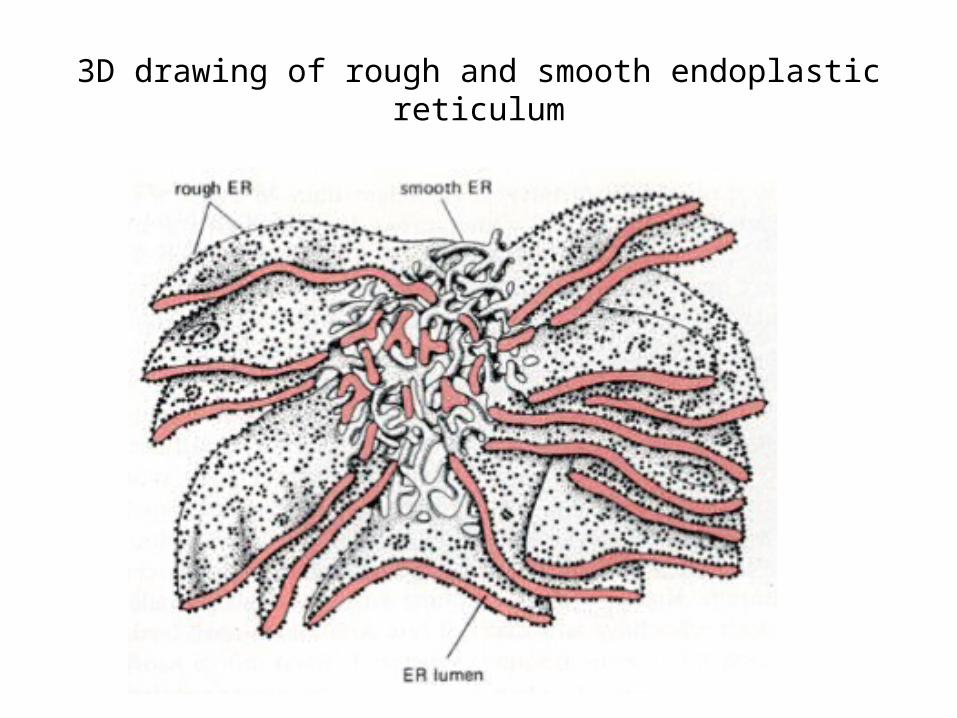
3D drawing of rough and smooth endoplastic reticulum

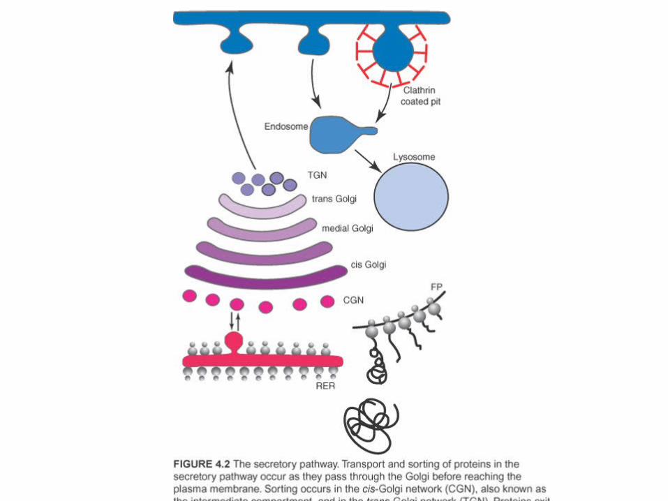
• (3) Golgi apparatus• Flat disk shaped structures with no obvious
knobs. • Packages the and distributes molecules with
the cell
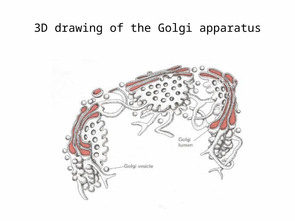
3D drawing of the Golgi apparatus
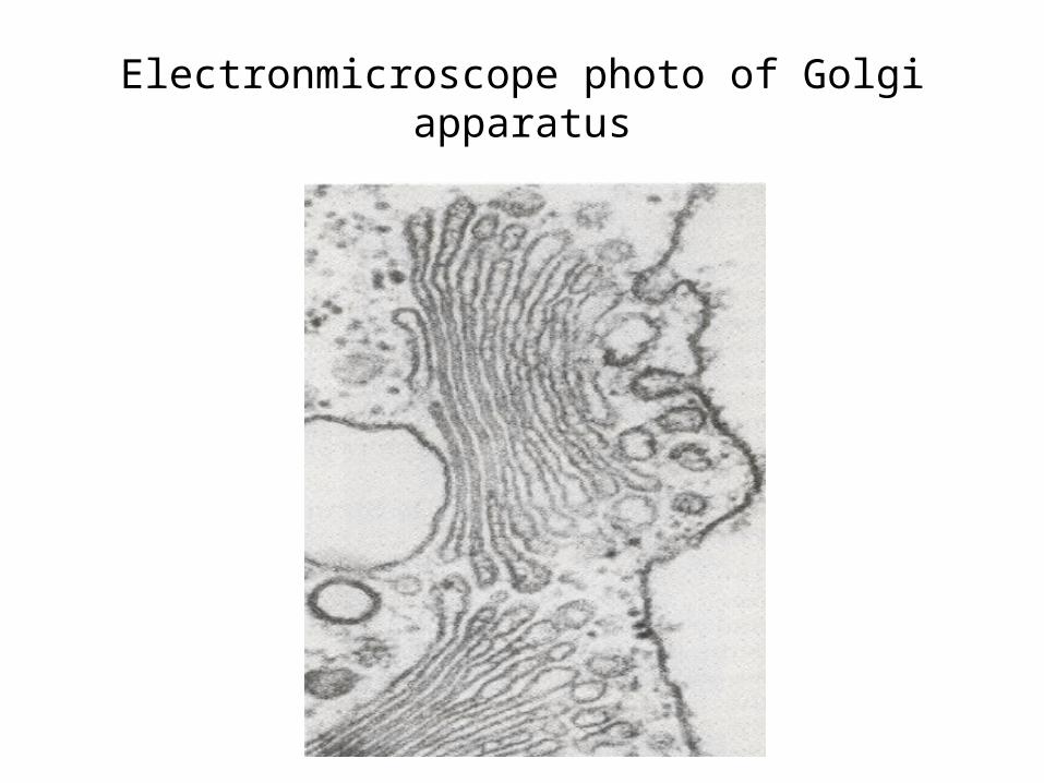
Electronmicroscope photo of Golgi apparatus
Secretory granules
• (4) Secretory granules: A small subcellular vesicle, surrounded by a membrane, that is formed from the Golgi apparatus and contains a highly concentrated protein destined for secretion. Secretory granules move towards the periphery of the cell and upon stimulation, their membranes fuse with the cell membrane, and their protein load is exteriorized. Processing of the contained protein may take place in secretory granules. Comment Note that the term 'secretory vesicle' is sometimes used in this sense, but can also mean 'transport vesicle.
Endosomes
• (5) Endosomes are granular membranes that contains endolithic material pinched off from the inside surface of the plasma membrane for transport to the lysosomes for degradation.
• Can be recycled if it is associated with the Golgi apparatus

Plasma membrane bi-leaflet phospholipid
• Surrounding membrane of the cell• Bi-leaflet structure:– a hydrophobic head group: choline, phosphate
and glycerol– A tail hydrophilic group: fatty acid– The head ends are contiguous with the
extracellular and intracellular material respectively.– The tail end point to one another

1/2 of a bi-leaflet plasma membrane

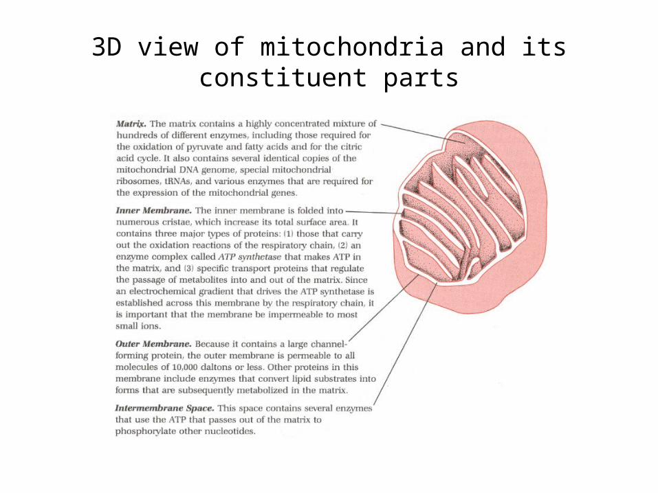
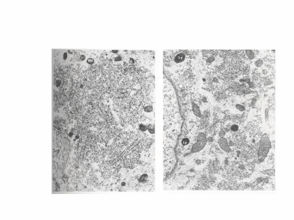
3D view of mitochondria and its constituent parts
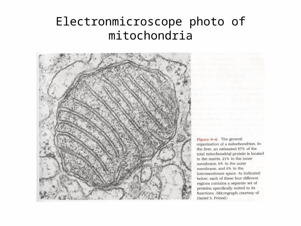
Electronmicroscope photo of mitochondria
III Mitochondria.
• This is an independent system that has been conserved evolutionary terms: low mutation rate.

All membrane systems are imbedded in the cytosol
• Cytosol: a gel like substance consisting of water-soluble proteins and a variety of insoluble filaments that form the cytoskeleton.

Essentially all macromolecules of a neuron are made in the cell body from
mRNA originating in the nucleus.• Exception: some few are made in the
mitochondria.
Large folded macromolecules
• Proteins are very large molecules made of serially places amino acids. All proteins are made from directions situated in the nucleus and brought to the ribosome by mRNA, which is the “work bench” of protein manufacture.
Each neuron makes only three classes of proteins
• 1. Proteins that are synthesized in the cytosol and remain there.
• 2. Proteins that are synthesized in the cytosol but are latter incorporated into the nucleus and mitochondria.
• 3. Proteins that are synthesized in association with the cell membrane system: 3 subcategories.

Proteins that remain attached to the basic three types of membranes but
can latter be detached
• Membrane-spanning proteins – integral proteins as they make up a part of the bi-leaflet structure of the plasma membrane
• Anchored proteins• Associated proteins
• Unattached proteins that remain in the lumen of the endoplasmic reticulum or Golgi sacs.

• Proteins that are transported by means of vesicles that pinch off from the Golgi apparatus and are distributed in secretory vesicles (used in the release of neurotransmitters) or other organelles – lysosomes
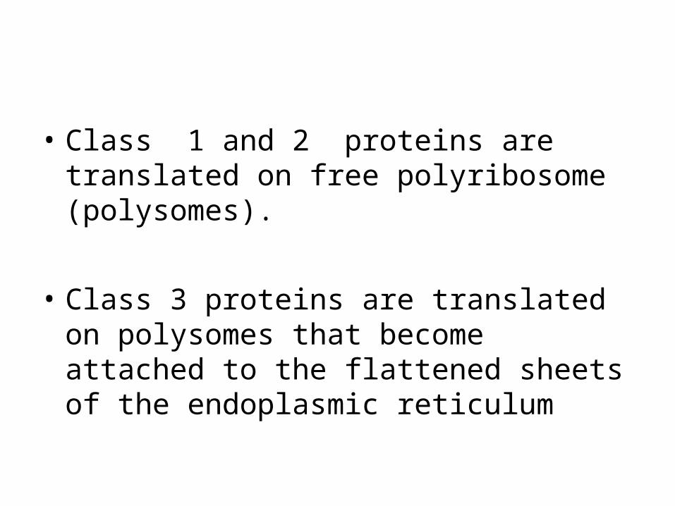
• Class 1 and 2 proteins are translated on free polyribosome (polysomes).
• Class 3 proteins are translated on polysomes that become attached to the flattened sheets of the endoplasmic reticulum
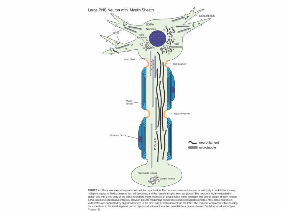
Microtubules
• Tubulin, subunits of microtubles, make up 10% of total brain proteins.
• Neuronal microtubule have biochemical specializations to meet unique demands imposed by the size and shape of neurons.

Intracellular transport and cell morphology are largley the
responsibility of microtubles.
• Microtubles, in vitro, are dynamic with polar ends (plus and minus) that correspond to the fast and slow growing ends respectively.
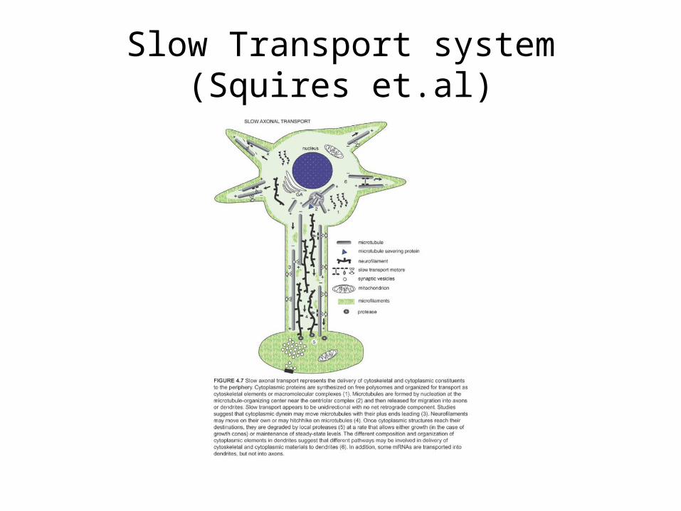
Slow Transport system (Squires et.al)

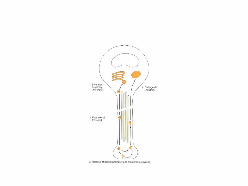
Fast axon Transport
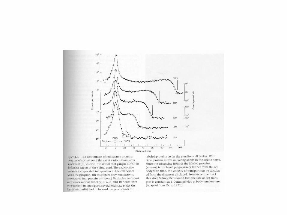
• Results: Ochs found that the rate of fast transport is about 410 mm (16.1 in) per day at body temperatures. Fast anterograde transport depends critically on the availability of ATP for energy; is not affected by inhibitors of protein synthesis; and is independent of the cell body (Fig 5-10).

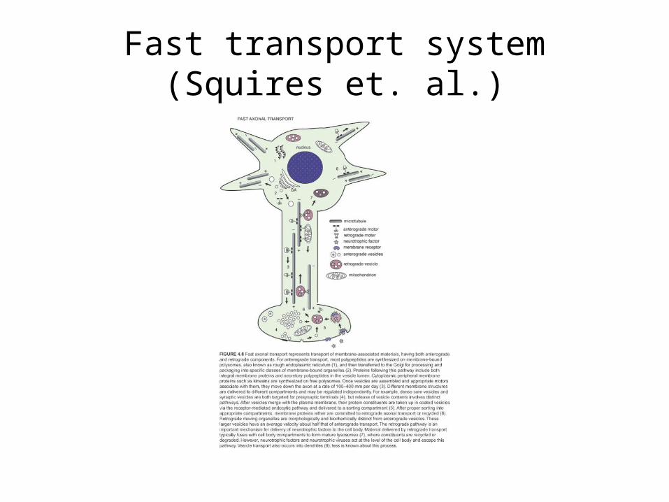
Fast transport system (Squires et. al.)