Embed Size (px)
Citation preview

Basic Cardiac Views & Interpretation of LV Function
Raj Dasgupta MD, FACP, FCCP, FAASM
Assistant Professor of Clinical Medicine
Department of Pulmonary / Critical Care / Sleep Medicine
University of Southern California (USC)
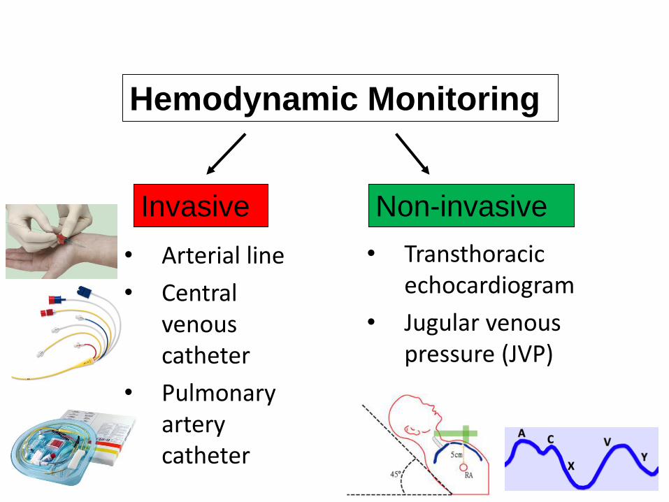
• Arterial line
• Central venous catheter
• Pulmonary artery catheter
Hemodynamic Monitoring
Invasive Non-invasive
• Transthoracic echocardiogram
• Jugular venous pressure (JVP)
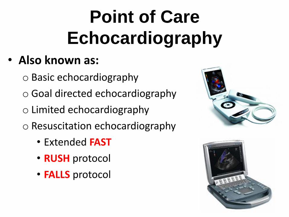
Point of Care
Echocardiography • Also known as:
o Basic echocardiography
o Goal directed echocardiography
o Limited echocardiography
o Resuscitation echocardiography
• Extended FAST
• RUSH protocol
• FALLS protocol

Extended FAST Protocol
• Focused assessment with sonography for trauma (FAST)
• The extended FAST (eFAST) allows for the examination of both lungs. This allows for the detection of a PNX

RUSH Protocol

Rapid Ultrasound for Shock and Hypotension (RUSH)
• RUSH Protocol was conceived in 2008 and looks are 3 basic aspects of physiology
• The Pump
o RV:LV
o Squeeze
o Pericardial effusion
• The Tank
o IVC
o Pleural effusions
o Pulmonary edema
• The Pipes
o AAA
o Aortic dissection
o DVT

FALLS Protocol
• Fluid Administration Limited by Lung Sonography
• For the assessment of patients with acute circulatory failure
• Relies on the evaluation of the pleura, lungs and pericardium

BLUE Protocol
• BLUE: “Bedside Lung Ultrasound in Emergency” • Upper BLUE point: upper lobes
• Lower BLUE point: middle lobe / lingula
• PLAPS point: lower lobes • Is the lowest point in the lung so
this is where you find pleural fluid
• PLAPS: “posterior-lateral alveolar pleural syndrome” • Posterior-lateral: around the back
• Alveolar syndrome: consolidation
• Pleural syndrome: pleural effusion
Lichtenstein DA, Mezière GA (2008) Relevance of lung ultrasound in the diagnosis of acute respiratory failure: the BLUE protocol.
CHEST July 2008 vol. 134 no. 1 117-125

BLUE Protocol
• 3 items were assessed:
1. Artifacts
• Horizontal A lines
• Vertical B lines
2. Lung sliding
3. Alveolar consolidation and/or pleural effusion
Lichtenstein DA, Mezière GA (2008) Relevance of lung ultrasound in the diagnosis of acute
respiratory failure: the BLUE protocol. CHEST July 2008 vol. 134 no. 1 117-125

All With One Common Theme
• Uses a limited number of echo views
• For rapid evaluation of hemodynamic failure
• For Identification of life threatening process
• For categorization of shock state
• To guide management of shock state
• To follow evolution of disease and response to therapy
• Not to replace full echo examination

Immediate Identification of Life
Threatening Conditions
• Hemodynamic failure has many causes
• Some may be imminently life threatening
• Goal directed echo allow immediate identification this subgroup
o Allows for “ruling out” of life threatening conditions
• Delay in performance of echo places the patient’s life at risk and prompts unnecessary testing
• All patients with shock should have an immediate goal directed echo

The Challenge of Critical Care Echo
• Cardiac transducer has small footprint to fit between ribs
• Ideally, patient is in the left lateral decubitus position with left arm abducted
• In the ICU, patient is difficult to position; supine position may be only possibility
• In supine position, subcostal view is often the best view
The footprint of the transducer refers to the area of skin that must be contacted to create an image

Integration of Scanning Results
• In order to make clinical decisions, one must integrate the information obtained from the echocardiogram along with knowledge of:
o Anatomy & function
o History & physical exam
o Labs & imaging
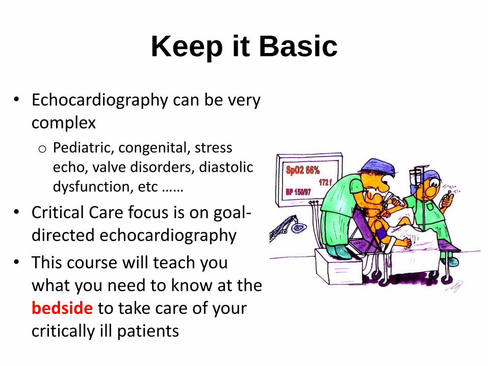
Keep it Basic
• Echocardiography can be very complex
o Pediatric, congenital, stress echo, valve disorders, diastolic dysfunction, etc ……
• Critical Care focus is on goal-directed echocardiography
• This course will teach you what you need to know at the bedside to take care of your critically ill patients

Transducer Movements
• Move: Transducer is shifted to another position on the thorax
• Angle: Transducer is angled to obtain adjacent tomographic planes
o 45, 90, 180 degree
• Rotate: Transducer is rotated without moving or tilting
• Tilt: The transducer is rocked along the tomographic plane

Goal Directed Echocardiography
• Step 1: Position the patient
• Step 2: Acquire the images
o Parasternal long axis
o Parasternal short axis
o Apical four chamber
o Subcostal view
o IVC view
• Step 3: Interpret the images
o Integrate knowledge about the patient
o LV & RV size and function
o Pericardial effusion • Tamponade
o Valvular disease • Regurgitation
• Stenosis
• Endocarditis
2D Echocardiography
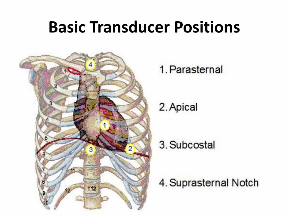
Basic Transducer Positions


5 Standard Views
I. Parasternal long axis view (PSL)
II. Parasternal short axis midventricular (PSS)
III. Apical four chamber view (AP4)
IV. Subcostal long axis view (SC long)
V. Inferior vena cave long axis view (IVC long)

Parasternal Long Axis (PLAX)
•3rd or 4th intercostal space
•Index mark to right shoulder
•Mark on monitor is on the right

Parasternal Long Axis (PLAX)

Parasternal Long Axis (PLAX)

Parasternal Long Axis (PLAX)

Parasternal Long Axis (PLAX)
•Primary uses:
o Gross LV size and function
o Pericardial effusion
o Pleural effusion • Can confuse with pericardial
effusion
o Estimate of RV size
o Visual inspection of valvular function • Mitral and aortic valve
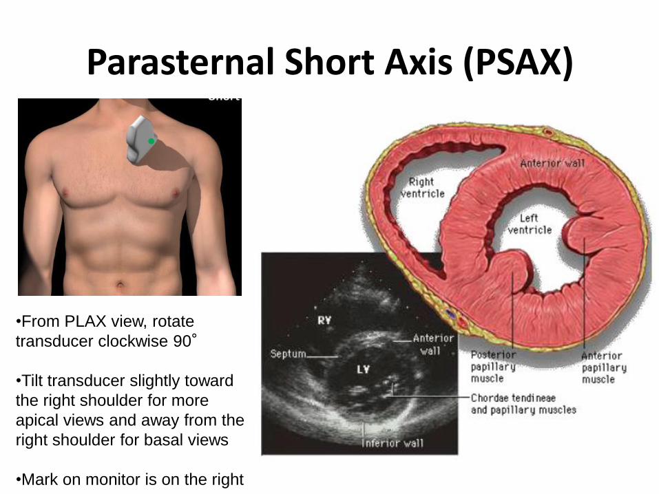
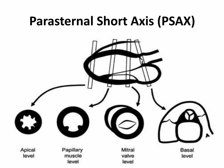
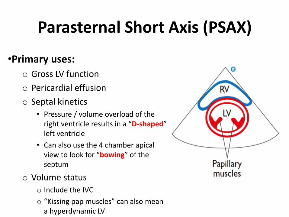
Parasternal Short Axis (PSAX)
•From PLAX view, rotate
transducer clockwise 90°
•Tilt transducer slightly toward
the right shoulder for more
apical views and away from the
right shoulder for basal views
•Mark on monitor is on the right
Parasternal Short Axis (PSAX)

Parasternal Short Axis (PSAX)

Parasternal Short Axis (PSAX)

Parasternal Short Axis (PSAX)
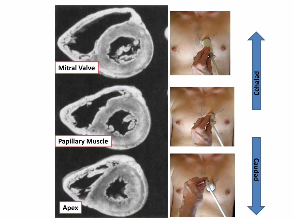
Mitral Valve
Papillary Muscle
Apex
Ce
hal
ad
Cau
dad

Parasternal Short Axis (PSAX)
Do not confuse the papillary muscles with vegetations
Parasternal Short Axis (PSAX)
•Primary uses:
o Gross LV function
o Pericardial effusion
o Septal kinetics • Pressure / volume overload of the
right ventricle results in a “D-shaped” left ventricle
• Can also use the 4 chamber apical view to look for “bowing” of the septum
o Volume status o Include the IVC
o “Kissing pap muscles” can also mean a hyperdynamic LV

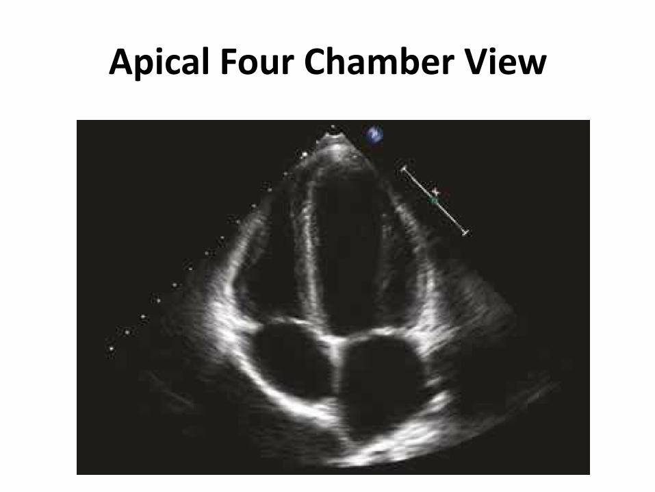
Apical Four Chamber View
•Place probe at apex lateral to nipple
line with index mark around 4 o’clock
for A4C
•Angle towards right shoulder: A5C
•Counter clockwise rotation: A2C

Apical Four Chamber View

Apical Four Chamber View
Apical Four Chamber View

Apical Four Chamber View
•Primary uses:
o RV/LV size, function, ratio
o Septal kinetics • “Bowing” of the LV
o Pericardial effusion
o TR and MR

Apical Two Chamber View

Apical Two Chamber View

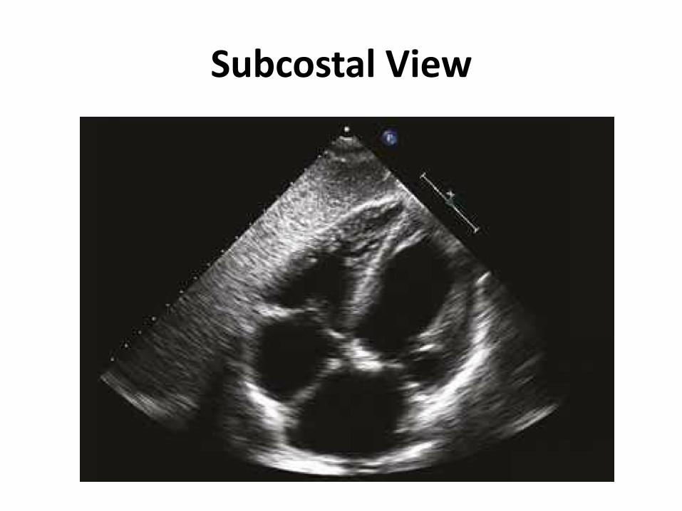
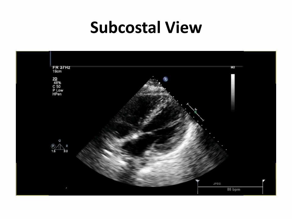
Subcostal View
•With patient lying supine,
place probe below xiphoid
process at a 45°angle
•Aim transducer toward
patient’s left shoulder
•Rotate probe counter
clockwise 90°to visualize
IVC
Subcostal View
Subcostal View

Subcostal View

Pericardial Effusion

Subcostal View
•Primary uses:
o “Sister” to A4C view
o RV/LV size
o RV/LV function
o Pericardial effusion
o Cardiac arrest • “sono pulse check”

Inferior Vena Cava

Inferior Vena Cava
•Primary uses:
o Volume status assessment
o Landmark is to measure 2 cm from the entrance of the IVC to the RA which is also where the hepatic vein connects to the IVC
o Use M-mode to measure the diameter of the IVC • > 2.1cm is dilated
o > 50% collapsibility

Inferior Vena Cava

Inferior Vena Cava

Color Doppler
• Higher velocity flow has progressively lighter shades of the same color
until the Nyquist limit is reached and then aliasing occurs

Severe Mitral Regurgitation

Goal Directed Approach to LV Function
• Not practical to measure ejection fraction
o Time consuming
o Exact number irrelevant and misleading
• Ex: Hyperdynamic LV can have EF of 70% but be underfilled such that SV is 20ml
• Ways to evaluate global LV function:
1. LV fractional area of change
2. MAPSE (Mitral annular plane systolic excursion )
3. LV outflow tract-shortening fraction (LVOT-SF)
4. “Eye balling”

Goal Directed LV Function Assessment
• Eyeball method
o Accurate with practice
o Is the LV:
• Really good
• Normal/Normalish
• Bad
• Terrible
Stam et al., 1982; Amico et al., 1989; Mueller et al., 1991

Wall Motion Abnormalities: What you’re looking for ?
• Endocardial excursion o LV systolic endocardial motion
represents the motion (excursion) of the endocardial segments towards the center of the ventricle
o Normal LV systolic function is expressed echocardiographically by a normal endocardial excursion in addition to myocardial thickening
• Myocardial thickening o Walls of the ventricle thicken during
systole and thin during diastole
o This is a visual assessment

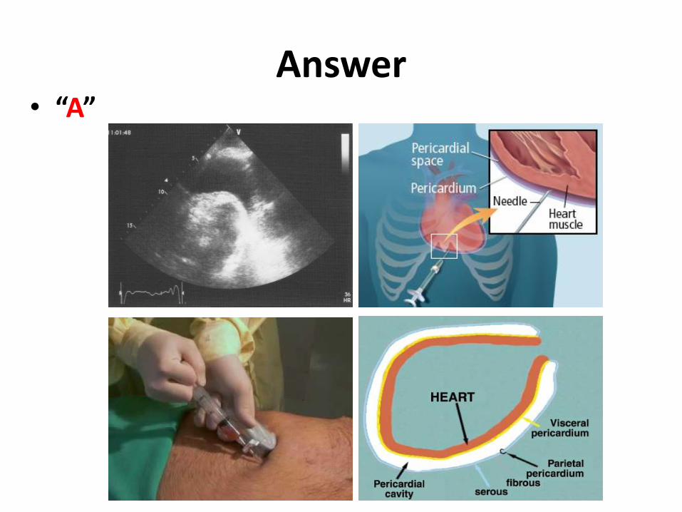
Bonus Question Part 1
• The critical care fellow is performing an emergent pericardiocentesis during a PEA arrest. After inserting the needle through the pericardium a significant amount of blood gushes out of the needle and the fellow thinks they have just punctured the ventricle. What should the fellow do next ?
A. Finish the procedure and insert the catheter
B. Remove the needle and attempt at a different location
C. Remove the needle and read Wikipedia
D. Remove the needle and pretend to have a syncopal episode

Bonus Question Part 2
• Which ventricle did the critical care fellow likely penetrate ?
A. Right ventricle
B. Left ventricle

Answer
• “B” o Right ventricular
punctures tends to bleed out versus left ventricle which a thick myocardium

Pearls for Technique
• Best position is left lateral decubitus
• Start wide and deep
• Small movements
• Thin patients may not get the best images because the rib space is too small
• Have a plan
1. Parasternal long axis
2. Parasternal short axis
3. Apical 4 chamber
4. Subcostal
o Bend the knees
5. Inferior vena cava
• In a stable patient do not rely on just 1 view to diagnose abnormalities

Conclusion
• Assessing global LV function requires eyeballs, not calipers
• Practice is important
• We can be very accurate
• This can help your patients and you through an undifferentiated shock state

Bonus Case #1
• 45 year old male with history of ESRD on HD who presented with fevers, chills and shortness of breath. Blood cultures positive for staph aureus. Patient started on IV antibiotics and acutely becomes short of breath and decompensates.

Parasternal Long Axis

Severe Aortic Regurgitation
Bonus Case #2
• 80 year old female with history of HTN, DM with history of 3 vessel CABG who presents with orthopnea, PND, and DOE for the past 2 days.

Parasternal Long Axis

Parasternal Short Axis
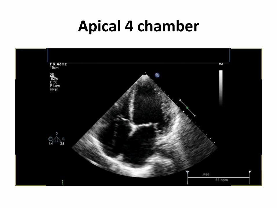
Apical 4 chamber

Bonus Case #3
• 80 year old female with history of coronary artery disease, DM, rheumatoid arthritis who presents with history exertional angina and shortness of breath. On exam, patient with a 3/6 systolic murmur at right 2nd intercostal space and crackles at bilateral base. On admission, patient given 40mg IV lasix and overnight becomes hypotensive.

Severe Aortic Stenosis

Bonus Case #4
• 54 year old female with history of breast cancer who is noted to have JVD, muffled heart sounds and low blood pressure.
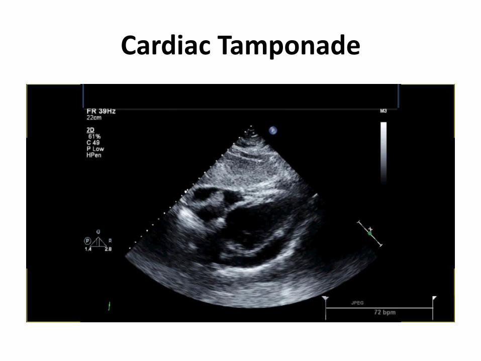
Cardiac Tamponade

Thank You
• E-mail: [email protected]
• Facebook: Raj Dasgupta MD
• Website: doctorrajd.com
• Twitter: @DoctorRajD
![· LV 01 - LV 02 - 14 - LV LV Of - LV - LV - LV - Skat Foru Out] 11 10 - 08 - 07 - Hiz tzht V HitÉ J Hilfe D.S. K : : Skat - : : Die PM Q Die 606 x)](https://img.dokumen.tips/doc/110x75/5e1f9008b175cd46915400c8/lv-01-lv-02-14-lv-lv-of-lv-lv-lv-skat-foru-out-11-10-08-07-.jpg)


![Visual Basic for Applications - schilk.at (Schilk).pdf · Visual Basic for Applications als Einstieg in das Programmieren [ARGE AINF HTL NÖ] LV-Nummer 351F4WWJ01 HTBLuVA Wiener Neustadt](https://img.dokumen.tips/doc/110x75/5a9dd6d17f8b9abd0a8dd16c/visual-basic-for-applications-schilkpdfvisual-basic-for-applications-als-einstieg.jpg)