Embed Size (px)
Citation preview

ipGmHf
Kg
BA
SICA
ND
TRA
NSLA
TION
AL
LIVER
GASTROENTEROLOGY 2013;144:1508–1517
BASIC AND TRANSLATIONAL—LIVER
GS-9620, an Oral Agonist of Toll-Like Receptor-7, Induces ProlongedSuppression of Hepatitis B Virus in Chronically Infected ChimpanzeesROBERT E. LANFORD,1,2,* BERNADETTE GUERRA,1 DEBORAH CHAVEZ,1 LUIS GIAVEDONI,1,2 VIDA L. HODARA,1
KATHLEEN M. BRASKY,2 ABIGAIL FOSDICK,3 CHRISTIAN R. FREY,4 JIM ZHENG,5 GRUSHENKA WOLFGANG,3
RANDALL L. HALCOMB,6 and DANIEL B. TUMAS3,4,*1 2
Department of Virology and Immunology, Texas Biomedical Research Institute, San Antonio, Texas; Southwest National Primate Research Center, San Antonio,Texas; and Departments of 3Drug Safety Evaluation, 4Biology, 5Drug Metabolism, and 6Medicinal Chemistry, Gilead Sciences, Inc, Foster City, CaliforniaatlalnlpHs(swa
eiimbr
See editorial on page 1342.
BACKGROUND & AIMS: Direct-acting antiviral agentssuppress hepatitis B virus (HBV) load, but they requirelife-long use. Stimulation of the innate immune systemcould increase its ability to control the virus and havelong-lasting effects after a finite regimen. We investigatedthe effects of immune activation with GS-9620 —a potentand selective orally active small molecule agonist of Toll-like receptor 7—in chimpanzees with chronic HBV infec-tion. METHODS: GS-9620 was administered to chim-panzees every other day (3 times each week) for 4 weeks at1 mg/kg and, after a 1-week rest, for 4 weeks at 2 mg/kg.We measured viral load in plasma and liver samples, thepharmacokinetics of GS-9620, and the following pharma-codynamics parameters: interferon-stimulated gene ex-pression, cytokine and chemokine levels, lymphocyte andnatural killer cell activation, and viral antigen expression.Clinical pathology parameters were monitored to deter-mine the safety and tolerability of GS-9620. RESULTS:Short-term oral administration of GS-9620 providedlong-term suppression of serum and liver HBV DNA. Themean maximum reduction of viral DNA was 2.2 logs,which occurred within 1 week of the end of GS-9620administration; reductions of �1 log persisted formonths. Serum levels of HBV surface antigen and HBV eantigen, and numbers of HBV antigen–positive hepato-cytes, were reduced as hepatocyte apoptosis increased.GS-9620 administration induced production of interfer-on-� and other cytokines and chemokines, and activatednterferon-stimulated genes, natural killer cells, and lym-hocyte subsets. CONCLUSIONS: The small moleculeS-9620 activates Toll-like receptor 7 signaling in im-une cells of chimpanzees to induce clearance ofBV-infected cells. This reagent might be developed
or treatment of patients with chronic HBV infection.
eywords: Innate Immunity; Interferon-�; Antiviral; Patho-en Recognition; TLR-7.
Therapeutic treatment of chronic hepatitis B virus(HBV) infection is currently limited to nucleos(t)ide
nalogues and pegylated interferon-(IFN)-�.1,2 First-lineherapy for HBV is limited to the 2 nucleos(t)ide ana-ogues tenofovir and entecavir, which are highly effectivet suppressing viral replication and can reduce serum viraload to undetectable levels. However, these agents doot lead to viral eradication, potentially requiring life-
ong use and possible emergence of resistance.3 Theotential for therapeutic immune modulation to treatBV chronic infection is illustrated by durable re-
ponses, normalization of alanine aminotransferaseALT), and sustained reduction in viremia attained in amall percentage (�20%) of patients treated for 1 yearith pegylated IFN-�.4 – 6 A key observation is that thepparent cure rate after long-term high-dose IFN-�
treatment increases for several years after treatment,based on loss of HBV surface antigen (HBsAg) andseroconversion for anti-HBsAg antibody. This supportsthe hypothesis that viral control is due to immunemodulation and slow induction of a protective antiviralimmune response. The low rate of HBsAg loss andseroconversion with current therapies illustrates theneed for new approaches to induce a protective antivi-ral immune response and durable cure in patients withchronic HBV.
Toll-like receptor (TLR) 7 is a pathogen recognitionreceptor predominantly expressed in lysosomal/endo-somal compartments of plasmacytoid dendritic cells(pDCs) and B lymphocytes that recognizes a pathogen-associated molecular pattern in viral single-stranded
*Authors share co-first authorship.
Abbreviations used in this paper: ALT, alanine aminotransferase;GGT, �-glutamyl transpeptidase; HBcAg, HBV core antigen; HBeAg, HBV
antigen; HBsAg, HBV surface antigen; HBV, hepatitis B virus; IFN,nterferon; IL, interleukin; IP-10, interferon-inducible protein 10; ISG,nterferon-stimulated genes; MCP, monocyte chemotactic protein; MIP,
acrophage inflammatory protein; NK, natural killer; PBMC, peripherallood mononuclear cells; pDC, plasmacytoid dendritic cell; TLR, Toll-likeeceptor.
© 2013 by the AGA Institute0016-5085/$36.00
http://dx.doi.org/10.1053/j.gastro.2013.02.003
tcn
9l
mrarss
t
pqs
ab
(aipT1a
3Pdi
BA
SIC
AN
DTR
AN
SLA
TIO
NA
LLI
VER
June 2013 GS-9620 SUPPRESSION OF CHRONIC HBV INFECTION 1509
RNA.7 Upon stimulation of TLR-7, pDCs produce IFN-�8,9 and other cytokines/chemokines and cause activa-ion of natural killer (NK) cells and cross-priming ofytotoxic lymphocytes,10 thereby orchestrating both in-ate and adaptive immune responses.11 For these rea-
sons, TLR-7 has been pursued as a therapeutic targetfor cancer, viral infections, and other diseases.12–15 GS-
620 is a potent, orally active TLR-7 agonist with se-ectivity for induction of IFN-� over proinflammatory
cytokines. Here, we demonstrate that a TLR-7 agonistprovides therapeutic efficacy for treatment of HBVchronic infection in chimpanzees, the only primatemodel of persistent HBV infection.16,17 The immune
odulation induced by activation of TLR-7 resulted inapid reduction of viremia, reduction in serum HBsAgnd HBV e antigen (HBeAg) levels, and an apparenteduction of the numbers of infected hepatocytes withhort-term therapy, and provided prolonged suppres-ion of viremia after termination of therapy.
MethodsAnimals and TreatmentChimpanzees were housed at the Southwest National
Primate Research Center at Texas Biomedical Research Institute.The animals were cared for in accordance with the Guide for theCare and Use of Laboratory Animals. Details on animal care andanimal histories are provided in the Supplementary Materials.The trial design included 4 weeks of prestudy evaluation (days�28, �13, and just before first dose) and 2 cycles of oralGS-9620 treatment every other day 3 times per week for 4 weeks,with one cycle at 1 mg/kg and, after a 1-week rest, a second cycleat 2 mg/kg. Animals were also intensely monitored for 14 weeksafter treatment to assess tolerability and durability of response.
Assays for HBV DNA and Viral AntigensHBV DNA levels were determined for the serum and
liver biopsy samples in multiple assays during the study period.Serum levels were measured by AmpliPrep/COBAS TaqManHBV Test, v2.0 and by an in-house TaqMan assay,18 see Supple-mentary Materials for details. Serum levels of HBsAg and HBeAgwere determined by enzyme-linked immunosorbent assay (ETI-MAK-2 PLUS and ETI-EBK PLUS, respectively; DiaSorin, Salug-gia, Italy). Immunohistochemical staining was performed onformalin-fixed liver tissue after antigen retrieval as describedpreviously19 and is further described in the Supplementary Ma-erials.
Quantitation of IFN-Stimulated GeneTranscript Levels by Reverse TranscriptionPolymerase Chain ReactionTranscript levels for 2=,5=-oligoadenylate synthetase
(OAS-1), MX1, interferon-stimulated gene (ISG) 15, interferoninduced T-cell alpha chemoattractant (I-TAC), IFN-inducibleprotein 10 (IP-10), TLR-7, and glyceraldehyde-3-phosphate de-hydrogenase (GAPDH) were determined by quantitative Taq-Man reverse transcription polymerase chain reaction as de-scribed previously.19 Briefly, 200 ng total cell RNA from liver or
eripheral blood mononuclear cells (PBMCs) was analyzed byuantitative reverse transcription polymerase chain reaction as-
ay using primers and probe from AB Gene Expression Assaysnd an ABI 7500 sequence analyzer (Applied Biosystems/Am-ion, Austin, TX).
Flow CytometryEvaluations of lymphocyte subpopulations were per-
formed using an 11-parameter CyAn ADP Flow Cytometer(Beckman-Coulter Inc, Fullerton, CA). All data were expressed aspercentage of lymphocytes that have the specified surface mark-ers. Detailed methods are provided in Supplementary Materials.
Cytokine and Chemokine AnalysisMonitoring of cytokines and chemokines was performed
by Luminex 100 with the xMAP (multi-analyte platform) systemusing a 39-plex human cytokine/chemokine kit (Millipore, Bil-lerica, MA). Dilutions of standards for each cytokine were eval-uated in each assay. Cytokines were evaluated in serum samplesat 0 and 8 hours post dose.
ResultsPharmacokinetics and Pharmacodynamics ofGS-9620 in Uninfected Chimpanzees:Induction of IFN Response and Cytokines-Chemokines by TLR-7 Agonist GS-9620
GS-9620, a potent selective TLR-7 agonist, wasdesigned to have rapid clearance and low-level systemicexposure after oral administration to allow for transientTLR-7 stimulation. Consistent with the selectivity of GS-9620 and the biology of TLR-7, chimpanzee PBMCs stim-ulated in vitro with GS-9620 displayed a lower minimumeffective concentration for IFN-�, chemokines CXCL10IP-10), CCL7 (monocyte chemotactic protein [MCP]-3),nd CCL4 (macrophage inflammatory protein [MIP]-1�),nterleukin (IL)-1 receptor antagonist, and IFN-� in com-arison to proinflammatory cytokines (Supplementaryable 1). In vivo, single, oral doses of GS-9620 at 0.3 andmg/kg in uninfected chimpanzees demonstrated a dose-
nd exposure-related induction of serum IFN-�, selectcytokines/chemokines, and ISG in the peripheral bloodand liver. After oral administration at 0.3 mg/kg (n � 3)and 1 mg/kg (n � 3 and n � 4), GS-9620 Cmax was 3.6 �
.5 nM, 36.8 � 34.5 nM, and 55.4 � 81.0 nM, respectively.eak serum interferon responses occurred at 8 hours postose and are shown in Figure 1. Mean peak levels of
nduced serum IFN-� were 66 pg/mL and 479 pg/mL atdoses of 0.3 mg/kg and 1 mg/kg, respectively (Figure 1).GS-9620 treatment induced ISG transcripts, includingISG15, OAS1 MX1, IP-10, and I-TAC in PBMC (Supple-mentary Table 2) at 0.3 mg/kg and in both PBMC and theliver at 1 mg/kg (Figure 2 and Supplementary Table 2).Serum levels of 42 different cytokines were evaluated. Themagnitude and breadth of cytokine induction correlatedwith GS-9620 dose (Figure 2 and Supplementary Table 3).The 0.3-mg/kg dose induced �3-fold increases in serumIL-7, IL-10, IP-10, fractalkine, IL-1�, IL-1 receptor antag-onist, and granulocyte colony-stimulating factor, whereasthe 1-mg/kg dose induced �3-fold increases in the samecytokines (except IL-7) and serum IL-12p40, IL-12p70,
MCP-1, MCP-3, MIP-1�, MIP-1�, IL-8, IL-1�, IL-6, tumor
acttmt
64s2aata
tdDtaos(vai
htDtaaf4ttwiaTaeti(
bhI
BA
SICA
ND
TRA
NSLA
TION
AL
LIVER
1510 LANFORD ET AL GASTROENTEROLOGY Vol. 144, No. 7
necrosis factor–�, and neopterin. GS-9620 was well toler-ted in uninfected chimpanzees; the only drug-relatedhanges were transient increases in peripheral blood neu-rophils and decreases in lymphocytes, consistent with cellrafficking induced by the cytokines and chemokines
entioned. Based on these data, 1 mg/kg was selected as
Figure 1. Pharmacokinetics and pharmacodynamics of GS-9620 inuninfected chimpanzees. Chimpanzees were dosed orally with GS-9620with 0.3 mg/kg (n � 3) or 1.0 mg/kg (group A, n � 3; group B, n � 4) and
lood levels of GS-9620 (A) and IFN-� (B) were determined over 24ours. Maximum concentration (Cmax) was determined for GS-9620 and
FN-� for each animal (C). The same 3 animals were used in each dosegroup, with a fourth animal added to group B.
he starting dose for treatment of HBV-infected animals. o
Antiviral Efficacy of TLR-7 Agonist GS-9620in HBV-Infected ChimpanzeesTherapeutic evaluation was performed in 3 chim-
panzees that had chronic HBV infections for �24 years.One chimpanzee (4x0139) had high baseline serum HBVDNA, while the other 2 chimpanzees (4x0328 and 4x0506)had lower HBV DNA levels at baseline (Figure 3 andSupplementary Table 4). Serum levels of HBV DNA de-clined gradually in all 3 animals during the first treatmentcycle, with a 1-log reduction in the high-titer animal(Figure 3A). The second treatment cycle caused a con-tinued but more rapid decline of viral DNA in all 3animals (Figure 3A–C), with a maximum viral reductionof 2.8 logs and a mean reduction of 2.2 logs (Figure 3and Supplementary Table 5). Suppression of serumviral DNA levels by �1 log persisted for a minimum of
4 days. The viral load in the high-titer animal (animalx0139) was 1.8 logs below baseline at the end of thetudy, day 121, and remained �1 log below baseline for80 days after initiation of dosing. The 2 low-viral–loadnimals returned to within 1 log of baseline within 100nd 71 days of the initiation of dosing, but continuedo be suppressed by approximately 1 log for 1 to 2 yearsfter this study.
Treatment also caused a decline in HBV viral DNA inhe liver of the high-titer animal (animal 4x0139). Theecline in liver HBV DNA paralleled the decline in serumNA, 1.0 and 2.1 logs at the end of the first and second
reatment cycles, respectively. The 2 low HBV DNA titernimals (animals 4x0328 and 4x0506) had very low levelsf hepatic HBV DNA at baseline and did not exhibit aignificant decline in viral DNA in the liver during therapydata not shown). The apparent lack of decline in hepaticiral DNA might have been due to limitations in the assaynd background in the assay imposed by the presence ofntegrated viral DNA.
HBsAg and HBeAg are secreted from HBV-infectedepatocytes independent of viral particles and are impor-ant clinical markers of infection independent of viralNA levels. In the high-titer animal (4x0139), GS-9620
reatment reduced HBsAg and HBeAg serum levels by 61%nd 93% from baseline, respectively (Figure 3C and D),nd levels remained suppressed through post-treatmentollow-up. Although the low-titer animals (4x0328 andx506) had low HBsAg levels at baseline, declines of 48%o 60% in HBsAg still occurred in both animals duringherapy (Figure 3C). One of the low-titer animals (4x0328)as HBeAg positive at baseline and had a decline of 55%
n HBeAg, while the other low-titer animal, 4x0506, wasnti-HBe–positive at baseline (Supplementary Table 4).he rapid declines in liver viral DNA and secreted viralntigens in the high-titer animal are consistent with anlimination of infected cells, thus we directly examinedhe elimination of infected cells by immunohistochem-cal staining of liver sections for HBV core antigenHBcAg). In the high-titer animal, approximately 30%
f hepatocytes were positive for HBcAg staining before
tle
kaI4d
I
osittupdTctras
aa(P
imqh
BA
SIC
AN
DTR
AN
SLA
TIO
NA
LLI
VER
June 2013 GS-9620 SUPPRESSION OF CHRONIC HBV INFECTION 1511
therapy (Figure 3E), and on the last day of dosing, whenHBV DNA levels were reduced by �100-fold, few core-positive cells were detected (Figure 3F). These resultsare in stark contrast to those observed in patientsduring therapy with nucleos(t)ide analogues, which canreduce serum HBV DNA by �4 logs, yet no significantreduction occurs in serum HBsAg or HBcAg-positivehepatocytes over 48 weeks of therapy.20 Unfortunately,he number of HBcAg-positive cells was too low in theow-titer animals to accurately determine the degree oflimination.
Induction of Cytokines and Chemokines andISGs by TLR-7 Agonist GS-9620 in HBV-Infected ChimpanzeesLevels of serum IFN-� and 38 other serum cyto-
ines and chemokines were evaluated at pretreatment andt regular intervals during each treatment cycle. PrestudyFN-� levels were below the limit of detection in animalsx0139 and 4x0328, and these animals had dose-depen-ent increases in IFN-� after administration of GS-9620
at 1 mg/kg (mean, 119 pg/mL) and 2 mg/kg (mean, 700pg/mL), although increases above baseline were not ob-served at every time point (Supplementary Table 6). Thehighest serum levels of IFN-� induced at the 2-mg/kgdose were 1396 pg/mL and 1545 pg/mL for animals4x0139 and 4x0328, respectively (Supplementary Tables 7and 8). The pretreatment baseline level of serum IFN-�was high in animal 4x0506 (1160 pg/mL) and was notfurther induced by GS-9620 treatment (SupplementaryTable 9). This animal also had an elevated pretreatmentbaseline level of serum IFN-�, yet GS-9620 treatmentinduced up to a 53-fold increase in serum levels of IP-10,a chemokine induced by IFN-� and IFN-�. Of the other38 cytokines and chemokines examined, during the firsttreatment cycle (1 mg/kg) only IL-10, MCP-3, and IL-1�were increased 5-fold above baseline, and during the sec-ond treatment cycle (2 mg/kg), 13 cytokines and chemo-
Figure 2. Fold induction of ISG transcripts and serum cytokines/chemncreases in ISG transcripts were quantified by TaqMan reverse transcrip
aximum mean fold increase from the baseline samples after a single dosuantified in the same animals by Luminex and are expressed as the mours post dose. Mean values are derived from 2 experiments with n �
1 additional animal present in the second study.
kines were induced �5-fold; with IL-7, MIP-1�, TNF-�,
and granulocyte colony-stimulating factor being induced�10-fold; and IFN-�, IL-10, IP-10, MCP-1, MCP-3, IL-8,L-1�, IL-1 receptor antagonist, and IL-6 being increased
�10-fold (Supplementary Table 6).Induction of ISG transcripts (ISG15, OAS1, MX1, IP-
10, and I-TAC) was evaluated in PBMC and liver biopsysamples, and each was increased in both compartments inresponse to GS-9620 at both the 1- and 2-mg/kg doselevels; however, induction was not consistently present atthe 1-mg/kg dose level for all days evaluated. ISG tran-scripts are rapidly up-regulated and down-regulatedwithin a few hours of stimulation.21 Variability in the level
f response may be technical and related to the use of aingle time point to measure a response that may bencreasing or decreasing from the maximum value at theime of sampling (8 hours post dose), although exposureo GS-9620 may have varied to some extent after individ-al doses. Induction of ISGs was both more consistentlyresent and the fold increases were greater at the 2-mg/kgose (Figure 4 and Supplementary Tables 7, 8, 9, and 10).he group mean increase in transcript levels of thehemokine IP-10 in PBMC was 49.6- and 194-fold duringhe first (1 mg/kg) and second (2 mg/kg) treatment cycles,espectively (Figure 4 and Supplementary Tables 7, 8, 9,nd 10). Interestingly, despite high pretreatment levels oferum IFN-� in animal 4x0506, and no apparent increase
in IFN-� levels after GS-9620 administration, GS-9620dministration caused increases in ISGs in both PBMCsnd the liver during both treatment cycles in this animalSupplementary Table 10). Because TLR-7 induction inBMCs by IFN-� was previously observed in chimpan-
zees,21 the induction of TLR-7 transcript was measured inthis study. At pretreatment, the relative expression ofTLR-7 in these chronically infected animals was 30-foldhigher in PBMC than liver. TLR-7 levels were increased atmultiple time points in the liver during treatment, with amean maximum induction of 11.9-fold, while increases in
es after a single oral dose of GS-9620 in uninfected chimpanzees. Thepolymerase chain reaction in PBMC and liver and are expressed as the
f 1 mg/kg GS-9620. Increases in serum cytokines and chemokines werefold increase from samples obtained before each dose (0 hours) and 8nd n � 4 animals. The same 3 animals were used in both studies, with
okintione o
ean3 a
PBMC were minimal at most time points, with a mean

BA
SICA
ND
TRA
NSLA
TION
AL
LIVER
1512 LANFORD ET AL GASTROENTEROLOGY Vol. 144, No. 7
maximum induction 4.4-fold compared with pretreat-ment levels (Figure 5).
Activation of T Cells and NK Cells by TLR-7GS-9620 Agonist in HBV-InfectedChimpanzeesBecause the stimulation of TLR-7 in pDCs can
result in the subsequent activation of immune effectorcells, we evaluated the activation status of peripheralblood lymphoid and NK cell subsets using cell surfaceCD69 expression as a biomarker. During the second treat-ment cycle (2 mg/kg), an increase in the percentage ofCD69-expressing CD8-positive T lymphocytes, NK, andNKT cells occurred, which was maximal after the firstdose (Supplementary Figure 1). Mean fold increases in thepercentage of CD69-positive cells ranged from 3.6 to 5.8(Supplementary Table 11). No significant increases oc-curred during the first treatment cycle at 1 mg/kg (Sup-
plementary Table 11).Histological Changes in the LiverIn general, the severity of hepatic inflammation in
chimpanzees associated with chronic HBV infection is lessthan that described in humans.18 A minimal to mildprimarily lymphocytic inflammatory infiltrate in the por-tal tracts was present in all 3 animals before treatment.Changes noted during treatment included an increasedmononuclear cell periportal infiltrate during the firsttreatment cycle, which, during the second treatment cycle,extended into adjacent hepatic parenchyma and sinu-soids. Additionally, there was increased single-cell hepa-tocyte apoptosis, which was often associated with mini-mal clusters of mononuclear cells. Histological changesnoted during treatment fully reversed within 3 to 5 weeksafter treatment. Immunohistochemistry of biopsies at pre-treatment and on the last day of therapy demonstrated amarked increase in hepatocellular expression of ISG15 pro-
Figure 3. Decline in HBV duringGS-9620 therapy in HBV-infectedchimpanzees. Three chimpan-zees chronically infected with HBVwere dosed orally 3 times perweek for 4 weeks with 1 mg/kg(days 1–25) and then for 4 weeksat 2 mg/kg (days 31–57) with a1-week rest between dosing cy-cles. HBV DNA in serum (IU/mL)and liver (genome equivalents[GE] per microgram of liver DNA;GE/�g) were determined byquantitative polymerase chain re-action for animal 4x0139 (A) and inserum for animals 4x0328 and4x0506 (B). HBsAg was deter-mined by enzyme-linked immu-nosorbent assay (ELISA) in the se-rum of all 3 animals (C) andHBeAg levels were determined byELISA in the serum of animal4x0139 (D). The level of serumHBV DNA from (A) is shown as adashed line in (D) for reference.Immunohistochemical staining ofHBcAg was performed on forma-lin-fixed sections of liver from4x0139 before dosing on day�28 (E) and on day 57 (F), the lastday of dosing at 2 mg/kg.
tein, a marker of IFN-� induction (Figure 6), and an in-

BA
SIC
AN
DTR
AN
SLA
TIO
NA
LLI
VER
June 2013 GS-9620 SUPPRESSION OF CHRONIC HBV INFECTION 1513
creased number of hepatocytes expressing activated caspase3, a marker for apoptosis. The latter was associated with acorrelative increase in hepatocellular regeneration and pro-liferation as determined by expression of Ki67.
Correlation of Viral Clearance and Elevationin Liver EnzymesGS-9620 therapy was generally well tolerated, and
no serious adverse events occurred during therapy. Clini-cal signs, body temperature, body weight, hematology,and blood chemistries were monitored throughout thestudy. Body weights were mildly decreased in all 3 animalsduring the study and recovered during the post-treatmentperiod. Adverse events in the study were limited to anemia
4™™™™™™™™™™™™™™™™™™™™™™™™™™™™™™™™™™™™Figure 4. Induction of ISG transcripts in liver and PBMC during GS-9620 therapy in HBV infected chimpanzees. The levels of transcriptsfor the ISGs; IP-10, ISG15, and I-TAC were determined by TaqManquantitative reverse transcription polymerase chain reaction in totalcell RNA from liver and PBMC during GS-9620 therapy. Animals weredosed orally 3 times per week for 4 weeks with 1 mg/kg or 2 mg/kgas described in the legend for Figure 3. Transcripts for the house-keeping gene GAPDH were determined as a control for nonspecificstimulation. Levels of transcripts are expressed as relative copy num-ber per microgram of total cell RNA and were determined at 8 hours
Figure 5. Induction of TLR-7 transcripts in liver and PBMC of HBV-infected chimpanzees during therapy with GS-9620. Three chimpan-zees chronically infected with HBV (4x0139, 4x0328, and 4x0506) weredosed orally 3 times per week for 4 weeks with 1 mg/kg or 2 mg/kg asdescribed in the legend for Figure 3. Increases in TLR-7 transcripts werequantified by TaqMan quantitative reverse transcription polymerasechain reaction in PBMC and liver and are expressed as copy number permicrogram of total cell RNA. Values for GAPDH are shown as a controlhousekeeping gene.
post dose.

mnn
cl
BA
SICA
ND
TRA
NSLA
TION
AL
LIVER
1514 LANFORD ET AL GASTROENTEROLOGY Vol. 144, No. 7
and transient increases in serum liver enzymes. Anemiawas mild to moderate in all 3 animals, maximal reduc-tions in red blood cell counts were 11% to 18%, and fullyor partially recovered by the end of the study (day 121).Increases in serum levels of the liver enzymes ALT, aspar-tate aminotransferase, and �-glutamyl transpeptidase(GGT) occurred during the second treatment cycle(2 mg/kg) (Figure 7). In the HBV high-titer animal (ani-mal 4x0139), a sharp increase in the level of serum ALTand GGT occurred after the first week of treatment at 2mg/kg, dosing was suspended for this animal for 1 weekduring which both ALT and GGT rapidly decreased, andthen treatment resumed. No further increases in ALT werenoted in this animal, however, GGT increases were noted(Figure 4). Liver enzyme elevations fully reversed aftertreatment; ALT and aspartate aminotransferase returnedto pretreatment baseline levels within 3 weeks and GGTby the end of the study. Mild, transient 2- to 3-foldincreases in serum total bilirubin occurred at single timepoints in 2 animals (day 43 in animal 4x0139 and day 57in animal 4x0506) during treatment at 2 mg/kg and were
Figure 6. Reduction of HBcAg and induction of ISG15, activatedaspase 3 and Ki67 in liver tissue after GS-9620 therapy. Formalin-fixed
iver sections from 4x0139 on day �28 (A, C, and E) and day 57 (B, F), orday 79 (D) were stained with antibodies to ISG15 (A, B) as a marker ofIFN-� induction, activated caspase 3 (C, D) as a marker for apoptosisand Ki67 (E, F) as a marker for proliferation. The ISG15 antibody hadsome staining of nuclei in the prestudy (day �28), as well as cytoplasmicstaining in a few cells in sinusoidal spaces, while tissue from day 57 hasvery intense staining across the entire section. Rare apoptotic cells pos-itive for activated caspase 3 could be detected in the prestudy samplebut were not present in this field (C), and numerous apoptotic cells arepresent at day 79 (D), and appear to represent both hepatocytes andmononuclear cells in sinusoidal spaces. A few Ki67-positive hepatocyteswere present in the prestudy sample (E), and positive hepatocytes werenumerous on day 57 (F).
concurrent with liver enzymes increases.
The transient and low-level single-incidence bilirubinincreases were not considered adverse, but warrant mon-itoring in future clinical trials.
DiscussionThe ultimate goal of therapy for HBV chronic
infection is viral eradication and cure of the underlyingliver disease.22 The greatest advances in therapy have been
ade with nucleos(t)ide analogues that are chain termi-ators of the reverse transcription process.1,23 Althoughucleos(t)ide therapies reduce circulating virus to unde-
Figure 7. Increase in serum levels of liver enzymes during therapy withGS-9620 in HBV-infected chimpanzees. Serum levels (U/L; left axis) ofthe liver enzymes ALT, aspartate aminotransferase (AST), and GGT areshown for 3 HBV-infected chimpanzees (4x0139, 4x0328, and 4x0506)during therapy with GS-9620 at 1 mg/kg and 2 mg/kg as described inthe legend for Figure 3. Levels of serum HBV DNA (IU/mL) from panels
A–C of Figure 3 are shown as dashed lines for reference.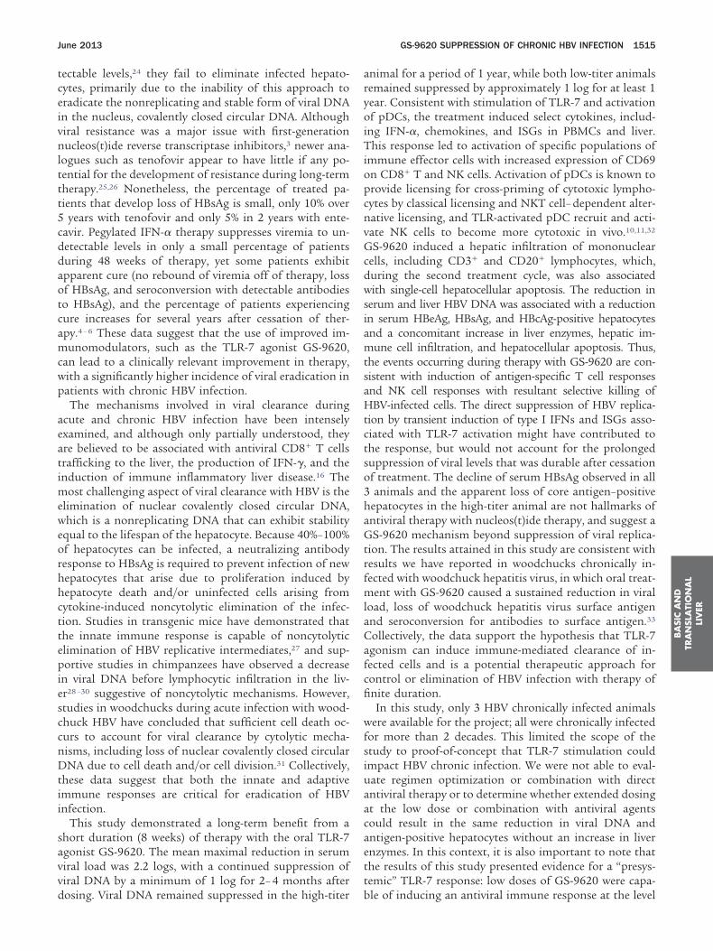
ltt
mcwp
aeat
pie
pcnv
dwsiamtsaHtctso3haGtrfmla
BA
SIC
AN
DTR
AN
SLA
TIO
NA
LLI
VER
June 2013 GS-9620 SUPPRESSION OF CHRONIC HBV INFECTION 1515
tectable levels,24 they fail to eliminate infected hepato-cytes, primarily due to the inability of this approach toeradicate the nonreplicating and stable form of viral DNAin the nucleus, covalently closed circular DNA. Althoughviral resistance was a major issue with first-generationnucleos(t)ide reverse transcriptase inhibitors,3 newer ana-ogues such as tenofovir appear to have little if any po-ential for the development of resistance during long-termherapy.25,26 Nonetheless, the percentage of treated pa-
tients that develop loss of HBsAg is small, only 10% over5 years with tenofovir and only 5% in 2 years with ente-cavir. Pegylated IFN-� therapy suppresses viremia to un-detectable levels in only a small percentage of patientsduring 48 weeks of therapy, yet some patients exhibitapparent cure (no rebound of viremia off of therapy, lossof HBsAg, and seroconversion with detectable antibodiesto HBsAg), and the percentage of patients experiencingcure increases for several years after cessation of ther-apy.4 – 6 These data suggest that the use of improved im-
unomodulators, such as the TLR-7 agonist GS-9620,an lead to a clinically relevant improvement in therapy,ith a significantly higher incidence of viral eradication inatients with chronic HBV infection.The mechanisms involved in viral clearance during
cute and chronic HBV infection have been intenselyxamined, and although only partially understood, theyre believed to be associated with antiviral CD8� T cellsrafficking to the liver, the production of IFN-�, and the
induction of immune inflammatory liver disease.16 Themost challenging aspect of viral clearance with HBV is theelimination of nuclear covalently closed circular DNA,which is a nonreplicating DNA that can exhibit stabilityequal to the lifespan of the hepatocyte. Because 40%–100%of hepatocytes can be infected, a neutralizing antibodyresponse to HBsAg is required to prevent infection of newhepatocytes that arise due to proliferation induced byhepatocyte death and/or uninfected cells arising fromcytokine-induced noncytolytic elimination of the infec-tion. Studies in transgenic mice have demonstrated thatthe innate immune response is capable of noncytolyticelimination of HBV replicative intermediates,27 and sup-
ortive studies in chimpanzees have observed a decreasen viral DNA before lymphocytic infiltration in the liv-r28 –30 suggestive of noncytolytic mechanisms. However,
studies in woodchucks during acute infection with wood-chuck HBV have concluded that sufficient cell death oc-curs to account for viral clearance by cytolytic mecha-nisms, including loss of nuclear covalently closed circularDNA due to cell death and/or cell division.31 Collectively,these data suggest that both the innate and adaptiveimmune responses are critical for eradication of HBVinfection.
This study demonstrated a long-term benefit from ashort duration (8 weeks) of therapy with the oral TLR-7agonist GS-9620. The mean maximal reduction in serumviral load was 2.2 logs, with a continued suppression ofviral DNA by a minimum of 1 log for 2– 4 months after
dosing. Viral DNA remained suppressed in the high-titeranimal for a period of 1 year, while both low-titer animalsremained suppressed by approximately 1 log for at least 1year. Consistent with stimulation of TLR-7 and activationof pDCs, the treatment induced select cytokines, includ-ing IFN-�, chemokines, and ISGs in PBMCs and liver.This response led to activation of specific populations ofimmune effector cells with increased expression of CD69on CD8� T and NK cells. Activation of pDCs is known to
rovide licensing for cross-priming of cytotoxic lympho-ytes by classical licensing and NKT cell– dependent alter-ative licensing, and TLR-activated pDC recruit and acti-ate NK cells to become more cytotoxic in vivo.10,11,32
GS-9620 induced a hepatic infiltration of mononuclearcells, including CD3� and CD20� lymphocytes, which,
uring the second treatment cycle, was also associatedith single-cell hepatocellular apoptosis. The reduction in
erum and liver HBV DNA was associated with a reductionn serum HBeAg, HBsAg, and HBcAg-positive hepatocytesnd a concomitant increase in liver enzymes, hepatic im-une cell infiltration, and hepatocellular apoptosis. Thus,
he events occurring during therapy with GS-9620 are con-istent with induction of antigen-specific T cell responsesnd NK cell responses with resultant selective killing ofBV-infected cells. The direct suppression of HBV replica-
ion by transient induction of type I IFNs and ISGs asso-iated with TLR-7 activation might have contributed tohe response, but would not account for the prolongeduppression of viral levels that was durable after cessationf treatment. The decline of serum HBsAg observed in allanimals and the apparent loss of core antigen–positive
epatocytes in the high-titer animal are not hallmarks ofntiviral therapy with nucleos(t)ide therapy, and suggest aS-9620 mechanism beyond suppression of viral replica-
ion. The results attained in this study are consistent withesults we have reported in woodchucks chronically in-ected with woodchuck hepatitis virus, in which oral treat-
ent with GS-9620 caused a sustained reduction in viraload, loss of woodchuck hepatitis virus surface antigennd seroconversion for antibodies to surface antigen.33
Collectively, the data support the hypothesis that TLR-7agonism can induce immune-mediated clearance of in-fected cells and is a potential therapeutic approach forcontrol or elimination of HBV infection with therapy offinite duration.
In this study, only 3 HBV chronically infected animalswere available for the project; all were chronically infectedfor more than 2 decades. This limited the scope of thestudy to proof-of-concept that TLR-7 stimulation couldimpact HBV chronic infection. We were not able to eval-uate regimen optimization or combination with directantiviral therapy or to determine whether extended dosingat the low dose or combination with antiviral agentscould result in the same reduction in viral DNA andantigen-positive hepatocytes without an increase in liverenzymes. In this context, it is also important to note thatthe results of this study presented evidence for a “presys-temic” TLR-7 response: low doses of GS-9620 were capa-
ble of inducing an antiviral immune response at the level
eaPI
1
1
1
1
BA
SICA
ND
TRA
NSLA
TION
AL
LIVER
1516 LANFORD ET AL GASTROENTEROLOGY Vol. 144, No. 7
of the liver, PBMC, and possibly gut-associated lymphoidtissue without induction of systemic IFN-� or its sideffects. At the low dose, in both uninfected and infectednimals, GS-9620 induced ISGs in the liver and/or inBMC with no detectable increase in serum levels ofFN-�. Ongoing clinical trials are evaluating whether ad-
ministration of GS-9620 at presystemic dose levels pro-vides similar antiviral benefits alone, as recognized in thisstudy, or in combination with direct-acting antivirals,without the side effects often associated with systemicadministration of IFN-�. Identification of a well-tolerated,finite therapeutic regimen, including the possible combi-nation of immune modulators, such as GS-9620, withdirect-acting antiviral agents for the treatment and cure ofchronic HBV infection in a significant and clinically rele-vant percentage of treated patients would be transforma-tive for this disease.
Supplementary Material
Note: To access the supplementary materialaccompanying this article, visit the online version ofGastroenterology at www.gastrojournal.org, and at http://dx.doi.org/10.1053/j.gastro.2013.02.003.
References
1. Kwon H, Lok AS. Hepatitis B therapy. Nat Rev GastroenterolHepatol 2011;8:275–284.
2. Sonneveld MJ, Janssen HL. Chronic hepatitis B: peginterferon ornucleos(t)ide analogues? Liver Int 2011;31(Suppl 1):78–84.
3. Locarnini S, Zoulim F. Molecular genetics of HBV infection. AntivirTher 2010;15(Suppl 3):3–14.
4. Moucari R, Korevaar A, Lada O, et al. High rates of HBsAgseroconversion in HBeAg-positive chronic hepatitis B patients re-sponding to interferon: a long-term follow-up study. J Hepatol2009;50:1084–1092.
5. Marcellin P, Bonino F, Lau GKK, et al. Sustained response ofhepatitis B e antigen-negative patients 3 years after treatmentwith peginterferon alpha-2a. Gastroenterology 2009;136:2169–2179.
6. Reijnders JG, Rijckborst V, Sonneveld MJ, et al. Kinetics of hep-atitis B surface antigen differ between treatment with peginter-feron and entecavir. J Hepatol 2011;54:449–454.
7. Barbalat R, Ewald SE, Mouchess ML et al. Nucleic acid recognitionby the innate immune system. Annu Rev Immunol 2011;29:185–214.
8. Gibson SJ, Lindh JM, Riter TR, et al. Plasmacytoid dendritic cellsproduce cytokines and mature in response to the TLR7 agonists,imiquimod and resiquimod. Cell Immunol 2002;218:74–86.
9. Hemmi H, Kaisho T, Takeuchi O, et al. Small anti-viral compoundsactivate immune cells via the TLR7 MyD88-dependent signalingpathway. Nat Immunol 2002;3:196–200.
0. Oh JZ, Kurche JS, Burchill MA, et al. TLR7 enables cross-presen-tation by multiple dendritic cell subsets through a type I IFN-dependent pathway. Blood 2011;118:3028–3038.
1. Colonna M, Trinchieri G, Liu YJ. Plasmacytoid dendritic cells inimmunity. Nat Immunol 2004;5:1219–1226.
2. Kanzler H, Barrat FJ, Hessel EM, et al. Therapeutic targeting ofinnate immunity with Toll-like receptor agonists and antagonists.Nat Med 2007;13:552–559.
3. Bergmann JF, de Bruine J, Hotho DM, et al. Randomised clinicaltrial: anti-viral activity of ANA773, an oral inducer of endogenousinterferons acting via TLR7, in chronic HCV. Aliment Pharmacol
Ther 2011;34:443–453.14. Pockros P, Guyager D, Patton H, et al. Oral resiquimod in chronicHCV infection: safety and efficacy in 2 placebo-controlled, double-blind phase IIa studies. J Hepatol 2007;47:174–182.
15. Fidock MD, Souberbielle BE, Laxton C, et al. The innate immuneresponse, clinical outcomes, and ex vivo HCV antiviral efficacy ofa TLR7 agonist (PF-4878691). Clin Pharmacol Ther 2011;89:821–829.
16. Chisari FV, Isogawa M, Wieland SF. Pathogenesis of hepatitis Bvirus infection. Pathol Biol (Paris) 2010;58:258–266.
17. Rehermann B, Nascimbeni M. Immunology of hepatitis B virusand hepatitis C virus infection. Nat Rev Immunol 2005;5:215–229.
18. Mason WS, Low HC, Xu C, et al. Detection of clonally expandedhepatocytes in chimpanzees with chronic hepatitis B virus infec-tion. J Virol 2009;83:8396–8408.
19. Lanford RE, Feng Z, Chavez D, et al. Acute hepatitis A virusinfection is associated with a limited type I interferon responseand persistence of intrahepatic viral RNA. Proc Natl Acad Sci U SA 2011;108:11223–11228.
20. Werle-Lapostolle B, Bowden S, Locarnini S, et al. Persistence ofcccDNA during the natural history of chronic hepatitis B anddecline during adefovir dipivoxil therapy. Gastroenterology 2004;126:1750–1758.
21. Lanford RE, Guerra B, Lee H, et al. Genomic response to interfer-on-� in chimpanzees: implications of rapid downregulation forhepatitis C kinetics. Hepatology 2006;43:961–972.
22. Lok AS. Hepatitis B infection: pathogenesis and management.J Hepatol 2000;32:89–97.
23. Locarnini SA, Yuen L. Molecular genesis of drug-resistant andvaccine-escape HBV mutants. Antivir Ther 2010;15:451–461.
24. Perry CM, Simpson D. Tenofovir disoproxil fumarate in chronichepatitis B. Drugs 2009;69:2245–2256.
25. Snow-Lampart A, Chappell B, Curtis M, et al. No resistance totenofovir disoproxil fumarate detected after up to 144 weeks oftherapy in patients monoinfected with chronic hepatitis B virus.Hepatology 2011;53:763–773.
26. Heathcote EJ, Marcellin P, Buti M, et al. Three-year efficacy andsafety of tenofovir disoproxil fumarate treatment for chronic hep-atitis B. Gastroenterology 2011;140:132–143.
27. Wieland SF, Guidotti LG, Chisari FV. Intrahepatic induction ofalpha/beta interferon eliminates viral RNA-containing capsidsin hepatitis B virus transgenic mice. J Virol 2000;74:4165–4173.
28. Guidotti LG, Rochford R, Chung J, et al. Viral clearance withoutdestruction of infected cells during acute HBV infection. Science1999;284:825–829.
29. Thimme R, Wieland S, Steiger C, et al. CD8(�) T cells mediateviral clearance and disease pathogenesis during acute hepatitis Bvirus infection. J Virol 2003;77:68–76.
30. Wieland SF, Spangenberg HC, Thimme R, et al. Expansion andcontraction of the hepatitis B virus transcriptional template ininfected chimpanzees. Proc Natl Acad Sci U S A 2004;101:2129–2134.
31. Summers J, Jilbert AR, Yang W, et al. Hepatocyte turnover duringresolution of a transient hepadnaviral infection. Proc Natl Acad SciU S A 2003;100:11652–11659.
32. Persson CM, Chambers BJ. Plasmacytoid dendritic cell-inducedmigration and activation of NK cells in vivo. Eur J Immunol 2010;40:2155–2164.
33. Menne S, Tennant BC, Liu KH, et al. Anti-viral efficacy and induc-tion of an antibody response against surface antigen with theTLR7 agonist GS-9620 in the woodchuck model of chronic HBVinfection. J Hepatol 2011;54:S441.
Author names in bold designate shared co-first authorship.Received October 10, 2012. Accepted February 6, 2013.
Reprint requests
Address requests for reprints to: Robert E. Lanford, PhD,
June 2013 GS-9620 SUPPRESSION OF CHRONIC HBV INFECTION 1517
Department of Virology and Immunology, Texas Biomedical ResearchInstitute, San Antonio, Texas 78227. e-mail: [email protected];fax: (210) 670-3329.
AcknowledgmentsThe authors thank L. Notvall-Elkey and H. Lee for excellent
technical assistance; the Southwest National Primate ResearchCenter, Immunology Core Laboratory for immunological analyses;and E. Dick for pathology evaluations.
Conflicts of interestThese authors disclose the following: Abigail Fosdick, Christian R.
Frey, Jim Zheng, Grushenka Wolfgang, Randall L. Halcomb, and
Daniel B. Tumas are employees of Gilead Sciences. Robert E.Lanford was funded by Gilead Sciences to conduct this research. Theremaining authors disclose no conflicts.
FundingSupported by a research contract from Gilead Sciences.
The Southwest National Primate Research Center is supportedby a National Institutes of Health Primate Center base grant(previously National Center for Research Resources grantP51 RR13986; currently Office of Research InfrastructurePrograms/OD P51 OD011133), and by Research FacilitiesImprovement Program Grants C06 RR 12087 and C06
RR016228.BA
SIC
AN
DTR
AN
SLA
TIO
NA
LLI
VER

ftbchHcsnot1sabwtTTtaeruHhhsvH
ts
tcupst
bipgmI
1517.e1 LANFORD ET AL GASTROENTEROLOGY Vol. 144, No. 7
Supplementary Methods
Animals and TreatmentThe care of chimpanzees housed at the Southwest
National Primate Research Center at Texas BiomedicalResearch Institute is detailed in the text. Animals weresedated for all procedures. Some aspects of the HBVinfections with animals 4x0139, 4x0328, and 4x0506have been described previously.1 The HBV genotype in-ecting these animals is unknown. However, a portion ofhe HBV genome isolated from 4x0328 and 4x0506 haveeen previously sequenced and clustered with otherhimpanzee isolates and were most closely related touman genotype F. Animal 4x0139 was infected withBV in 1979, HCV in 1986, and human immunodefi-
iency virus (HIV) in 1986. At the time of enrollment, thetatus of the animal was chronic HBV positive, HCVegative, and HIV negative. This animal was enrolled inther investigational studies in the past and has receivedreatments, including recombinant plasmid IL-12 in999, short-term lamivudine treatment in 2002, and aingle dose of pegylated IFN-� treatment in 2006. Thisnimal was also exposed to investigational HBV vaccinesetween 1999 and 2001. Animal 4x0328 was infectedith an unknown source of HBV infection before initial
esting for HBsAg in 1986 and is a chronic HBV carrier.his animal has had no prior exposures to HCV or HIV.his animal has in the past been enrolled in other inves-
igational studies and has received treatments includingntibodies to HBsAg in 1990, 1994, and 2001, and anxperimental antiviral for HBV in 2005, which did noteduce viral load. Animal 4x0506 was infected with annknown source of HBV before the initial testing forBsAg in 1983 and is a chronic HBV carrier. This animalas had no prior exposures to HCV or HIV. This animalas in the past been enrolled in other investigationaltudies and has received treatments, including an HBVaccine in 1995 and a human monoclonal antibody toBsAg in 1998.
Clinical Pathology and UrinalysisAt each sedation, an extensive safety profile was
performed on each animal, including complete bloodcount with differential, whole panel blood chemistries,and urinalysis. Coagulation profiles with prothrombintime and activated partial thromboplastin time were per-formed at selected times during high- and low-dose pe-riods. Blood chemistries were determined with a UnicelDxC 600 Analyzer (Beckman Coulter, Inc, and DiagnosticChemicals Ltd, Oxford, CT). Coagulation parameterswere determined with an ACL 8000 Analyzer using PT-Fibrinogen Kit for prothrombin time, and Simplastin foractivated partial thromboplastin time determinations.Values from uninfected animals from the same colony
were used to establish normal ranges. Urinalysis was by amanual techniques using common dip-stick technology.No drug-related alteration in urinalysis was observed.
Liver BiopsiesNeedle biopsies from the liver were taken in anes-
thetized animals by a standard procedure. Biopsy mate-rial was divided immediately into a fraction for histopa-thology and fractions snap frozen for RNA and DNAextraction. The sections for histopathology were pro-cessed for fixation in 10% formalin in phosphate-bufferedsaline, paraffin embedded, and stained with H&E. Histo-pathological evaluations were performed by a board-cer-tified veterinarian pathologist (Dr Edward Dick) atSouthwest National Primate Research Center under code.
Drug Formulation and DosingFinal dose formulations for each animal were pre-
pared based on individual animal body weight and thetarget dose. GS-9620 was diluted to 100 mL in water andadministered to a sedated animal by oral gavage. Twodose levels were utilized in the study, 1 and 2 mg/kg. The1 mg/kg dose was administered 3 times per week for 4weeks (12 doses; days 1–25), and after a 6-day rest, 2mg/kg was administered 3 times per week for 4 weeks (12doses; days 31–57). The exception was animal 4x0139which, due to increases in serum levels of ALT, was notadministered GS-9620 on days 43, 45, and 47; dosing wasreinitiated on day 50 in this animal.
Determination of GS-9620 Concentrations inChimpanzee SerumAn aliquot of 50 �L of each serum sample was
reated with 100 �L of acetonitrile containing internaltandard. After the protein precipitation, 100 �L of the
supernatant was mixed with 100 �L water, and 20 �L ofhe solution was injected into a HyPurity C18 HPLColumn (30x2.1 mm; ThermoHypersil; #22105-032130)sing a TSQ Ultra Quantum LC/MS/MS system. Mobilehases A and B contained 1% and 80% acetonitrile, re-pectively, in 10 mM ammonium formate aqueous solu-ion containing 1% formic acid.
Assays for Serum and Liver HBV DNAHBV DNA levels were determined for the serum
and liver biopsy samples in multiple assays during thestudy period. Serum levels were measure by AmpliPrep/COBAS TaqMan HBV Test, v2.0 by the Scott & WhiteHospital Molecular Pathology Laboratory (Temple, TX)and by an in-house TaqMan assay.1 Liver DNA could not
e determined by COBAS and was measured using then-house TaqMan assay. The Roche COBAS assay com-utes HBV DNA levels based on the World Health Or-anization reference standard in international units perilliliter of serum with a lower limit of detection of 20
U/mL. The in-house TaqMan assay expresses HBV DNA
s genome equivalents (GE) per microgram of liver DNA.
cGGFEpdfcsGafu6
blsrcC
N
eeBPe(e(F
June 2013 GS-9620 SUPPRESSION OF CHRONIC HBV INFECTION 1517.e2
For comparison of serum and liver DNA values, 1 IU inthe Roche Assay is approximately 5 GE for the in-houseTaqMan assay. For the in-house TaqMan assay, DNA waspurified from liver biopsies by homogenization in 20 mMTris pH 8/20 mM EDTA/20 mM NaCl/1% sodium do-decyl sulfate and digestion with Proteinase K for 2 hoursat 65°C. Digested samples were sequentially extractedwith phenol/chloroform/amyl-alcohol followed by chlo-roform, and then precipitated with 100% ethanol. DNAsamples were analyzed by real-time polymerase chainreaction using TaqMan technology as described previ-ously,1 with primers and probe designed against the HBVore gene (forward primer, HBV core F-CGAGGCAG-TCCCCTAGAAG; reverse primer, HBV core R-TGC-ACGCGGYGATTG; probe, HBV core probe 5=-/56-AM/AGAACTCCCTCGCCTCGCAGACG/36-TAMSp/3=).ach serum sample was extracted in duplicate for DNAurification. If sufficient liver biopsy sample was available,uplicate extractions were performed for liver, and DNArom each extraction was run in duplicate. A plasmidontaining an HBV DNA insert was used to generate atandard curve for each TaqMan assay ranging from 10E to 1 million GE. Samples were analyzed in TaqMan
ssays using an ABI 7500 sequence detector using theollowing cycle parameters: 2 minutes at 50°C/10 min-tes at 95°C/45 cycles of 15 seconds at 95°C/1 min at0°C.
Quantitation of ISG Transcript Levels byReverse Transcription Polymerase ChainReactionThe transcript levels for OAS, MX1, ISG15, I-TAC,
IP-10, TLR-7, and GAPDH were determined by quantita-tive reverse transcription polymerase chain reaction(qRT-PCR) as described previously.2 Briefly, 200 ng oftotal cell RNA from liver or PBMC was analyzed byqRT-PCR assay using primers and probe from AB GeneExpression Assays and an ABI 7500 TaqMan sequenceanalyzer (Applied Biosystems/Ambion). The qRT-PCRwas performed using reagents from the RNA UltraSenseOne-Step Quantitative RT-PCR System (Invitrogen Cor-poration, Carlsbad, CA), and the following cycle settings:48°C, 30 minutes; 95°C, 10 minutes; and 95°C, 15 sec-onds; and 60°C, 1 minute, the latter 2 for 45 cycles. RNAwas isolated from liver at 8 hours post dose and PBMCsat 0 and 8 hours post dose on days 7, 14, 25, 31, 45, and57. The fold increase of ISGs was determined by compar-ison to a day �28 prestudy sample for liver and anaverage of the day �28 and day �13 prestudy samplesfor PBMC. PBMCs were purified from whole blood sam-ples collected into cell separation tubes (Vacutainer CPTtubes; Becton Dickinson and Company, Franklin Lakes,NJ). Isolated PBMCs were suspended in a solution ofRNA-Bee (Tel-Test, Inc, Friendswood, TX) and processed
for total cell RNA. Liver biopsies were immediately placedin RNAlater Stabilization Reagent and processed as de-scribed by the manufacturer.
Flow CytometryEvaluations of lymphocyte subpopulations were
performed on days �28, 7, 14, 25, 45, and 57 by theSouthwest National Primate Research Center Immunol-ogy Core Laboratory using an 11-parameter CyAn ADPFlow Cytometer (Beckman–Coulter Inc, Fullerton, CA).All data were expressed as percentage of lymphocytes thathave the specified surface markers. For example, T cellswere expressed as the percentage of lymphocytes thatwere CD3�. The lymphocyte population was determined
ased on forward and side scatter and this gated popu-ation was used for all subsequent analysis. The followingubpopulations were examined for activation based oneactivity to a panel of monoclonal antibodies: B lympho-ytes: CD20� cells for increased expression of CD69,D86, and HLA-DR (MFI); NK cells: CD16�CD56� cells
for increased expression of CD69; NKT cells: CD16�CD56�
CD3� cells for increased expression of CD69 and CD25;K8 cells: CD16�CD56�CD3�CD8� cells for increased
expression of CD69; T lymphocytes: CD3�CD4�CD8�
for increased expression of CD69, CD25, and FoxP3. Thefollowing antibody reagents were utilized for fluores-cence-activated cell sorting: CD25�APC (BC96; Bioleg-nd, San Diego, CA), CD3-Alx700 (Sp34-2; BD Biosci-nces, San Jose, CA), CD4-PerCP-Cy5.5 (SK3; BDiosciences), CD56-Alx488 (B159; BD Biosciences), CD69-acBlue (FN50; Biolegend), CD8-FITC (SK1; BD Biosci-nces), CD8-PerCP-Cy5.5 (SK1; BD Biosciences), CD56-PEB159; BD Biosciences), CD86-PE (2331, FUN-1; BD Biosci-nces), CD16-FITC (3G8; BD Biosciences), HLA-DR-Alx700L243; Biolegend), CD20-APC (2H7; BD Biosciences), andOXp3-PE (259D; Biolegend).
Immunohistochemical StainingLiver biopsies were fixed in buffered-formalin, par-
affin embedded, and sectioned at 4 �m. Slides werede-paraffinized in EZ-DeWax (BioGenex; HK 585-5K) 2times for 5 minutes and rinsed with water. Antigen re-trieval was performed in a microwave pressure cooker for15 minutes at 1000 watts and 15 minutes at 300 watts incitrate buffer (antigen retrieval solution; BioGenex; HK086-9K) as described previously.2 Cooled slides wererinsed with water and phosphate-buffered saline andtreated sequentially with peroxidase suppressor, universalblock, and avidin (all reagents from Pierce 36000 Immu-nohisto Peroxidase Detection Kit; Pierce, Rockford, IL).Slides were incubated sequentially for 1 hour at roomtemperature with primary antibody diluted in universalblock containing a biotin block, for 0.5 hour with bio-tinylated goat anti-mouse IgG, and for 0.5 hour withavidin-biotin complex. Slides were developed with ImmpactNova Red peroxidase substrate (SK-4805; Vector, Burlin-
game, CA), counterstained with Mayers (Lillie’s) hematoxy-
1517.e3 LANFORD ET AL GASTROENTEROLOGY Vol. 144, No. 7
lin (DAKO, Glostrup, Denmark; S3309), dehydrated, andmounted in nonaqueous mounting media (VectaMount,H-5000; Vector). Rabbit anti-HBV core was prepared frompurified core particles expressed in baculovirus.3 Rabbitmonoclonal antibody 5A1E (Cell Signaling, Danvers, MA;9964) was used at 1/50 dilution to detect activatedcaspase 3. Mouse monoclonal M1B-1(DAKO; #7240) wasused at 1/100 dilution to detect Ki67. Rabbit polyclonalH-150 (Santa Cruz Biotechnology, Santa Cruz, CA; sc-
50366) was used at 1/400 dilution to detect ISG15.Supplementary References
1. Mason WS, Low HC, Xu C, et al. Detection of clonally expandedhepatocytes in chimpanzees with chronic hepatitis B virus infec-tion. J Virol 2009;83:8396–8408.
2. Lanford RE, Feng Z, Chavez D, et al. Acute hepatitis A virusinfection is associated with a limited type I interferon responseand persistence of intrahepatic viral RNA. Proc Natl Acad Sci U SA 2011;108:11223–11228.
3. Beames B, Lanford RE. Carboxy-terminal truncations of the HBVcore protein affect capsid formation and size of the encapsidated
HBV RNA. Virology 1993;194:597–607.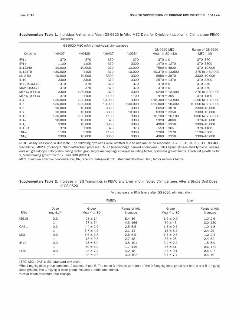
IIsII
MMIIII
I
TT
standard deviation; TNF, tumor necrosis factor.
I
O
M
I
I
I
June 2013 GS-9620 SUPPRESSION OF CHRONIC HBV INFECTION 1517.e4
Supplementary Table 1. Individual Animal and Mean GS-962Cultures
Cytokine
GS-9620 MEC (nM) of individual chim
4x0427 4x0428 4x0437
IFN-� 370 370 370IFN-� 1100 1100 370L-12p40 10,000 10,000 370L-12p70 �30,000 1100 370IL-2 RA 10,000 10,000 3300L-10 3300 3300 370P-10 (CXCL10) 370 370 370MCP-3 (CCL7) 370 370 370
IP-1� (CCL3) 3300 �30,000 370IP-1� (CCL4) 370 1100 1100
L-4 �30,000 �30,000 10,000L-5 �30,000 �30,000 10,000L-6 10,000 10,000 3300L-7 10,000 10,000 3300IL-13 �30,000 �30,000 1100L-1� 10,000 10,000 370IL-1� 3300 10,000 3300IL-1 RA 370 1100 370NF-� 1100 3300 1100NF-� 3300 10,000 3300
NOTE. Assay was done in duplicate. The following cytokines were omfractalkine, MCP-1 (monocyte chemoattractant protein-1), MDC (maeotaxin, granulocyte colony-stimulating factor, granulocyte-macrophage2, transforming growth factor 2, and GRO (CXCL1).MEC, minimum effective concentration; RA, receptor antagonist; SD,
Supplementary Table 2. Increase in ISG Transcripts in PBMCof GS-9620
RNADose
(mg/kg)a
Fold
PMBCs
GroupMeanb � SD
SG15 0.3 23 � 141 77 � 74
AS-1 0.3 4.4 � 2.01 6.7 � 4.3
X1 0.3 8.6 � 3.81 13 � 9.3
P-10 0.3 45 � 501 50 � 40
-TAC 0.3 9.8 � 7.21 33 � 40
-TAC; MX1; OAS-1; SD, standard deviation.aThe 1-mg/kg dose group combined 2 studies, A and B. The same 3 adose groups. The 1-mg/kg B dose group included 1 additional anima
0 In Vitro MEC Data for Cytokine Induction in Chimpanzee PBMC
panzeesGS-9620 MEC
Mean � SD (nM)Range of GS-9620
MEC (nM)4x0364
370 370 � 0 370–3703300 1470 � 1270 370–3300
10,000 7590 � 4820 370–10,00010,000 10,370 � 13,800 370 to �30,000
3300 6650 � 3870 3300–10,0003300 2570 � 1470 370–3300
370 370 � 0 370–370370 370 � 0 370–370
3300 9240 � 13,900 370 to �30,0001100 918 � 365 370–11003300 �18,300 � 13,800 3300 to �30,000
�30,000 �25,000 � 10,000 10,000 to �30,0003300 6650 � 3870 3300–10,000
10,000 8330 � 3350 3300–10,0003300 16,100 � 16,100 1100 to �30.0003300 5920 � 4860 370–10,0003300 4980 � 3350 3300–10,000370 553 � 365 370–1100
3300 2200 � 1270 1100–33003300 4980 � 3350 3300–10,000
itted due to minimal or no response, IL-2, -3, -8, -9, -15, -17, sCD40L,crophage derived chemokine), Flt-3 ligand (fms-related tyrosine kinase),colony-stimulating factor, epidermal growth factor, fibroblast growth factor
and Liver in Uninfected Chimpanzees After a Single Oral Dose
increase in RNA levels after GS-9620 administration
Liver
Range of foldincrease
GroupMeanb � SD
Range of foldincrease
8.4–36 1.9 � 0.9 1.0–2.64.3–166 60 � 47 3.0–1302.5–6.5 1.5 � 0.5 1.0–1.82.1–14 16 � 8.9 2.4–292.5–6.5 1.7 � 0.8 1.0–2.42.7–18 35 � 28 2.4–823.5–101 3.4 � 2.3 1.5–5.91.7–116 46 � 61 0.6–1714.2–18 0.6 � 0.1 0.5–0.73.0–102 8.7 � 7.7 0.4–19
nimals were part of the 0.3-mg/kg dose group and both A and B 1-mg/kgl.
bGroup mean maximum fold change.

I
A
ad
1517.e5 LANFORD ET AL GASTROENTEROLOGY Vol. 144, No. 7
Supplementary Table 3. Increase in Serum Cytokine and Chemokine Levels in Uninfected Chimpanzees After a Single OralDose of GS-9620
Cytokine
0.3 mg/kg dose groupa 1 mg/kg dose groupb
Mean cytokineCmax � SD
(pg/mL)
Fold increase(Mean Cmax Compared
to Pre-dose Mean)
Mean cytokineCmax � SD
(pg/mL)
Fold increase(Mean Cmax Compared
to Pre-dose Mean)
Antiviral CytokinesIFN-� 66 � 69 4 479 � 372 23
mmunomodulatory CytokinesIFN-� 0 � 0 �3 6.9 � 5.6 3IL-7 6.0 � 10 4 89 � 88 �3IL-10 12 � 8.3 4 86 � 125 11IL-12p40 4.5 � 7.3 �3 27 � 49 27IL-12p70 0.3 � 0.5 �3 1.4 � 2.1 7
ChemokinesIP-10 1003 � 831 8 4950 � 5960 37Fractalkine (CX3CL1) 92 � 110 5 651 � 516 3MCP-1 (CCL2) 452 � 268 �3 4100 � 4700 12MCP-3 (CCL7) 3.7 � 1.7 �3 45 � 50 4MIP-1� (CCL3) 0 � 0 �3 8.7 � 13 3MIP-1� (CCL4) 44 � 8.1 �3 842 � 1680 10IL-8 (CXCL8) 14 � 5.9 �3 732 � 1660 30
cute Phase Response CytokinesIL-1� 140 � 91 4 506 � 435 6IL-1 Receptor Antagonist 20 � 11 4 3900 � 5870 250IL-1� 0.1 � 0.1 �3 5.5 � 9.8 11IL-6 9.2 � 5.9 NC 2230 � 4490 300TNF-� 0.1 � 0.2 NC 3.8 � 8.1 19
Hematopoietic CytokinesG-CSF 44 � 18 3 1520 � 2870 43
OtherNeopterin 2.2 � 0.2 �3 3.5 � 1.9 3
BLD, below limit of detection; G-CSF, granulocyte colony-stimulating factor; NC, not calculated due to division by 0; baseline sample below limitof detection; TNF, tumor necrosis factor.aMean and standard deviation from n � 3 animals.bMean and standard deviation from n � 7 animals for combined experiments A and B. The same 3 animals were part of the 0.3-mg/kg dose groupnd both A and B 1-mg/kg dose groups. The 1-mg/kg B dose group included 1 additional animal, for a total of 7 animals in the combined 1-mg/kg
ose groups.Supplementary Table 4. Baseline Characteristics of HBV-Infected Chimpanzees
Animal ID HBV DNA (GE/mL) HBeAg Anti-HBeAg Anti-HBcAg Sex HBV infection (years)
4x0139 6.5x10⁷ � � � F 304x0328 2.5x10⁵ � � � M �244x0506 1.6x10⁴ � � � F �27
NOTE. HBV DNA levels were determined using TaqMan quantitative polymerase chain reaction as described in Methods and are expressed asgenome equivalents per mL (GE/mL). HBeAg levels and antibody to HBeAg and HBcAg were determined by enzyme-linked immunosorbent assay.The exact times of infection of 4x0328 and 4x0506 are unknown and are stated as before the first time the animals were tested in an HBsAg
assay.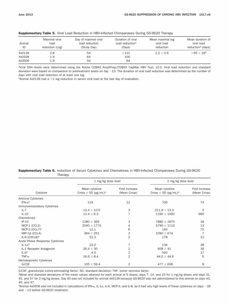
4
I
C
A
H
June 2013 GS-9620 SUPPRESSION OF CHRONIC HBV INFECTION 1517.e6
Supplementary Table 5. Viral Load Reduction in HBV-Infected Chimpanzees During GS-9620 Therapy
AnimalID
Maximal viralload
reduction (Log)
Day of maximal viralload reduction
(Study Day)
Duration of viralload reductiona
(Days)
Mean maximal logviral loadreduction
Mean duration ofviral load
reductiona (days)
x0139 2.8 54 �121 2.2 � 0.6 �95 � 29b
4x0328 1.9 64 1004x0506 1.9 54 64
aViral DNA levels were determined using the Roche COBAS AmpliPrep/COBAS TaqMan HBV Test, v2.0. Viral load reduction and standarddeviation were based on comparison to pretreatment levels on day �13. The duration of viral load reduction was determined as the number ofdays with viral load reduction of at least one log.
bAnimal 4x0139 had a �1 log reduction in serum viral load at the last day of evaluation.Supplementary Table 6. Induction of Serum Cytokines and Chemokines in HBV-Infected Chimpanzees During GS-9620Therapy
Cytokine
1 mg/kg dose level 2 mg/kg dose level
Mean cytokineCmax � SD (pg/mL)a
Fold increase(Mean Cmax)
Mean cytokineCmax � SD (pg/mL)a
Fold increase(Mean Cmax)
Antiviral CytokinesIFN-�b 119 12 700 74
mmunomodulatory CytokinesIL-7 14.4 � 10.5 3 211.9 � 13.3 5IL-10 12.4 � 6.3 7 1160 � 1060 660
hemokinesIP-10 1280 � 369 3 7880 � 1870 18MCP-1 (CCL2) 2040 � 1770 4 5790 � 1110 13MCP-3 (CCL7)b 12.1 6 140 70MIP-1� (CCL4) 364 � 251 2 1090 � 674 7IL-8 (CXCL8)b 52.3 2 178 12
cute Phase Response CytokinesIL-1�b 23.2 7 134 38IL-1 Receptor Antagonist 26.4 � 30 2 608 � 41 39IL-6b 4.5 2 500 17TNF-� 16.6 � 8.4 2 44.0 � 44.6 6
ematopoietic CytokinesG-CSF 105 � 59.4 2 477 � 608 8
G-CSF, granulocyte colony-stimulating factor; SD, standard deviation; TNF, tumor necrosis factor.aMean and standard deviations of the mean values attained for each animal at 6 doses; days 7, 14, and 25 for 1 mg/kg doses and days 31,45, and 57 for 2 mg/kg doses. Day 45 was not included for animal 4x0139 because GS-9620 was not administered to this animal on days 43,45, and 47.bAnimal 4x0506 was not included in calculations of IFN-�, IL-1�, IL-6, MCP-3, and IL-8, as it had very high levels of these cytokines on days �28
and �13 before GS-9620 treatment.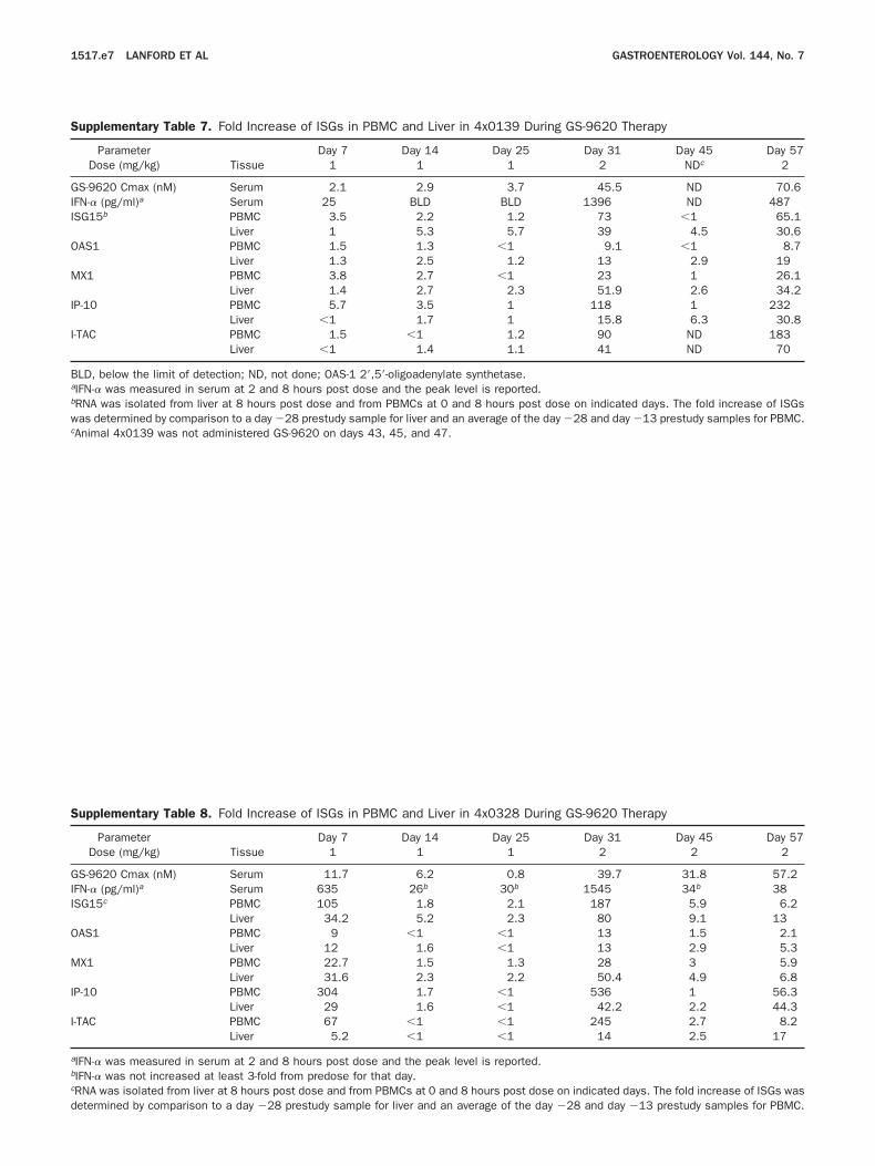
GI
O
M
I
I
d
1517.e7 LANFORD ET AL GASTROENTEROLOGY Vol. 144, No. 7
Supplementary Table 7. Fold Increase of ISGs in PBMC and Liver in 4x0139 During GS-9620 Therapy
ParameterTissue
Day 7 Day 14 Day 25 Day 31 Day 45 Day 57Dose (mg/kg) 1 1 1 2 NDc 2
S-9620 Cmax (nM) Serum 2.1 2.9 3.7 45.5 ND 70.6FN-� (pg/ml)a Serum 25 BLD BLD 1396 ND 487ISG15b PBMC 3.5 2.2 1.2 73 �1 65.1
Liver 1 5.3 5.7 39 4.5 30.6OAS1 PBMC 1.5 1.3 �1 9.1 �1 8.7
Liver 1.3 2.5 1.2 13 2.9 19MX1 PBMC 3.8 2.7 �1 23 1 26.1
Liver 1.4 2.7 2.3 51.9 2.6 34.2IP-10 PBMC 5.7 3.5 1 118 1 232
Liver �1 1.7 1 15.8 6.3 30.8I-TAC PBMC 1.5 �1 1.2 90 ND 183
Liver �1 1.4 1.1 41 ND 70
BLD, below the limit of detection; ND, not done; OAS-1 2=,5=-oligoadenylate synthetase.aIFN-� was measured in serum at 2 and 8 hours post dose and the peak level is reported.bRNA was isolated from liver at 8 hours post dose and from PBMCs at 0 and 8 hours post dose on indicated days. The fold increase of ISGswas determined by comparison to a day �28 prestudy sample for liver and an average of the day �28 and day �13 prestudy samples for PBMC.
cAnimal 4x0139 was not administered GS-9620 on days 43, 45, and 47.Supplementary Table 8. Fold Increase of ISGs in PBMC and Liver in 4x0328 During GS-9620 Therapy
ParameterTissue
Day 7 Day 14 Day 25 Day 31 Day 45 Day 57Dose (mg/kg) 1 1 1 2 2 2
GS-9620 Cmax (nM) Serum 11.7 6.2 0.8 39.7 31.8 57.2IFN-� (pg/ml)a Serum 635 26b 30b 1545 34b 38ISG15c PBMC 105 1.8 2.1 187 5.9 6.2
Liver 34.2 5.2 2.3 80 9.1 13AS1 PBMC 9 �1 �1 13 1.5 2.1
Liver 12 1.6 �1 13 2.9 5.3X1 PBMC 22.7 1.5 1.3 28 3 5.9
Liver 31.6 2.3 2.2 50.4 4.9 6.8P-10 PBMC 304 1.7 �1 536 1 56.3
Liver 29 1.6 �1 42.2 2.2 44.3-TAC PBMC 67 �1 �1 245 2.7 8.2
Liver 5.2 �1 �1 14 2.5 17
aIFN-� was measured in serum at 2 and 8 hours post dose and the peak level is reported.bIFN-� was not increased at least 3-fold from predose for that day.cRNA was isolated from liver at 8 hours post dose and from PBMCs at 0 and 8 hours post dose on indicated days. The fold increase of ISGs was
etermined by comparison to a day �28 prestudy sample for liver and an average of the day �28 and day �13 prestudy samples for PBMC.
GI
d
IOMII
June 2013 GS-9620 SUPPRESSION OF CHRONIC HBV INFECTION 1517.e8
Supplementary Table 9. Fold Increase of ISGs in PBMC and Liver in 4x0506 During GS-9620 Therapy
ParameterTissue
Day 7 Day 14 Day 25 Day 31 Day 45 Day 57Dose (mg/kg) 1 1 1 2 2 2
S-9620 Cmax (nM) Serum 4.5 1.8 35.9 30 14 104FN-� (pg/ml)a Serum 1214b 956b 972b 1127b 644b 647b
ISG15c PBMC 7.9 5.8 12 40 2.3 41.5Liver 3.6 3.8 6.1 14 6.6 18.7
OAS1 PBMC 4.1 3.2 3.4 8.6 1.7 8.2Liver 2.4 2.4 2.2 5.4 2.5 8.1
MX1 PBMC 6.4 5 6 15 2 24.2Liver 3.3 3.1 3.4 13.6 4.1 26.2
IP-10 PBMC 19.9 22.8 87 80 3 548Liver �1 1 1.1 7.9 4.4 47.6
I-TAC PBMC 2.1 1.7 3.3 8.8 2.2 100Liver �1 �1 �1 6 6.3 58
aIFN-� was measured in serum at 2 and 8 hours post dose and the peak level is reported.bIFN-� was 1160 pg/mL at baseline and was not increased by GS-9620 administration.cRNA was isolated from liver at 8 hours post dose and from PBMCs at 0 and 8 hours post dose on indicated days. The fold increase of ISGs was
etermined by comparison to a day �28 prestudy sample for liver and an average of the day �28 and day �13 prestudy samples for PBMC.Supplementary Table 10. Mean Fold Increase in ISGs in PBMC and Liver During GS-9620 Therapy
ISG
ISG mean fold increase � SD
GS-9620 1 mg/kga GS-9620 2 mg/kgb
PBMC Liver PBMC Liver
SG15 16 � 18 7.5 � 5.6 54.5 � 23.0 27.3 � 12.3AS1 2.8 � 1.4 3.0 � 1.7 6.9 � 1.8 9.5 � 5.7X1 5.6 � 3.0 5.8 � 5.4 16.9 � 6.7 26.1 � 15.0
P-10 49.6 � 49.7 4.3 � 5.4 194 � 17.9 24.3 � 4.9-TAC 8.9 � 12.3 1.5 � 0.8 86.3 � 49.8 30.0 � 22.9
aDays 7, 14, and 25.bDays 31, 45, and 57. Values for animal 4x0139 from day 45 were not included in these calculations as the animal was not administeredGS-9620 on days 43, 45, and 47.
For values in Supplementary Tables 6, 7, and 8 for which fold increases were �1, a value of 1 was used for the purpose of mean calculation.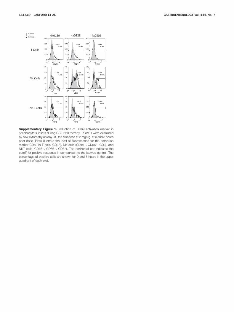
cpq
1517.e9 LANFORD ET AL GASTROENTEROLOGY Vol. 144, No. 7
T Cells
NK Cells
NKT Cells
0 Hours
8 Hours
5.63%
31.49%
5.44%
6.28%
3.69%
14.78%
4x0139 4x0328 4x0506
14.03%
63.44%
7.02%
30.48%
5.87%
36.01%
3.36%
36.67%
3.46%
7.00%
6.13%
27.3%
Supplementary Figure 1. Induction of CD69 activation marker inlymphocyte subsets during GS-9620 therapy. PBMCs were examinedby flow cytometry on day 31, the first dose at 2 mg/kg, at 0 and 8 hourspost dose. Plots illustrate the level of fluorescence for the activationmarker CD69 in T cells (CD3�), NK cells (CD16�, CD56�, CD3), andNKT cells (CD16�, CD56�, CD3�). The horizontal bar indicates theutoff for positive response in comparison to the isotype control. Theercentage of positive cells are shown for 0 and 8 hours in the upper
uadrant of each plot.
T
T
T
B
N
N
T
NC
June 2013 GS-9620 SUPPRESSION OF CHRONIC HBV INFECTION 1517.e10
Supplementary Table 11. Increase in CD69� Positive Lymphocytes in PBMC during GS-9620 Therapy
Cell type and markerGS-9620
Dose (mg/kg) Animal Day
Percent CD69� cells
FoldIncrease
Group mean foldIncrease � SD
0 hrPre-Dose
8 hrPost-Dose
Lymphocytes CD3� 1 4x0139 7 2.93 3.67 1.3 1.2 � 0.24x0328 7 3.66 4.60 1.34x0506 7 4.85 4.75 1.0
2 4x0139 31 3.69 14.78 4.0 3.6 � 2.24x0328 31 5.63 31.49 5.64x0506 31 5.44 6.28 1.2
Lymphocytes CD4� 1 4x0139 7 3.57 2.59 0.7 0.9 � 0.24x0328 7 3.44 3.82 1.14x0506 7 4.17 3.63 0.9
2 4x0139 31 3.20 1.83 0.6 2.7 � 3.14x0328 31 4.55 28.25 6.24x0506 31 4.27 4.96 1.2
Lymphocytes CD8� 1 4x0139 7 3.61 3.39 0.9 1.1 � 0.24x0328 7 2.96 3.81 1.34x0506 7 3.88 4.29 1.1
2 4x0139 31 3.75 13.24 3.5 3.1 � 1.84x0328 31 7.27 34.45 4.74x0506 31 6.07 6.67 1.1
Lymphocytes CD20� 1 4x0139 7 2.39 3.94 1.6 3.4 � 1.94x0328 7 1.15 3.67 3.24x0506 7 0.60 3.18 5.3
2 4x0139 31 3.71 9.09 2.5 1.8 � 0.74x0328 31 4.35 5.03 1.24x0506 31 6.03 9.55 1.6
K CD16� CD56� CD3� 1 4x0139 7 9.15 7.87 0.9 1.4 � 0.54x0328 7 12.72 22.37 1.84x0506 7 8.31 11.25 1.4
2 4x0139 31 5.87 36.01 6.1 5.0 � 1.04x0328 31 14.03 63.44 4.54x0506 31 7.02 30.48 4.3
KT CD16� CD56� CD3� 1 4x0139 7 4.79 2.84 0.6 1.0 � 0.44x0328 7 2.20 3.36 1.54x0506 7 2.23 2.20 1.0
2 4x0139 31 6.13 27.03 4.4 5.8 � 4.64x0328 31 3.36 36.67 10.94x0506 31 3.46 7.00 2.0
reg CD4� CD25� FoxP3� 1 4x0139 7 3.56 5.01 1.4 1.8 � 0.44x0328 7 2.90 5.95 2.14x0506 7 1.55 2.72 1.8
2 4x0139 31 1.74 3.86 2.2 2.3 � 1.34x0328 31 3.99 14.69 3.74x0506 31 1.76 1.95 1.1
OTE. Peripheral blood mononuclear cells were examined by flow cytometry at 0 and 8 hours post dose to determine the increase in percentD69� cells in different lymphocyte subsets in comparison to an isotype control.
SD, standard deviation; Treg, regulatory T cells.