Embed Size (px)
Citation preview
Basal Ganglia Organizationin Amphibians: Development
of Striatal and Nucleus AccumbensConnections With Emphasis
on the Catecholaminergic Inputs
OSCAR MARIN,1,2 WILHELMUS J.A.J. SMEETS,3 AND AGUSTIN GONZALEZ1*1Departamento de Biologıa Celular, Facultad de Biologıa, Universidad Complutense,
28040 Madrid, Spain2Departamento de Ciencias Morfologicas y Fisiologıa, Universidad Europea, 28670 Madrid, Spain
3Graduate School of Neurosciences of Amsterdam, Research Institute of Neurosciences andDepartment ofAnatomy and Embryology, Vrije Universiteit,Amsterdam 1081 BT, TheNetherlands
ABSTRACTTo broaden our insight into the organization of the basal ganglia of amphibians, the
development of the connections of the striatum and the nucleus accumbens was studied bymeans of tract-tracing techniques based on the transport of biotinylated dextran amines. In anumber of experiments, these techniques were combined with tyrosine hydroxylase immuno-histochemistry to identify the sources of catecholaminergic inputs to the striatum and thenucleus accumbens. Already at late embryonic stages, the basal telencephalon receives inputsfrom cells located in the amygdala, the thalamus, the suprachiasmatic nucleus, the raphenucleus, and the rhombencephalic reticular formation. At these stages, the rostral part of theposterior tubercle seems to be the only source of the dopaminergic input to the basaltelencephalon. During premetamorphosis, not only a differentiation between connections ofthe striatum and the nucleus accumbens could be made, but new sources of inputs were alsodetected in the mesencephalic and isthmic tegmentum, the parabrachial nucleus, and thenucleus of the solitary tract. Double-labeling experiments revealed that, at these stages, inaddition to the posterior tubercle, cells within the mesencephalic tegmentum, the locuscoeruleus, and the solitary tract nucleus contribute to the catecholaminergic innervation ofthe basal forebrain. During prometamorphic stages, a gradual increase occurs in the numberof cells that project to the basal telencephalon. At the beginning of the metamorphic climax,the organization of the basal ganglia afferents largely resembles the pattern observed injuveniles and adults. Remarkably, during larval stages, the cells that contribute to thedopaminergic innervation of the basal forebrain show a rostrocaudal gradient in time ofappearance. Moreover, the dopaminergic fibers reach the striatum earlier than the nucleusaccumbens, and they precede markedly the development of the efferent connections of bothbrain structures. These developmental aspects are easily correlated with the situation inamniotes; therefore, the notion that amphibians share an essentially similar pattern of basalganglia organization with other tetrapods is further strengthened. J. Comp. Neurol. 383:349–369, 1997. r 1997 Wiley-Liss, Inc.
Indexing terms: striatum; substantia nigra; nucleus of the solitary tract; locus coeruleus; evolution
In part of a research program that aims to answer thequestion to what extent the basal ganglia organization ofamphibians is comparable with that of amniotes, theafferent and efferent connections of the striatum and thenucleus accumbens have been studied in the brains ofadult anurans and urodeles (Marın et al., 1995, 1996,1997a–c). Significant differences in connections between
Grant sponsor: Spanish DGICYT; Grant number: PB96-0606; Grantsponsor: NATO (Collaborative Grant); Grant number: CRG 910970.*Correspondence to: Dr. Agustın Gonzalez, Departamento de Biologıa
Celular, Facultad de Biologıa, Universidad Complutense, 28040 Madrid,Spain. E-mail: [email protected] 6 November 1996; Revised 4 February 1997; Accepted 13
February 1997
THE JOURNAL OF COMPARATIVE NEUROLOGY 383:349–369 (1997)
r 1997 WILEY-LISS, INC.
the nucleus accumbens and the striatum were demon-strated, suggesting the presence of functional subdivi-sions. Moreover, the results of those studies stronglysupport the notion that a common pattern of catecholamin-ergic inputs to the basal forebrain exists among tetrapodvertebrates.A comparison of the development of basal ganglia struc-
tures of amphibians with that of amniotes would certainlycontribute to answering the question whether these struc-tures are basically similarly organized among vertebrates.A notable feature of the embryonic development of thebasal ganglia of amniotes is the early appearance ofdopaminergic fibers in the developing striatum (Olson andSeiger, 1972; Specht et al., 1981; Voorn et al., 1988; Medinaet al., 1994a,b). It has been hypothesized that the earlypresence of dopaminergic fibers in the striatum indicatesan important role in organizing the development of thisbrain region and directly influences the maturation ofstriatal neurons (Tennyson et al., 1973; Specht et al., 1981;Voorn et al., 1988; Medina et al., 1994a,b). Moreover, thecatecholaminergic innervation of the striatum precedesthe formation of the long descending projections to themidbrain tegmentum (Fishell and van der Kooy, 1987).Recently, the ontogeny of the catecholamine systems in
amphibians has been studied immunohistochemically bymeans of antibodies against tyrosine hydroxylase (TH)and dopamine (Gonzalez et al., 1994a,b, 1995). Thesestudies revealed that, as in amniotes, dopaminergic fibersreach the developing striatum and nucleus accumbens atearly stages of development. However, the origin andprogressive organization of this innervation during theembryonic and larval stages remained unknown, becausedata about the formation of connections of the basalganglia of amphibians were not available. A major reasonfor the absence of such information was the lack ofsensitive tracers that could be injected in vivo at restrictedsites in the developing basal telencephalon of amphibians.With the development of an in vitro approach in which thetracer biotinylated dextran amine (BDA) is applied to
restricted brain areas, this problem has been largelyovercome (Luksch et al., 1996; Munoz et al., 1996).The aims of the present study were 1) to establish the
temporal sequence of appearance of striatal and accumbalconnections in amphibians, with emphasis on the catechol-aminergic inputs; and 2) to compare the development ofbasal ganglia connections of amphibians with that re-ported for amniotes in order to gain more insight into theevolution of the basal ganglia of vertebrates. To reachthose goals, the SouthAfrican clawed toad,Xenopus laevis,has been selected as the core species because of theavailability of an accurate time table of development(Nieuwkoop and Faber, 1967) and the existence of recenthodological data of basal ganglia connections in adultbrains of this species (Marın et al., 1997a–c). Low-weight(3 kD) BDA has been used to selectively study the connec-tions of the developing striatum and nucleus accumbens.BDA, depending on the method of application, is both ananterograde and a retrograde tracer (Fritzsch, 1993;Munozet al., 1996, Marın et al., 1997a–c).
MATERIALS AND METHODS
For the present study, a total of 88 Xenopus laevisembryos and larvae ranging from developmental stage 45to developmental stage 65 (Nieuwkoop and Faber, 1967)were used (Table 1). The original research reported hereinwas performed under guidelines established by the Span-ish Royal Decree 223/1988. Animals were obtained byPregnyl-induced (Organon) breeding and were maintainedin tap water at 20°C throughout their development. Atappropriate times, embryos and tadpoles were deeplyanesthetized in a 0.3% solution of tricaine methanesulpho-nate (MS222; Sandoz, Basel, Switzerland) in distilledwater, pH 7.4, and then processed for tracing experimentsunder in vitro conditions, as previously described (Lukschet al., 1996). Briefly, under anesthesia, the animals werecooled to a body temperature of 4°C and were perfusedtranscardially with iced Ringer’s solution (75mMNaCl, 25
Abbreviations
A anterior thalamic nucleusAcc nucleus accumbensAd anterodorsal tegmental nucleusaob accessory olfactory bulbApl amygdala, pars lateralisApm amygdala, pars medialisAv anteroventral tegmental nucleusC central thalamic nucleusCb cerebellumDp dorsal palliumEa anterior entopeduncular nucleusgl glomerular layer of the olfactory bulbgr granular layer of the olfactory bulbHab habenulaHyp hypothalamusIII nucleus nervi oculomotoriIp interpeduncular nucleusLa lateral thalamic nucleus, anterior divisionLc locus coeruleuslfb lateral forebrain bundleLp lateral palliumLpv lateral thalamic nucleus, posteroventral divisionLs lateral septumm medial tegmental regionmfb medial forebrain bundleml mitral cell layer of the olfactory bulbMp medial pallium
Ms medial septumob olfactory bulboc optic chiasmP posterior thalamic nucleusPb parabrachial nucleuspc posterior commissurePd posterodorsal tegmental nucleusPOa preoptic areaPv posteroventral tegmental nucleusRa raphe nucleusRi nucleus reticularis inferiorRm nucleus reticularis mediusRs nucleus reticularis superiorSC suprachiasmatic nucleusSIR superficial isthmic reticular nucleussol solitary tractStr striatumtect tectumtegm tegmentumThal thalamusTor torus semicircularisTP posterior tuberclev ventricleVH ventral hypothalamusVm nucleus motorius nervi trigeminiVM ventromedial thalamic nucleusvt ventral thalamus
350 O. MARIN ET AL.

mM NaHCO3, 2 mM CaCl2, 2 mM KCl, 0.5 mM MgCl2, 11mM glucose; Merck, Darmstadt, Germany), which wasoxygenated with carbogen (95% O2, 5% CO2) to a pH of 7.3(Straka and Dieringer, 1993). Subsequently, the brain andspinal cord were rapidly isolated, and, after removal of thedura mater and the choroid plexuses, they were trans-ferred to fresh iced Ringer’s solution.Applications of 3 kD BDA (D-7135; Molecular Probes,
Eugene, OR) were made unilaterally within the ventrolat-eral (striatal) and ventromedial (nucleus accumbens) sub-regions of the basal telencephalon. The tracer was appliedby impaling the selected regions with a very sharp tung-sten needle. On the tip of the needle, BDA was recrystal-lized from a saturated solution in distilled water. Thebrains weremaintained for 15–24 hours at 15°C in continu-ously oxygenated Ringer’s solution. They were then fixedfor 3–5 hours in 4% paraformaldehyde in 0.1 M phosphatebuffer (PB), pH 7.4, blocked in a solution of 15% gelatinand 30% sucrose in PB, and stored for 5 hours at 4°C in asolution containing 4% formaldehyde and 30% sucrose inPB. Sections were cut on a freezingmicrotome at 30–40 µmthickness in the frontal or the sagittal plane and werecollected in cold PB.In a first series of experiments, BDAwas visualized with
an avidin biotin complex (Vectastain ABC standard kit;Vector Laboratories, Burlingame, CA) and peroxidase ac-tivity with 3,38-diaminobenzidine (DAB)-nickel as chromo-gen (for details, see Marın et al., 1997a). The sections weremounted on glass slides (mounting medium: 0.2% gelatinin Tris buffer, pH 7.6) and dried overnight. After ethanoldehydration and xylene cleaning, they were coverslippedwith Entellan (Merck). Some sections were counterstainedwith cresyl violet. In a second set of experiments, visualiza-tion of BDA was combined with indirect immunofluores-cence for TH. Briefly, brain sections were first incubatedfor 48 hours at 4°C with a mouse anti-TH antibody(Incstar, Stillwater, MN) diluted 1:1,000. They were thenincubated with a fluorescein isothiocyanate (FITC)-conjugated mouse-immunoglobulin (IgG) complex (Inc-star) diluted 1:150 for 90 minutes. BDA was visualized byincubation with a Texas red-conjugated streptavidin com-plex (Vector Laboratories; diluted 1:200) together with thesecondary antibody. The sections were then mounted onglass slides and coverslipped with Vectashield (VectorLaboratories). Alternating the appropriate filter combina-tions in a Zeiss fluorescence microscope allowed the identi-fication of BDA retrogradely labeled cells and TH-immunoreactive (THir) cells.The distribution of labeled cells and fibers in the brains
of Xenopus embryos and larvae was charted in representa-tive transverse sections by means of a camera lucida or acomputer-aided X-Y plotting system (MD-2 digitizer andsoftware;Minnesota Datametrics). The nomenclature usedin this study is largely the same as that used in ourprevious studies of amphibians (Gonzalez et al., 1994a,b).
RESULTS
In the present study, BDAwas applied to the developingstriatum and nucleus accumbens in Xenopus embryos andlarvae. The developmental sequence of characteristic stagesin the embryonic and larval periods of Xenopus has beensummarized in Figure 1. For each stage, the pattern oflabeling was consistent among animals treated identically.The different methods for visualizing BDA, as reportedpreviously for adult amphibians (Marın et al., 1997c), gaveessentially the same results. In the following sections,attention is paid to the development of the afferent connec-tions of the striatum and the nucleus accumbens and, inparticular, to their catecholaminergic inputs. Further-more, because BDA is also anterogradely transported, thedevelopment of the main efferent projections of the stria-tum and the nucleus accumbens was also studied.
Development of the afferent connections
Late embryonic stages. The earliest tadpoles ofXeno-pus that were analyzed corresponded to developmentalstages 44–45, which are the last stages of the embryonicperiod. At these stages, the hemispheric evaginationsleading to the formation of the lateral ventricles have nottaken place yet, and subregions within the subpallium arestill not recognizable. Nevertheless, applications of BDArestricted to the telencephalic hemisphere revealed sev-eral projections already at these stages (Fig. 2). Numerousretrogradely labeled neurons were found bilaterally in themedial amygdala in the caudal part of the telencephalon(Fig. 2B). In the rostral diencephalon, labeled cells wereseen in the suprachiasmatic nucleus, mainly in its ventro-lateral part (Fig. 2C). More caudally, scattered cells wereobserved in the hypothalamus (Fig. 2D,E,a). In addition,numerous labeled cells were found throughout the rostro-caudal extent of the thalamus and the posterior tubercle(Fig. 2D,E,a). Already at stage 45, three brainstem cellgroups were found to project to the telencephalon. Thelatter groups included strongly labeled cells in the parabra-chial region and the raphe at caudal isthmic levels (Fig.2F,b) and a few weakly labeled cells in the reticularformation at more caudal brainstem levels (Fig. 2G).Larval stages: Striatum. At larval stages 46–47,
three distinct regions were identified within the telence-phalic hemisphere, i.e., a rostral area bulbaris and lateraland medial hemispheric walls. Starting from these stages,tracer applications could be restricted to the ventrolateral(striatum) or the rostral ventromedial (nucleus accum-bens) regions of the telencephalic hemisphere.Premetamorphic stages. The development of the stria-
tal afferent projections during premetamorphic stages ischaracterized not only by a progressive maturation ofexisting connections but also by the appearance of new cellgroups projecting to the striatum (Fig. 3). Already at stage47, numerous cell groups became retrogradely labeled
TABLE 1. Number of Animals Investigated at Different Stages of Development With Tracer Applicationin the Striatum (Str) or the Nucleus Accumbens (Acc)1
Developmental stages
Embryonic Premetamorphic Prometamorphic Metamorphic climax
n45 46 47 50 51 52 53 54 55 56 57 58 59 61 63 64 65
6 Str 4 3 4 3 2 1 2 4 2 3 2 2 2 3 2 3 42Acc 3 2 3 2 3 2 2 3 4 2 4 1 3 1 2 3 40
1Staging of the embryos and larvae according to Nieuwkoop and Faber (1967).
DEVELOPMENT OF AMPHIBIAN BASAL GANGLIA CONNECTIONS 351

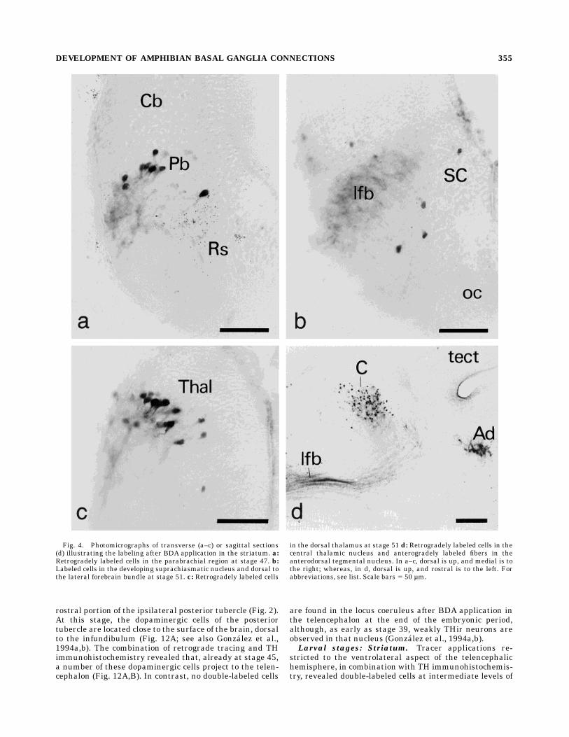
after tracer applications to the ventrolateral telencephalicwall. Labeled cells were found in the olfactory bulb, medialpallium, amygdala, suprachiasmatic nucleus, dorsal thala-mus, posterior tubercle, parabrachial region, raphe, andreticular formation (Fig. 4a). At stage 50, retrogradelylabeled cells in the suprachiasmatic nucleus after BDAapplication in the developing striatum were still restrictedto its lateral aspect (Figs. 3D, 4b). At about the same time,two distinct populations of labeled cells that correspondedto the anterior division of the lateral thalamic nucleus andthe central thalamic nucleus were recognized in the dorsalthalamus at intermediate levels (Fig. 3E, 4c). In addition,starting at stages 50–51, labeled cells were found in theventral thalamus just dorsal to the lateral forebrainbundle (Figs. 3D, 4b) and in the nucleus of the solitarytract (Figs. 3J). At the end of premetamorphosis, a notableincrease in the number of cells projecting to the striatumoccurred in the dorsal thalamus (Fig. 4d).Prometamorphic stages. During the prometamorpho-
sis, the distribution of cell bodies that projected to thestriatum showed a clear resemblance to the pattern ob-served in adults (Fig. 5; see alsoMarın et al., 1997a). In thecaudal telencephalon, numerous retrogradely labeled cellsconstituted a rather compact group in the anterior entope-duncular nucleus, whereas a few cells occurred in thepreoptic area (Fig. 5C,D). In the hypothalamus, labeledneurons were mainly restricted to the rostral aspect of theventral hypothalamic nucleus (Fig. 5F). In the dorsalthalamus, cells in the anterior division of the lateralthalamic nucleus were now clearly segregated from thoseof the central thalamic nucleus (Figs. 5E–G, 6a). Finally,from stage 54, tracer applications in the striatum consis-tently labeled cells in the rostral aspect of the mesence-phalic tegmentum (Fig. 5I).Metamorphic climax. This period of development is
characterized by a general maturation of the brain and asteady increase in the number of cells projecting to thestriatum (Fig. 7). For example, the number of cells in thecaudal extent of the ventral striatum and in the anteriorentopeduncular nucleus that projected to rostral striatalterritories became considerably larger (Figs. 6b, 7C,D).Similarly, the number of labeled cells located in the caudalaspect of the central thalamic nucleus and in the posteriortubercle region increased substantially during these stages(Figs. 6c, 7H,I). Finally, during the metamorphic climax,cells in the posteroventral division of the lateral thalamicnucleus became labeled for the first time after BDAapplication to the striatum (Fig. 7H).Larval stages: Nucleus accumbens
Premetamorphic stages. Restricted tracer applicationsto the ventromedial telencephalic wall of larvae at latepremetamorphic stages revealed the existence of severalwell-developed afferent projections to the nucleus accum-bens (Fig. 8). Labeled cells were found in the olfactorybulb, medial amygdala, preoptic area, the rostral aspect ofthe suprachiasmatic nucleus, anterior thalamic nucleus,ventromedial thalamic nucleus, posterior tubercle, parabra-chial nucleus, raphe, reticular formation, and nucleus ofthe solitary tract (Figs. 8, 9a,b). Labeling was bilateral inthe amygdala, suprachiasmatic nucleus, anterior thalamicnucleus, and posterior tubercle, although the ipsilateralprojection was always predominant (Fig. 8C,D,G).Prometamorphic stages. In the prometamorphic pe-
riod, the distribution of cells that projected to the nucleusaccumbens was largely comparable with that observed in
Fig. 1. Developmental stages ofXenopus laevis from egg to juvenileafter Nieuwkoop and Faber (1967). Numbers on the left indicatedevelopmental stages. Drawings represent Xenopus at selected devel-opmental stages. Lower part of drawing at stage 53 is an enlargementof the hindlimb.
352 O. MARIN ET AL.
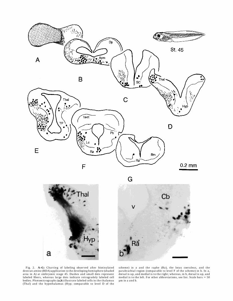
Fig. 2. A–G: Charting of labeling observed after biotinylateddextran amine (BDA) application to the developing hemisphere (shadedarea in A) at embryonic stage 45. Dashes and small dots representlabeled fibers, whereas large dots indicate retrogradely labeled cellbodies. Photomicrographs (a,b) illustrate labeled cells in the thalamus(Thal) and the hypothalamus (Hyp; comparable to level D of the
scheme) in a and the raphe (Ra), the locus coeruleus, and theparabrachial region (comparable to level F of the scheme) in b. In a,dorsal is up, and medial is to the right; whereas, in b, dorsal is up, andmedial is to the left. For other abbreviations, see list. Scale bars 5 50µm in a and b.

adults (Fig. 10; see also Marın et al., 1997a). Additionalretrogradely labeled cells occurred in the caudal aspect ofthe suprachiasmatic nucleus (Fig. 10E) as the prometamor-phosis proceeded. Moreover, cells in the posterior tubercleregion reached a high degree of organization (Fig. 9c).Starting at stages 52–53, labeled cells were found in theanteroventral tegmental nucleus (Fig. 10I). At isthmiclevels, only a few cells were present in the parabrachialregion, whereas the number of neurons in the adjacentreticular formation increased (Fig. 10J). Another notablefeature of this period was the rise in the number of labeledcells in the raphe, which remained restricted mainly to theupper rhombencephalon (Fig. 9d). At the end of theprometamorphic period, the main afferent projections tothe nucleus accumbens arose from the medial amygdala,
the anterior thalamic nucleus, and the posterior tubercle-mesencephalic tegmentum continuum (Figs. 9e,f, 10C–H).Metamorphic climax. Already at early metamorphic
climax stages, the afferent connections to the nucleusaccumbens were highly organized (Fig. 11). Major eventsthat take place during these stages concern the matura-tion of inputs from the ventral hypothalamus, posteriortubercle, and midbrain tegmentum (Fig. 11F–J).
Development of the catecholaminergicinnervation
Late embryonic stages. Unilateral applications ofBDA to the telencephalic hemisphere of Xenopus embryosat stage 45 revealed retrogradely labeled cells in the
Fig. 3. A–J: Charting of labeling in the brain of Xenopus at stage 50 after BDA application to thestriatum (shaded area in B). Dashes and small dots represent labeled fibers, whereas large dots indicateretrogradely labeled cell bodies. For abbreviations, see list.
354 O. MARIN ET AL.
rostral portion of the ipsilateral posterior tubercle (Fig. 2).At this stage, the dopaminergic cells of the posteriortubercle are located close to the surface of the brain, dorsalto the infundibulum (Fig. 12A; see also Gonzalez et al.,1994a,b). The combination of retrograde tracing and THimmunohistochemistry revealed that, already at stage 45,a number of these dopaminergic cells project to the telen-cephalon (Fig. 12A,B). In contrast, no double-labeled cells
are found in the locus coeruleus after BDA application inthe telencephalon at the end of the embryonic period,although, as early as stage 39, weakly THir neurons areobserved in that nucleus (Gonzalez et al., 1994a,b).Larval stages: Striatum. Tracer applications re-
stricted to the ventrolateral aspect of the telencephalichemisphere, in combination with TH immunohistochemis-try, revealed double-labeled cells at intermediate levels of
Fig. 4. Photomicrographs of transverse (a–c) or sagittal sections(d) illustrating the labeling after BDA application in the striatum. a:Retrogradely labeled cells in the parabrachial region at stage 47. b:Labeled cells in the developing suprachiasmatic nucleus and dorsal tothe lateral forebrain bundle at stage 51. c: Retrogradely labeled cells
in the dorsal thalamus at stage 51 d: Retrogradely labeled cells in thecentral thalamic nucleus and anterogradely labeled fibers in theanterodorsal tegmental nucleus. In a–c, dorsal is up, and medial is tothe right; whereas, in d, dorsal is up, and rostral is to the left. Forabbreviations, see list. Scale bars 5 50 µm.
DEVELOPMENT OF AMPHIBIAN BASAL GANGLIA CONNECTIONS 355

Fig. 5. A–L: Charting of labeling in the brain of Xenopus at stage 56 after BDA application to thestriatum (shaded area in B). Dashes and small dots represent labeled fibers, whereas large dots indicateretrogradely labeled cell bodies. For abbreviations, see list.
356 O. MARIN ET AL.

the posterior tubercle at premetamorphic stages 46–47(Fig. 12C,D). At about the same time, a few double-labeledcells were also found in the locus coeruleus. The number ofdouble-labeled cells in the posterior tubercle increasedstrikingly at stage 50, and the cell group extended farthercaudally to the border between the diencephalon and themesencephalon (Fig. 3G). In addition, a distinct cell popu-lation of striatal afferent cells that did not contain dopamin-ergic neurons was located in the dorsomedial aspect of theposterior tubercle, mainly at rostral levels (Fig. 3F). Fromstages 50–51, a few weakly THir cells in the nucleus of thesolitary tract were found to project to the ventrolateralaspect of the telencephalic hemisphere.During the prometamorphic period, the previously de-
scribed catecholaminergic cell groups mature primarily byincreasing their number of cells (Fig. 12E,F). In addition,beginning at stage 55, double-labeled cells are found in therostral aspect of the mesencephalic tegmentum. Duringthemetamorphic climax, a basic pattern of catecholaminer-gic innervation is observed that is already similar to thatfound in the adult brain (Marın et al., 1997c).Larval stages: Nucleus accumbens. From premeta-
morphic stages 46–47, a population of dopaminergic neu-rons in the posterior tubercle already projects to theimmature nucleus accumbens. Starting with stage 47,dopaminergic neurons in the caudal posterior tubercleextend to the diencephalic-mesencephalic transition area,where numerous cells are found to project to the nucleusaccumbens (Fig. 12G,H). Slightly later (stage 48), largenondopaminergic cells that project to the nucleus accum-bens are located in the rostral aspect of the posteriortubercle, where a few double-labeled cells are also present(Fig. 13A,B). At about the same time, double-labeled cellsare found in the locus coeruleus after tracer applicationinto the nucleus accumbens (Fig. 13C,D). At stages 50–51,
double-labeling experiments show that most of the cells inthe solitary tract nucleus that were labeled after applica-tion of BDA to the nucleus accumbens are also THir. At theend of the premetamorphic period, considerably densercatecholaminergic plexuses are found in the nucleus accum-bens (see also Gonzalez et al., 1994a), which is directlyrelated to the increase in number of double-labeled cellsfound in the posterior tubercle region.During the prometamorphic stages, dopaminergic cells
are found in the mesencephalic tegmentum. From stages57–58, most of the double-labeled cells found after tracerapplications in the nucleus accumbens are located in thecaudal aspect of the posterior tubercle and, predominantly,in the midbrain tegmentum (Fig. 13E,F). Finally, thedevelopment of the catecholaminergic innervation of thenucleus accumbens during the metamorphic climax ischaracterized by an increase in the number of catechol-aminergic cells projecting to this nucleus. However, nosignificant changes in the organization of these afferentsystems are observed.
Development of the efferent connections
Previous studies have revealed significant differences inafferent and efferent connections between the nucleusaccumbens and the striatum in adult amphibians (Marınet al., 1997a–c). From those studies, it became clear thatdextran amines can be preferentially transported retro-gradely or anterogradely, depending on the method ofapplication and the size of the injection. Therefore, be-cause BDA is also distributed anterogradely, data on thedevelopment of the main basal ganglia efferent connec-tions could also be studied.Striatum. The major efferent systems of the striatum,
as observed in adult brains, are formed by ipsilateraldescending projections to the anterior entopeduncular
Fig. 6. Photomicrographs of transverse sections through the brain of Xenopus tadpoles illustrat-ing retrogradely labeled cells after BDA application in the striatum. a: Dorsal thalamus at stage 56.b:Anterior entopeduncular nucleus at stage 58. c:Caudal pole of the central thalamic nucleus at stage 58.In all photomicrographs, dorsal is up, and medial is to the right. For abbreviations, see list. Scale bars 5100 µm.
DEVELOPMENT OF AMPHIBIAN BASAL GANGLIA CONNECTIONS 357

Fig. 7. A–M: Charting of labeling in the brain of Xenopus at stage 59 after BDA application to thestriatum (shaded area in B). Dashes and small dots represent labeled fibers, whereas large dots indicateretrogradely labeled cell bodies. For abbreviations, see list.

nucleus, the pretectum, and the mesencephalic and isth-mic reticular formation (Marın et al., 1997b). During thepremetamorphic stages, striatal efferent projections arevery restricted. Only minor projections reach the imma-ture caudal portion of the ventral striatum and its caudalcontinuation, i.e., the anterior entopeduncular nucleus. Inaddition, no anterogradely labeled fibers are present in thepretectum (Fig. 14A), and only minor projections reach therostral mesencephalic tegmentum (Fig. 15A). The striatalefferent systems, as the prometamorphosis proceeds, reacha high degree of organization. A prominent bundle oflabeled fibers projects to the caudal extent of the ventralstriatum-anterior entopeduncular continuum. Further-more, starting at stage 53, anterogradely labeled fibers arefound in the pretectum (Fig. 14B,C). Considerably denserplexuses extend caudally in themesencephalic tegmentum
(Fig. 15B,C) as the prometamorphic period continues. Thedevelopment of the efferent projections of the striatumduring the metamorphic climax is characterized by anincrease in the number of fibers that reach the pretectumand, remarkably, the mesencephalic and isthmic reticularformation (Figs. 14D,E, 15D–G).Nucleus accumbens. In adult amphibians, the nucleus
accumbens projects mainly to the preoptic area, the ven-tral hypothalamus, and the posterior tubercle-medial teg-mental region continuum (Marın et al., 1997b). Starting atpremetamorphic stages 49–50, efferent fibers are recog-nized in the anterior aspect of the preoptic area and theventral hypothalamus. An increase in the number ofefferent fibers from the nucleus accumbens is observed asthe larval development proceeds. From prometamorphicstage 55, numerous anterogradely labeled fibers are found
Fig. 8. A–J: Charting of labeling in the brain of Xenopus at stage 50 after BDA application to thenucleus accumbens (shaded area in B). Dashes and small dots represent labeled fibers, whereas large dotsindicate retrogradely labeled cell bodies. For abbreviations, see list.
DEVELOPMENT OF AMPHIBIAN BASAL GANGLIA CONNECTIONS 359

Fig. 9. Photomicrographs of transverse (a,b,e,f) or sagittal (c,d)sections through the brain ofXenopus tadpoles after tracer applicationinto the nucleus accumbens illustrating labeled cells in the nucleus ofthe solitary tract at stage 50–51 (a), in the raphe and parabrachialregions at stage 51 (b), in the posterior tubercle region at stage 52–53(c), in the rostral raphe at stage 52–53 (d), in the medial amygdala
above the medial forebrain bundle at stage 55 (e), and in the anteriorthalamic nucleus at stage 55 (f). In a, b, e, and f, dorsal is up, andmedial is to the right; whereas, in c and d, dorsal is up, and rostral is tothe left. For abbreviations, see list. Scale bars 5 50 µm in a–d, 100 µmin e,f.
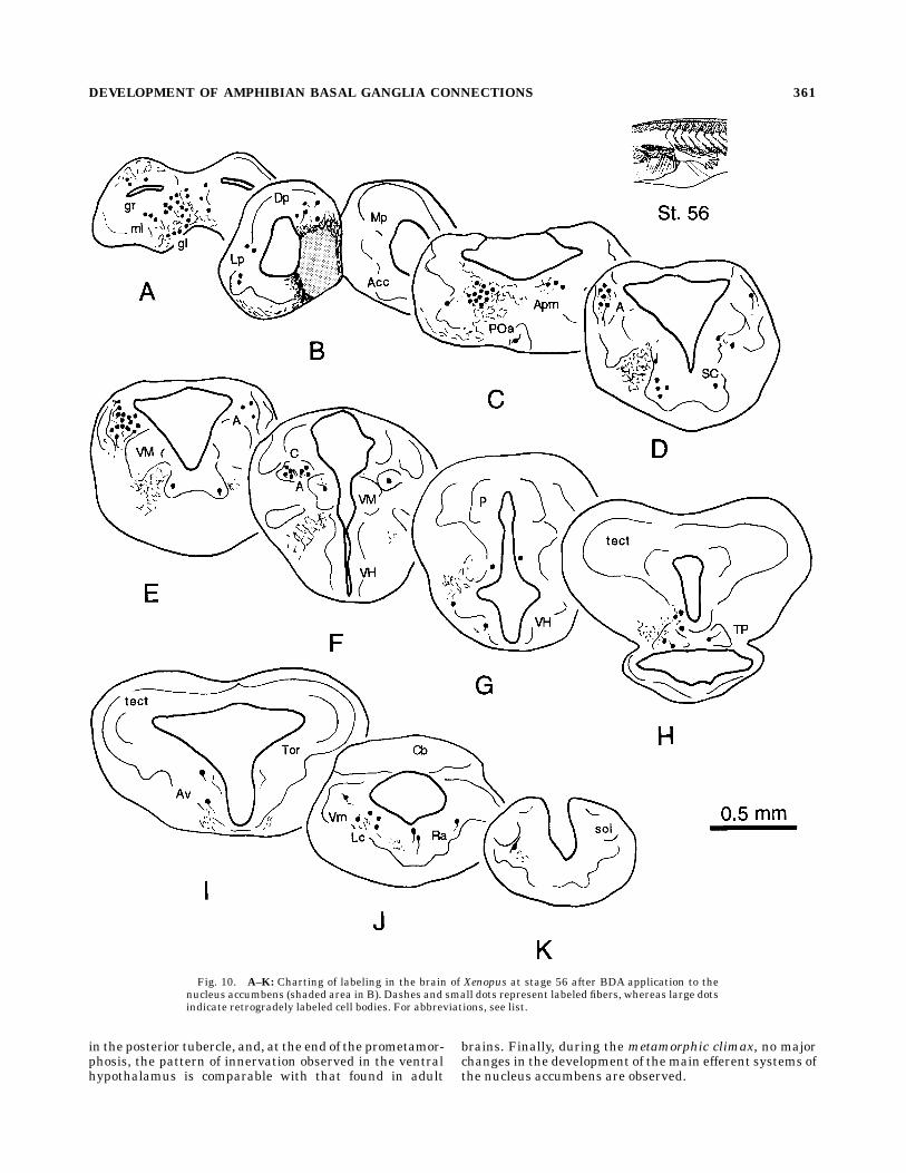
in the posterior tubercle, and, at the end of the prometamor-phosis, the pattern of innervation observed in the ventralhypothalamus is comparable with that found in adult
brains. Finally, during the metamorphic climax, no majorchanges in the development of themain efferent systems ofthe nucleus accumbens are observed.
Fig. 10. A–K: Charting of labeling in the brain of Xenopus at stage 56 after BDA application to thenucleus accumbens (shaded area in B). Dashes and small dots represent labeled fibers, whereas large dotsindicate retrogradely labeled cell bodies. For abbreviations, see list.
DEVELOPMENT OF AMPHIBIAN BASAL GANGLIA CONNECTIONS 361

Fig. 11. A–L: Charting of labeling in the brain of Xenopus at stage 59 after BDA application to thenucleus accumbens (shaded area in B). Dashes and small dots represent labeled fibers, whereas large dotsindicate retrogradely labeled cell bodies. For abbreviations, see list.
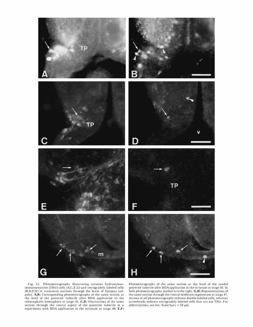
Fig. 12. Photomicrographs illustrating tyrosine hydroxylase-immunoreactive (THir) cells (A,C,E,G) and retrogradely labeled cells(B,D,F,H) in transverse sections through the brain of Xenopus tad-poles. A,B: Corresponding photomicrographs of the same section atthe level of the posterior tubercle after BDA application to thetelencephalic hemisphere at stage 45. C,D: Illustrations of the samesection through the rostral aspect of the posterior tubercle in aexperiment with BDA application in the striatum at stage 48. E,F:
Photomicrographs of the same section at the level of the caudalposterior tubercle after BDAapplication in the striatum at stage 56. Inboth photomicrographs, medial is to the right.G,H:Representations ofthe same section through the rostral midbrain tegmentum at stage 47.Arrows in all photomicrographs indicate double-labeled cells, whereasarrowheads indicate retrogradely labeled cells that are not THir. Forabbreviations, see list. Scale bars 5 50 µm.

Fig. 13. Photomicrographs illustrating THir cells (A,C,E) andretrogradely labeled cells (B,D,F) in transverse sections through thebrain of Xenopus tadpoles after BDA applications into the nucleusaccumbens.A,B:Corresponding photomicrographs of the same sectionthrough the rostral aspect of the posterior tubercle at stage 48. C,D:The same section at the level of the locus coeruleus at stage 48. E,F:
Illustrations of the same section through the midbrain tegmentum atstage 57. In both photomicrographs, medial is to the right. Arrows inall photomicrographs indicate double-labeled cells, whereas arrow-heads indicate retrogradely labeled cells that are not THir. Forabbreviations, see list. Scale bar 5 50 µm.

Fig. 14. A–E: Schematic drawings of transverse sections through the brain of Xenopus tadpoles atlevels of the pretectal region. The progressive innervation of the pretectal areas from the striatum isillustrated for stages 51, 56, and 59. Note that B and D are rostral to C and E, respectively. Forabbreviations, see list.
Fig. 15. A–G: Schematic drawings of transverse sections through the brain of Xenopus tadpolesillustrating the progressive innervation of the mesencephalic and isthmic tegmental areas from thestriatum at stages 51, 56, and 59. Note that B is rostral to C, and D–G are ordered from rostral to caudal.For abbreviations, see list.
DEVELOPMENT OF AMPHIBIAN BASAL GANGLIA CONNECTIONS 365

DISCUSSION
In the present study, the developing connections of thebasal ganglia have been investigated in amphibians. Itshould be noted that the afferent connections have beenrevealed by means of retrograde tracing, and, in somecases, minor spread of the tracers into bordering areascould not be avoided. In those cases, the verification ofafferent striatal and/or nucleus accumbens projectionsfrom brainstem regions awaits confirmation by means ofanterograde tracing.
General features of the developmentof striatal and accumbalconnections in Xenopus
From the present account, it is clear that, already at thebeginning of the larval period, distinct sets of projectionscharacterize the ventrolateral and ventromedial regions ofthe hemispheres. In that perspective, the present studyprovides further evidence for an anatomical and functionalsubdivision of the amphibian basal telencephalon into astriatum (caudatoputamen in mammals) and a nucleusaccumbens, as proposed previously (Gonzalez and Smeets,1991; Marın et al., 1997a–c).The present study demonstrates that the inputs to the
striatum and the nucleus accumbens develop before theend of the embryonic period and show an almost adultappearance at late prometamorphic stages. Moreover, theingrowth of afferent fibers into the striatum slightlyprecedes that of the afferent connections to the nucleusaccumbens, which is in agreementwith the overall develop-ment of the hemispheres (Clairambault, 1976). Accord-ingly, the first catecholaminergic fibers innervating thetelencephalon are found in caudal and lateral regions(amygdala and striatum) followed by rostral and medialterritories (nucleus accumbens and septum; Gonzalez etal., 1994a,b; present study).Compared with the afferent connections, the main effer-
ent connections of the striatum and the nucleus accum-bens developmuch later. Their development starts at earlyprometamorphic stages, and the adult-like pattern ofefferents is recognized only at metamorphic climax stages.However, as in the case of the afferent connections, theprojections of the striatum develop slightly earlier thanthose of the nucleus accumbens.
Temporal correlation between thedevelopment of striatal and accumbal
connections and other developmental eventsin the central nervous system of anurans
Diencephalic afferents. The majority of the earlydeveloping inputs to the basal telencephalon of Xenopus,as shown by the present study, arises from the diencepha-lon. Previous studies by Clairambault (1976) and Tay andStraznicky (1982) have shown that the development of theanuran diencephalon starts in its caudoventrolateral por-tion and then proceeds in a rostrodorsomedial direction.Accordingly, the cells of the posterior tubercle region aregenerated early during development, from stages 20 on-ward (Tay and Straznicky, 1982). Therefore, it was notsurprising that a projection from the posterior tubercleregion to the basal telencephalon could already be recog-nized before the end of the embryonic period (stages44–45) in the present study. Because cells within theposterior tubercle become immunoreactive for TH and
dopamine as early as stage 39, and the basal telencephaloncontains dopaminergic fibers already at stage 41 (Gonzalezet al., 1994a,b), it seems likely that the first dopaminergicprojections from the posterior tubercle reach the telen-cephalon at that early stage.Another remarkable finding of the present study is the
early development of thalamic projections to the basaltelencephalon. A study on the development of somatosen-sory systems of Xenopus (Munoz et al., 1993) revealed thatprojections from the dorsal root ganglion cells to the dorsalcolumn nucleus (DCN) develop slightly earlier than thoseof the DCN to the diencephalon, which can be recognizedat stage 51. The projection from the DCN to the thalamusreaches primarily the ventral thalamus, although somefibers terminate in dorsal thalamic regions (Neary andWilczynski, 1977; Munoz et al., 1995). The present studyprovides evidence that a projection from the ventral thala-mus to the striatum is present at stages 50–51, approxi-mately at the same time when fibers from the DCN reachthe diencephalon (Munoz et al., 1993). On the other hand,the present study also reveals that projections from thedorsal thalamus to the striatum are established appar-ently before the first fibers originating from the DCNarrive at the mesodiencephalic border.The suprachiasmatic nucleus constitutes another source
of early developing inputs to the basal telencephalon. Cellslocated in the lateral aspect of the nucleus are generatedearly during the development of the diencephalon accord-ing to Tay and Straznicky (1982), which is in agreementwith the results of the present study. In addition, thepresent study has demonstrated that the telencephalicprojections arising from cells in the rostral part of thesuprachiasmatic nucleus develop remarkably earlier thanthose originating from its caudal part, thus supporting thenotion that the anuran suprachiasmatic nucleus may becomprised of at least two distinct components (Puelles etal., 1996).Brainstem afferents. At late embryonic stages, two
major sources of inputs to the basal forebrain are noted,i.e., the raphe nucleus and the parabrachial nucleus. Theearly appearance of projections from the raphe nucleus tothe basal telencephalon, as in the case of dopamine,suggests that serotonin plays an important role in thedevelopment of the striatum. On the basis of Nissl-stainedmaterial, only one raphe nucleus could be recognized inanurans (Nikundiwe and Nieuwenhuys, 1983). However,an immunohistochemical study of the development of theserotoninergic system in Xenopus by van Mier et al. (1986)revealed the existence of two nuclei, i.e., a nucleus raphessuperior extending between the level of the octaval cranialnerve and the midbrain and a nucleus raphes inferiorlocated between the level of the octaval cranial nerve andthe obex. The serotoninergic input to the spinal cordoriginates primarily from the inferior raphe nucleus,whereas the superior raphe nucleus gives rise to predomi-nantly ascending projections (van Mier et al., 1986; Tanand Miletic, 1990). The early appearance of telencephalicprojection neurons within the superior raphe nucleus hasbeen established by the results of the present study.However, the study by vanMier et al. (1986) indicates thatthese projectionsmay existmuch earlier, because serotonin-ergic ascending projections were already observed at stage32, reaching the basal telencephalon at stages 35–36 (seeFig. 1 in van Mier et al., 1986).
366 O. MARIN ET AL.

Apart from the raphe nucleus, the parabrachial nucleusis among the first brainstem cell populations innervatingthe basal telencephalon. In adult amphibians, the projec-tions from the parabrachial nucleus terminate primarilyin the caudal division of the striatum (Marın et al., 1997a),which, on the basis of its connections, has been comparedwith the mammalian central nucleus of the amygdala(Bruce and Neary, 1995; Marın et al., 1997a,b). Theparabrachial nucleus also projects to the caudal aspect ofthe ventromedial telencephalic wall, but it projects onlysparsely to the nucleus accumbens proper. The observedmassive input from the parabrachial nucleus into thebasal telencephalon during the embryonic period is in linewith the development of the telencephalic hemispheres,which follows a caudorostral gradient (Clairambault, 1976).The high numbers of retrogradely labeled cells at lateembryonic stages may be explained by the involvement inthe injection site of relatively large portions of the caudalstriatum and the amygdala. The size and differentiation ofthe telencephalon increase as the larval period proceeds.This enables more restricted applications of tracers toeither the striatum or the nucleus accumbens, which, inturn, lead to lower numbers of labeled cells within theparabrachial nucleus.Efferent connections. Striatal projections to the pre-
tectum as well as to the mesencephalic and isthmicreticular formation are considered to have an indirect,modulatory effect on the optic tectum of anurans (Wilczyn-ski and Northcutt, 1983; Marın et al., 1997b), which playsa key role in the control of visually triggered orientingmovements (for review, see Ewert, 1987). The presentstudy has demonstrated that the development of striatalprojections to the mesencephalic and isthmic reticularformation follows a rostrocaudal gradient, in parallel tothe general development of the brainstem (Straznicky andGaze, 1972; Lewis and Straznicky, 1979). Striatal fibersreach the mesencephalic tegmentum during the premeta-morphic period, but the adult-like pattern is not reacheduntil the end of the prometamorphosis. Striatal projectionsto the pretectum develop slightly later than striatotegmen-tal projections, reaching a high degree of organization alsoat the end of the prometamorphic period. Remarkably,Debski and Constantine-Paton (1993) found that the pro-jection from the mesencephalic and isthmic tegmentum tothe optic tectum of Rana pipiens precedes the formation ofpretectotectal connections, but both afferent connectionsdevelop during the premetamorphic period, apparentlyprior to the arrival of the striatal fibers to the brainstemtegmentum and pretectum (present study). Taken to-gether, the available data suggest that the neuronalcircuitry underlying striatal modulation of visuomotorbehavior is already established at the end of the prometa-morphosis. Accordingly, behavioral tests have demon-strated that, at the end of the metamorphosis, the neuro-nal mechanisms underlying visually oriented behavior inanurans are functional (Ewert, 1987).
Comparative aspects of the developmentof the catecholaminergic innervation
of the basal forebrain
In contrast to data from adult brains, results fromstudies of developmental aspects of striatal connections invertebrates are scarce. Most of these studies have beencarried out in neonatal animals that already possess fairlymature striatal connections and, thus, do not provide
substantial information about the ontogeny of these path-ways (Morris et al., 1977, 1979; Jacobowitz et al., 1980).However, developmental studies in opossums and rats,animals that are born with rather immature striatalconnections, suggest that projections from the thalamus,midbrain dopaminergic groups, and raphe are among thefirst to reach the striatal anlage (Martin et al., 1989;Iniguez et al., 1990), similar to what occurs in amphibians(present study). On the other hand, the development of thedopaminergic projections to the striatum of amniotes hasbeen studied immunohistochemically in detail (Specht etal., 1981; Voorn et al., 1988; Medina et al., 1994a,b),enabling a comparison with the development of suchpathways in amphibians.In adult amphibians, the dopaminergic cells in the
posterior tubercle and the adjacent medial tegmentalregion constitute a continuous field along the rostrocaudalaxis, extending from the retromammillary region (afterPuelles et al., 1996) to the level of the exit of the oculomo-tor nerve (Gonzalez and Smeets, 1991, 1994). The genera-tion of these dopaminergic cells starts in the lateral aspectof the posterior tubercle region at the end of the embryonicperiod and continues in caudomedial direction by rapidlyincreasing the number of cells (Gonzalez et al., 1994a,b).Thus, at the beginning of the prometamorphic period, theorganization of the dopaminergic cells in the caudal dien-cephalon and mesencephalon largely resembles that ob-served in adult amphibians. This continuum of cells formsa V shape along the rostrocaudal axis, with the tip of the Vpointing caudallywithin themesencephalicmedial tegmen-tal region (Gonzalez and Smeets, 1994; Gonzalez et al.,1994a).Comparison of the dopaminergic cell populations in
adult amphibians with those of amniotes has commonlyled to the conclusion that important differences exist inbasal ganglia organization.Moreover, because the dopamin-ergic cell groups in the ventral tegmental area (A10), thesubstantia nigra (A9), and the retrorubral area (A8) havebeen classically considered to originate within the ventralmesencephalon of mammals (Specht et al., 1981), reptiles,and birds (Parent et al., 1986), fundamental differencesbetween amphibians and amniotes appear to exist alreadyduring the development of the midbrain dopaminergic cellgroups. However, recent reviews by Puelles and Medina(1994) and Smeets and Reiner (1994) have shown that adifferent conclusion is reached when a segmental approachis applied to the localization of the catecholaminergic cellgroups in the brains of vertebrates. Comparison of thedopaminergic cell groups located in the diencephalic andmesencephalic basal plate suggests close topographic simi-larities among tetrapods (Medina et al., 1994a,b; Puellesand Medina, 1994; Marın et al., 1997c). In that perspec-tive, the A8–A10 complex of amniotes stretches acrossseveral segments and is constituted by distinct dience-phalic (retromammillary, posterior tubercle, and basalsynencephalic), mesencephalic, and isthmic cell groups(Medina et al., 1994a,b; Puelles and Medina, 1994). Simi-larly, the dopaminergic cell field corresponding to the A10complex is almost completely present in amphibians,although they seem to lack the isthmic portion of the A10complex and a laterally migrated substantia nigra andretrorubral area (Marın et al., 1997c).Misunderstanding of the boundary between the dien-
cephalon and the mesencephalon has also contributed tothe confusion about the site of origin ofA8–A10 dopaminer-
DEVELOPMENT OF AMPHIBIAN BASAL GANGLIA CONNECTIONS 367

gic cell groups during development. Thus, as it wasrecently established by Puelles and Medina (1994) in theirelegant study of the segmental development of catechol-aminergic neurons in chicks, there is evidence that thedevelopment of the dopaminergic cells in the diencephalicand mesencephalic basal plate is roughly similar in tetra-pods. Reassessment of the actual limits of themesencepha-lon during development suggests that a precocity of thedopaminergic cell groups in the diencephalic prosomeres,compared with those in the mesencephalon, is a commonfeature of amphibians, birds, and mammals (Voorn et al.,1988; Gonzalez et al., 1994a,b; Puelles and Medina, 1994).Furthermore, the expression of the TH gene starts rostralto the cephalic flexure and extends afterward into thecaudal mesencephalon (Burgunder and Young, 1990).The present study has provided further evidence for the
existence of a common pattern of the development of thedopaminergic innervation of the basal forebrain amongtetrapods. First, as in the rat (Voorn et al., 1988), the firstcells projecting to the prosencephalon are located in therostrolateral portions of the future A9–A10 complex. Sec-ond, early generated dopaminergic cells appear to migrateto their final position after their axons have invaded thetarget areas in both amphibians and amniotes (Tennysonet al., 1973; Voorn et al., 1988; Medina et al., 1994a,b;Marın et al., 1997c). Third, in all tetrapods studied, thedopaminergic innervation of the dorsal striatum precedesthat of the nucleus accumbens (Voorn et al., 1988; Gonza-lez et al., 1994a,b; Medina et al., 1994a,b). Fourth, thearrival of dopaminergic fibers in the striatum precedes theformation of long, descending projections arising from thestriatum (Fishell and van der Kooy, 1987; present study).Finally, at themetamorphic climax, the organization of theamphibian dopaminergic cell groups strikingly resemblesthat of such groups at the beginning of the last week in therat embryonic development. These groups, as in amphib-ians, form a V shape along the rostrocaudal axis (see Fig.38 in Voorn et al., 1988). During the last week of embryonicdevelopment, the rat dopaminergic cell groups expandcaudally and laterally to occupy the entire lateral zone ofthe substantia nigra (Voorn et al., 1988). Taken together,all of these data strengthen the notion that the amphibianequivalent of theA9–A10 groups of amniotes represents anearly stage in the evolution of these groups, which, mostprobably, have developed further by increasing their num-ber of neurons and expanding caudally and laterally(Marın et al., 1997c).
Functional considerations
The present study has shown that the early develop-ment of catecholaminergic projections to the basal telen-cephalon appears to be a common feature not only foramniotes but probably for all tetrapod vertebrates. Numer-ous studies have suggested that dopamine signaling couldplay a role in establishing synaptic connections (Lankfordet al., 1987, 1988; Rodrigues and Dowling, 1990; Lauder,1993). It has also been suggested that the arrival ofdopaminergic fibers in the striatum may influence thematuration of the striatal neurons (Tennyson et al., 1973;Specht et al., 1981; Voorn et al., 1988; Medina et al.,1994a,b). Nevertheless, depletion of dopamine does notproduce substantial cell death of striatal neurons and doesnot prevent the fundamental organization of the striatum(Lanca et al., 1986; Zhou and Palmiter, 1995). On the otherhand, an intact connection between the developing substan-
tia nigra and the striatum is crucial for the normalexpression of substance P, dynorphin, and opiate receptorsin the striatum (van der Kooy and Fishell, 1992; Zhou andPalmiter, 1995). Although dopamine appears to be criti-cally involved in the expression of some striatal markers, anumber of experiments have suggested that another factorreleased by dopaminergic neurons may be implicated inthe induction of some striatal markers (Moon, 1984; vander Kooy, 1984). Furthermore, the early appearance ofserotoninergic cells in the superior raphe nucleus andtheir projections to the basal forebrain indicate thatserotonin may also play a key role in the developingstriatum (Whitaker-Azmitia et al., 1995). In that perspec-tive, anurans like the clawed toad, Xenopus laevis, offer aunique opportunity to study ontogenic aspects of neuronalconnections, because their embryonic development occursover a prolonged period of time, during which the animal isaccessible for experimental studies. Experimental lesionson the developing mesostriatal system in amphibianswould certainly contribute to understanding the role ofthis pathway in the development of striatal function invertebrates.
ACKNOWLEDGMENTS
We are grateful to Mr. D. de Jong for preparing thephotomicrographs.
LITERATURE CITED
Bruce, L.L., and T.J. Neary (1995) The limbic system of tetrapods: Acomparative analysis of cortical and amygdalar populations. BrainBehav. Evol. 46:224–234.
Burgunder, J.-M., andW.S. Young, III (1990) Ontogeny of tyrosine hydroxy-lase and cholecystokinin gene expression in the rat mesencephalon.Dev. Brain Res. 52:85–93.
Clairambault, P. (1976) Development of the prosencephalon. In R. Llinasand W. Precht (eds): Frog Neurobiology. Heidelberg: Springer-Verlag,pp. 924–944.
Debski, E.A., andM. Constantine-Paton (1993) The development of nonreti-nal afferent projections to the frog optic tectum and the substance Pimmunoreactivity of tectal connections. Dev. Brain Res. 72:21–29.
Ewert, J.-P. (1987) Neuroethology of releasing mechanisms: Prey-cachingin toads. Behav. Brain Sci. 10:337–405.
Fishell, G., and D. van der Kooy (1987) Pattern formation in the striatum:Developmental changes in the distribution of striatonigral neurons. J.Neurosci. 7:1969–1978.
Fritzsch, B. (1993) Fast axonal diffusion of 3000 molecular weight dextranamines. J. Neurosci. Methods 50:95–103.
Gonzalez, A., and W.J.A.J. Smeets (1991) A comparative analysis ofdopamine-immunoreactivity in the brain of two amphibians, the an-uran Rana ridibunda and the urodele Pleurodeles waltlii. J. Comp.Neurol. 303:457–477.
Gonzalez, A., and W.J.A.J. Smeets (1994) Catecholamine systems in theCNS of amphibians. In W.J.A.J. Smeets and A. Reiner (eds): Phylogenyand Development of Catecholamine Systems in the CNS of Vertebrates.Cambridge: Cambridge University Press, pp. 77–102.
Gonzalez, A., O. Marın, R. Tuinhof, and W.J.A.J. Smeets (1994a) Ontogenyof catecholamine systems in the CNS of anuran amphibians. Animmunohistochemical study with antibodies against tyrosine hydroxy-lase and dopamine. J. Comp. Neurol. 346:63–79.
Gonzalez, A., O. Marın, R. Tuinhof, and W.J.A.J. Smeets (1994b) Develop-mental aspects of catecholamine systems in the brain of amphibians. InW.J.A.J. Smeets and A. Reiner (eds): Phylogeny and Development ofCatecholamine Systems in the CNS of Vertebrates. Cambridge: Cam-bridge University Press, pp. 343–360.
Gonzalez, A., O. Marın, and W.J.A.J. Smeets (1995) Development ofcatecholamine systems in the central nervous system of the newtPleurodeles waltlii as revealed by tyrosine hydroxylase immunohisto-chemistry. J. Comp. Neurol. 360:33–48.
368 O. MARIN ET AL.
Iniguez, C., J. De Juan, A. Al-Majdalawi, and M.J. Gayoso (1990) Postnataldevelopment of striatal connections in the rat: A transport study withwheat germ agglutinin-horseradish peroxidase. Dev. Brain. Res. 57:43–53.
Jacobowitz, D.M., M. Kato, R.R. O’Neill, S.G. Speciale, A. Gottesfeld, andW.F. Veness (1980) Discrete regional analysis of norepinephrine, dopa-mine, choline, acetyltransferase and glutamic acid decarboxylase in thebrain of the newborn and pubescent monkey. Brain Res. Bull. 5:461–468.
Lanca, A.J., S. Boyd, B. Kolb, and D. van der Kooy (1986) The developmentof a patchy organization of the rat striatum. Dev. Brain Res. 27:1–10.
Lankford, K., F.G. DeMello, and W.L. Klein (1987) A transient embryonicdopamine receptor inhibits growth conemotility and neurite out growthin a subset of avian retina neurons. Neurosci. Lett. 75:169–174.
Lankford, K., F.G. DeMello, and W.L. Klein (1988) D1-type dopaminereceptors inhibit growth cone motility in cultured retina neurons:Evidence that neurotransmitters act as morphogenic growth regulatorsin the developing central nervous system. Proc. Natl. Acad. Sci. USA85:4567–4571.
Lauder, J.M. (1993) Neurotransmitters as growth regulatory signals: Roleof receptors and second messengers. TINS 16:233–240.
Lewis, S., and C. Straznicky (1979) The time of origin of the mesencephalictrigeminal neurons in Xenopus. J. Comp. Neurol. 183:633–646.
Luksch, H., W. Walkowiak, A. Munoz, and H.J. ten Donkelaar (1996) Theuse of in vitro preparations of the isolated amphibian central nervoussystem in neuroanatomy and electrophysiology. J. Neurosci. Methods70:91–102.
Marın, O., A. Gonzalez, and W.J.A.J. Smeets (1995) Evidence for amesolimbic pathway in anuran amphibians. A combined tract-tracing/immunohistochemical study. Neurosci. Lett. 190:183–186.
Marın, O., W.J.A.J. Smeets, and A. Gonzalez (1996) Do amphibians have atrue locus coeruleus? Neuroreport 7:1447–1451.
Marın, O., A. Gonzalez, and W.J.A.J. Smeets (1997a) Basal gangliaorganization in amphibians: Afferent connections to the striatum andthe nucleus accumbens. J. Comp. Neurol. 378:16–49.
Marın, O., A. Gonzalez, and W.J.A.J. Smeets (1997b) Basal gangliaorganization in amphibians: Efferent connections of the striatum andthe nucleus accumbens. J. Comp. Neurol. 380:23–50.
Marın, O., W.J.A.J. Smeets, and A. Gonzalez (1997c) Basal ganglia organi-zation in amphibians: Catecholaminergic innervation of the striatumand the nucleus accumbens. J. Comp. Neurol. 378:50–69.
Martin, G.F., R.H. Ho, and J.C. Hazlett (1989) The early development ofmajor projections to the dorsal striatum in the North Americanopossum. Dev. Brain Res. 47:161–170.
Medina, L., L. Puelles, and W.J.A.J. Smeets (1994a) Development ofcatecholamine systems in the brain of the lizard Gallotia galloti. J.Comp. Neurol. 350:41–62
Medina, L., L. Puelles, and W.J.A.J. Smeets (1994b) Ontogenesis ofcatecholamine systems in the brain of the lizard Gallotia galloti. InW.J.A.J. Smeets and A. Reiner (eds): Phylogeny and Development ofCatecholamine Systems in the CNS of Vertebrates. Cambridge: Cam-bridge University Press, pp. 361–379.
Moon, S.L. (1984) Prenatal haloperidol alters striatal dopamine and opiatereceptors. Brain Res. 323:109–113.
Morris, R., D.R.G. Fuller, C.D. Hull, and N.A. Buchwald (1977) Develop-ment of caudate neuronal response to stimulation of the midbrain,thalamus, and cortex in the kitten. Exp. Neurol. 57:121–131.
Morris, R., M.S. Levine, E. Cherubini, N.A. Buchwald, and C.D. Hull (1979)Intracellular analysis of the development of responses of caudateneurons to stimulation of the cortex, thalamus and substantia nigra inthe kitten. Brain Res. 173:471–487.
Munoz, A., R. de Boer van Huizen, I. Bergervoet-Vernooy, and H.J. tenDonkelaar (1993) Early development of dorsal column-medial lemniscalprojections in the clawed toad, Xenopus laevis. Dev. Brain Res. 74:291–294.
Munoz, A., M. Munoz, A. Gonzalez, and H.J. ten Donkelaar (1995) Anurandorsal column nucleus: Organization, immunohistochemical character-ization and fiber connections in Rana perezi and Xenopus laevis. J.Comp. Neurol. 363:197–220.
Munoz,A., M. Munoz,A. Gonzalez, and H.J. ten Donkelaar (1996) Evidencefor an anuran homologue of the mammalian spinocervicothalamic
system: An in vitro tract-tracing study in Xenopus laevis. Eur. J.Neurosci. 8:1390–1400.
Neary, T.J., and W. Wilczynski (1977) Ascending thalamic projections fromthe obex region in ranid frogs. Brain Res. 138:529–533.
Nieuwkoop, P.D., and J. Faber (1967) Normal table of Xenopus laevis(Daudin). Amsterdam: North-Holland.
Nikundiwe, A.M., and R. Nieuwenhuys (1983) The cell masses in thebrainstem of the SouthAfrican clawed frogXenopus laevis:A topographi-cal and topological analysis. J. Comp. Neurol. 213:199–219.
Olson, L., and A. Seiger (1972) Early prenatal ontogeny of central mono-amine neurons in the rat: Fluorescence histochemical observations. Z.Anat. Entwickl. Gesch. 137:301–316.
Parent, A. (1986) Comparative Neurobiology of the Basal Ganglia. NewYork: JohnWiley and Sons, Inc.
Puelles, L., and L. Medina (1994) Development of neurons expressingtyrosine hydroxylase and dopamine in the chicken brain:A comparativesegmental analysis. In W.J.A.J. Smeets and A. Reiner (eds): Phylogenyand Development of Catecholamine Systems in the CNS of Vertebrates.Cambridge: Cambridge University Press, pp. 381–404.
Puelles, L., F.J. Milan, andM.Martınez-de la Torre (1996)Asegmental mapof subdivisions in the diencephalon of the frogRana perezi: Acetylcholin-esterase-histochemical observations. Brain Behav. Evol. 47:279–310.
Rodrigues, P.S., and J.E. Dowling (1990) Dopamine induces neurite retrac-tion in retinal horizontal cells via diacylglycerol and protein kinase C.Proc. Natl. Acad. Sci. USA 87:9693–9697.
Smeets, W.J.A.J., and A. Reiner (1994) Catecholamines in the CNS ofvertebrates: Current concepts of evolution and functional significance.In W.J.A.J. Smeets and A. Reiner (eds): Phylogeny and Development ofCatecholamine Systems in the CNS of Vertebrates. Cambridge: Cam-bridge University Press, pp. 463–481.
Specht, L.A., V.M. Pickel, T.H. Joh, and D.J. Reis (1981) Light-microscopicimmunocytochemical localization of tyrosine hydroxylase in prenatalrat brain. J. Comp. Neurol. 199:233–253.
Straka, H., and N. Dieringer (1993) Electrophysiological and pharmacologi-cal characterization of vestibular inputs to identified frog abducensmotoneurons and internuclear neurons in vitro. Eur. J. Neurosci.5:251–260.
Straznicky, K., and R.M. Gaze (1972) The development of the tectum inXenopus laevis: An autoradiographic study. J. Embryol. Exp. Morphol.28:87–115.
Tan, H., and V. Miletic (1990) Bulbospinal serotoninergic pathways in thefrog Rana pipiens. J. Comp. Neurol. 292:291–302.
Tay, D., and C. Straznicky (1982) The development of the diencephalon inXenopus. An autoradiographic study. Anat. Embryol. 163:371–388.
Tennyson, V.M., C. Mytilineou, and R.E. Barrett (1973) Fluorescence andelectron microscopic studies of the early development of the substantianigra and area ventralis tegmenti in the fetal rabbit. J. Comp. Neurol149:233–258.
van der Kooy, D. (1984) Developmental relationships between opiatereceptors and dopamine in the formation of caudate-putamen patches.Dev. Brain Res. 14:300–303.
van der Kooy, D., and G. Fishell (1992) Embryonic lesions of the substantianigra prevent the patchy expression of opiate receptors, but not thesegregation of patch and matrix compartment neurons, in the develop-ing rat striatum. Dev. Brain Res. 66:141–145.
van Mier, P., H.W.J. Joosten, R. van Rheden, and H.J. ten Donkelaar (1986)The development of serotoninergic raphe spinal projections in Xenopuslaevis. Int. J. Dev. Neurosci. 4:465–475.
Voorn, P., A. Kalsbeek, B. Jorritsma-Byham, and H.J. Groenewegen (1988)The pre-and postnatal development of the dopaminergic cell groups inthe ventral mesencephalon and the dopaminergic innervation of thestriatum of the rat. Neuroscience 25:857–887.
Whitaker-Azmitia, P.M., M. Druse, P. Walker, and J.M. Lauder (1995)Serotonin as a developmental signal. Behav. Brain Res. 73:19–29.
Wilczynski, W., and R.G. Northcutt (1983) Connections of the bullfrogstriatum: Efferent projections. J. Comp. Neurol. 214:333–343.
Zhou, Q.-Y., and R.D. Palmiter (1995) Dopamine-deficient mice are severelyhypoactive, adipsic and aphagic. Cell 83:1197–1209.
DEVELOPMENT OF AMPHIBIAN BASAL GANGLIA CONNECTIONS 369