Embed Size (px)
Citation preview
2013 Research Journal of Bangladesh Phytopathological Society
ISSN 2412-7558 (Online)
ISSN 1012-9279 (Print)
Bangladesh Journalof
Plant PathologyVolume 29, No. 1&2, 2013
CONTENT
1 Chemical control of bacterial soft rot of onioncaused by Burkholderia cepacia
M. M. Rahman, A. A. Khan, A. M. Akanda,I. H. Mian and M. Z. Alam
5 First record on Botrytis Blight (Botrytisgladiolorum) of gladiolus from Bangladesh
S. S. Siddique, A. U. Ahmed, M. S. Begum,M. M. Islam and I. H. Mian
11 Pestalotiopsis guepinii (desm.) stay. – a pathogenof black spot disease of rose in Bangladesh
Shamim Shamsi and Anita Ghosh
15 In-vitro evaluation of antifungal activity of plantextracts against Rhizoctonia oryzae-sativaecausing aggregated sheath spot of rice
S. B. Jahan, M. A. Ali, S. Alam,Z. R. Moni and M. A. Alam
21 Identification of pathotypes ofXanthomonas oryzae pv. oryzae causingbacterial blight of rice in Bangladesh
S. M. K. H. Chowdhury, I. H. Mianand M.A.I. Khan
29 Effect of tuber-borne inoculum of Rhizoctoniasolani on the development of stem canker andblack scurf of potato
M. M. Rahman, M. A. Ali,M. U. Ahmad and T. K. Dey
33 Integrated application of insecticide and fungicideto control OkYVCMV and Pseudocercospora leafspot of okra seed crops
G. Kibria, I. H. Mian, A. M. Akanda and M.K. Alam Bhuiyan
39 Characterization of Orobanche species occurringin Bangladesh
Manisha Shaha and I. H. Mian
2013 Bangladesh Phytopathological Society iii
Bangladesh Journalof
Plant Pathology(Bangladesh J. Plant Pathol.)
Research Journal of Bangladesh Phytopathological Society
Vol. 29 No. 1&2 2013
EDITORIAL BOARD
Dr. Hamizuddin AhmedChief Editor
Prof. Dr. Ismail Hossain MianExecutive Editor
Prof. Dr. M. A. Wadud MianProf. Dr. M. Ashrafuzzaman SalimProf. Dr. M. A. Mannan AkandaProf Dr. M. A. KhairProf. Dr. M. K. Alam BhuiyanDr. M. A. BakrDr. Tapan Kumar Dey
MemberMemberMemberMemberMemberMemberMember
Address for correspondence:Executive EditorBangladesh Journal of Plant PathologyDepartment of Plant PathologyBangabandhu Sheikh Mujibur Rahman AgriculturalUniversity, Gazipur-1706, Bangladesh
Subscription for each volume:
IndividualInstitution
BangladeshTk. 500.00Tk. 1500.00
OverseasUS $ 10US$ 25
ii 2013 Bangladesh Phytopathological Society
Bangladesh Phytopathological Society.
Executive Committee(2011-2013)President: Dr. M. A. Bari, Director (Retd.), Bangladesh Agricultural Research Institute
(BARI), Gazipur, BangladeshSenior Vice President: Professor Dr. Ashrafuzzaman Salim, Department of Plant Pathology, Bangladesh
Agricultural University, Mymensingh, Bangladesh
Vice President: Prof. Dr. Md. Khurshed Alam Bhuiyan, Department of Plant Pathology,Bangabandhu Sheikh Mujibur Rahman Agricultural University, Gazipur -1706
Treasurer: Dr. M. Anser Ali, Chief Scientific Officer, Plant Pathology Division, BangladeshRice Research Institute, Gazipur-1701, Bangladesh
General Secretary Dr. Tapan Kumer Dey, Chief Scientific Officer, Plant Pathology Division, BARIGazipur-1701, Bangladesh
Joint Secretary-1 Prof. Dr. Salahuddin Mahmud Chowdhury, Department of Plant Pathology,Sher-E-Bangla Agricultural University, Dhaka, Bangladesh
Joint Secretary-2 Dr. Sakhawat Hossain, Chief Scientific Officer, Oilseed Research Center, BARI,Gazipur, Bangladesh
Office Secretary Md. Mynul Islam, Senior Scientific Officer, Plant Pathology Division, BARIGazipur, Bangladesh
Members: Prof. Dr. Ismail Hossain Mian, Department of Plant Pathology and Treasurer,BSMRAU, Gazipur-1706, Bangladesh
Prof. Dr. M. A. Mannan Akanda, Department of Plant Pathology and Vice-Chancellor, Exim Bank Agricultural University, Chapainawabganj, Bangladesh
Prof. Dr. Ismail Hossain, Department of Plant Pathology, BAU, Mymensingh,Bangladesh
Dr. Md. Abu Taher Mia, CSO (Ret’d), Plant Pathology Division, BRRI, Gazipur
Dr. Mohammad Hossain, CSO, Tuber Crops Research Center, BARI, Gazipur
Dr. Md. Ibrahim Talukder, CSO, Plant Pathology Division, BangladeshSugarcane Research Institute, Ishurdi, Pabna
Prof. Dr. Aminuddin Mridha, Department of Botany, Chittagong UniversityChittagong
Dr. A. Muqit, SSO, Plant Pathology Division, BARI, Gazipur
Dr. Mostafa Kamal, PSO, Plant Pathology Division, BRRI. Gazipur
Ex-Officio Member Dr. M. A. Wadud Mian, Professor (Ret’d.), Bangladesh Agricultural UniversityMymensingh
Dr. M.A. Bakr, Project Director (Ret’d.), Pulses and Oilseeds, BARI, Gazipur
Distinguished Member Prof. Dr. Golam Ali Fakir, Ex-Vice Chancellor, Khulna University Khulna;Vice-Chancellor, Bangladesh University, Dhaka
Dr. Hamizuddin Ahmed, Director (Ret’d), BARI, Gazipur
2013 Bangladesh Phytopathological Society iii
INFORMATION FOR CONTRIBUTORS
Membership of the Bangladesh Phytopathological Society is a prerequisite for publishing papers in the BangladeshJournal of Plant Pathology (Bangladesh J. Plant Pathol.), or at least one of the authors must be a member of the Society.The Editorial board, however, may relax this rule in case of contribution of exceptionally merit and contributor fromabroad. The journal publishes original research that signify advancement of knowledge in plant diseases, the causalagents, the factors that influence diseases, and the methods which are be used to control them. The journal is published inboth print form (ISSN1012-9279) and online (ISSN 2412-7558). Materials accepted for publication must be scientificallysound and suitable for the designated readers. The manuscript should be prepared according to the guidelines providedbelow.
(1) Subject matters of the papers for publication should fall in the following categories:
a. Full length papers: Original research on any branch of plant pathology;
b. Review papers: Developing a new concept, hypothesis, theory, or other investigation in the field of plantpathology;
c. Phytopathological notes: Accounts of plant pathological techniques, original research or new records of plantdiseases or plant pathogens.
(2) The manuscript should be typed double-space on one side of A4 size good quality paper in 12-point preferably inTimes New Roman font. Margins should be 4.0 cm from the left and right, and 2.5 cm from the top and bottom ofeach page. Submit two copies of the manuscript to the Executive Editor, Bangladesh Journal of Plant Pathology,Plant Pathology Division, Bangladesh Agricultural Research Institute, Gazipur-1701, Bangladesh along with hardcopy, a soft copy of the manuscript in word 97-2003/XP format written in CD should be submitted.
(3) A full length paper should be arranged under the section heading: TITLE, ABSTRACT, INTRODUCTION,MATERIALS AND METHODS, RESULTS AND DISCUSSION, ACKNOWLEDGEMENT (if any) andLITERATURE CITED. Phytopathological notes should be sufficiently brief (less than 4 pages) and arrangedwithout section headings of INTRODUCTION, MATERIALS AND METHODS, and RESULTS ANDDISCUSSION.
a. Title page – Title page should be independent and contain only Title, authors’ name and their address. The titleshould be typed in capital letters and must accurately identify and describe the contents of the manuscript.Scientific name (if any) must be in capital letter and italic. The title should be followed by authors’ names. Theauthors’ names should be followed by author’s addresses. A person who did not take part in research, analysisof data and writing of the paper and who cannot assume responsibility for the technical content of the papercannot be considered an author. Include each author's complete mailing address and institutional affiliation inthe title page. Indicate the author to whom correspondence should be addressed and the author’s e-mail address.Indicate whether the research is the portion of a thesis or dissertation.
b. Abstract - Start abstract from a separate page. Type the title again on this page but no authors’ name andaddress. After the title, type a 150–200 words abstract containing the objectives, very short methodology,important findings of the research and your conclusion.
c. Keywords –The keywords and phrases following the Abstract should be alphabetically arranged and shouldreflect the contents of the paper. Give keywords only in one line.
(4) The author(s) are responsible to check that all references in the text appear in the section ‘LITERATURE CITED’and vice versa. References should come primarily from papers published in professional journals. A small numberof abstracts or text book-type references may be included; however, these should not make up the bulk of thereferences. Personal communication references are permitted, but, again should be kept to a minimum. In textreferences should be cited by giving the author's name followed by the year without any coma in between.
a. For two authors’ paper, give both authors' last names.
i) Ahmed and Hamid (1995) reported the losses due to late blight of potato.
ii) The loss due to late blight of potato has been reported by Ahmed and Hamid (1995).
iii) Loss due to late blight of potato has been well documented (Ahmed and Hamid 1955).
b. Articles with more than two authors are cited by the first authors last name followed "et al.", and then theyear:
i) Effect of environmental factors on disease development is well documented (MacHardy et al. 2001)instead of (MacHardy, W. E., Gadoury and Gessler. C. 2001).

iv 2013 Bangladesh Phytopathological Society
ii) Disease development was recorded by MacHardy et al. (2001). At least 12 diseases of pulses are inBangladesh (Ahmed et al. 1985) or Ahmed et al. (1985) recorded at least 12 diseses of pulses.
c. A string of citations should be separated by coma.
i) Effect of environmental factors on pathogens is recorded by many researchers (Rowe and Beute 1975,Sutton 1981, Pinkerton et el. 1998, MacHardy et al. 2001).
ii) Ahmed and Hamid (1955, 1989), Mian (1985), Fakir (1985) reported losses due to potato late blight.
iii) Loss due to late blight was recorded by Ahmed and Hamid (1955, 1989) and Mian (1985).
d. When a book, paper, or article has no definite author, cite it as Anon. and year, e.g., (Anon. 1996) (Anon. isthe abbreviation for anonymous).
e. If you want to refer a paper found in another article, do so as (Driblick 1983 In: Oobleck 1978).
(5) In the section LITERATURE CITED, references should be listed alphabetically according to the author’s name. Ifthe same author(s) are cited for more than one paper having the same order of authors' names, the papers should belisted in chronological sequence by year of publication. Anon. should be listed as ‘Anonymous’.a. Journal article with single author e.g.- Bisessar, S. 1982. Effect of heavy metals on microorganisms in soil near
secondary lead smelter. Wat. Air Soil Poll. 17:305-308.
b. Journal article with two authors e.g.- Sivan, A. and Chet, I. 1993. Integrated control of Fusarium crown androot rot of tomato with Trichoderma harzianum in combination with methyl bromide or soil solarization. CropProtec. 12:380-386
c. Journal article with multiple authors e.g.- Schneider, R. W. Williams, R. J., and Sinclair, J. B. 1976.Cercospora leaf spot of cowpea, models for estimating yield loss. Phytopathology 66(5): 384-388.
d. Author(s) Unknown or Not Named - If the authorship of a paper or other document is not provided, cite theauthor using the word "Anonymous" in the place of the author’s name(s) e.g. Anonymous. 2004. AnnualInternal Review. Bangladesh Rice Research Institute, Joydebpur, Gazipur. pp. 39-41.
e. Book with two authors – Dhingra, P. and Sinclair, A. 1985. Basics plant pathology methods. CRC press. Inc.Boca Raton Florida. pp.13-44 [(pp. 13-44 page means on 13 to 44), p.13, (p. 13 means on page 13), (250 ppmeans total page of the book)].
f. Book with multiple authors - Huth, J., Brogan, M., Dancik, B., Kommedahl, T., Nadziejka, D., Robinson, P.and W. Swanson.1994. Scientific format and style: The CBE manual for authors, editors, and publishers. 6thed. Cambridge: Cambridge University Press. 825 pp.
g. Book: authors contributing a specific chapter - Kuret, J. and Murad. F. 1990. Adenohypophyseal hormones andrelated substances. In: Gilman, A., Rall, T., Nies, A., Taylor, P., edited. “The pharmacological basis oftherapeutics”. 8th ed. New York: Pergamon. pp. 1334-60.
(6) Table and graphs must be typed and drawn respectively on separate sheet. Same data must not be given in bothtables and graphs. Data based on which a graph is created should be enclosed on separate sheet.
(7) All quantitative data must be in metric units. Abbreviations should be as per "Style Manual for Biological editors".
(8) Picture should be submitted on separate file in JPEG format.
(9) Manuscript not prepared following the style of the journal will not be accepted.(10) For more information contact the Executive Editors (Email:[email protected]) or consult its latest issue.(11) Due to the high cost of publishing, page charges are required. The rates are subject to change without notice. Present
page charges areare shown below:
Paper type Black and white ColourFor Bangladeshi (Tk/article)
a. Full paper having 4 pages 2000.00 2500.00b. Full paper having 5-8 pages 2500.00 3000.00c. Phytopathological Note (4 pages) 1000.00 1500.00
For foreigners ($/article)d. Full paper having 4 pages 20.00 30.00f. Full paper having 5-8 pages 25.00 40.00g. Phytopathological Note (4 pages) 15.00 20.00

Vol. 29, No. 1&2, 2013 1
CHEMICAL CONTROL OF BACTERIAL SOFT ROT OF ONION CAUSEDBY BURKHOLDERIA CEPACIA
M. M. Rahman1, A. A. Khan1, A. M. Akanda1, I. H. Mian1 and M. Z. Alam2
1Department of Plant Pathology, 2Department of Entomology, Bangabadhu Sheikh Mujibur RahmanAgricultural University, Gazipur-1706, Bangladesh Email of first author: [email protected]
(This is a part of Ph.D thesis of first author)
ABSTRACT
M. M. Rahman, A. A. Khan, A. M. Akanda, I. H. Mian and M. Z. Alam. 2013. Chemical control of bacterialsoft rot of onion caused by Burkholderia cepacia. Bangladesh J. Plant Pathol. 29 (1&2): 1-4.
Bactericidal properties of eight chemicals were tested in-vitro against onion soft rot bacteria (Burkholderiacepacia). Among the chemicals, acetic acid, boric acid andbleaching powder showed bactericidal activity againstonion soft rot bacteria, B. cepacia O-15. These threechemicals were tested to treat onion bulbs against soft rotdisease in storage. For treatment, fresh onion bulbs weredipped in 0.00, 0.02, 0.05 and 1.00% solutions of aceticacid, boric acid, and bleaching powder for 30 min. Thetreated bulbs were inoculated by spraying B. cepacia
suspension and stored for 2-22 weeks. During the storageperiod, the chemicals caused 0.0-18.2% reduction in softrot incidence and 0.00-18.0% reduction in weight loss ofonion bulbs over to control. The lowest incidence of softrot as well as weight loss was achieved with bleachingpowder followed by boric acid and acetic acid. Results ofboth in vitro test and onion treatment experiment indicatethat bleaching powder, boric acid and acetic acid may beused to control soft rot of onion bulbs during storage for aperiod of 2-22 weeks.
Key words: Bacterial soft rot, Burkholderia cepacia, onion, chemical control
INTRODUCTION
Bacterial soft rot (Burkholderia cepacia) is acommon post harvest disease of onion and manyother vegetables throughout the world. The diseasecauses severe loss of onion bulb in storage (Bdliyaand Haruna 2007). Normally, chemical bactericidesare not recommended for the control of soft rotdisease because of high risk of health hazards(Agrios 1997). However, many scientists testedvarious bactericides including chemicals andmicrobial pesticides to control the soft rot bacteria(Chen and Lin 2000, Abd-El-Khair 2004, Wright etal. 2005). Researchers identified some chemicalswith antimicrobial activity, which increase resistancein potato and onion against soft rot disease(Hammerschmidt and Smith 1997). Benzothiodiazole(BTH) has been idenfied as a systemic resistanceinducer in many plants and effective against variousplant pathogens (Gorlach et al. 1996, Bokshi et al.2003). Increased resistance in potato tubers againstE. carotovora subsp. carotovora was observed whentubers were dipped in acetyl salicylic acid (Abd-El-Sayed et al. 1996, Bokshi et al. 2003). Salttreatments also can inhibit plant pathogens orsuppress their toxin production (Olivier et al. 1998).Salts including calcium propionate and calciumchloride reduced tissue maceration of potato tuberscaused by E. carotovora (McGuire and Kelman1986, Biggs et al. 1997, Droby et al. 1997).
2013 Bangladesh Phytopathological Society
Suppression of bacterial soft rot in potatotubers by application of an antibiotic ‘kasugamycin’was investigated by Bartz (1999). Reports onchemical control of soft rot bacteria are not availablein Bangladesh. Search for selection of chemicalswithout health hazard to human is necessary tocontrol soft rot of onion.
Considering the above facts the presentinvestigation was conducted to test some chemicalsubstances for their effectiveness to control soft rotcausing bacterial pathogens of onion.
MATERIALS AND METHODS
In vitro evaluation of eight chemicals against softrot bacteria
An in vitro experiment was conducted to evaluateeight chemicals for their bactericidal activity againstsoft rot pathogen, B. cepacia O-15 of onion. Thechemicals were acetic acid, boric acid, bleachingpowder, lactic acid, calcium hydroxide, calciumchloride, potassium chloride and sodiumhypochloride. Acetic acid, boric acid, lactic acid,bleaching powder and sodium hypo-chloride weretested at 0.02, 0.05 and 0.10% (w/w). Otherchemicals were tested at 0.05, 0.10 and 0.20%. Yeastpeptone dextrose agar (YPDA) was used as basalmedium.
The YPDA was prepared following a standardmethod as described by Tuite (1969). After cookingthe medium was amended with appropriate quantityof each chemical to have desired levels of
2 Bangladesh J. Plant Pathol.
concentrations. Each chemical was added to YPDA,mixed thoroughly and autoclaved for 20 min at 121Cunder 1.1 kg/cm2 pressures. YPDA without anychemical amendment served as control. Aftersterilization, the medium was poured into 90 mmglass Petri dishes at 20 ml/plate and allowed tosolidify.
To prepare the inocula, B. cepacia O-15 wasgrown on YPDA at 28C for 24 hr. Bacterial cellswere collected from the culture and suspended insterilized distilled water to a concentration of ca.108
cfu/ml. After solidification, YPDA in the plates wasinoculated with bacterial suspension and incubated at30C in an incubator. The plates were arranged in anincubator following completely randomized designwith three replications. Three additional plateshaving YPDA without any chemical were maintainedas control. Growth of the test bacteria in the plateswas observed up to 14 days of inoculation andantibacterial activity of the chemicals wasdetermined based on initiation of colony growth.
Efficacy of the chemicals to control soft rot
Based on the results of the in vitro test anotherexperiment was conducted to evaluate the efficacy ofacetic acid, boric acid and bleaching powder tocontrol soft rot disease of onion in storage.Apparently healthy bulbs of onion variety‘Taherpuri’ were selected and treated with thechemicals at 0.00, 0.20, 0.05 and 1.00% concen-trations. For each concentration of every chemical,700 g of fresh onion bulbs were treated by dipping insolution of each chemical separately for 30 min andthen air dried.
Fresh cultures of B. cepacia O-15 grown on
YPDA were suspended in sterilized distilled water to
prepare inocula at a concentration of ca.108 cfu/m.
Onion bulb treated with chemicals were inoculated
with the inoculum suspension of the bacteria using
an atomizers and air dried again. The onion bulbs
were packed in net bags and stored at room
temperature for 22 weeks. For control treatment
onion bulbs were treated with plain water, air dried
and inoculated with the pathogen. The bulbs were
checked for soft rot incidence on 2, 6, 10, 14, 18 and
22 weeks after inoculation. Data on soft rot infection
and loss of weight due to soft rot in storage were
recorded and expressed as percentage using the
following formula described by Abd-El-Khair and
Karima (2007):
Number of infected bulbsInfection %= -----------------------------------X100
Total number of bulbs
I-WLoss of weight %= --------------------X100,
IWhere I= Initial weight of bulbs and
W= weight after discarding the infected tubers
Percentage of disease reduction (PDR) wascalculated using the following formula (Hajhamed etal. 2007):
Ack - Atr
PDR= ---------------- X 100,Ack
Where Ack = loss in weight in control bulbs andAtr = loss in weight of treated bulbs.
RESULTS AND DISCUSSION
In vitro evaluation of eight chemicals against softrot bacteria
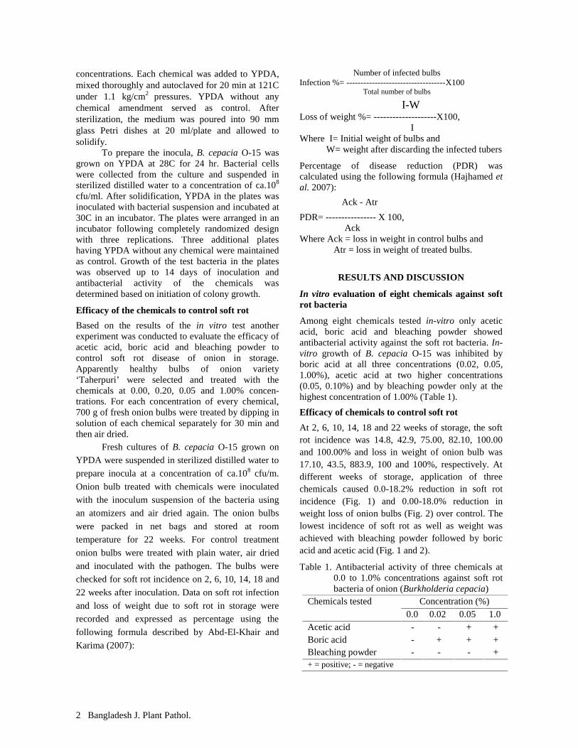
Among eight chemicals tested in-vitro only aceticacid, boric acid and bleaching powder showedantibacterial activity against the soft rot bacteria. In-vitro growth of B. cepacia O-15 was inhibited byboric acid at all three concentrations (0.02, 0.05,1.00%), acetic acid at two higher concentrations(0.05, 0.10%) and by bleaching powder only at thehighest concentration of 1.00% (Table 1).
Efficacy of chemicals to control soft rot
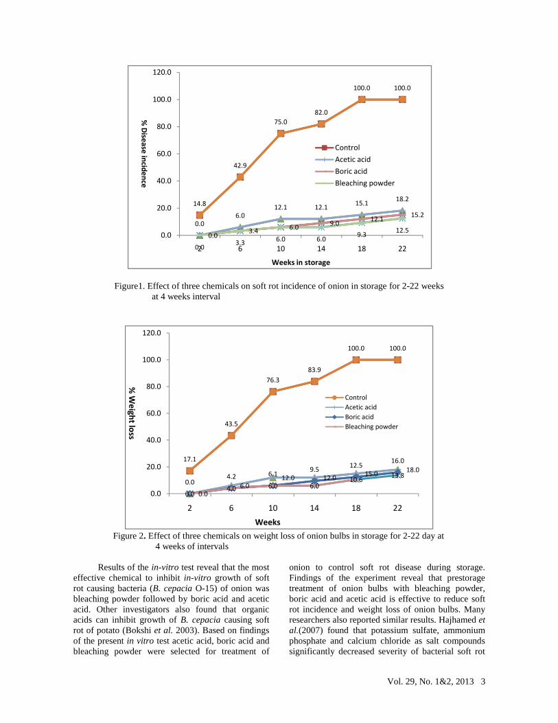
At 2, 6, 10, 14, 18 and 22 weeks of storage, the softrot incidence was 14.8, 42.9, 75.00, 82.10, 100.00and 100.00% and loss in weight of onion bulb was17.10, 43.5, 883.9, 100 and 100%, respectively. Atdifferent weeks of storage, application of threechemicals caused 0.0-18.2% reduction in soft rotincidence (Fig. 1) and 0.00-18.0% reduction inweight loss of onion bulbs (Fig. 2) over control. Thelowest incidence of soft rot as well as weight wasachieved with bleaching powder followed by boricacid and acetic acid (Fig. 1 and 2).
Table 1. Antibacterial activity of three chemicals at0.0 to 1.0% concentrations against soft rotbacteria of onion (Burkholderia cepacia)
Chemicals tested Concentration (%)0.0 0.02 0.05 1.0
Acetic acid - - + +Boric acid - + + +Bleaching powder - - - ++ = positive; - = negative
Vol. 29, No. 1&2, 2013 3
Figure1. Effect of three chemicals on soft rot incidence of onion in storage for 2-22 weeksat 4 weeks interval
Figure 2. Effect of three chemicals on weight loss of onion bulbs in storage for 2-22 day at4 weeks of intervals
Results of the in-vitro test reveal that the mosteffective chemical to inhibit in-vitro growth of softrot causing bacteria (B. cepacia O-15) of onion wasbleaching powder followed by boric acid and aceticacid. Other investigators also found that organicacids can inhibit growth of B. cepacia causing softrot of potato (Bokshi et al. 2003). Based on findingsof the present in vitro test acetic acid, boric acid andbleaching powder were selected for treatment of
onion to control soft rot disease during storage.Findings of the experiment reveal that prestoragetreatment of onion bulbs with bleaching powder,boric acid and acetic acid is effective to reduce softrot incidence and weight loss of onion bulbs. Manyresearchers also reported similar results. Hajhamed etal.(2007) found that potassium sulfate, ammoniumphosphate and calcium chloride as salt compoundssignificantly decreased severity of bacterial soft rot
0.0 3.3 6.0 6.0 9.3 12.5
14.8
42.9
75.082.0
100.0 100.0
0.06.0
12.1 12.1 15.1 18.2
0.0 3.4 6.0 9.0 12.1 15.2
0.0
20.0
40.0
60.0
80.0
100.0
120.0
2 6 10 14 18 22
% Disease incidence
Weeks in storage
ControlAcetic acidBoric acidBleaching powder
17.1
43.5
76.383.9
100.0 100.0
0.06.0
12.0 12.0 15.0 18.0
0.04.2 6.1 9.5 12.5
16.0
0.04.0 6.0 6.0
10.6 13.8
0.0
20.0
40.0
60.0
80.0
100.0
120.0
2 6 10 14 18 22
% W
eight loss
Weeks
ControlAcetic acidBoric acidBleaching powder

4 Bangladesh J. Plant Pathol.
disease of potato. Saleh and Huang (1997) reportedthat benzoic acid and sodium benzoate at 1, 5 and 10mM inhibited soft rot bacterial growth and wereeffective in controlling the disease in both tomatofruits and potato tubers. Salts including calciumpropionate and calcium chloride reduced tissuemaceration of potato tubers due to attack of E.carotovora (Biggs et al. 1997, Droby et al. 1997,Olivier et al. 1998). Findings of the two experimentsindicate that acetic acid, boric acid and bleachingpowder may be recommended to control soft rot ofonion caused by B. cepacia.
LITERATURE CITED
Abd El-Sayed, Wafaa M., Abd El-Ghaffar, N. Y. andShehata, A. M. 1996. Application of salicylicacid and aspirin for induction of resistance totomato plants against bacterial wilt and itseffect on endogenous hormones. Ann. Agric.Sci., Ain Shams Univ., Cairo, 41:1007-1020.
Abd-El-Khair, H. 2004. Efficacy of starner incontrolling the bacterial soft rot in onion. Ann.Agril. Sci., Cairo. 49(2): 721-731.
Abd-El-Khair, H. and Karima, H.E.H. 2007.Application of some bactericides andbioagents for controlling the soft rot disease inpotato. Res. J. Agric. and Bio. Sci., 3(5): 463-473.
Agrios, G. N. 1997. Control of plant diseases. InPlant Pathology, 4th edn. California:Academic Press. pp. 200-216.
Bartz, J. A. 1999. Suppression of bacterial soft rot inpotato tubers by application of kasugamycin.American J. Potato Res. 76(3): 127-136.
Bdliya, B. S. and Haruna, H. U. 2007. Efficacy ofsolar heat in the control of bacterial soft ofpotato tubers caused by Erwinia carotovorasubsp. carotovora. J. Plant Protec. Res.,Nigeria Vol. 47 No. 1.
Biggs, A. R., El-Kholi, M. M., El-Neshawy, S. andNickerson, R. 1997. Effect of calcium salts ongrowth, polygalacturonase activity, andinfection of peach Fruit by Moniliniafructicola. Plant Dis. 81: 399-403.
Bokshi, A. I, Morris, S. C. and Deverall, B. J. 2003.Effects of benzothiadiazole and acetylsalicylicacid on b-1,3-glucanase activity and disease
resistance in potato. J. Plant Pathol. 52: 22–27.
Chen, C. W. and Lin, C. Y. 2000. Control of Erwiniasoft rot disease of Calla lily. Pl. Pathol. Bull.2000. 9(3): 107-114.
Droby, S., Wisniewski, M. E., Cohen, L., Weiss, B.,Touitou, D., Eilam, Y. and Chalutz, E. 1997.Influence of CaCl2 on Penicillium digitatumgrapefruit peel tissue and biocontrol activityof Pichia guilliermondii. Plant Dis.87: 310-315.
Gorlach, J., Sandra, V., Gertrud, K., Georges, H.,Uli, B., Karl-Heinz, K., Oostendrop, M.,Staub, T., Ward, E., Kessmann, H. andRayals, J. 1996. Benzothiadiazole, a novelclass of inducers of systemic acquiredresistance in wheat. Plant cell8: 629-643.
Hajhamed, A. A., Sayed, W. M. A. E., Yazied, A. A.E. and Ghaffar, N. Y. A. E. 2007. Suppressionof bacterial soft rot disease of potato.Egypt J.Phytopathol.35(2): 69-80.
Hammerschmidt, R. and Smith, J. B. 1997. Acquiredresistance to disease in plants. Hort. Rev. 18:247-289.
McGuire, R. G. and Kelman, A. 1986. Calcium inpotato cell wall in relation to tissue macerationby Erwinia carotovora. Phytopathology76:401-406.
Olivier, C., Halseth, D. E., Mizubuti, E. S. G. andLoria, R. 1998. Postharvest application oforganic and inorganic salts for suppression ofsilver scurf on potato tubers. Plant Dis. 82:213-217.
Saleh, O. I. and Huang, J. S. 1997. Bacterial soft rotdisease of tomato fruits in Florida, USA:Identification, response of some American andEgyptian cultivars of solanaceous plants andchemical control. Assiut J. Agril. Sci. 28(2):11-26.
Tuite, J. 1969. Plant pathoglogical methods. Fungiand Bacteria. Bur.Pub. Co. Minneapolis,Minn. USA. 293 pp.
Wright, P. J., Triggs, C. M., and Burge, G. K. 2005.Control of bacterial soft rot in calla(Zantedeschia spp.) by pathogen exclusion,elimination and removal. New-Zealand J.Crop Hort. Sci. 33(2):117-123.
Vol. 29, No. 1&2, 2013 5
FIRST RECORD ON BOTRYTIS BLIGHT (BOTRYTIS GLADIOLORUM)OF GLADIOLUS FROM BANGLADESH
S. S. Siddique1, A. U. Ahmed2, M. S. Akter1, M. M. Islam1 and I. H. Mian3
1Scientific Officer, Plant Pathology Division, Bangladesh Agricultural Research Institute (BARI), RARS, Jessore;2Principal Scientific Officer, Plant Pathology Division, BARI, 3Professor, Department of Plant Pathology,
Bangabandhu Sheik Mujibur Rahman Agricultural University, Gazipur, Bangladesh
ABSTRACT
S. S. Siddique, A. U. Ahmed, M. S. Akter, M. M. Islam and I. H. Mian. 2013. First report on Botrytis Blight(Botrytis gladiolorum) of gladiolus from Bangladesh. Bangladesh J. Plant Pathol. 29 (1&2):5-10.
Botrytis gray mold disease like symptoms appeared ongladiolus during 2012 and 2013 crop season grown in Jessoreregions of Bangladesh. The disease caused spots on leaves,flower buds and inflorescence. In severe infection, thedisease caused both flower and leaf blight and corm rot.Botrytis gladiolorum was consistently isolated from infectedgladiolus plants. For confirmation of the disease, Koch’spostulate was performed through artificial ino-culation ofhealthy leaves of gladiolus grown in pots in a glass house.
Conidial suspension of B. gladiolorum isolated fromnaturally infected plants used as inocula for inoculation.Characteristic symptoms of Botrytis blight developed oninoculated gladiolus plants were identical as recorded fromthe field. Based on inoculation test it was confirmed that thedisease was Botrytis blight of gladiolus and the causal funguswas B. gladiolorum. This is the first record on the occurrenceof Botrytis blight and its causal pathogen, B. gladiolorum inBangladesh.
Key words: Botrytis gray, Botrytis gladiolorum, incidence
INTRODUCTION
Gladiolus (Gladiolus hortulanus), also known as queenof the bulbous plants, is cultivated in Bangladesh for itsbeautiful flower spikes having a long life as cut flower.Its magnificent inflorescence with variety of colors andnumber of pretty florets makes it attractive to thegrowers and cut flower users (Chanda et al. 2000, Boseet al. 2003, Pant 2005). The cut flower is one of themost important commercial crops in Jessore region ofBangladesh. The requirements of cut flower in thecountry are supplied by the growers of these regions.
Gladiolus plant is attacked by a number ofdiseases throughout the world. Of them Botrytis blightcaused by B. gladiolorum is very destructive one. Thedisease is manifested by spots on leaf, flower bud,inflorescence and stem, and corm rot. Drayton (1928)reported Botrytis disease of gladiolus from Canada in1928. The disease has also been reported from Holland(Drayton 1929), England (Moore 1939), New York(Dodge and Laskaris 1941), Australia (Wade 1945),India (Sohi (1992, Singh et al. 2005), Pakistan (Mirzaand Shakir 1991) and Iran (Mirzaei et al. 2008). Mirzaand Shakir (1991) reported B. gladiolorum from cormand leaves of gladiolus in Pakistan. Sohi (1992)worked on diseases of ornamental plants and reportedB. gladiolorum from corms and leaves of gladiolus inIndia. Blight caused by B. gladiolorum is noted as themajor threat for gladiolus production in India (Singh etal. 2005).
2013 Bangladesh Phytopathological Society
The disease has not yet been reported fromBangladesh. In recent years, disease problems appearedin Jessore regions of Bangladesh as one of the majorlimiting factors for growing gladiolus. In 2013-2014crop season, a new disease appeared in farmer’s fieldsof the regions. The disease was manifested bycharacteristic symptoms of Botrytis blight as spots onleaf, flower bud, flower and stem and rotting of corm.The disease severity was very high and caused leaf andinflorescence blight. Almost all plants in a field werefound to be infected by the disease. Moreover, themarket price of flower sticks was reduced. Thesymptoms appeared in the field was recorded andcompared with the symptoms reported by otherworkers (Mirza and Shakir 1991, Sohi 1992, Singh etal. 2005 and Mirzaei et al. 2008). The comparisonreveals that the disease may be Botrytis blight. Toidentify the disease an investigation was conductedduring the flower season of 2013 in Jessore region ofBangladesh.
MATERIALS AND METHODS
Diseased samples of leaf, flower buds and stem ofgladiolus were collected from farmers’ fields of Jessoreregions. The fungus associated with the specimens wasisolated following tissue planting methods on potatodextrose agar (PDA) (Mian 1995). Collected leafspecimens were cut into small pieces, sterilized with1.0% chlorox (NaHCl) solution for 1 min, rinsed insterile distilled water for three times and placed in Petridishes containing PDA. The isolated fungus waspurified following hyphal tip method (Mian 1995). Toidentify the fungus, morphological characters such as

6 Bangladesh J. Plant Pathol.
conidiophore length, conidial and sclerotial dimensionswere recorded, and the associated fungus wasidentified based on the morphology (Mirzaei et al.2008).
Pathogenicity of the isolated fungus wasperformed under control conditions by inoculatinghealthy gladiolus with spore suspension of B.gladiolorum isolates. Gladiolus plants were grown inearthen pots (20 cm height and 20 cm rim diameter).The isolates were multiplied on PDA in Petri dishes.Ten days after incubation, conidia were harvested fromthe cultures by flooding the plates with sterilizeddistilled water and scraping with sterilized glass slides.The conidial suspension was filtered through muslincloth to remove mycelium fragments. The suspensionwas adjusted to 6x10⁴ conidia ml-1 using sterilizeddistilled water. At flowering stage, apparently healthygladiolus leaves were inoculated with the conidialsuspension. For inoculation, the inoculum suspensionwas sprayed over the plants. Plants under control weresprayed with plain water. Both inoculated and controlplants were covered with polythene sheet to keep theplants humid for 48 hours. The pots with plants wereplaced in a glass house having ambient temperature of20-22C until development of symptoms. Characteristicsymptoms of the disease appeared within 12 days ofinoculation. The inoculated fungus was re-isolatedfrom the inoculated plant parts showing characteristicsymptoms following the procedures as mentionedearlier. Pieces of leaf specimens were also plated onmoist blotting paper in Petri dishes and incubated at21C. The fungus grew on the leaf samples wereisolated, purified and morphological characteristics ofthe fungus were recorded.
RESULTS AND DISCUSSION
Symptoms of Botrytis blight of gladiolus observed onleaf, stem, flower and corm in the farmer’s fields andin inoculated plants of gladiolus are described below:
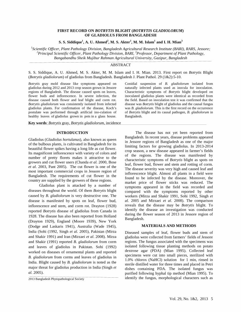
Symptoms on leafInitially, reddish brown tiny spots appeared on theleaves which became round to oval and sometimesirregular in shape (Plate 1 A). The spots enlargedgradually and turned into pale brown in color withreddish brown margin and dark yellow center (Plate 1B). In case of severe infection, several spots coalescedtogether and formed large lesions and a blightedsymptom appeared (Plate 1 C). At later stage ofinfection, moldy structure of mycelium, conidiophoresand spores appeared on the blighted leaves (Plate 1 D&E). Severe lesions appeared on leaf sheath whichgirdled the sheath around the stem (Plate 1 F).Symptoms on stemInfection of stem started from leaf sheath (Plate 1 F).From the sheath, lesions moved downwards and
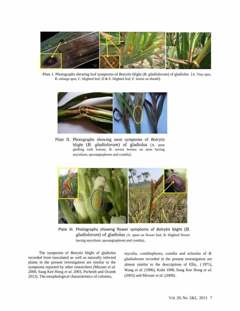
reached the stem causing stem girdling. The lesionencircles the stem and soft rot symptoms appeared.Sometime stem girdling occurred at the point ofinfection (Plate 2 A). Grayish fungal growth alsoobserved at the point of infection of stem (Plate II B).
Symptoms on flower
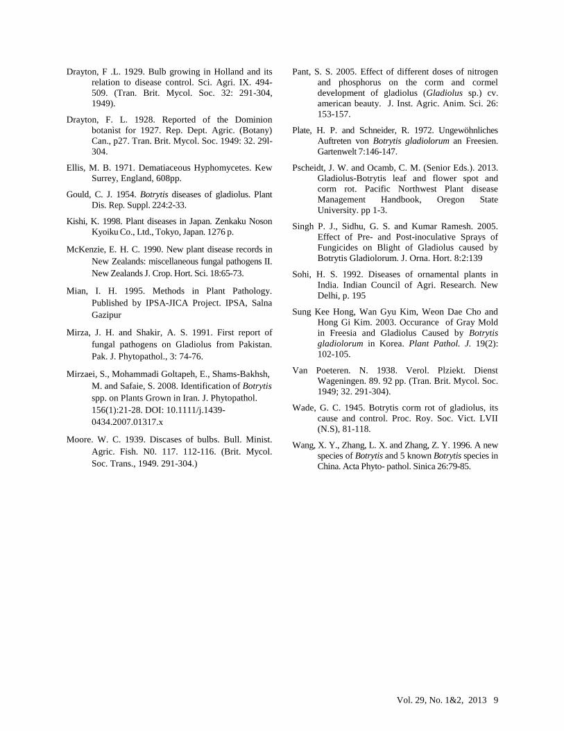
Minute water-soaked lesions developed on flower budsand flowers of infected plants at 72 hr after inoculation.Lesions increased in size and coalesced to form patcheswithin 7 to 10 days of infection. Shoots becameblighted and died in 12-14 days. All the infected organswere covered with gray mold within 16-18 days. Re-isolation of the causal fungus from the inoculatedplants consistently yielded the inoculated fungus. Onpetals and sepals translucent water soaked spotsappeared with light brown margin and pale coloredcentre (Plate III A). As the spots enlarged, dead tissueturned into brown and the flowers became rotten (PlateIII B). In severe cases entire flower can be rotted andgray mass of spore appeared on rotted portion (Plate IIIC). Plants under control plants did not show anysymptom of the disease.
Symptoms on corms
Initially, small reddish brown lesions appeared on
corms of gladiolus (Plate IV A). Gradually, several
lesions coalesced together and turned into large black
lesion (Plate IV B). Sometime large black mummified
areas appeared on the neck region of the corm. In the
neck region, large brown spots were observed (Plate IV
C).
Morphological characteristics of the causal fungus
After 48 hr of incubation, whitish mycelial growthappeared on the infected leaf pieces of gladiolus usedas inocula and plated on moist blotting paper in Petridish (Plate V A). Similarly, after 5 days of incubationcolonies of B. gladiolorum grew from pieces ofgladiolus leaf were placed on PDA in Petri dishes(Plate V B). Conidiophores bearing conidia (Plate II C)appeared on the colonies produced on leaf pieces aswell as PDA (Plate V A and C). Later on, the colonyturned into brown and produced black sclerotia after14-16 days of incubation (Plate V D and E).Conidiophores were dark brown and twisted (Plate VA, B, D and E). Conidia were ellipsoid and ovoid oroval in shape, pale brown in color and 14.2-20.7 mm x9.3-12.9 mm in dimension (Plate V F).
Vol. 29, No. 1&2, 2013 7
The symptoms of Botrytis blight of gladiolusrecorded from inoculated as well as naturally infectedplants in the present investigation are similar to thesymptoms reported by other researchers (Mirzaei et al.2008, Sung Kee Hong et al. 2003, Pscheidt and Ocamb2013). The morphological characteristics of colonies,
mycelia, conidiophores, conidia and sclerotia of B.
gladiolorum recorded in the present investigation are
almost similar to the descriptions of Ellis, ( 1971),
Wang et al. (1996), Kishi 1998, Sung Kee Hong et al.
(2003) and Mirzaei et al. (2008).
Plate II. Photographs showing stem symptoms of Botrytisblight (B. gladiolorum) of gladiolus [A. stemgirdling with lesions, B. severe lesions on stem having
mycelium, sporangiophores and conidia].
BA
A
B
C
Plate III. Photographs showing flower symptoms of Botrytis blight (B.gladiolorum) of gladiolus [A. spots on flower bud, B. blighted flower
having mycelium, sporangiophores and conidia].
F
Plate 1. Photographs showing leaf symptoms of Botrytis blight (B. gladiolorum) of gladiolus [A. Tiny spot,B. enlarge spot, C. blighted leaf, D & E. blighted leaf, F. lesion on sheath].

8 Bangladesh J. Plant Pathol.
It has been recorded that B. gladiolorum infectsdifferent flowers under Iridaceae family like gladiolus,freesia, ixia, crocus, and iris in North America, Europe,Africa, New Zealand, China, and Japan (Boerema andHamers 1989, Gould 1954, Kishi 1998, Mckenzie1990, Plate and Schneider 1972, Wang et al. 1996, SungKee Hong et al. 2003, Mirzaei et al. 2008).
Findings of the present investigation clearly revealthat Botrytis blight caused by B. gladiolorum regularlyattacks the gladiolus plants in Jessore regions ofBangladesh. However, the disease has not yet beenreported from the country. So, the present report may beconsidered as the new record of Botrytis blight ofgladiolus and its causal fungus, B. gladiolorum inBangladesh.
LITERATURE CITED
Boerema, G. H. and Hamers, M. E. C. 1989. Checklistfor scientific names of common parasitic fungi.Neth. J. Plant Pathol. 95 (Suppl.3):4-5.
Bose, T. K., Yadav, L. P., Pat P., Parthasarathy, V. A.and Das, P. 2003. Commercial Flowers, NayaUdyog, Kolkata, India. Vol. 2.
Chanda, S., Barma, G. and Roychowdhury, N. 2000.Influence of different levels of nitrogen,phosphorus and potassium on growth andflowering of gladiolus. Hort. J. 13 (1): 76-86.
Dodge, B. O. and Laskaris, T. 1941. Botrytis corm rotof Gladiolus. J.N.Y. bot. Gdn. 42, 92-5. (Tran.Brit. Mycol. Soc. 1949; 32. 291-304).
Plate IV. Photographs showing corm symptomsof Botrytis blight (B. gladiolorum) ofgladiolus [A. spots on flower bud, B.blighted flower having mycelium,conidiophores and conidia, C. large brown spot
on neck region].
C
B
A
Plate V. Photographs showing corm symptoms of Botrytis blight (B. gladiolorum) of gladiolus [A.mycelium grew on leaves placed on moist blotter, B. mycelium grew on leaves placed on PDA, D and E. sclerotia
formation, C & F. conidiophores bearing conidia, F. Conidia].
A
F CB
E
D
Vol. 29, No. 1&2, 2013 9
Drayton, F .L. 1929. Bulb growing in Holland and itsrelation to disease control. Sci. Agri. IX. 494-509. (Tran. Brit. Mycol. Soc. 32: 291-304,1949).
Drayton, F. L. 1928. Reported of the Dominionbotanìst for 1927. Rep. Dept. Agric. (Botany)Can., p27. Tran. Brit. Mycol. Soc. 1949: 32. 29l-304.
Ellis, M. B. 1971. Dematiaceous Hyphomycetes. KewSurrey, England, 608pp.
Gould, C. J. 1954. Botrytis diseases of gladiolus. PlantDis. Rep. Suppl. 224:2-33.
Kishi, K. 1998. Plant diseases in Japan. Zenkaku NosonKyoiku Co., Ltd., Tokyo, Japan. 1276 p.
McKenzie, E. H. C. 1990. New plant disease records inNew Zealands: miscellaneous fungal pathogens II.New Zealands J. Crop. Hort. Sci. 18:65-73.
Mian, I. H. 1995. Methods in Plant Pathology.Published by IPSA-JICA Project. IPSA, SalnaGazipur
Mirza, J. H. and Shakir, A. S. 1991. First report offungal pathogens on Gladiolus from Pakistan.Pak. J. Phytopathol., 3: 74-76.
Mirzaei, S., Mohammadi Goltapeh, E., Shams-Bakhsh,M. and Safaie, S. 2008. Identification of Botrytisspp. on Plants Grown in Iran. J. Phytopathol.156(1):21-28. DOI: 10.1111/j.1439-0434.2007.01317.x
Moore. W. C. 1939. Discases of bulbs. Bull. Minist.Agric. Fish. N0. 117. 112-116. (Brit. Mycol.Soc. Trans., 1949. 291-304.)
Pant, S. S. 2005. Effect of different doses of nitrogenand phosphorus on the corm and cormeldevelopment of gladiolus (Gladiolus sp.) cv.american beauty. J. Inst. Agric. Anim. Sci. 26:153-157.
Plate, H. P. and Schneider, R. 1972. UngewöhnlichesAuftreten von Botrytis gladiolorum an Freesien.Gartenwelt 7:146-147.
Pscheidt, J. W. and Ocamb, C. M. (Senior Eds.). 2013.Gladiolus-Botrytis leaf and flower spot andcorm rot. Pacific Northwest Plant diseaseManagement Handbook, Oregon StateUniversity. pp 1-3.
Singh P. J., Sidhu, G. S. and Kumar Ramesh. 2005.Effect of Pre- and Post-inoculative Sprays ofFungicides on Blight of Gladiolus caused byBotrytis Gladiolorum. J. Orna. Hort. 8:2:139
Sohi, H. S. 1992. Diseases of ornamental plants inIndia. Indian Council of Agri. Research. NewDelhi, p. 195
Sung Kee Hong, Wan Gyu Kim, Weon Dae Cho andHong Gi Kim. 2003. Occurance of Gray Moldin Freesia and Gladiolus Caused by Botrytisgladiolorum in Korea. Plant Pathol. J. 19(2):102-105.
Van Poeteren. N. 1938. Verol. Plziekt. DienstWageningen. 89. 92 pp. (Tran. Brit. Mycol. Soc.1949; 32. 291-304).
Wade, G. C. 1945. Botrytis corm rot of gladiolus, itscause and control. Proc. Roy. Soc. Vict. LVII(N.S), 81-118.
Wang, X. Y., Zhang, L. X. and Zhang, Z. Y. 1996. A newspecies of Botrytis and 5 known Botrytis species inChina. Acta Phyto- pathol. Sinica 26:79-85.

10 Bangladesh J. Plant Pathol.
Vol. 29, No. 1&2, 2013 11
PESTALOTIOPSIS GUEPINII (DESM.) STAY. – A NEW PATHOGEN OFBLACK SPOT DISEASE OF ROSE IN BANGLADESH
Shamim Shamsi1 and Anita Ghosh2
1Professor and 2 Post Graduate studentDepartment of Botany, University of Dhaka, Dhaka-1000, Bangladesh
E-mail address: [email protected]
ABSTRACT
Shamim Shamsi and Anita Ghosh. 2013. Pestalotiopsis guepinii (desm.) Stay. – a new pathogen of black spotdisease of rose in Bangladesh. Bangladesh J. Plant Pathol. 29 (1&2): 11-14.
An investigation was conducted during November 2009 toOctober 2010 to determine causal fungi of black spot diseaseof rose (Rosa centifolia L.). Black spot infected leaf sampleswere collected from different locations of Dhaka city. Thefungi associated with the samples were isolated andidentified. The principal fungal pathogen associated with thediseased specimens was Diplocarpon rosae Wolf (imperfectstage Marssonina rosae). Other fungal pathogens associatedwith the disease were Pestalotiopsis guepinii and its twoculture types (Pestalotiopsis guepinii-1, P. guepinii-2). Thefungi belong to the class Coelomyecetes under the Division
Deuteromycota. The fungi were frequently isolated fromblack spot infected leaf samples of the rose. After inoculationof detached leaves and seedlings of rose, P. guepinii and itstwo culture types developed characteristics symptoms ofblack spot. The pathogen was reisolated from black spotinfected inoculated leaves to fulfill Koch’s postulate. Thefindings of the investigation indicate that P. guepinii isanother fungal pathogen of black spot of rose in addition toD. rosae. This is a new record about causal agents of blackspot disease of the cut flower.
Keywords: Black spot, rose, Pestalotiopsis guepinii
INTRODUCTION
Rose (Rosa centifolia L.) is grown throughout theworld for their beautiful flower and fragrance. Theornamental plant is also grown in Bangladesh.Diplocarpon rosae Wolf and its imperfect stageMarssonina rosae (Lib.) Died is well documentedpathogen of black spot of rose. Black spots are circularwith a perforated edge and reach a diameter of 14 mm.Severely affected plants however, do not show thecircular spots as they coalesced together and form largelesion (Debner 1988). From India, Mukerji and Bhasin(1986) reported leaf spot of rose caused byPestalotiopsis versicolor. Islam et al. (2010) reportedseven diseases of rose from Bangladesh. The diseases,in order of their prevalence, are Botrytis blight(Botrytis cinerea), Cercospora leaf spot (Cercosporapuderi), rose mosaic (Rose Mosaic Virus), black spot(Diplocarpon rosae), die-back (Botryodiplodiatheobromae), Alternaria leaf spot (Alternariaalternata) and stem canker (Crytosporella umbrina(Speg.) Stey. In Bangladesh, reports on association offungal pathogens with black spot symptoms are notavailable. The present investigation was undertaken toidentify the fungal pathogens associated with blackspot disease of rose plant other than D. rosae.
MATERIALS AND METHODS
Healthy and diseased leaves of rose were collectedfrom different locations of Dhaka city duringNovember 2009 to October 2010. Fungi wereisolated from black spot infected leaf samples
2013 Bangladesh Phytopathological Society
following “Tissue Planting method” on PDA mediumand pure culture of the isolated fungi were preparedfollowing single spore method (Tuite 1969).Morphological characteristics of the fungi wererecorded under a compound microscope andidentified using standard key books (Barnett andHunter 2000, Booth 1971, Ellis 1971, 1976, Sutton1980, Ellis and Ellis 1997). All diseased specimens andassociated fungi were preserved in the Herbarium of theDepartment of Mycology and Plant Pathology,University of Dhaka.
Pathogenicity test of the isolated fungi wasperformed following modified detached leaf assaytechnique (Azad and Shamsi 2011). Fresh, healthyand mature leaves of rose were collected, washedwith distilled water, surface sterilized with 1.0%Clorox for five minutes and rinsed in sterilized water.Ventral and dorsal sides of the leaflets with andwithout pricking with needles were inoculated with 2mm diameter mycelial block of the isolated fungipreviously grown on PDA medium for seven days.Another set of leaves with and without pricking andwithout inoculation were maintained, which served ascontrols. Three replicated leaflets were used for eachtreatment. The inoculated leaflets were placed in Petridishes containing water soaked filter paper and cottonball to maintain sufficient humidity to initiate infection.The plates were incubated at 25-28C. The inoculatedand non inoculated leaflets were checked for symptomdevelopment starting from 3 days of inoculation andcontinued up to 7-10 days.
12 Bangladesh J. Plant Pathol.
Seedling inoculation method was also followed toconfirm pathogenicity of the isolated fungi. Healthyseedling of rose plant was transplanted in pots (30cm Diam.) containing sterilized soil at three seedlingsper pot and allow to grow for three month in anethouse providing necessary water and nutrients.Healthy leaves of seedlings were washed withsterilized water and surface sterilized with 1.0%Chlorox and rinsed with sterilized distilled water.
Surface sterilized leaves were pricked withsterilized needle. Pricked and unpricked leaves wereinoculated with the test fungi by rubbing sporulatingPDA culture of the test fungus. Leaves under controlreceived only fresh PDA medium without fungalinoculum. Plants were covered with polythene bags tomaintain proper humidity level and to avoidcontamination. Three seedlings were inoculated foreach treatment. The inoculated plants were placed in aclean bench. The plants were examined daily and
continued up to 10 days to record the development ofsymptoms. Symptoms developed on artificialinoculated leaves were recorded and compared withthe symptoms of those observed on naturallyinoculated leaves. The fungus was resonated fromthe inoculated leaves of rose to fulfill Koch’spostulates.
RESULTS AND DISCUSSION
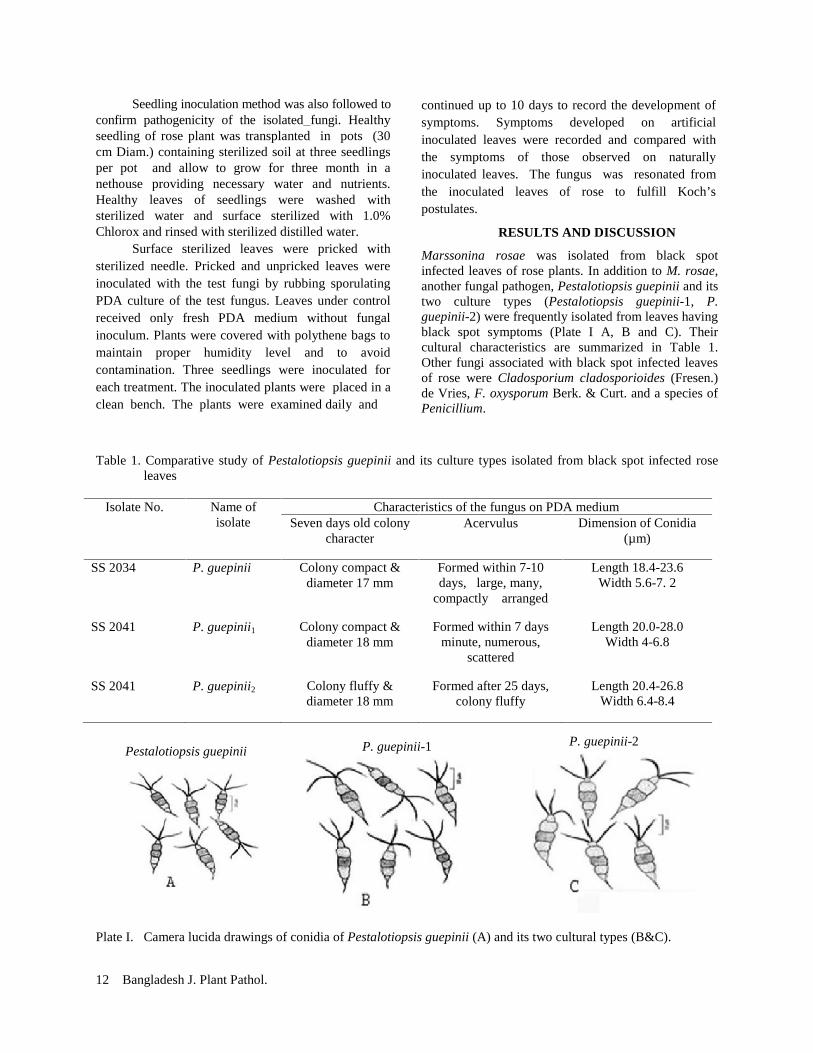
Marssonina rosae was isolated from black spotinfected leaves of rose plants. In addition to M. rosae,another fungal pathogen, Pestalotiopsis guepinii and itstwo culture types (Pestalotiopsis guepinii-1, P.guepinii-2) were frequently isolated from leaves havingblack spot symptoms (Plate I A, B and C). Theircultural characteristics are summarized in Table 1.Other fungi associated with black spot infected leavesof rose were Cladosporium cladosporioides (Fresen.)de Vries, F. oxysporum Berk. & Curt. and a species ofPenicillium.
Table 1. Comparative study of Pestalotiopsis guepinii and its culture types isolated from black spot infected roseleaves
Isolate No. Name ofisolate
Characteristics of the fungus on PDA mediumSeven days old colony
characterAcervulus Dimension of Conidia
(µm)
SS 2034 P. guepinii Colony compact &diameter 17 mm
Formed within 7-10days, large, many,
compactly arranged
Length 18.4-23.6Width 5.6-7. 2
SS 2041 P. guepinii1 Colony compact &diameter 18 mm
Formed within 7 daysminute, numerous,
scattered
Length 20.0-28.0Width 4-6.8
SS 2041 P. guepinii2 Colony fluffy &diameter 18 mm
Formed after 25 days,colony fluffy
Length 20.4-26.8Width 6.4-8.4
Plate I. Camera lucida drawings of conidia of Pestalotiopsis guepinii (A) and its two cultural types (B&C).
Pestalotiopsis guepiniiP. guepinii-2P. guepinii-1
Vol. 29, No. 1&2, 2013 13
Taxonomic enumeration of Pestalotiopsis guepinii
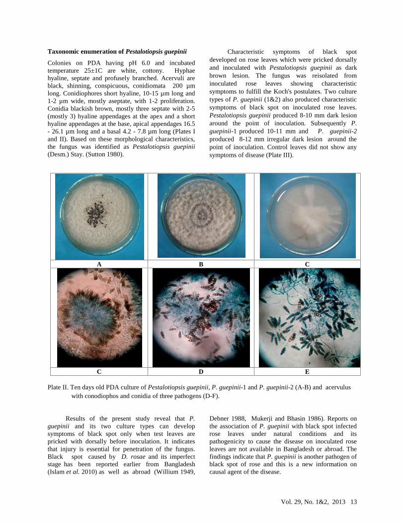
Colonies on PDA having pH 6.0 and incubatedtemperature 25±1C are white, cottony. Hyphaehyaline, septate and profusely branched. Acervuli areblack, shinning, conspicuous, conidiomata 200 µmlong. Conidiophores short hyaline, 10-15 µm long and1-2 µm wide, mostly aseptate, with 1-2 proliferation.Conidia blackish brown, mostly three septate with 2-5(mostly 3) hyaline appendages at the apex and a shorthyaline appendages at the base, apical appendages 16.5- 26.1 µm long and a basal 4.2 - 7.8 µm long (Plates Iand II). Based on these morphological characteristics,the fungus was identified as Pestalotiopsis guepinii(Desm.) Stay. (Sutton 1980).
Characteristic symptoms of black spotdeveloped on rose leaves which were pricked dorsallyand inoculated with Pestalotiopsis guepinii as darkbrown lesion. The fungus was reisolated frominoculated rose leaves showing characteristicsymptoms to fulfill the Koch's postulates. Two culturetypes of P. guepinii (1&2) also produced characteristicsymptoms of black spot on inoculated rose leaves.Pestalotiopsis guepinii produced 8-10 mm dark lesionaround the point of inoculation. Subsequently P.guepinii-1 produced 10-11 mm and P. guepinii-2produced 8-12 mm irregular dark lesion around thepoint of inoculation. Control leaves did not show anysymptoms of disease (Plate III).
Plate II. Ten days old PDA culture of Pestalotiopsis guepinii, P. guepinii-1 and P. guepinii-2 (A-B) and acervuluswith conodiophos and conidia of three pathogens (D-F).
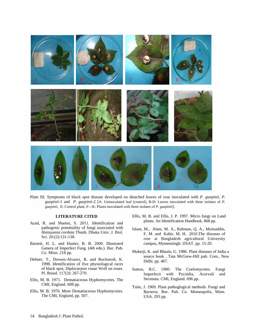
Results of the present study reveal that P.guepinii and its two culture types can developsymptoms of black spot only when test leaves arepricked with dorsally before inoculation. It indicatesthat injury is essential for penetration of the fungus.Black spot caused by D. rosae and its imperfectstage has been reported earlier from Bangladesh(Islam et al. 2010) as well as abroad (Willium 1949,
Debner 1988, Mukerji and Bhasin 1986). Reports onthe association of P. guepinii with black spot infectedrose leaves under natural conditions and itspathogenicity to cause the disease on inoculated roseleaves are not available in Bangladesh or abroad. Thefindings indicate that P. guepinii is another pathogen ofblack spot of rose and this is a new information oncausal agent of the disease.
A B C
C D E
Vol. 29, No. 1&2, 2013 13
Taxonomic enumeration of Pestalotiopsis guepinii
Colonies on PDA having pH 6.0 and incubatedtemperature 25±1C are white, cottony. Hyphaehyaline, septate and profusely branched. Acervuli areblack, shinning, conspicuous, conidiomata 200 µmlong. Conidiophores short hyaline, 10-15 µm long and1-2 µm wide, mostly aseptate, with 1-2 proliferation.Conidia blackish brown, mostly three septate with 2-5(mostly 3) hyaline appendages at the apex and a shorthyaline appendages at the base, apical appendages 16.5- 26.1 µm long and a basal 4.2 - 7.8 µm long (Plates Iand II). Based on these morphological characteristics,the fungus was identified as Pestalotiopsis guepinii(Desm.) Stay. (Sutton 1980).
Characteristic symptoms of black spotdeveloped on rose leaves which were pricked dorsallyand inoculated with Pestalotiopsis guepinii as darkbrown lesion. The fungus was reisolated frominoculated rose leaves showing characteristicsymptoms to fulfill the Koch's postulates. Two culturetypes of P. guepinii (1&2) also produced characteristicsymptoms of black spot on inoculated rose leaves.Pestalotiopsis guepinii produced 8-10 mm dark lesionaround the point of inoculation. Subsequently P.guepinii-1 produced 10-11 mm and P. guepinii-2produced 8-12 mm irregular dark lesion around thepoint of inoculation. Control leaves did not show anysymptoms of disease (Plate III).
Plate II. Ten days old PDA culture of Pestalotiopsis guepinii, P. guepinii-1 and P. guepinii-2 (A-B) and acervuluswith conodiophos and conidia of three pathogens (D-F).
Results of the present study reveal that P.guepinii and its two culture types can developsymptoms of black spot only when test leaves arepricked with dorsally before inoculation. It indicatesthat injury is essential for penetration of the fungus.Black spot caused by D. rosae and its imperfectstage has been reported earlier from Bangladesh(Islam et al. 2010) as well as abroad (Willium 1949,
Debner 1988, Mukerji and Bhasin 1986). Reports onthe association of P. guepinii with black spot infectedrose leaves under natural conditions and itspathogenicity to cause the disease on inoculated roseleaves are not available in Bangladesh or abroad. Thefindings indicate that P. guepinii is another pathogen ofblack spot of rose and this is a new information oncausal agent of the disease.
A B C
C D E
14 Bangladesh J. Plant Pathol.
Plate III. Symptoms of black spot disease developed on detached leaves of rose inoculated with P. guepinii, P.guepinii-1 and P. guepinii-2 [A: Uninoculated leaf (control), B-D: Leaves inoculated with three isolates of P.guepinii, E: Control plant, F—K: Plants inoculated with three isolates of P. guepinii].
LITERATURE CITED
Azad, R. and Shamsi, S. 2011. Identification andpathogenic potentiality of fungi associated withHuttuyania cordata Thunb. Dhaka Univ. J. Biol.Sci. 20:(2):131-138.
Barnett, H. L. and Hunter, B. B. 2000. IllustratedGenera of Imperfect Fung. (4th edn.). Bur. Pub.Co. Minn. 218 pp.
Debner, T., Drewes-Alvarez, R. and Rockstroh, K.1998. Identification of five physiological racesof black spot, Diplocarpon rosae Wolf on roses.Pl. Breed. 117(3): 267-270.
Ellis, M. B. 1971. Dematiaceous Hyphomycetes. TheCMI, England. 608 pp.
Ellis, M. B. 1976. More Dematiaceous Hyphomycetes.The CMI, England, pp. 507.
Ellis, M. B. and Ellis, J. P. 1997. Micro fungi on Landplants. An Identification Handbook. 868 pp.
Islam, M., Alam, M. A., Rahman, Q. A., Moinuddin,F. M. and Kabir. M. H. 2010.The diseases ofrose at Bangladesh agricultural Universitycampus, Mymensingh. IJSAT. pp. 15-20.
Mukerji, K. and Bhasin, G. 1986. Plant diseases of India asource book . Tata McGrew-Hill pub. Com., NewDelhi. pp. 467.
Sutton, B.C. 1980. The Coelomycetes. FungiImperfecti with Pycnidia, Acervuli andStromata. CMI, England. 696 pp.
Tuite, J. 1969. Plant pathoglogical methods. Fungi andBacteria. Bur. Pub. Co. Minneapolis, Minn.USA. 293 pp.
A B DC
E F G H
I J K
Vol. 29, No. 1&2, 2013 15
IN-VITRO EVALUATION OF ANTIFUNGAL ACTIVITY OF PLANT EXTRACTS AGAINSTRHIZOCTONIA ORYZAE-SATIVAE CAUSING AGGREGATED SHEATH SPOT OF RICE
S. B. Jahan1, M. A. Ali2, S. Alam1, Z. R. Moni3 and M. A. Alam3
1Department of Botany, Rajshahi University, Rajshahi; 2Plant Pathology Division, 3Plant Breeding division,Bangladesh Rice Research Institute, Joydebpur, Gazipur-1701, Bangladesh.
Email of first author: [email protected]
(The paper is a part of M.Ph thesis of the first author)
ABSTRACT
S. B. Jahan, M. A. Ali, S. Alam, Z. R. Moni and M. A. Alam. 2013. In-vitro evaluation of antifungal activity ofplant extracts against Rhizoctonia oryzae-sativae causing aggregated sheath spot of rice. Bangladesh J. Plant Pathol.29 (1&2):15-19.
In-vitro experiments were conducted to test theantifungal activity of garlic clove (Allium sativam),ginger rhizome (Zingiber officinales), and leaves ofhenna (Lawsonia inermis), water pepper (Poligonumhydropiper), ivy gourd (Coccinia cordifolia) and neem(Azadirachta indica) against mycelia growth ofRhizoctonia oryzae-sativae, the causal fungus ofaggregated sheath spot of rice. Aqueous extracts of allthe botanicals were prepared, mixed with liquid potatodextrose agar (PDA) medium at 0, 5, 10, 15, 20 and25% concentrations and poured into 90 mm Petridishes at 20 ml per dish. After solidification of PDA,90 mm glass Petri dishes were inoculated with myceliablocks of Rhizoctonia oryzae-sativae cut from 5 days
old PDA culture of the pathogen at one block per plate.It was found that the plant extract reduced radialcolony diameter of the pathogen appreciably atdifferent concentrations of the botanicals compared tocontrol (0%). Among the tested plant extracts, garlicand henna was the most effective material against R.oryzae-sativae showing 50% reduction in colonydiameter at 3.25% concentration, which indicatedlowest LD (lethal dose) 50 value followed by hennaextract at 3.75%. Lowest LD 90 value also showed bygarlic extracts at 17.25% followed by henna extract at19%. Garlic and henna considerably decreasedsclerotia germination at all the concentrations tested inthe present study.
Key words: Rhizoctonia oryzae-sativae, aggregate sheath spot, in-vitro screening, plant extracts
INTRODUCTION
Aggregated sheath spot caused by Rhizoctonia oryzae-sativae is usually considered as a minor disease of ricebut it can be a very aggressive disease of the cropunder favourable conditions (Lanoiselet et al. 2007). Itoccurs in many of the rice growing countries of theworld (Gunnel et al. 1984, 1992, Cedeno et al. 1998,Lanoiselet et al. 2001). In Bangladesh, aggregatesheath spot was reported for the first time in 1988 byShahjahan et al. (1988). The disease caused yieldlosses of 20% in Australia, 4 to 9% in Uruguay(Lanoiselet et al. 2005) and in Bangladesh the diseasemay reduce rice yield by 14.74%.
Chemical, physical and cultural methods arerecommended to control aggregated sheath spot of rice.Available reports reveal that extracts of many plantspecies possess antifungal and antibacterial properties(Hasan et al. 2005, Ogbo and Oyibo 2008, Dubey et al.2009). Several higher plants and their constituents maybe used successfully in plant disease control (Singh etal. 1993). Adityachaudhury (1991) mentioned that useof plant extracts and phytoproducts is gaining attentiondue to their bio-degradability, low toxicity and mini-2013 Bangladesh Phytopathological Society
mum residual toxicity in the ecosystem. Antifungalactivities were found in Eucalyptus, Syzygiumaromticum, Azadirachta indica, Rosmarinus officinalisagainst Rhizoctonia solani, R. oryzae, R. oryzae-sativaeand Sclrotium hydrophilum (Aye et al. 2011).Cinnamon oil was found efficient plant product thatinhibited in vitro colony diameter of R. oryzae-sativaeas well as suppression of aggregate sheath spot diseaseof rice under greenhouse condition. The present pieceof research was undertaken to evaluate efficacy of sixplant extracts to inihiting invitro vegetative growth andsclerotia germination of R. oryzae-sativae.
MATERIALS AND METHODS
Six locally available plant species namely garlic(Allium sativam), ginger (Zingiber officinales), henna(Lawsonia inermis), water pepper (Poligonumhydropiper), ivy gourd (Coccinia cordifolia) and neem(Azadirachta indica) were collected. Water extracts ofgarlic clove, ginger rhizome, and leaves of henna,water pepper, ivy gourd and neem were prepared. Forpreparation of the plant extracts, 100 g of each materialwas washed in sterile distilled water, 100 ml sterilewater was added (1:1 w/v), crushed in a mortar and
16 Bangladesh J. Plant Pathol.
pestle and passed throught 2-ply cheese cloth. Eachplant extract was filtered through filter paper(Whatman no.1), which was considered as standardplant extract of each plant species and tested forantifungal activity.
Poison food technique was followed to test theplant extracts using potato dextrose agar (PDA) as thebasic medium (Dhingra and Sinclair 1985, Ali andArcher 2003). The aqueous extracts were thoroughlymixed with warm PDA (40C) at 5, 10, 15, 20 and 25%concentration and poured into sterile Petri plates (90mm). PDA without any extract served as the control.An isolate (No. MY-1) of R. oryzae-sativae wasobtained from Plant Pathology Division, BangladeshRice Research Institute (BRRI), Gazipur and multipliedon fresh PDA. Mycelial disc were cut from the activelygrowing section of 3-day old culture of the pathogen.The disc was placed at the center of each Petri platecontaining amended or unamended PDA at one discper plate. The inoculated plates were incubated at roomtemperature (25-28C). The plates were arranged on thedesks in the Laboratory of Plant Pathology Division,BRRI following completely randomized design. Fourreplicated plates were used for each treatment. Theredial colony diameter was measured in all treatmentswhen the mycelium reached the rim of the Petri plateunder control. Percent growth inhibition was calculatedbased on diameter of colony under control plates.
Inhibition of sclerotia germination wasdetermined by the method of Chaizuckam and Davis(2010). PDA medium was amended with the plantextracts at 0, 5, 10, 15, 20 and 25% following theprocedures as mentioned earlier. Ten sclerotia of thepathogen (isolate MY-1) were collected from 14 daysold PDA culture, soaked in sterile water for 10 min.Water soaked sclerotia were transferred to amendedPDA plates. After tree day of incubation at roomtemperature number of germinated sclerotia wascounted. The experiment was repeated once.
Data on radial mycelial growth were subjectedto statistical analysis using CropStat (version 7.2)computer software. Paired-t test was performed toassess the effect of plant extract on sclerotialgermination comparing with control.
RESULTS AND DISCUSSION
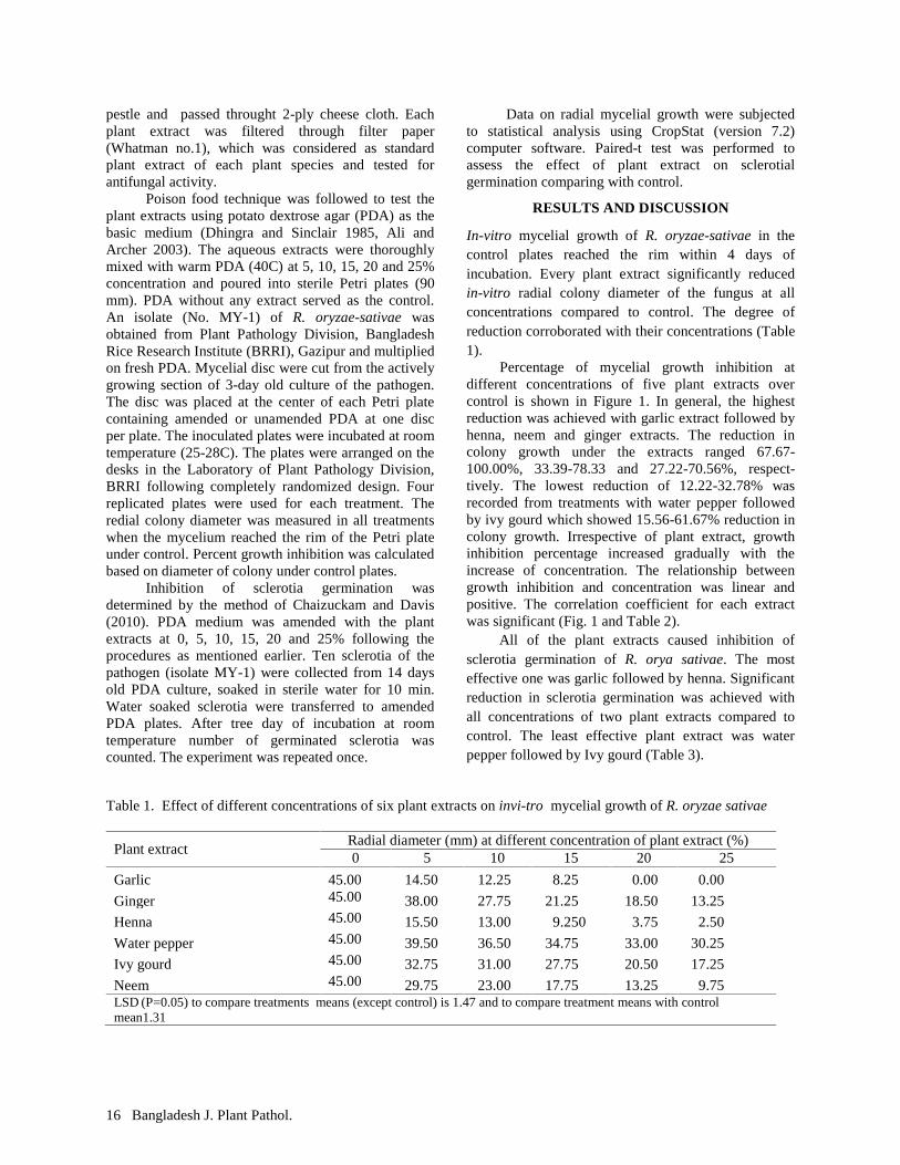
In-vitro mycelial growth of R. oryzae-sativae in thecontrol plates reached the rim within 4 days ofincubation. Every plant extract significantly reducedin-vitro radial colony diameter of the fungus at allconcentrations compared to control. The degree ofreduction corroborated with their concentrations (Table1).
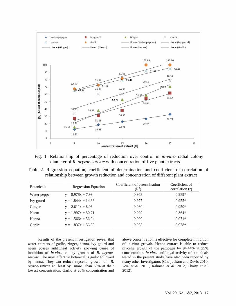
Percentage of mycelial growth inhibition atdifferent concentrations of five plant extracts overcontrol is shown in Figure 1. In general, the highestreduction was achieved with garlic extract followed byhenna, neem and ginger extracts. The reduction incolony growth under the extracts ranged 67.67-100.00%, 33.39-78.33 and 27.22-70.56%, respect-tively. The lowest reduction of 12.22-32.78% wasrecorded from treatments with water pepper followedby ivy gourd which showed 15.56-61.67% reduction incolony growth. Irrespective of plant extract, growthinhibition percentage increased gradually with theincrease of concentration. The relationship betweengrowth inhibition and concentration was linear andpositive. The correlation coefficient for each extractwas significant (Fig. 1 and Table 2).
All of the plant extracts caused inhibition ofsclerotia germination of R. orya sativae. The mosteffective one was garlic followed by henna. Significantreduction in sclerotia germination was achieved withall concentrations of two plant extracts compared tocontrol. The least effective plant extract was waterpepper followed by Ivy gourd (Table 3).
Table 1. Effect of different concentrations of six plant extracts on invi-tro mycelial growth of R. oryzae sativae
Plant extractRadial diameter (mm) at different concentration of plant extract (%)0 5 10 15 20 25
Garlic 45.00 14.50 12.25 8.25 0.00 0.00
Ginger 45.00 38.00 27.75 21.25 18.50 13.25
Henna 45.00 15.50 13.00 9.250 3.75 2.50
Water pepper 45.00 39.50 36.50 34.75 33.00 30.25
Ivy gourd 45.00 32.75 31.00 27.75 20.50 17.25
Neem 45.00 29.75 23.00 17.75 13.25 9.75LSD (P=0.05) to compare treatments means (except control) is 1.47 and to compare treatment means with controlmean1.31
Vol. 29, No. 1&2, 2013 17
Table 2. Regression equation, coefficient of determination and coefficient of correlation ofrelationship between growth reduction and concentration of different plant extract
Botanicals Regression EquationCoefficient of determination
(R2)Coefficient ofcorrelation (r)
Water pepper y = 0.978x + 7.99 0.963 0.989*
Ivy gourd y = 1.844x + 14.88 0.977 0.955*
Ginger y = 2.611x + 8.06 0.980 0.950*
Neem y = 1.997x + 30.71 0.929 0.864*
Henna y = 1.566x + 56.94 0.990 0.971*
Garlic y = 1.837x + 56.85 0.963 0.928*
Results of the present investigation reveal thatwater extracts of garlic, zinger, henna, ivy gourd andneem posses antifungal activity showing cause ofinhibition of in-vitro colony growth of R. oryzae-sativae. The most effective botanical is garlic followedby henna. They can reduce mycelial growth of R.oryzae-sativae at least by more than 60% at theirlowest concentration. Garlic at 20% concentration and
above concentration is effective for complete inhibitionof in-vitro growth. Henna extract is able to reducemycelia growth of the pathogen by 94.44% at 25%concentration. In-vitro antifungal activity of botanicalstested in the present study have also been reported bymany other investigators (Chaijuckam and Devis 2010,Aye et al. 2011, Rahman et al. 2012, Chaity et al.2012).
Fig. 1. Relationship of percentage of reduction over control in in-vitro radial colonydiameter of R. oryzae-sativae with concentration of five plant extracts.

18 Bangladesh J. Plant Pathol.
Table 3. Antifungal activity of different plant extracts with different concentrations on sclerotia germination of R.oryzae-sativae
Plants ExtractConcentration of plant extracts (%)
5 10 15 20 25
Garlic 8.0*** 7.25*** 7.0*** 0 0
IVY Gourd 9.5 9.0 9.0 8.75 8.25**
Ginger 8.25** 8.25** 8.0*** 7.5*** 7.25***
Henna 9.5 9.5 9.5 9.0 9.0
Water pepper 9.5 9.5 9.25 8.75 8.5*
Neem 9.25 9.0 8.75 8.5* 8.0***
Control (without plant extract) 9.5
Significant difference between plant extracts with different concentration and the controls at 0.1%, 1% and 5% level is indicatedby ***, ** and * respectively
The factors responsible for antifungal activity ofthe botanical tested have not been studied in presentinvestigation. However, other investigators reportedthat sulfur rich protein Ajoene derived from garlic hadantifungal activities against Aspergillus niger, Candidaalbicans (Yoshida et al.1987). Other workers alsoshowed the presences of antifungal properties in A.sativam (Misra and Dixit 1976, Agarwal 1978). Garlichas already been reported to have antifungal activityagainst R. oryzae-sativae (Chaijuckam et al. 2010).The sensitivity of fungi and even isolates of the samespecies to plant extracts may vary. Such as, garlicextract at 5% completely inhibited vegetative growth ofCalifornia isolates of R. oryzae-sativae (Chaijuckam etal. 2010) but in case of Bangladeshi isolate of R.oryzae-sativae, it required 20% concentration.
In the present experiment R. oryzae-sativaewas added in the list of sensitivity to the henna extract.The henna leaf extract caused 65.56% - 94.44%inhibition at 5 - 25% concentrations. Leaf extract ofhenna completely controlled the growth of Drechsleraoryzae, Sclerotium oryzae, S. rolfsii and Rhizoctoniasolani at 20% (w/v) concentration. The presence ofantifungal compound (2- hydroxyl- 1, 4 napthoquinine)in the leaf extract of henna had been identified whichmight be responsible for microbial growth inhibition(Tripathi et al. 1978).
Neem leaf extract showed 33.89 - 78.33%inhibition at 5 - 25% concentration. The plant extractinhibited the mycelial growth of R. solani, R. oryzaesativae R. oryzae, and Sclerotium hydrophilum by 87.5,80.0, 92.5 and 49.2% respectively (Aye and
Matsumoto 2011). Similarly, findings of the presentinvestigation showed satisfactory reduction in colonygrowth on R. oryzae-sativae by using neem leafextract. In present test, ginger showed moderateinhibition against R. oryzae-sativae. Pakrashi (2003)demonstrated that Ginferenone A, a diarylheptenoneconstituent of ginger, showed strong antifungal actionagainst Pyricularia oryzae and moderate anticoccidiumeffect in vitro.
Lower inhibition percentage were noted in thisinvestigation with P. hidropiper and Cocciniacordifolia. Hasan et al. (2009) observed that P.hidropiper root extract on chloroform had strongantifungal activities against A. niger, A. fumigatus, A.flavus, C. albicans, Rhizopus oryzae and Tricophytonrubrum. Garlic and henna extracts significantlydecreased sclerotia germination at all concentrationswhile, neem extract at 20% and 25% reduced sclerotiagermination significantly compared to the control. Thefindings of the present investigation reveal that garlicand henna contain effective properties against R.oryzae-sativae (Table 3).
ACKNOWLEDGEMENT
This work was supported by Plant Pathology Division,Bangladesh Rice Research Institute, Gazipur. Firstauthor appreciates the Ph.D. grant by University GrantCommission, Bangladesh.

Vol. 29, No. 1&2, 2013 19
LITERATURE CITED
Adityachaudhury, N. 1991. Phytochemicals: theirpotency as fungicides and insecticides and theirprospects of manipulating natural production. In:Sen and Dutta (eds.) Biotechnology in cropprotection. Kalynana, India.
Agarwal, P. 1978. Effect of root and bulb extracts ofAllium spp. on fungal growth. Trans. Brit.Mycol. Soc., 70 (3): 439-441.
Ali, M. A. and Archer, A. 2003. Evolution of somenew fungicides against sheath blight disease ofrice caused by Rhizoctonia solani. BangladeshJ. Pl. Pathol. 19 (1&2):13-20.
Aye, S. S. and Matsumoto, M. 2011. Effect of someplant extracts on Rhizoctonia spp. andSclerotium hydrophillum. J. Medi. Pl. Res.5:3751-3757.
Cedeno, L., Nass, H., Carrero, C., Cardona, R.,Rodriguez, H., and Aleman, L. 1998.Rhizoctonia oryzae-sativae, agent of theaggregated stain of rice in Venezuela.Interciencia 23: 248-251.
Chaijuckam, P. and Davis, R.M. 2010. Efficacy ofnatural plant products on the control ofaggregate sheath spot of rice. Pl. Dis..94(8):986-992.
Chaity, S. A., Khan, A. A. and Mian, I. H. 2012. In vitroevaluation of fungicides and botanicals againstFusarium oxysporum and Macrophominaphaseolina isolated from soybean seeds.Bangladesh J. Plant Pathol. 28 (1&2): 59-62.
Dhingra, O. D. and Sinclair, J. B. 1985. Basic plantmethod. CRP Press, Inc.Boca Raton, Florida,132 pp.
Dubey, R. C., Kumar, H. and Pandey, R. R. 2009.Fungitoxic effect of Neem xtracts on growth andsclerotial survival of Macrophomina phaseolinain vitro. J. Am. Sci., 5: 17-24.
Gunnel, P. S. and Webster, R. K. 1984. Aggregatesheath spot of rice in California, Pl. dis. 68, 529-531.
Hasan, M. M., Chowdhry, S. P., Alam, S., Hossain, B.and Alam, M. S. 2005. Antifungal effect of plantextracts on seed-borne fungi of wheat seedregarding seed germination. Pak. J. Biol. Sci., 8:1284-1289.
Hasan, M. F., Das, R., Khan, A., Hossain, M. S. andRahman, M. 2009. The Determination ofantibacterial and antifungal activities ofPolygonum hidropiper root extract. Adv. Biol.Res. 3(1-2): 53-56.
Lanoiselet, V. M., Ash, G. J., Cother, E. J., Priest, M. J.and Watson, A. 2001. First report of Waiteacircinata causing sheath spot and Rhizoctoniaoryzae-sativae causing aggregate sheath spot onrice in south-eastern Australia. Plant Pathol.30:369-370.
Lanoiselet, V.M., Cother E. J., Ash, G. J., and Harper,J. D. I. 2005. Yield loss in rice caused byRhizoctonia oryzae-sativae in Australia.Australian Pl. Pathol. 34: 175-179.
Mistra, S. B. and Dixit, S. N. 1976. Fungicidalspectrum of the leaf extract of Allium sativum.Indian Phytopath. 29(4): 208-231.
Ogbo, E. M. and Oyibo, A. E., (2008). Effects of threeplant extracts (Ocimum gratissimum,Acalypha wilkesiana ans Acalyphamacrostachya) on post harvest pathogen ofPersea americana. J. Med. plants Res., 2: 311-314.
Pakrashi, S. C., Pakrashi, A. 2003. Ginger: A VersatileHealing Herb. P. 42.
Rahman, M. Z., Mafuzul Haque, A. H. M., Zaman, M.A., Amin, M. F. and Das, A. K. 2012. Efficacyof two fungicides and two botanicals to controlfoot and root rot disease (Sclerotium rolfsii) ofcowpea. Bangladesh J. Plant Pathol. 28 (1&2): 29-32.
Singh, H. N. P., Prasad, M. and Shinha, K. K. 1993.
Efficacy of leaf extracts of some medicinal
plants against disease development in banana.
Lett. Microbial., 17: 269-271.
Tripathi, R. D., Srivastava, H. S., and Dixit, S. N.
1978. A fungistosic principle from the leaves
of Lawsonia inermis Linn. Experientia, 24:51-
52.
Yoshida, S., Kasuga, S., Hayashi, N., Ushiroguchi, T.,Matsmura, H. and Nakagawa, S. 1987.Antifungal Activity of Ajoene Derived fromGarlic. Appl. Environ. Biol. 53(3): 615-617.

20 Bangladesh J. Plant Pathol.
Vol. 29, No. 1&2, 2013 21
IDENTIFICATION OF PATHOTYPES OF XANTHOMONAS ORYZAE PV. ORYZAECAUSING BACTERIAL BLIGHT OF RICE IN BANGLADESH
S. M. K. H. Chowdhury2, I. H. Mian1 and M.A.I. Khan3
1Professor and 2Graduate Student, Department of Plant PathologyBangabandhu Sheikh Mujibur Rahman Agricultural University, Gazipur-1706, Bangladesh, and
3Senior Scientific Officer, Bangladesh Rice Research Institute, Gazipur-1701, Bangladesh
(This is a part of MS thesis of first author)
ABSTRACT
S. M. K. H. Chowdhury, I. H. Mian and M.A.I. Khan. 2013. Identification of pathotypes of Xanthomonas oryzae pv.oryzae causing bacterial blight of rice in Bangladesh. Bangladesh J. Plant Pathol. 29 (1&2):21-27.
An investigation was conducted to identify pathotypes orraces of Xanthomonas oryzae pv. oryzae causing bacterialblight of rice depending on the reaction patterns to a set ofnine near-isogenic lines (NILs) or differentials. Ten isolatesof X. oryzae pv. oryzae collected from different locations ofBangladesh were tested on the NILs namely IRBB2, IRBB4,IRBB5, IRBB7, IRBB10, IRBB11, IRBB13, IRBB14 andIRBB21 harboring the resistance genes Xa2, Xa4, xa5, Xa7,Xa10, Xa11, xa13, Xa14 and Xa21, respectively. Theresistance frequency was maximal in IRBB21 followed byIRBB4, IRBB5 and IRBB7. Among the differentials (NILs),the resistance frequency was the lowest in IRBB13 followed
by IRBB10, IRBB2, IRBB14 and IRBB11. Based onsusceptibility of the differentials and virulence of the isolatesin terms of lesion length and infection frequency the isolateswere grouped into 8 different pathotypes or races. Of themthree isolates (ISO3, ISO4 and ISO6) showed identicalvirulence on the differentials and they were grouped intopathotype 1. Other seven isolates were grouped into 7different pathotypes (Pathotype 2 to 8). The most avirulentisolate was ISO-1 followed by ISO-5 and ISO-10. Only onedifferential, IRBB21 showed resistant reaction to all isolateswhereas IRBB13 was susceptible to all isolates except ISO1.
Key word: Xanthomonas oryzae pv. oryzae, pathotype, rice differential.
INTRODUCTION
Bacterial blight (BB) caused by Xanthomonas oryzaepv. oryzae is one of the major diseases of rice (Oryzasativa L.) in Bangladesh. The disease appears everyyear with different degree of severity (Jalaluddin et al.2005). It is noted as the most destructive and widespread bacterial disease in the tropics and sub-tropics(Mew 1989, Ezuka 2000). The average yield of rice inBangladesh is lower compared to the other ricegrowing countries of the world. There are manyconstraints responsible for low yielding, among themdisease are considerable as the most important one.Bacterial blight disease has been recognized as one ofthe most damaging disease of rice in Bangladesh. But,comprehensive report on the effects of BB on rice yieldin Bangladesh is not available. Reports from other ricegrowing countries reveal that yield loss due to BBvaried from 2% to 74% depending on location, season,crop growth stage and genotype (Kumar et al. 2013).
Present recommendations for the managementof bacterial blight of rice are use of host resistance,modifications in cultural practices, use of biologicalcontrol agents, botanical extracts, natural products, andconventional and non-conventional chemicals. Ricegenotypes showing resistance to bacterial blightindifferent countries are also recorded Ou 1985, Anon.
2013 Bangladesh Phytopathological Society
(Ou 1985, Anon. 1988). Use of resistant varieties isthe most effective and economic management practiceagainst bacterial blight of rice. When different strainsof a bacterial pathogen are present, it is recommendedto grow resistant varieties possessing field resistantgenes to overcome development of new races in thepathogen populations (Khan et al. 2009). Khan et al.(2009) identified virulence differentiation of 41 isolatesof X. oryzae pv. oryzae collected from different areasof Bangladesh, based on strain-cultivar interactionusing 9 near isogenic lines of rice with differentresistance genes. Leaves were inoculated following leafclipping method. The virulence of the strains wasdifferentiated distinctly. Weak interaction patternswere found between bacterial isolates and rice cultivars,but there were specific interactions. They suggestedthat X. oryzae pv. oryzae could be divided into 6pathotypes.
Environment, rice cultivation with new varieties,BB incidence/severity might cause a shift in pathogen-host interaction. Therefore, regular investigationsespecially before development of durable resistant ricevarieties to BB, is necessary to identify the existingpathotypes or races of the causal pathogen.Considering the above point, the present investigationwas undertaken to identify pathotypes or races of X.oryzae pv. oryzae (Xoo) isolates collected fromselected areas of Bangladesh.

22 Bangladesh J. Plant Pathol.
MATERIALS AND METHODS
Leaf samples of rice infected with Xanthomonas oryzaepv oryzae (BB) were collected from farmers’ fields ofGazipur, Comilla, Chittagong, Rangpur and Dinajpurdistricts in Bangladesh during 2010-2011. Ten isolatesof X. oryzae pv. oryzae were established from thecollected samples using peptone sucrose agar (PSA)medium (Jalaluddin et al. 2005). Single colony of pureculture of each isolate was prepared and maintained inPSA medium slants. Bacteria were mixed in sterilizedclay suspension and stored under room condition forlong term use (Hossain 2001).
The isolates were designated using anabbreviation ‘ISO’ for isolate and a serial number.Pathogenicity of the isolates was tested on a set of ninenear-isogenic lines (NILs) namely IRBB2, IRBB4,IRBB5, IRBB7, IRBB10, IRBB11, IRBB13, IRBB14and IRBB21 harboring the resistance genes Xa2, Xa4,xa5, Xa7, Xa10, Xa11, xa13, Xa14 and Xa21,respectively (Huang et al. 1997). This set of NILs wasdeveloped by IRRI, Philippines using background ofIR24.
Seeds of the NILs were collected fromBangladesh Rice Research Institute (BRRI). The BRRIobtained them from International Rice ResearchInstitute (IRRI), Manila, Philippines. The experimentwas conducted in the Experimental Farm of BRRI,Gazipur during T. Aman season, 2011. Seedlings ofeach differential were raised in earthen pots containingsandy loam soils mixed with well decomposed cowdung. Thirty days old seedling was transplanted in thefield at one seedling per hill maintaining row to rowand hill to hill distances of 20 cm. Ten sets of nineNILs were planted for ten isolates maintaining 40 cmset to set distance.
The isolates of X. oryzae pv. oryzae were grownon PSA slants separately for 72 hours at 30°C.Inoculum of each isolate was prepared by mixing thecultured bacteria in 10 ml sterile distilled water in a testtube. Before inoculation, the concentration of thebacterial suspension was adjusted to 108 CFU/ml usingsterile distilled water. At the maximum tillering stage,8-10 leaves/hill were inoculated by clipping with sterilescissors dipped in the bacterial suspensions. Three hillsof each of the NILs were inoculated with individualisolate (Khan et al. 2009).
For data collection, a total of 20 leaves infectedwith BB were collected after 21 days of inoculation.Data on lesion length on each leaf was measured.Disease reactions of the NILs to the isolates werecategorized based on lesion length, where less than 3cm was considered as resistant (R) and more than 3 cmwas rated as susceptible (S) according to Li et al.(2008). The isolates of X. oryzae pv. oryzae producedmore than 3 cm long lesion on inoculated leaves were
considered as virulent and the NILs were considered assusceptible (S), and those produced less than 3 cm longlesion were noted as avirulent and the NILs as resistant(R).
RESULTS AND DISCUSSION
Reactions of nine near-isogenic lines (NILs)
Mean lesion length of bacterial blight developed onnine near-isogenic lines (NILs) owing to inoculationwith ten isolates of X. oryzae pv. oryzae varied greatly.Each of ten isolates produced a range of lesion size,showing the largest size lesion on a single NIL and theshortest on another NIL. These two extreme valueswere significantly different. The intermediate valuessome were significantly different from the highestvalue and some were different from the lowest value(Table 1).
Significantly the highest mean lesion length wasrecorded from NIL IRBB10. The second highest meanlesion length was found on IRBB11 followed byIRBB4 and IRBB2. The lesion length of three riceNILs was statistically similar but significantly highercompared to other NILs except IRBB10. The averagelesion length on leaves of IRBB5, IRBB13 and IRBB7was also statistically similar and significantly highercompared to only IRBB14 and IRBB21. The lowestlesion length was found in IRBB21 followed byIRBB14. The response of two NILs to the pathogenwas significantly different (Fig. 1).
The mean lesion length on nine-rice NILscaused by isolates ISO4, ISO6 and ISO3 of X. oryzaepv. oryzae was statistically similar and significantlyhigher compared to other seven isolates. The meanlesion length developed by ISO2, ISO8 and ISO9 wasalso statistically similar and significantly highercompared to ISO1, ISO5 and ISO10. The differences inaverage lesion length of later three isolates were notsignificant (Fig. 2).Virulence diversity among ten isolatesThe isolates of X. oryzae pv. oryzae were polymorphicfor virulence to nine NILs of rice possessing theresistance genes Xa2, Xa4, xa5, Xa7, Xa10, Xa11, xa13,Xa14 and Xa21. Ten isolates collected from naturallyBB infected rice plant grown in the districts of Rangpur,Dinajpur, Gazipur, Jhalokati, Comilla and Chittagongwere classified into 8 pathogenic groups, which weretentatively designated as pathotype 1 to 8 on the basisof their pathogenicity on nine NILs. The reactionpatterns of the 8 pathogenic groups were respectivelySSSSSSSSR, SRRRSRSRR, RRRRSRSRR,RSSSSSSSR, SRRRSSSRR, RRRRSRSSR,RRRRRRSRR and RRRRRRRRR. None of the isolatesshowed virulence effect on all the NILs (Table 2).

Vol. 29, No. 1&2, 2013 23
Table 1. Lesion length of bacterial blight on nine near isogenic lines (NILs) and a variety of rice caused by tenisolates of Xanthomonas oryzae pv. oryzae after inoculation.
NILs Mean lesion length (cm) developed by ten isolates
ISO1 ISO2 ISO3 ISO4 ISO5 ISO6 ISO7 ISO8 ISO9 ISO10 Mean
IRBB2 0.71 13.86 19.81 23.73 0.51 7.37 2.57 8.50 0.83 1.60 7.95
IRBB4 0.41 0.54 17.81 25.43 0.60 22.10 10.70 0.83 0.55 0.60 7.96
IRBB5 0.36 2.40 15.03 13.63 0.72 19.33 10.74 0.39 0.94 0.62 6.42
IRBB7 0.42 0.88 11.60 11.42 0.77 19.28 8.09 1.14 1.13 0.85 5.56
IRBB10 1.18 10.24 17.37 26.23 4.02 20.17 7.65 11.57 12.95 1.20 11.26
IRBB11 0.67 1.77 17.55 17.57 0.73 17.46 21.21 4.44 2.79 1.53 8.57
IRBB13 1.60 5.94 8.92 9.06 4.62 9.01 3.70 8.98 3.65 3.47 5.90
IRBB14 1.43 0.99 9.61 3.13 0.82 13.37 5.50 1.79 4.47 1.76 4.29
IRBB21 0.55 2.71 1.72 1.09 1.62 2.66 1.15 1.07 0.72 2.42 1.57
Mean 0.81 4.37 13.27 14.59 1.60 14.53 7.92 4.30 3.11 1.56 -
11.26
8.57
7.96
7.95 6.42 5.9
5.56 4.29
1.57
0
2
4
6
8
10
12
14
IRBB10 IRBB11 IRBB4 IRBB2 IRBB5 IRBB13 IRBB7 IRBB14 IRBB21Near isogenic lies of rice
Mea
n le
sion
leng
th (c
m)
Figure 1. Mean lesion length of bacterial blight on nine near-isogenic lines of rice due to inoculation with isolates(Xanthomonas oryzae pv. oryzae)

24 Bangladesh J. Plant Pathol.
14.59
14.53
13.27
7.92
4.37
4.3 3.11 1.6
1.56
0.81
0
2
4
6
8
10
12
14
16
18
ISO4 ISO6 ISO3 ISO7 ISO2 ISO8 ISO9 ISO5 ISO10 ISO1Isolates of Xanthomonas oryzae pv. oryzae
Mea
n le
sion
leng
th (c
m)
Figure 2. Mean lesion length of bacterial blight caused by ten isolates of Xanthomonas oryzae pv. oryzae on ninenear-isogenic lines of rice
Table 2. Pathogenic diversity of the 10 isolates of Xanthomonas oryzae pv. oryzae based on the reaction of ninenear-isogenic lines (NILs)
Patho type
Representative
isolates
Near-isogenic lines (NILs) and known resistance genesa
IRB
B2 (X
a2)
IRB
B4 (X
a4)
IRB
B5 (xa5)
IRB
B7 (X
a7)
IRB
B10 (X
a10)
IRB
B11(X
a11)
IRB
B13 (xa13)
IRB
B14 (X
a14)
IRB
B21 (X
a21)
1 ISO3, ISO4, ISO6 S S S S S S S S R
2 ISO2 S R R R S R S R R
3 ISO5 R R R R S R S R R
4 ISO7 R S S S S S S S R
5 ISO8 S R R R S S S R R
6 ISO9 R R R R S R S S R
7 ISO10 R R R R R R S R R
8 ISO1 R R R R R R R R Ra Near-isogenic lines (NILs) harboring a single gene for resistance were used to characterize pathotypes among 10 isolates of X.oryzae pv. oryzae. S = susceptible and R = resistant. Note: A mean lesion length of < 3 cm was considered as resistance and >3as susceptible (*Li et al. 2008).

Vol. 29, No. 1&2, 2013 25
All tested isolates were avirulent to only onedifferential (IRBB21). Isolates ISO3, ISO4 and ISO6were virulent to all NILs except IRBB21 which weregrouped into pathotype 1. The isolate, ISO1 wasavirulent to all the NILs which was placed in anotherpathotype designated as pathotype 8. Isolate ISO7 wasavirulent to IRBB2 and IRBB21 containing resistancegenes Xa2 and Xa21 respectively. Other isolates (ISO2,ISO5, ISO7, ISO8, ISO9 and ISO10) showed variablereaction patterns on the NILs and they were classifiedinto different pathotypes and designated as pathotype 2to 7, respectively (Table 2 and Fig. 3).
Differential characteristics of near-isogenic lines
Ten isolates were tested to assess the differentialcharacteristics of nine NILs. Genotype IRBB21showed resistant reaction to all isolates showing lesionlength of 0.55-2.71 cm with mean 1.57 cm. IRBB13(xa13) showed resistant reaction to only ISO1 havinglesion length of 1.60 cm and IRBB10 possessingresistant gene Xa10 was resistant to ISO1 and ISO10showing lesion length of 1.18 cm and 1.20 cm,
respectively. Three genotypes, IRBB4, IRBB5 andIRBB7 containing the resistant genes Xa4, xa5, Xa7respectively were resistant to 6 isolates (ISO1, ISO2,ISO5, ISO8, ISO9 and ISO10). In other NILs, IRBB2,IRBB11 and IRBB14 showing resistant reactionagainst 5 different isolates and lesion length ranged0.51-23.73, 0.67-21.21 and 0.82-13.37 cm with meansof 7.95, 8.57 and 4.29 cm, respectively (Tables 1&2and Fig. 1).
Resistance frequency of isolates on rice near-isogenic lines (NILs)
The resistance frequency of the isolates on the ricenear-isogenic lines having specific gene for resistanceis shown in Table 3 and Fig. 2. The BB resistance geneshowed 10 to 100% resistant reaction to the isolates ofX. oryzae pv. oryzae tested in the present study. Thehighest resistance frequency was found in IRBB21(Xa21) followed by IRBB4 (Xa4), IRBB5 (xa5) andIRBB7 (Xa7). The lowest resistance frequency wasfound in IRBB13 (xa13) followed by IRBB10 (Xa10),IRBB2 (Xa2), IRBB14 (Xa14) and IRBB11 (Xa11).
Figure 3. Pictorial views of reaction pattern of nine near-isogenic lines (NILs) to BB isolates ofPathotype 1.
Near-isogenic lines (NILs) and known resistance genesa
IRB
B2 (X
a2)
IRB
B4 (X
a4)
IRB
B5 (xa5)
IRB
B7 (X
a7)
IRB
B10 (X
a10)
IRB
B11(X
a11)
IRB
B13 (xa13)
IRB
B14 (X
a14)
IRB
B21 (X
a21)
S S S S S S S S Ra Near-isogenic lines (NILs) containing a single gene for resistanceS = susceptible and R = resistant. A mean lesion length of < 3 cm was consideredas resistance and >3 as susceptible.

26 Bangladesh J. Plant Pathol.
Table 3. Resistance genes of rice near-isogenic lines(NILs) and their resistance frequency to 10Bangladeshi isolates of X. oryzae pv. oryzae
NILs Resistancegene
Resistancefrequency (%)a
IRBB2 Xa2 50
IRBB4 Xa4 60
IRBB5 xa5 60
IRBB7 Xa7 60
IRBB10 Xa10 20
IRBB11 Xa11 50
IRBB13 xa13 10
IRBB14 Xa14 50
IRBB21 Xa21 100
aResistance frequency was calculated as the ratio of isolatesincluding resistance reaction vs. total isolates tested on eachNILs.
Pathotypes or races of bacterial plant pathogensare generally grouped on the basis of virulencephenotypes on a set of differential varieties of rice(Jeung et al. 2006, Liu et al. 2007, Khan et al. 2009).In the present investigation, nine NILs of rice withdistinct genes for bacterial blight resistance were testedin determining virulence of ten isolates of X. oryzae pv.oryzae collected from different locations of Bangladesh.The symptoms developed by those isolates were clearto distinguish virulent and avirulent isolates (Fig. 3).Based on susceptibility of the differentials andvirulence of the isolates, in terms of lesion length andinfection frequency, the isolates were grouped into 8different pathotypes. Similar results were reported byother investigators who worked in other countries withother isolates of the pathogen (Noda et al. 2001, Khanet al. 2009). Khan et al. (2009) tested 41 Bangladeshiisolates of X. oryzae pv. oryzae using the same set ofdifferentials and grouped them into 6 pathotypes.Pathotypes 1 contained 33 isolates, 2 contained 3isolates and pathotype 3 contained 2 isolates. Each ofpathotypes 4, 5 and 6 contained only one isolate. TheBB resistance gene showed 9.09 to 63.64% resistantreaction to the isolates used by them. In the presentstudy, 8 pathotypes were identified. Among them ISO3,ISO4 and ISO6 were most virulent. Among thedifferentials IRBB13 (xa13) and IRBB10 (Xa10) weresusceptible to almost all the tested isolates. In China,xa5 and xa13 genes were found resistant to almost allthe isolates tested by Noda et al. (2001). It indicates
that Bangladeshi isolates tested in the presentinvestigation are different from those in China (Li et al.2008, Liu et al. 2007). The NIL IRBB21 (Xa21) wasfound resistant to all pathotypes tested in the presentstudy. IRBB5 and IRBB7 showed complete resistanceto all isolates, and IRBB2 and IRBB11 weresusceptible to all isolates in Vietnam, indicating thatBangladeshi isolates are different from those inVietnam (Lai Van et al. 1999).
Based findings of te investigation it may beconcluded that wide variations exist in the populationsof X. oryzae pv. oryzae that cause bacterial blight ofrice in Bangladesh. There are at least 8 differentpathotypes in the populations of the BB pathogensoccur in the country. The isolate ISO4, along withISO3 and ISO6, was the most virulent whereasISO1showed the most avirulent pathotype. All isolateswere avirulent to only one differential, IRBB21 (Xa21)tested in the present study.
ACKNOWLEDGEMENTS
The authors express their gratefulness to ResearchManagement Committee (RMC) of BangabandhuSheikh Mujibur Rahman Agricultural University(BSMRAU), Gazipur-1706, Bangladesh for providingfund to undertake the research work. The authors alsoexpress their indebtedness to Plant Pathology Division,Bangladesh Rice Research Institute (BRRI),Bangladesh for providing research materials andlaboratory facilities to undertake the study.
Literature Cited
Anonymous. 1988. Bacterial √blight of rice.In:proceedings of the International Workshop onBacterial Blight of Rice, 14-18 Mar 1988. LosBaños, Laguna : IRRI. 235 pp.
Ezuka, A. A. 2000. A Historical Review of BacterialBlight of Rice. Bull. Natl. Ins. Agro. Res.15:211.
Farooq, H. M. and Ahmad, N. 2007. Bacterial leafblight — a serious rice disease. DAWN,November 05, 2007, Pakistan.
Hossain, M. A. 2001. Rice cultivar- Xanthomonasoryzae pv. oryzae interactions for developmentof bacterial blight disease in Bangladesh. Ph. D.Thesis. Department of Botany, University ofDhaka, Dhaka-1000, Bangladesh. 151 pp.
Huang, N., Angeles, ER., Domingo, J., Magpantay, G.,Singh, S., Zhang, G., Kumaravadivel, N.,Bennett, J. and Khush. GS. 1997. Pyramiding ofbacterial leaf blight resistance genes in rice:marker-assisted selection using RFLP and PCR.Theor Appl Genet 95: 313-320.

Vol. 29, No. 1&2, 2013 27
Jalaluddin, M., Yamamoto, T., Nakai, H. and Tsuyumu,S. 2005. Pathogenic variability and DNAfingerprinting of Xanthomonas oryzae pv.oryzae from Bangladesh. SABRAO J. Breed.Gene. 37(1): 1-10.
Jeung, J. C., Heu, S.K., Shin, M.S., Vera Cruz, C. M.and Jena, K. K. 2006. Dynamics ofXanthomonas oryzae pv. oryzae populations inKorea and their relationship to know bacterialblight resistance gene. Phytopathology 96: 867-875.
Khan, M.A.I., Mansur, M. A., Ali, M.A and Mia,M.A.T. 2009. Pathogenic diversity ofXanthomonas oryzae pv. oryzae in Bangladesh.Bangladesh J. Plant Pathol. 25(1&2): 1-6.
Kumar, A., Guha, A., Bimolata, W., Reddy, A.R., Laha,G.S., Sundaram, R.M., Pandey, M.K., Ghazi,I.A. 2013. Leaf gas exchange physiology in ricegenotypes infected with bacterial blight: Anattempt to link photosynthesis with diseaseseverity and rice yield. AJCS 7(1): 32-39.
Li, G., Song, C. F., Pang, X. M., Yang, Y. and Wang, J.S. 2008. Analysis of pathotypic and genotypic
diversity of Xanthomonas oryzae pv. oryzae inChina. J. Phytopathology 157: 208-218.
Lai Van E., T. Noda, and Pham Van Du. 1999.Resistance assessment of rice cultivars toXanthomonas oryzae pv. oryzae andpathogenicity testing of bacterial leaf blightisolates in Vietnam. Japan Intl. Res. Cen. Agric.Sci. Tsukuba, Ibaraki, Japan pp. 120-130.
Liu, H., Yang, W., Hu, B. and Liu, F. 2007. Virulenceanalysis and race classification of Xanthomonasoryzae pv. oryzae in China. J. Phytopath. 155:129-135.
Mew, T.W. 1989. An overview of the world bacterialblight situation. In: T. Ogawa and G.S. Kush(eds). Bacterial blight of rice . International RiceResearch Institute, Manila, Philippines. pp 7-12.
Noda, T., Li, C., Li, J., Ochiai, H., Ise, K. and Kaku, H.2001. Pathogenic diversity of Xanthomonasoryzae pv. oryzae strains from Yunnan province,China. JARQ 35(2): 97-103.
Ou, S. H. 1985. Rice diseases. 2nd ed. Great Britain(UK). CMI. 380 pp.

28 Bangladesh J. Plant Pathol.

Vol. 29, No. 1&2, 2013 29
EFFECT OF TUBER-BORNE INOCULUM OF RHIZOCTONIA SOLANI ON THEDEVELOPMENT OF STEM CANKER AND BLACK SCURF OF POTATO
M. M. Rahman1, M. A. Ali2, M. U. Ahmad2 and T. K. Dey3
1Principal Scientific officer, TCRSC, BARI, Bogra, 2Professor, Department of Plant Pathology, BAU,Mymensingh. 3Director, PRC, BARI, Ishurdi, Pabna Banladesh
Email of first author: [email protected]
(This is a part of Ph.D. dissertation of the first author)
ABSTRACT
M. M. Rahman, M. A. Ali, M. U. Ahmad and T. K. Dey. 2013. Effect of tuber-borne inoculum of Rhizoctoniasolani on the development of stem canker and black scurf of potato. Bangladesh J. Plant Pathol.29 (1&2):29-32.
An experiment was conducted to determine the effect oflevel of Rhizoctonia solani infected seed tubers ongermination, plant growth and development of stem cankerand black scurf diseases of potato. The levels of infectedseed tubers tested in the experiment were 0, 5, 10, 20, 50,and 100%. Germination, stem number per hill, plantheight, disease incidence and percent disease incidence(PDI) under control (0% infected seed tuber) were 98.33%,5.1, 60.58 cm, 18.33% and 7.29, respectively. At 5-100%infected seed tubers, the three growth parametersdecreased to 86.33-94.08%, 4.43-4.70/hill, 54.33-59.55cm, whereas DSI and PDI increased to 27.08-51.24% and9.16-21.25, respectively. Decrease in growth parametersand increase in DSI as well as PDI were corroborated with
the levels of infected seed tubers. Planting 5-100% infectedseed tubers increased production of diseased tubers bynumber as well as by weight compared 100% healthy seedtubers (control). At 100% healthy seed, number of russet,deformed and sclerotia bearing tubers were 8.25, 5.50 and4.00/plot. Use of potato seeds mixed with 5-100% R.solani infected tubers reduced the three parameters withinthe range of 12.75-20.00, 9.75-17.00 and 90.50/plot,respectively. The results revealed that increase in inoculumlevel caused increased in infection on progeny tubers.Number of russet, deformed and sclerotia bearing tuberswere higher when 100% infected tubers were sown. Theother levels of tuber-borne R. solani incoculum showedlow of infection.
Key words: Rhizoctonia solani, stem canker, black scurf, potato, seed tuber
INTRODUCTIONPotato is the major vegetable crop in Bangladesh. Italso plays an important role for food security in thecountry (Hashem 1990). The area under potatocultivation is increasing year after year and farmersare adopting it as a cash crop. During 2008-2009,about 6.89 million tons of potato was produced from0.46 million hectares of land with an average yield of14.86 t/ha (Uddin et al. 2010). The national averageyield of potato is much lower as compared to otherpotato growing countries like the Netherlands, wherethe average potato yield is 41.3 t/ha (Chadha 1995,Swaminathan 2000).
The major constraint of potato production inBangladesh is prevalence of epidemic diseases andshortage of quality seed tuber. A total of 39 diseases(both biotic and abiotic) of potato have beenrecorded in the country (Ali and Khan 1990). Themajor soil and tuber-borne diseases of potato areblack scurf and stem canker, bacterial wilt andcommon scab. Among them stem canker and blackscurf caused by Rhizoctonia solani (Kuhn) is themost common and widespread disease throughoutBangladesh (Ali and Dey 1994).
Rhizoctonia solani infects the undergroundstem and produces necrosis called stem canker,whereas tuber infection produces symptoms on skinin the form of black sclerotia called black scurf. The
2013 Bangladesh Phytopathological Society
pathogen is also involved in the early dyingsyndrome of potato plants (Kotcon et al. 1985).Tuber-borne inocula act as the main source forintroducing the disease into the new areas (Wicks etal. 1996). The highest level of inoculum causes thehighest infection resulting in maximum reduction inplant growth and tuber yield (Rahman et al. 1996a).So, it is essential to know the effect of tuber-borneinoculum on disease development. The present studywas undertaken to determine the effect of inoculumlevels of seed tuber infected with R. solani on thedevelopment of stem canker and black scurf ofpotato.
MATERIALS AND METHODSHealthy as well as R. solani infected seed tubers of asusceptible variety, Diamant were collected from theBreeder Seed Production Centre (BSPC) Debiganjunder Panchagarh district. Both healthy and infectedpotato seed tubers were mixed before planting at 0,5, 10, 20, 50 and 100% infected seed tubers. Theexperiment was conducted at the Tuber CropsResearch Sub-Centre (TCRSC) of BangladeshAgricultural Research Institute (BARI), atChalopara, Bogra district during 2007-2008 potatogrowing season. The experiment was laid out in arandomized complete block design with fourreplications. The unit plot size was 3 m × 3 m. Block
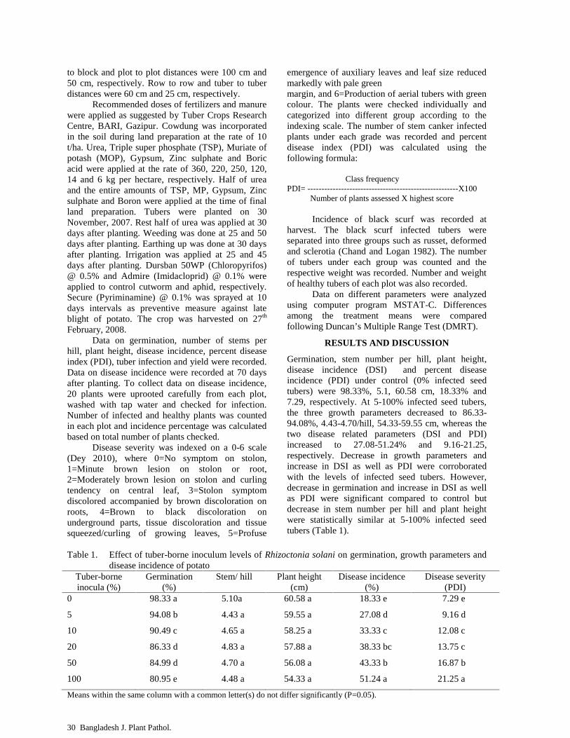
30 Bangladesh J. Plant Pathol.
to block and plot to plot distances were 100 cm and50 cm, respectively. Row to row and tuber to tuberdistances were 60 cm and 25 cm, respectively.
Recommended doses of fertilizers and manurewere applied as suggested by Tuber Crops ResearchCentre, BARI, Gazipur. Cowdung was incorporatedin the soil during land preparation at the rate of 10t/ha. Urea, Triple super phosphate (TSP), Muriate ofpotash (MOP), Gypsum, Zinc sulphate and Boricacid were applied at the rate of 360, 220, 250, 120,14 and 6 kg per hectare, respectively. Half of ureaand the entire amounts of TSP, MP, Gypsum, Zincsulphate and Boron were applied at the time of finalland preparation. Tubers were planted on 30November, 2007. Rest half of urea was applied at 30days after planting. Weeding was done at 25 and 50days after planting. Earthing up was done at 30 daysafter planting. Irrigation was applied at 25 and 45days after planting. Dursban 50WP (Chloropyrifos)@ 0.5% and Admire (Imidacloprid) @ 0.1% wereapplied to control cutworm and aphid, respectively.Secure (Pyriminamine) @ 0.1% was sprayed at 10days intervals as preventive measure against lateblight of potato. The crop was harvested on 27th
February, 2008.Data on germination, number of stems per
hill, plant height, disease incidence, percent diseaseindex (PDI), tuber infection and yield were recorded.Data on disease incidence were recorded at 70 daysafter planting. To collect data on disease incidence,20 plants were uprooted carefully from each plot,washed with tap water and checked for infection.Number of infected and healthy plants was countedin each plot and incidence percentage was calculatedbased on total number of plants checked.
Disease severity was indexed on a 0-6 scale(Dey 2010), where 0=No symptom on stolon,1=Minute brown lesion on stolon or root,2=Moderately brown lesion on stolon and curlingtendency on central leaf, 3=Stolon symptomdiscolored accompanied by brown discoloration onroots, 4=Brown to black discoloration onunderground parts, tissue discoloration and tissuesqueezed/curling of growing leaves, 5=Profuse
emergence of auxiliary leaves and leaf size reducedmarkedly with pale greenmargin, and 6=Production of aerial tubers with greencolour. The plants were checked individually andcategorized into different group according to theindexing scale. The number of stem canker infectedplants under each grade was recorded and percentdisease index (PDI) was calculated using thefollowing formula:
Class frequencyPDI= ------------------------------------------------------X100
Number of plants assessed X highest score
Incidence of black scurf was recorded atharvest. The black scurf infected tubers wereseparated into three groups such as russet, deformedand sclerotia (Chand and Logan 1982). The numberof tubers under each group was counted and therespective weight was recorded. Number and weightof healthy tubers of each plot was also recorded.
Data on different parameters were analyzedusing computer program MSTAT-C. Differencesamong the treatment means were comparedfollowing Duncan’s Multiple Range Test (DMRT).
RESULTS AND DISCUSSION
Germination, stem number per hill, plant height,disease incidence (DSI) and percent diseaseincidence (PDI) under control (0% infected seedtubers) were 98.33%, 5.1, 60.58 cm, 18.33% and7.29, respectively. At 5-100% infected seed tubers,the three growth parameters decreased to 86.33-94.08%, 4.43-4.70/hill, 54.33-59.55 cm, whereas thetwo disease related parameters (DSI and PDI)increased to 27.08-51.24% and 9.16-21.25,respectively. Decrease in growth parameters andincrease in DSI as well as PDI were corroboratedwith the levels of infected seed tubers. However,decrease in germination and increase in DSI as wellas PDI were significant compared to control butdecrease in stem number per hill and plant heightwere statistically similar at 5-100% infected seedtubers (Table 1).
Table 1. Effect of tuber-borne inoculum levels of Rhizoctonia solani on germination, growth parameters anddisease incidence of potato
Tuber-borneinocula (%)
Germination(%)
Stem/ hill Plant height(cm)
Disease incidence(%)
Disease severity(PDI)
0 98.33 a 5.10a 60.58 a 18.33 e 7.29 e
5 94.08 b 4.43 a 59.55 a 27.08 d 9.16 d
10 90.49 c 4.65 a 58.25 a 33.33 c 12.08 c
20 86.33 d 4.83 a 57.88 a 38.33 bc 13.75 c
50 84.99 d 4.70 a 56.08 a 43.33 b 16.87 b
100 80.95 e 4.48 a 54.33 a 51.24 a 21.25 a
Means within the same column with a common letter(s) do not differ significantly (P=0.05).
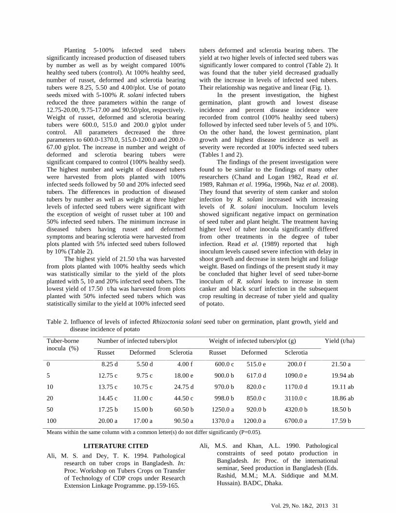
Vol. 29, No. 1&2, 2013 31
Planting 5-100% infected seed tuberssignificantly increased production of diseased tubersby number as well as by weight compared 100%healthy seed tubers (control). At 100% healthy seed,number of russet, deformed and sclerotia bearingtubers were 8.25, 5.50 and 4.00/plot. Use of potatoseeds mixed with 5-100% R. solani infected tubersreduced the three parameters within the range of12.75-20.00, 9.75-17.00 and 90.50/plot, respectively.Weight of russet, deformed and sclerotia bearingtubers were 600.0, 515.0 and 200.0 g/plot undercontrol. All parameters decreased the threeparameters to 600.0-1370.0, 515.0-1200.0 and 200.0-67.00 g/plot. The increase in number and weight ofdeformed and sclerotia bearing tubers weresignificant compared to control (100% healthy seed).The highest number and weight of diseased tuberswere harvested from plots planted with 100%infected seeds followed by 50 and 20% infected seedtubers. The differences in production of diseasedtubers by number as well as weight at three higherlevels of infected seed tubers were significant withthe exception of weight of russet tuber at 100 and50% infected seed tubers. The minimum increase indiseased tubers having russet and deformedsymptoms and bearing sclerotia were harvested fromplots planted with 5% infected seed tubers followedby 10% (Table 2).
The highest yield of 21.50 t/ha was harvestedfrom plots planted with 100% healthy seeds whichwas statistically similar to the yield of the plotsplanted with 5, 10 and 20% infected seed tubers. Thelowest yield of 17.50 t/ha was harvested from plotsplanted with 50% infected seed tubers which wasstatistically similar to the yield at 100% infected seed
tubers deformed and sclerotia bearing tubers. Theyield at two higher levels of infected seed tubers wassignificantly lower compared to control (Table 2). Itwas found that the tuber yield decreased graduallywith the increase in levels of infected seed tubers.Their relationship was negative and linear (Fig. 1).
In the present investigation, the highestgermination, plant growth and lowest diseaseincidence and percent disease incidence wererecorded from control (100% healthy seed tubers)followed by infected seed tuber levels of 5, and 10%.On the other hand, the lowest germination, plantgrowth and highest disease incidence as well asseverity were recorded at 100% infected seed tubers(Tables 1 and 2).
The findings of the present investigation werefound to be similar to the findings of many otherresearchers (Chand and Logan 1982, Read et al.1989, Rahman et al. 1996a, 1996b, Naz et al. 2008).They found that severity of stem canker and stoloninfection by R. solani increased with increasinglevels of R. solani inoculum. Inoculum levelsshowed significant negative impact on germinationof seed tuber and plant height. The treatment havinghigher level of tuber inocula significantly differedfrom other treatments in the degree of tuberinfection. Read et al. (1989) reported that highinoculum levels caused severe infection with delay inshoot growth and decrease in stem height and foliageweight. Based on findings of the present study it maybe concluded that higher level of seed tuber-borneinoculum of R. solani leads to increase in stemcanker and black scurf infection in the subsequentcrop resulting in decrease of tuber yield and qualityof potato.
Table 2. Influence of levels of infected Rhizoctonia solani seed tuber on germination, plant growth, yield anddisease incidence of potato
Tuber-borneinocula (%)
Number of infected tubers/plot Weight of infected tubers/plot (g) Yield (t/ha)
Russet Deformed Sclerotia Russet Deformed Sclerotia
0 8.25 d 5.50 d 4.00 f 600.0 c 515.0 e 200.0 f 21.50 a
5 12.75 c 9.75 c 18.00 e 900.0 b 617.0 d 1090.0 e 19.94 ab
10 13.75 c 10.75 c 24.75 d 970.0 b 820.0 c 1170.0 d 19.11 ab
20 14.45 c 11.00 c 44.50 c 998.0 b 850.0 c 3110.0 c 18.86 ab
50 17.25 b 15.00 b 60.50 b 1250.0 a 920.0 b 4320.0 b 18.50 b
100 20.00 a 17.00 a 90.50 a 1370.0 a 1200.0 a 6700.0 a 17.59 b
Means within the same column with a common letter(s) do not differ significantly (P=0.05).
LITERATURE CITED
Ali, M. S. and Dey, T. K. 1994. Pathologicalresearch on tuber crops in Bangladesh. In:Proc. Workshop on Tubers Crops on Transferof Technology of CDP crops under ResearchExtension Linkage Programme. pp.159-165.
Ali, M.S. and Khan, A.L. 1990. Pathologicalconstraints of seed potato production inBangladesh. In: Proc. of the internationalseminar, Seed production in Bangladesh (Eds.Rashid, M.M.; M.A. Siddique and M.M.Hussain). BADC, Dhaka.

32 Bangladesh J. Plant Pathol.
Chadha, K.L. 1995. Integrated management ofbacterial wilt (Eds, Hardly, B and E.R.French). CIP, Lima, Peru. 19 (1-2): 13-20.
Chand, T. and Logan, C. 1982. Reaction of tenpotato cultivars to stem canker and black scurfof potato caused by Rhizoctonia solani.Annals of Applied Biology. 100: 102-103.
Hashem, A. 1990. An introduction to the potato seedindustary of Bangladesh. Internationalseminar. Seed potato in Bangladesh, BADC,Dhaka, Bangladesh. 1-15.
Dey, T. K. 2010. Tuber crops diseases-theiridentification, method of recording, ratingscale and grading system. Oilseed Res.Center, Bangladesh Agril. Res. Inst. Gazipur.pp. 28-31.
Kotcon, J. B., Rouse, O.I. and Mitchell, J. E. 1985.Interactions of Verticillium dahliae,Colletotrichum coccodes, Rhizoctonia solaniand Pratylenchus penetrans in the early dyingsyndrome of Russet Burbank potatoes.Phytopathol. 75: 68-74.
Naz, Farah, Rauf, C. A., Abbasi, N. A., Haque, I. U.and Ahmad, I. 2008. Influence of inoculumlevels of Rhizoctonia solani and susceptibilityon new potato germplasms. Pakistan. J. Bot.40 (5): 2199-2209.
Rahman, M.L., Hossain, M.M., Ashrafuzzaman, M.
and Islam, T. 1996a. Effect of inoculum levelsof Rhizoctonia solani on the incidence ofblack scurf disease of potato. Bangladesh J.Plant Pathol. 12 (1-2): 21-22.
Rahman, M.L., Hossain, M.M., Islam, T. andAshrafuzzaman, M. 1996b. Effect of inoculumlevels of Sclerotium rolfsii on thedevelopment of wilt in potato. Bangladesh J.Plant Pathol. 12 (1-2): 27-28.
Read, P. J., Hide, G. A., Firmaer, J.P. and Hall, S.M.1989. Growth and yield of potatoes as affectedby severity of stem canker (Rhizoctoniasolani). Potato Res. 32: 9-15.
Swaminathan, M.S. 2000. Potato for global security.In: Potato Global Research and Development.Indian Potato Assoc, CPRI, Shimla, H.P,India. 302 p.
Uddin, M. A., Yasmin, S., Rahman, M. L., Hossain,S. M. B. and Choudhury, R.U. 2010.Challenges of potato cultivation inBangladesh and developing digital databasesof potato. Bangladesh J. Agril. Res. 35 (3):453-463.
Wicks, T, Morgan, J. B. and Hall, B. 1996. Influenceof soil fumigation and seed tuber treatment onthe control of Rhizoctonia solani on potatoes.Australian J. Exp. Agric. 36: 339-345.
21.519.94 19.11 18.86 18.5 17.59
y = -0.689x + 21.66R² = 0.920
0
5
10
15
20
25
0 5 10 20 50 100
Yield of healthy tubers (t/ha)
Level of infected seed tubers (%)
Figurer 1. Relationship between levels of seed tubers infected with Rhizoctonia solaniand yield of healthy tubers.
Vol. No. 1&2, 2013 33
INTEGRATED APPLICATION OF INSECTICIDE AND FUNGICIDE TO CONTROL OkYVCMVAND PSEUDOCERCOSPORA LEAF SPOT OF OKRA SEED CROPS
M. G. Kibria1 and I. H. Mian2
1Senior Scientific Officer, Plant Pathology Section, HRC, BARI, 2Professor, Department of Plant Pathology,Bangabandhu Sheikh Mujibur Rahman Agricultural University, Gazipur, Bangladesh
(The paper is a part of Ph. D. thesis of the first author)
ABSTRACT
M. G. Kibria and I. H. Mian. 2013. Integrated application of insecticide and fungicide to control OkYVCMVand Pseudocercospora leaf spot of okra seed crops. Bangladesh J. Plant Pathol. 29 (1&2):33-38.
A field experiment was conducted to test the efficacy ofintegrated application of pesticides to control Okra yellowvein clearing mosaic virus (OkYVCMV) and Pseudo-cercospora leaf spot caused by Pseudocercosporaabelmoschi (PLS) of okra seed crop, and to increase yieldand quality of okra seeds. All pesticidal treatments gaveconsiderable reduction over control in incidence ofOkYVCMV, severity of PLS, and population of whitefly(Bemisia tabaci). The reduction over control was 55.95,43.33, 35.35 and 21.02% in OkYVCMV incidence and89.18, 72.45, 62.36 and 31.52% in PLS due to treatmentsT1= pre-sowing seed treatment with Gaucho 72 WS(Imidacloprid) at 5 g/kg seed + soil treatment with Furadan5G (Carbofuran) at 30 kg/ha)+ 4 foliar sprays with Admire
(Imidacloprid) at 1 ml/L+ 4 foliar sprays with Emivit 50WP at 3 g/liter, T2=seed treatment with Gaucho 72 WS+ 4foliar sprays with Admire + 4 foliar sprays with Emivit 50WP, T3=seed treatment with Gaucho 72 WS+ 4 foliarsprays with Emivit 50 WP, and T4=seed treatment withGaucho 72 WS, respectively. Reduction in population ofwhitefly due to the pesticidal treatments were 69.06, 58.39and 17.78% at 30 days after sowing, and 73.90, 65.76 and40.29% at 60 days after sowing. The treatments improvedplant growth, pod and seed yield and seed qualityconsiderably. The seed yield was increased by 133.22,80.54, 64.91 and 23.05% over control due to thetreatments. The best treatment was T1 to control bothdiseases and to improve yield and quality of seeds.
Key words: Okra, OkYVCMV, Pseudocercospora leaf spot.
INTRODUCTION
Okra, also known as lady’s finger (Abelmoschusesculentus L.) is one of the popular vegetable cropsin Bangladesh. It is cultivated throughout the yearexcept 3-4 cool months. It suffers from a number ofdiseases caused by fungi, bacteria, nematode andviruses (Talukdar 1974, Fakinr 2000, Rangaswamiand Mahadevan 2006). Among them, Okra YellowVein Clearing Mosaic Virus (OkYVCMV) andPsudocercospora leaf spot (P. abelmoschi) are twomajor diseases. OkYVCMV infects at all growingstages of okra causing severe reduction in plantgrowth and yield. The disease is transmitted by thevector whitefly (Bemisia tabaci). Plants infected atearly stages remain stunted. The fruits of the infectedplant exhibit pale yellow color, deformed, small andtough in texture. Seeds of infected plants are smallerand shriveled. If the plants are infected within 20days after germination (DAG), the yield loss may beup to 94%. Infection of plants at 50 and 60 DAGmay cause 84 and 49% yield losses, respectively(Sastry and Singh 1974).Attempt was made by many researchers to control
the disease through insect vector control and foundthat application of insecticide minimized whiteflypopulation and the incidence of OkYVCMV (Pun etal. 2005a, 2005b, Gowdar et al. 2007). Significantreduction in OkYVCMV incidence and whitefly
2013 Bangladesh Phytopathological Society
populations was achieved with foliar spray of Aceta-miprid, Dimethoate, Imidacloprid, Meta-systox,Monocrotophos, Nuvacron, Thiamethoxam, TriazoAhmed et al. 2001, Misra 2005, Gowdar et al. 2007).Pesticidal seed treatment with insecticide(Imidacloprid) (Praveen et al. 2007) and soilapplication of Carbofuran at the time of sowing areeffective measures to control OkYVCMV through itsvector suppression (Murthy and Reddy 1992,Praveen et al. 2007).
Pseudocercospora leaf spot (PLS) of okracaused by Pseudocercospora abelmoschi is prevalentin late summer and early winter. The disease maycause severe problem in seed crop causing smallerfruits and shriveled seeds. Spraying okra field with500 g Zineb or Maneb suspended in 250 liters ofwater at 15 days interval after the first appearance ofthe symptoms proved effective for controllingPseudocercospora leaf blight disease. The bestcontrol of P. abelmoschi was obtained from foliarspray of Copper oxychloride, followed byCarbendazim and Mancozeb. Carbendazim gave thehighest yield increase, followed by CopperOxychloride and Mancozeb (Srivastava et al. 1992,Rahman et al. 2000).
Both OkYVCMV and Pseudocercospora leafspot are common in Bangladesh (Talukdar 1974).These are considered as serious diseases of okra,

34 Bangladesh J. Plant Pathol.
especially when it is grown in late summer.Comprehensive research has not yet been accom-plished in the country to find out effective controlmethods against the diseases. Available literaturereveal that effective control of OkYVCMV andPseudocercospora leaf spot may be achievedthrough vector control using insecticides and foliarspray with fungicides, respectively. Integratedapplication of the pesticides may give better controlcompared to application of either insecticides orfungicides. Present piece of research was conductedto test the efficacy of integrated application ofinsecticide and fungicide to control OkYVCMV andPsuedocercospora leaf spot of okra.
MATERIALS AND METHODS
An experiment was conducted to test the efficacy ofthree insecticides and a fungicide applied in anintegrated approach to control Okra Yellow VeinClearing Mosaic Virus (OkYVCMV) andPsuedocercospora leaf spot (Pseudocercosporaabelmoschi) of okra. Three insecticides namelyGaucho 72 WS (Imidacloprid), Furadan 5G(Carbofuran) and Admire (Imidacloprid) wereapplied to control OkYVCMV through vectorcontrol, and the fungicide Emivit 50 WP was used tocontrol Psuedocercospora leaf spot. Individualcomponents of treatments in the present experimentwere: C1 = Pre-sowing insecticidal seed treatmentwith Gaucho 72 WS (Imidacloprid) @ 5 g/kg seed,C2 = Pre-sowing soil treatment with Furadan 5G(Carbofuran) @ 30 kg/ha), C3 = Four foliar sprayswith Admire (Imidacloprid) @ 1 ml/L, and C4 = Fourfoliar sprays with Emivit 50 WP @ 3 g/liter.Spraying of Admire was started from 15 DAS andcontinued for 4 times at 15 days interval. Furadan 5Gwas applied in the furrows at sowing. Seeds weretreated with the insecticide just before sowing. Sprayof fungicide was started when symptoms of PLSappeared and continued for 4 times with 7 daysinterval. The integrated treatments, T1= C1+ C2+ C3+C4, T2= was C1+C3+ C4, T3= C1+ C4 and T4= C1. Anadditional treatment T5 was maintained whichreceived neither insecticide nor fungicide and servedas control.
The experiment was conducted in theexperimental farm of Bangabandhu Sheikh MujiburRahman Agrilcultural Univeristy (BSMRAU),Gazipur during 2009. The experiment was laid outfollowing randomized complete block design withthree replications. The unit plot size was 4.5 m × 1.2m. Drains of 50 cm width and 30 cm depth were dugaround each unit plot for maintaining isolationdistance as well as facilitating irrigation and drainage
of excess water. The experimental field was preparedproperly for good tilth following standard practices(Razzaque et al. 2000). Fertilizers were applied @50-24-30-10-1-0.5 kg of N-P-K-S-Zn-B per hectare,respectively (Anon. 2005). Manure was applied ascowdung at the rate of 4 t/ha. The entire quantity ofcowdung, P, K, S, Zn and B and half of N wereapplied at the time of final land preparation.Remaining N was applied around the base of theplant as top dressing and incorporated with soil at3rd and 5th week after sowing. A previously selectedsusceptible okra genotype was used in theexperiment. Seeds of the genotype were soaked intap water for 12 hours, air dried and treated withGaucho 72 WS. Treated seeds were sown in the fieldmaintaining plant to plant and row to row distancesof 60 and 50 cm, respectively. After 6 to 7 days ofgermination, the seedlings were thinned to have oneplant per hill. If any seedling died within three weeksof germination it was replaced by seedling of sameage raised in polyethylene bags (9 x 15 cm).Irrigation was applied after each top dressing of ureaand whenever necessary. Every time, irrigation wasfollowed by mulching and weeding. Other necessaryintercultural operations were done throughout thecropping season for proper growth and fruitproduction (Razzaque et al. 2000).
Data on incidence of OkYVCMV wasrecorded on 60 days after sowing (DAS). Number ofdiseased and healthy plants in each unit plot wascounted. Incidence of OkYVCMV was expressed inpercentage based on total number of plants checked.The disease incidence (DI) was computed followinga formula as shown below:
Number of infected plants% DI = ------------------------------------------X 100
Total number of plants checked
Data on whitefly population was alsorecorded. To count population of adult whitefly, fiveplants were selected randomly from each unit plotand 5 leaves were selected from each plant atdifferent nodal positions (one at the top, two in themiddle and two at the bottom). The counts weretaken in the morning when whiteflies are less activeas suggested by Basu (1995). Number of whitely perleaf was counted at 30 and 60 DAS. Precaution wastaken not to disturb the insect at the time of counting.Severity of Pseudocercospora leaf spot (PLS) wasindexed on a 0-8 scale as suggested by Rahman andNahar (1990) at 60 DAS. Percent Disease Index(PDI) for the leaf spot was computed according tothe following formula as described by Mian (1995):
Sum of all disease ratingsPDI = --------------------------------------------------------------------X 100
Total number of leaves checked X maximum disease grade

Vol. No. 1&2, 2013 35
Data on plant growth, seed yield, selectedyield contributing parameters, and seed quality werealso recorded. Quality of seeds in terms ofgermination, seedling growth and vigor index weredetermined following methods of International SeedTesting Association (ISTA) (Anon. 1996). Seedlingvigour was determined using a standard formula(Baki and Anderson, 1972) viz.
Vigour Index = (mean root length + mean shootlength) x percent emergence.
Fully ripened pods were collected from each of theunit plot and seeds were separated from the pods, sundried and cleaned. Seed yield per plant werecomputed. Recorded data were subjected tostatistical analysis following standard procedure(Gomez and Gomez 1984) using MSTAT-Cstatistical software. Whenever necessary, data weretransformed using ArcSin transformation methodbefore performing analysis of variance.
RESULTS AND DISCUSSION
Incidence of OkYVCMV
Incidence of OkYVCMV on okra seed crop variedfrom 28.31 to 64.27% under different treatmentsincluding control (T1 to T5). The highest incidence ofthe disease was recorded from control plot. Allpesticidal treatments (T1 to T4) caused significantreduction in disease incidence over control. Thereduction over control was 55.95, 43.33, 35.35 and21.02% respectively under treatments T1, T2, T 3 andT 4 (Table 1).
Severity of Pseudocercospora leaf spot (PLS)
The percent disease index (PDI) of PLS on okra inplots under different treatments including controlranged 5.64-52.12. The highest disease severity wasfound under control. Like OkYVCMV, PDI of PLSwas also significantly reduced due to applicationpesticidal treatments (T1 to T4) compared to control.The reductions over control were 89.18, 72.45, 62.36and 31.52% under T1, T2, T 3 and T 4 treated plots,respectively (Table 1).
Population of whitefly
Average populations of whitefly on okra seed cropranged from 0.91-2.98 per leaf at 30 DAS and 1.25-4.79 per leaf per plant at 60 DAS recorded on fivetreatment approaches (T1 to T4) including control(T5). Treatments T1, T2 and T3 significantly reducedthe whitefly populations over control (T5). The maxi-mum reduction was achieved by T1 treated plotfollowed by T2 and T3 showing 69.06, 58.39 and17.78% reduction over control at 30 DAS, and 73.90,65.76 and 40.29% at 60 DAS, respectively (Table 2).
Plant height and Fruit yield per plant
Plant height of the seed crop was 105.89, 97.90,93.80, 90.01 and 84.12 cm found in the plots treatedwith T1 to T5, respectively. All pesticidal treatmentincreased plant height significantly over control. Themaximum increase was obtained in T1 treated plotfollowed by T2, T3 and T4 treated ones (Table 3).
Number of fruits per plant under the T2, T 3,T4 and T5 was 16.11, 13.83, 13.15, 11.91 and 10.44,respectively. Significant increase in fruit yield wasobtained with all the pesticidal treatments (T1 to T4).Differences in number of fruit per plant weresignificant (Table 3).
Seed number per fruit and per plant, seed yield,seed size and Seed quality
All qualitative parameters of seeds in terms ofgermination, seedling growth and seed vigor wereimproved tremendously due to application of allpesticidal treatments. The lowest number of seedsper fruit was found in treated control plots (T5). Thefruit number increased significantly over control dueto application of the treatments T1 to T4. The highestfruit yield was obtained from T1 treated plotfollowed by T2 and T4 treated plots showing 59.96,56.39, 55.16 and 49.59 seeds per fruit. The lowestseed number of 476.63 seeds per plant was harvestedfrom control plot. It was increased to 965.90, 779.81,725.17 and 590.82 due to pesticidal treatments T1,T2, T3 and T4, respectively. The increase wassignificant compared to control (Table 3).
Seed yield was 56.44, 43.69, 39.91, 31.23 and24.20 g/plant in plots treated with differenttreatments (T1, T2, T3 and T4). Significantly thehighest seed yield was achieved with all pesticidaltreatments over control. The yield increase in T1, T2,T3 and T4 treated plots was 133.22, 80.54, 64.91 and23.05%, respectively. Seed size in terms of 1000-seed weight increased significantly over control (T5)due to four pesticidal treatments (T1,- T4). Themaximum increase in 1000-seed weight wasachieved with T1 followed by T2, T3 and T4 showing1000-seed weight of 58.43, 56.03, 55.03 and 52.85 g,respectively. (Table 4).
Shoot and root length and Seed vigour index
The root and shoot length ranged from 6.04-13.05cm and 5.32-9.59 cm, respectively obtained indifferent treated plots. Both the parameters weresignificantly higher in all four pesticidal treated plotcompared to untreated control plot. The highestshoot and root length were found in the plot treatedwith T1 followed by T2, T3, T4 and T5 treated plots(Table 5).
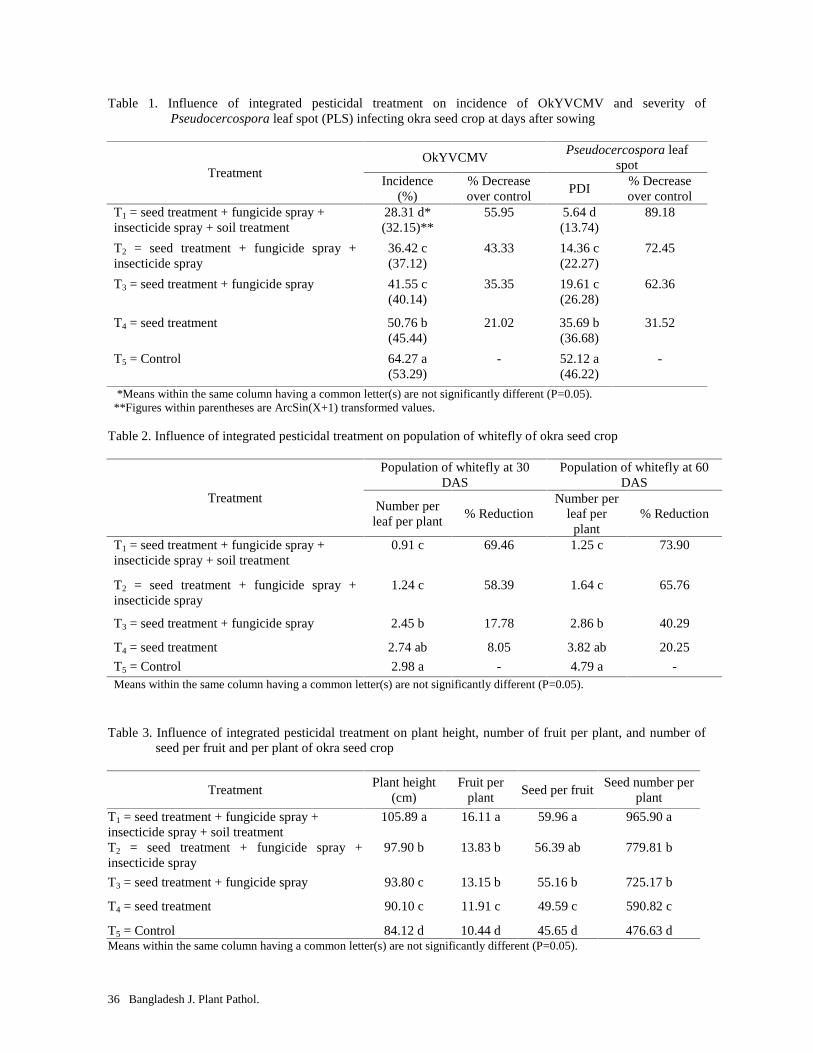
36 Bangladesh J. Plant Pathol.
Table 1. Influence of integrated pesticidal treatment on incidence of OkYVCMV and severity ofPseudocercospora leaf spot (PLS) infecting okra seed crop at days after sowing
TreatmentOkYVCMV
Pseudocercospora leafspot
Incidence(%)
% Decreaseover control
PDI% Decreaseover control
T1 = seed treatment + fungicide spray +insecticide spray + soil treatment
28.31 d*(32.15)**
55.95 5.64 d(13.74)
89.18
T2 = seed treatment + fungicide spray +insecticide spray
36.42 c(37.12)
43.33 14.36 c(22.27)
72.45
T3 = seed treatment + fungicide spray 41.55 c(40.14)
35.35 19.61 c(26.28)
62.36
T4 = seed treatment 50.76 b(45.44)
21.02 35.69 b(36.68)
31.52
T5 = Control 64.27 a(53.29)
- 52.12 a(46.22)
-
*Means within the same column having a common letter(s) are not significantly different (P=0.05).**Figures within parentheses are ArcSin(X+1) transformed values.
Table 2. Influence of integrated pesticidal treatment on population of whitefly of okra seed crop
Treatment
Population of whitefly at 30DAS
Population of whitefly at 60DAS
Number perleaf per plant
% ReductionNumber per
leaf perplant
% Reduction
T1 = seed treatment + fungicide spray +insecticide spray + soil treatment
0.91 c 69.46 1.25 c 73.90
T2 = seed treatment + fungicide spray +insecticide spray
1.24 c 58.39 1.64 c 65.76
T3 = seed treatment + fungicide spray 2.45 b 17.78 2.86 b 40.29
T4 = seed treatment 2.74 ab 8.05 3.82 ab 20.25
T5 = Control 2.98 a - 4.79 a -Means within the same column having a common letter(s) are not significantly different (P=0.05).
Table 3. Influence of integrated pesticidal treatment on plant height, number of fruit per plant, and number ofseed per fruit and per plant of okra seed crop
TreatmentPlant height
(cm)Fruit per
plantSeed per fruit
Seed number perplant
T1 = seed treatment + fungicide spray +insecticide spray + soil treatment
105.89 a 16.11 a 59.96 a 965.90 a
T2 = seed treatment + fungicide spray +insecticide spray
97.90 b 13.83 b 56.39 ab 779.81 b
T3 = seed treatment + fungicide spray 93.80 c 13.15 b 55.16 b 725.17 b
T4 = seed treatment 90.10 c 11.91 c 49.59 c 590.82 c
T5 = Control 84.12 d 10.44 d 45.65 d 476.63 dMeans within the same column having a common letter(s) are not significantly different (P=0.05).
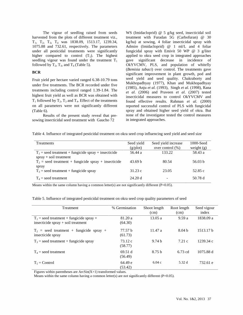
Vol. No. 1&2, 2013 37
The vigour of seedling raised from seedsharvested from the plots of different treatment viz.,T1, T2, T3, T4 was 1838.09, 1513.17, 1239.34,1075.88 and 732.61, respectively. The parametersunder all pesticidal treatments were significantlyhigher compared to control (T5). The highestseedling vigour was found under the treatment T1
followed by T2, T3, and T4 (Table 5).
BCR
Fruit yield per hectare varied ranged 6.38-10.79 tonsunder five treatments. The BCR recorded under fivetreatments including control ranged 1.39-1.84. Thehighest fruit yield as well as BCR was obtained withT1 followed by T2, T3 and T4. Effect of the treatmentson all parameters were not significantly different(Table 6).
Results of the present study reveal that pre-sowing insecticidal seed treatment with Gaucho 72
WS (Imidacloprid) @ 5 g/kg seed, insecticidal soiltreatment with Furadan 5G (Carbofuran) @ 30kg/ha) at sowing, 4 foliar insecticidal spray withAdmire (Imidacloprid) @ 1 ml/L and 4 foliarfungicidal spray with Emivit 50 WP @ 3 g/literapplied to okra seed crop in integrated approachesgave significant decrease in incidence ofOkYVCMV, PLS, and population of whitefly(Bemisia tabaci) over control. The treatments gavesignificant improvement in plant growth, pod andseed yield and seed quality. Chakraborty andMukhopadhyay (1977), Khan and Mukhopadhyay(1985), Anju et al. (1993), Singh et al. (1998), Ranaet al. (2006) and Praveen et al. (2007) testedinsecticidal measures to control OkYVCMV andfound effective results. Rahman et al. (2000)reported successful control of PLS with fungicidalspray and obtained higher seed yield of okra. Butnone of the investigator tested the control measuresin integrated approaches.
Table 4. Influence of integrated pesticidal treatment on okra seed crop influencing seed yield and seed size
Treatments Seed yield(g/plot)
Seed yield increaseover control (%)
1000-Seedweight (g)
T1 = seed treatment + fungicide spray + insecticidespray + soil treatment
56.44 a 133.22 58.43 a
T2 = seed treatment + fungicide spray + insecticidespray
43.69 b 80.54 56.03 b
T3 = seed treatment + fungicide spray 31.23 c 23.05 52.85 c
T4 = seed treatment 24.20 d - 50.78 d
Means within the same column having a common letter(s) are not significantly different (P=0.05).
Table 5. Influence of integrated pesticidal treatment on okra seed crop quality parameters of seed
Treatment % Germination Shoot length(cm)
Root length(cm)
Seed vigourindex
T1 = seed treatment + fungicide spray +insecticide spray + soil treatment
81.20 a(64.30)
13.05 a 9.59 a 1838.09 a
T2 = seed treatment + fungicide spray +insecticide spray
77.57 b(61.73)
11.47 a 8.04 b 1513.17 b
T3 = seed treatment + fungicide spray 73.12 c(58.77)
9.74 b 7.21 c 1239.34 c
T4 = seed treatment 69.51 d(56.49)
8.75 b 6.73 cd 1075.88 d
T5 = Control 64.49 e(53.42)
6.04 c 5.32 d 732.61 e
Figures within parentheses are ArcSin(X+1) transformed values.Means within the same column having a common letter(s) are not significantly different (P=0.05).
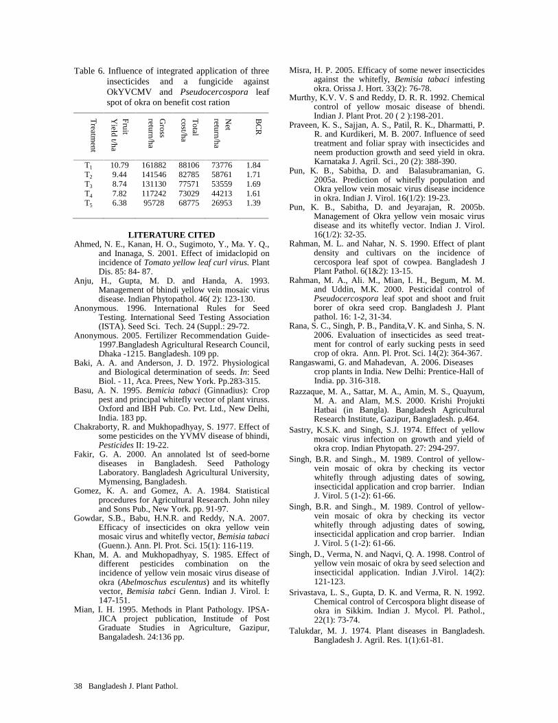
38 Bangladesh J. Plant Pathol.
Table 6. Influence of integrated application of threeinsecticides and a fungicide againstOkYVCMV and Pseudocercospora leafspot of okra on benefit cost ration
Treatm
ent
Fruit
Yield t/ha
Gross
return/ha
Total
cost/ha
Net
return/ha
BC
R
T1
T2
T3
T4
T5
10.799.448.747.826.38
16188214154613113011724295728
8810682785775717302968775
7377658761535594421326953
1.841.711.691.611.39
LITERATURE CITEDAhmed, N. E., Kanan, H. O., Sugimoto, Y., Ma. Y. Q.,
and Inanaga, S. 2001. Effect of imidaclopid onincidence of Tomato yellow leaf curl virus. PlantDis. 85: 84- 87.
Anju, H., Gupta, M. D. and Handa, A. 1993.Management of bhindi yellow vein mosaic virusdisease. Indian Phytopathol. 46( 2): 123-130.
Anonymous. 1996. International Rules for SeedTesting. International Seed Testing Association(ISTA). Seed Sci. Tech. 24 (Suppl.: 29-72.
Anonymous. 2005. Fertilizer Recommendation Guide-1997.Bangladesh Agricultural Research Council,Dhaka -1215. Bangladesh. 109 pp.
Baki, A. A. and Anderson, J. D. 1972. Physiologicaland Biological determination of seeds. In: SeedBiol. - 11, Aca. Prees, New York. Pp.283-315.
Basu, A. N. 1995. Bemicia tabaci (Ginnadius): Croppest and principal whitefly vector of plant viruss.Oxford and IBH Pub. Co. Pvt. Ltd., New Delhi,India. 183 pp.
Chakraborty, R. and Mukhopadhyay, S. 1977. Effect ofsome pesticides on the YVMV disease of bhindi,Pesticides II: 19-22.
Fakir, G. A. 2000. An annolated lst of seed-bornediseases in Bangladesh. Seed PathologyLaboratory. Bangladesh Agricultural University,Mymensing, Bangladesh.
Gomez, K. A. and Gomez, A. A. 1984. Statisticalprocedures for Agricultural Research. John nileyand Sons Pub., New York. pp. 91-97.
Gowdar, S.B., Babu, H.N.R. and Reddy, N.A. 2007.Efficacy of insecticides on okra yellow veinmosaic virus and whitefly vector, Bemisia tabaci(Guenn.). Ann. Pl. Prot. Sci. 15(1): 116-119.
Khan, M. A. and Mukhopadhyay, S. 1985. Effect ofdifferent pesticides combination on theincidence of yellow vein mosaic virus disease ofokra (Abelmoschus esculentus) and its whiteflyvector, Bemisia tabci Genn. Indian J. Virol. I:147-151.
Mian, I. H. 1995. Methods in Plant Pathology. IPSA-JICA project publication, Institude of PostGraduate Studies in Agriculture, Gazipur,Bangaladesh. 24:136 pp.
Misra, H. P. 2005. Efficacy of some newer insecticidesagainst the whitefly, Bemisia tabaci infestingokra. Orissa J. Hort. 33(2): 76-78.
Murthy, K.V. V. S and Reddy, D. R. R. 1992. Chemicalcontrol of yellow mosaic disease of bhendi.Indian J. Plant Prot. 20 ( 2 ):198-201.
Praveen, K. S., Sajjan, A. S., Patil, R. K., Dharmatti, P.R. and Kurdikeri, M. B. 2007. Influence of seedtreatment and foliar spray with insecticides andneem production growth and seed yield in okra.Karnataka J. Agril. Sci., 20 (2): 388-390.
Pun, K. B., Sabitha, D. and Balasubramanian, G.2005a. Prediction of whitefly population andOkra yellow vein mosaic virus disease incidencein okra. Indian J. Virol. 16(1/2): 19-23.
Pun, K. B., Sabitha, D. and Jeyarajan, R. 2005b.Management of Okra yellow vein mosaic virusdisease and its whitefly vector. Indian J. Virol.16(1/2): 32-35.
Rahman, M. L. and Nahar, N. S. 1990. Effect of plantdensity and cultivars on the incidence ofcercospora leaf spot of cowpea. Bangladesh JPlant Pathol. 6(1&2): 13-15.
Rahman, M. A., Ali. M., Mian, I. H., Begum, M. M.and Uddin, M.K. 2000. Pesticidal control ofPseudocercospora leaf spot and shoot and fruitborer of okra seed crop. Bangladesh J. Plantpathol. 16: 1-2, 31-34.
Rana, S. C., Singh, P. B., Pandita,V. K. and Sinha, S. N.2006. Evaluation of insecticides as seed treat-ment for control of early sucking pests in seedcrop of okra. Ann. Pl. Prot. Sci. 14(2): 364-367.
Rangaswami, G. and Mahadevan, A. 2006. Diseasescrop plants in India. New Delhi: Prentice-Hall ofIndia. pp. 316-318.
Razzaque, M. A., Sattar, M. A., Amin, M. S., Quayum,M. A. and Alam, M.S. 2000. Krishi ProjuktiHatbai (in Bangla). Bangladesh AgriculturalResearch Institute, Gazipur, Bangladesh. p.464.
Sastry, K.S.K. and Singh, S.J. 1974. Effect of yellowmosaic virus infection on growth and yield ofokra crop. Indian Phytopath. 27: 294-297.
Singh, B.R. and Singh., M. 1989. Control of yellow-vein mosaic of okra by checking its vectorwhitefly through adjusting dates of sowing,insecticidal application and crop barrier. IndianJ. Virol. 5 (1-2): 61-66.
Singh, B.R. and Singh., M. 1989. Control of yellow-vein mosaic of okra by checking its vectorwhitefly through adjusting dates of sowing,insecticidal application and crop barrier. IndianJ. Virol. 5 (1-2): 61-66.
Singh, D., Verma, N. and Naqvi, Q. A. 1998. Control ofyellow vein mosaic of okra by seed selection andinsecticidal application. Indian J.Virol. 14(2):121-123.
Srivastava, L. S., Gupta, D. K. and Verma, R. N. 1992.Chemical control of Cercospora blight disease ofokra in Sikkim. Indian J. Mycol. Pl. Pathol.,22(1): 73-74.
Talukdar, M. J. 1974. Plant diseases in Bangladesh.Bangladesh J. Agril. Res. 1(1):61-81.
Vol. 29, No. 1&2, 2013 39
CHARACTERIZATION OF OROBANCHE SPECIES OCCURRING IN BANGLADESH
Manisha Saha1 and I. H. Mian2
1Graduate Student and 2Professor, Department of Plant Pathology, BangabandhuSheikh Mujibur Rahman Agricultural University, Gazipur, Bangladesh
Email of corresponding author: [email protected]
(The paper is a part of M.S thesis of the first author)
ABSTRACT
Manisha Shaha and I. H. Mian. 2013. Characterization of Orobanche species occurring in Bangladesh.Bangladesh J. Plant Pathol. 29 (1&2): 39-44.
A survey was conducted in the district of Jessore, Kushtia,Pabna and Rajshahi to identify and characterize Orobanchespecies occurring in those locations of Bangladesh. Twodistinct species of Orobanche were found to occur in thecountry. The first one was found to attack mustard,cabbage and cauliflower, and the second one attackedsunflower, tomato and tobacco. Orobanche sp. collectedfrom mustard showed higher shoot length, canopydiameter, sepal number, petal length, ovary length, ovarydiameter, and style length. Flowers were with 2 bractlets,calyx divided at the base posteriorly, and entire anteriorly,stamen number was 4 (all equal), with wooly anthers. On
the other hand, Orobanche sp. collected from tomato hadhigher stem girth, leaf length, leaf width, sepal length,stigma diameter and stamen length. Flowers were withoutbractlets, calyx divided at the base anteriorly as well asposteriorly, stamen number was 4 (2 longer than other 2),with glabrous anthers. The morphological charactersclearly showed that the two Orobanche observed on twohosts species were distinctly different species. Themorphological characters were compared with a standardkey and Orobanche collected from mustard was identifiedas O. aegyptiaca L. and the parasite attacking tomato wasidentified as O. cernua L.
Key words: Orobanche species, host range, survey.
INTRODUCTION
Orobanche, commonly known as broomrape, is agenus of parasitic flowering plants. It is aholoparasite affecting several hundred crop speciesincluding rapeseed-mustard, tobacco, potato, brinjal,cabbage, cauliflower, turnip and many otherSolanaceous and Cruciferous plants (Musselman1980). They lack chlorophyll and their leaves arevestigial. Above-ground stems are produced only forthe purpose of flowering and setting seeds. Afterripening of the fruits, seeds of Orobanche shed insoil, they undergo a conditioning period of severalweeks. After conditioning, seeds germinate inresponse to host stimulant and radicle (germtube)emerges. Germination is followed by haustorialinitiation, attachment of the host roots, and lastlypenetration of the vascular tissues (Musselman 1980,Kreutz 1995).
Species of Orobanche are aggressive anddamaging parasitic weeds which have a tremendousimpact on agriculture causing considerable yieldlosses, in many countries, especially in the drier andwarmer areas of Europe, Africa and Asia (Dhanapalet al. 2009). The losses may vary from 5 to 100%(Stewart and Press 1990). The average crop loss,across all broomrape species, is approximately 34%(Linke et al. 1989, Diaz et al. 2006).
A comprehensive review published in India(Dhanapal et al. 2009) reveal that practices to control
Bangladesh Phytopathological Society
broomrape include physical methods (weeding, soil
tillage, flooding, irrigation, pola-rization, flaming),
chemical methods (soil fumigation, herbicide
application, use of germination stimulants), bio-
logical methods (use of resistant or tolerant
varieties), cropping systems with trap and catch
crops, intercropping, and biological control (with
insects or fungi).In Bangladesh, Orobanche attack different
crops. However, list of the susceptible crops are notavailable in the country. At at least two species of thepest namely O. aegyptiaca and O. cernua occur inBangladesh (Begum and Huq 1983). Comprehensivereport on crop loss due to the pest is not available inthe country. A recent hand book on agriculturaltechnologies published by Bangladesh AgriculturalResearch Institute reveals that Orobanche is acommon pest of mustard. In Bangladesh,recommendations to control the pest include removalof Orobanche plants before flowering, avoidance ofgrowing mustard in known infested fields,destruction of the weed with 2,4-D and use ofappropriate amount of TSP fertilizer (Islam et al.2004). Alam et al. (2008) identified some resistantgermplasm of mustard in a screening test conductedduring 2007-2008.
The present paper reported the results of aninvestigation conducted to characterize the species ofOrobanche attacking crop plants in Bangladesh.
40 Bangladesh J. Plant Pathol.
MATERIALS AND METHODS
The investigation was conducted in the district ofJessore, Kushtia, Pabna and Rajshahi. Orobancheinfested fields of mustard, sunflower, tomato,cabbage, cauliflower and tobacco were visited andhost range of the parasite was recorded. Apparentlyhealthy and matured Orobanche plants werecollected from each of the host crops. Canopydiameter covered by Orobanche plants and growthhabit of shoot were recorded.
To collect data on plant characters, theselected Orobanche plants were uprooted by diggingwith a spade along with root systems of respectivehosts. They were washed with tap water carefully tominimize root damage of host plants. Whole plantsand their individual parts were checked with nakedeyes or under a hand lens or using a compoundmicroscope according to necessity. Data oncharacteristics of shoot, leaf, flower and fruit wererecorded.
For recording leaf characters, 20 fully openleaves were selected randomly from the plants.Lamina length from base attached with the stem tothe tip, lamina width at the widest point and laminashape were recorded.
A total of 20 fully open flowers were checkedand data on flowers types, flowers arrangement(solitary or clustered), color of petals and anther,number and length of sepal, petal and stamen, lengthof style and ovary, diameter of stigma and ovary, andshape and diameter of pollen were recorded.
RESULTS AND DISCUSSION
Orobanche and their hosts identified
During the investigation, mustard, sunflower,tomato, cabbage, cauliflower and tobacco werefound to be infected with Orobanche. Themorphological appearance of the pests infectingsunflower, tomato and tobacco was distinctlydifferent from the parasites that attacked mustard,cabbage and cauliflower (Plate-IA&B). Morpho-logical characters of stem, leaf and flowers recordedduring the investigation were compared with astandard key and Orobanche from mustard familywas identified as Orobanche aegyptiaca L. and theparasite infecting other crops was identified asOrobanche cernua L (Lie Dang Shu 1998).
In mustard fields, O. aegyptiaca infected up to100% host plants. In severe cases, infected hostplants died before maturation (Plate-IC). In infectedtomato fields, Orobanche shoot emerged in clustershowing up to 42% host plants infection. Infectedtomato plants gradually died and weathered beforematuration of fruits (Plate-ID).
Both O. aegyptiaca and O. cernua were foundto grow closed to the stem base or a little far from itdepending on length of root systems of their hosts.The shoot of O. cernua attacking tomato is robust ascompared to O. aegyptiaca that attacks mustard. Theappressorium of the Orobanche developed at thebase of stems was thicker on tomato than thoseinfecting mustard (Plate-IE&F). The number ofparasite shoots per square meter was higher inmustard fields than tomato fields. It might be due tothe fact that the parasite attacking mustard has highertendency to branch than those of tomato.
Patrick and Miller (1994) also found similarresults. They reported that in some species (such asO. aegyptiaca, O. ramosa) branching of the shoot isnormal, whereas in others (O. cernua, O. minor, Ocrenata) branching is rare.
Reports from various sources reveal that seedsof Orobanche germinate in response to the hoststimulant and the radicle, also known as germtube,emerges. If the radicle reaches the host root, its tipthickens and attaches itself to root surface. Thethickening is known as appressorium. Finally,haustorium initiates from the appressorium, whichengulfs the host roots and penetrates the vasculartissues of the host and makes parasitic relationshipwith the host (Plate-IIA&B). With this organ, theparasite withdraws water, minerals and organiccompound from the hosts (Visser and Dorr 1986,Stewart and Press 1990, Nun and Mayer 1993).
Morphology of Orobanche aegyptiaca
Plant: The canopy diameters of O. aegyptiaca inmustard fields ranged 1.40-3.80 cm with a mean of2.44 cm and standard deviation 0.65 cm. Stems wereerect, roundish, thinner, purplish white and glandularpubescent. The shoots were branched above themiddle point but some were unbranched. They weresmooth having crowded leaves and flowers. Theplant height ranged 17.00-31.00 cm with a mean of23.88 cm and standard deviation 3.71 cm. The rangeof stem girth at the widest point was 1.00-2.00 cmwith a mean of 1.69 cm and standard deviation of0.35 cm (Table 1).
Leaf: The leaves were alternate, acute, andlanceolate. The base of leaves was wide and tip waspointed. Stem under and around a leaf was lightgreenish yellow. Leaf length ranged 0.60-1.10 cmwith a mean of 0.77 cm and standard deviation 0.15cm. The maximum leaf width ranged 0.30-0.60 cmwith a mean of 0.41 and standard deviation 0.09 cm(Plates I&II and Table 1).
Flower: Flowering initiated in January and ended inMarch. Inflorescence was spicate, bract ovate-lanceolate, with 2 bractlets, flowers sub-sessile,bisexual, alternate, axial and attached with the stemat an acute angle (Plate I&II). Calyx was campanu-
Orobanche
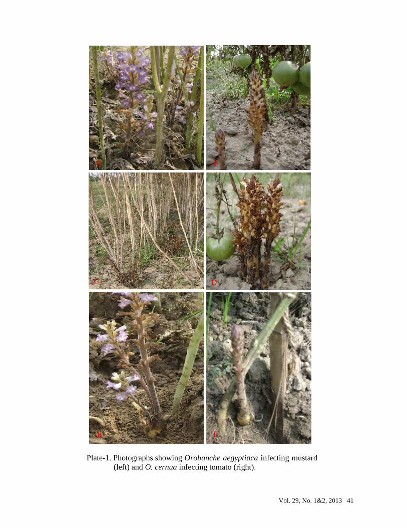
Vol. 29, No. 1&2, 2013 41
A
Plate-1. Photographs showing Orobanche aegyptiaca infecting mustard(left) and O. cernua infecting tomato (right).
A B
C D
E F
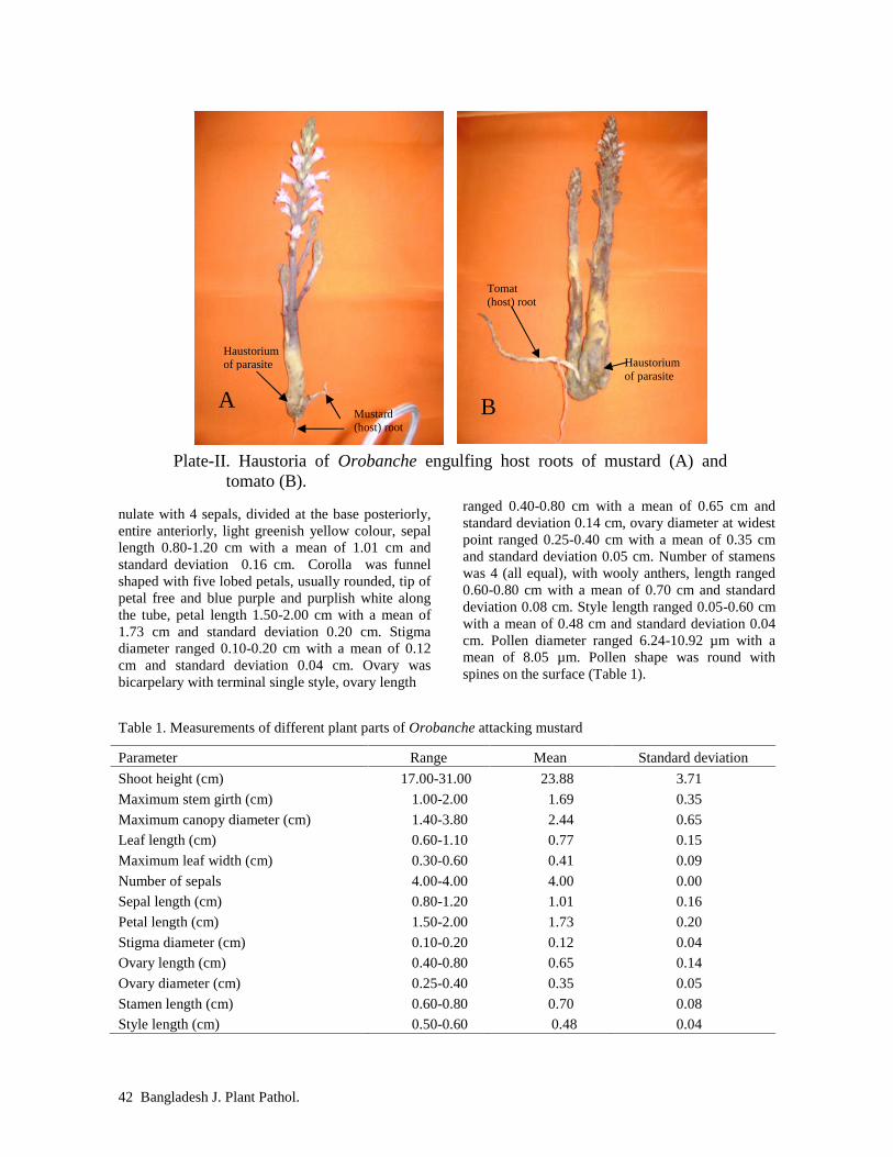
42 Bangladesh J. Plant Pathol.
nulate with 4 sepals, divided at the base posteriorly,entire anteriorly, light greenish yellow colour, sepallength 0.80-1.20 cm with a mean of 1.01 cm andstandard deviation 0.16 cm. Corolla was funnelshaped with five lobed petals, usually rounded, tip ofpetal free and blue purple and purplish white alongthe tube, petal length 1.50-2.00 cm with a mean of1.73 cm and standard deviation 0.20 cm. Stigmadiameter ranged 0.10-0.20 cm with a mean of 0.12cm and standard deviation 0.04 cm. Ovary wasbicarpelary with terminal single style, ovary length
ranged 0.40-0.80 cm with a mean of 0.65 cm andstandard deviation 0.14 cm, ovary diameter at widestpoint ranged 0.25-0.40 cm with a mean of 0.35 cmand standard deviation 0.05 cm. Number of stamenswas 4 (all equal), with wooly anthers, length ranged0.60-0.80 cm with a mean of 0.70 cm and standarddeviation 0.08 cm. Style length ranged 0.05-0.60 cmwith a mean of 0.48 cm and standard deviation 0.04cm. Pollen diameter ranged 6.24-10.92 µm with amean of 8.05 µm. Pollen shape was round withspines on the surface (Table 1).
Table 1. Measurements of different plant parts of Orobanche attacking mustard
Parameter Range Mean Standard deviation
Shoot height (cm) 17.00-31.00 23.88 3.71
Maximum stem girth (cm) 1.00-2.00 1.69 0.35
Maximum canopy diameter (cm) 1.40-3.80 2.44 0.65
Leaf length (cm) 0.60-1.10 0.77 0.15
Maximum leaf width (cm) 0.30-0.60 0.41 0.09
Number of sepals 4.00-4.00 4.00 0.00
Sepal length (cm) 0.80-1.20 1.01 0.16
Petal length (cm) 1.50-2.00 1.73 0.20
Stigma diameter (cm) 0.10-0.20 0.12 0.04
Ovary length (cm) 0.40-0.80 0.65 0.14
Ovary diameter (cm) 0.25-0.40 0.35 0.05
Stamen length (cm) 0.60-0.80 0.70 0.08
Style length (cm) 0.50-0.60 0.48 0.04
Plate-II. Haustoria of Orobanche engulfing host roots of mustard (A) andtomato (B).
A BMustard(host) root
Haustoriumof parasite
Tomat(host) root
Haustoriumof parasite

Vol. 29, No. 1&2, 2013 43
Pollen diameter (µm) 6.24-10.92 8.05 1.09Morphology of Orobanche cernua
Plant: The stems were erect, robust, rigid, thicker,light brown at immature stage, dark brown at maturestage, glabrous near stem base or minutely pubescentfrom middle upwards, and unbranched with crowdedleaves and flowers. The plant height ranged 14.00-32.00 cm with a mean of 23.13 cm and standarddeviation 5.27 cm. Maximum stem girth ranged1.70-4.00 cm with a mean of 3.26 cm and standarddeviation 0.85 cm, and the canopy diameters ranged1.40-2.00 cm with a mean of 1.68 cm and standarddeviation 0.25 cm (Plate-III and Table 2).
Leaf: The leaves were scale like, alternate, acute,ovate-lanceolate along with bracts, bractlets andcalyx. The base of leaves was swollen due todevelopment of flower bud, and pointed tip. Leaflength ranged 1.00-1.50 cm with a mean of 1.27 cmand standard deviation 0.20 cm. The maximum leafwidth ranged 0.40-0.70 cm with a mean of 0.58 andstandard deviation 0.10 cm (Plate III and Table 2).
Flower: Flowering initiated in January and ended inMarch. Inflorescence was spicate, bract ovate-lanceolate, without bractlets, acuminate, flower
sub-sessile, bisexual, alternate, axial and attachedwith the stem at an acute angle. Calyx wascampanulate with 2 distinct sepals, divided at thebase both anteriorly and posteriorly, greenish brown,length 0.7-1.0 cm with a mean of 1.88 cm andstandard deviation 0.11 cm. Corolla was funnelshaped with five lobed petals, usually rounded, tip ofpetal free and white with blue tint at the tip, purplishwhite along the tube, length 1.30-2.00 cm with amean of 1.68 cm and standard deviation 0.25 cm.Stigma diameter ranged 0.10-0.20 cm with a mean of0.16 cm and standard deviation 0.05 cm. Ovary wasbicarpelary with terminal single style, length ranged0.30-0.70 cm with a mean of 0.57 cm and standarddeviation 0.13 cm, diameter 0.20-0.30 cm with amean of 0.27 cm and standard deviation 0.03 cm.Number of stamens was 4 (2 longer than other 2),length ranged 0.70 -0.90 cm with a mean of 0.81 cmand standard deviation 0.07 cm. Style length ranged0.04-0.50 cm with a mean of 0.48 cm and standarddeviation 0.04 cm. Pollen diameter ranged 7.80-10.92 µm with a mean 9.05 µm and standarddeviation 1.57 µm. Pollen of O. cernua collectedfrom tomato was round (Plate III and Table 2).
Table 2. Measurements of different plant parts of Orobanche attacking tomato
Parameter Range Mean Standard deviation
Shoot height (cm) 14.00-32.0 23.13 5.27
Maximum stem girth (cm) 1.70-4.0 3.26 0.85
Maximum canopy diameter (cm) 1.40-2.0 1.68 0.25
Leaf length (cm) 1.00-1.5 1.27 0.20
Maximum leaf width (cm) 0.40-0.7 0.58 0.10
Number of sepals 2.00-2.0 2.00 -
Sepal length (cm) 0.70-1.0 1.88 0.11
Petal length (cm) 1.30-2.0 1.68 0.25
Stigma diameter (cm) 0.10-0.2 0.16 0.05
Ovary length (cm) 0.30-0.7 0.57 0.13
Ovary diameter (cm) 0.20-0.30 0.27 0.03
Stamen length (cm) 0.70-0.9 0.81 0.07
Style length (cm) 0.40-0.5 0.48 0.04
Pollen diameter (µm) 7.80-10.92 9.05 1.57
Comparison of O. aegyptiaca and O. cernua
Morphological differences between two Orobanchespecimens collected from mustard and tomato areshown in Table 3. Orobanche collected from
mustard showed higher shoot length, canopydiameter, sepal number, petal length, ovary length,ovary diameter, and style length. Flowers were with2 bractlets, calyx divided at the base posteriorly, and

44 Bangladesh J. Plant Pathol.
entire anteriorly, stamen number was 4 (all equal),with wooly anthers. On the other hand, Orobanchecollected from tomato had higher stem girth, leaflength, leaf width, sepal length, stigma diameter andstamen length. Flowers were without bractlets, calyxdivided at the base anteriorly as well as posteriorly,stamen number was 4 (2 longer than other 2), withglabrous anthers. The morphological charactersclearly showed that the two Orobanche observed ontwo hosts species were distinctly different species.The morphological characters were compared with astandard key published in China for identifying them
up to species level (Lie Dang Shu 1998). Accordingto the key, Orobanche from mustard was identifiedas Orobanche aegyptiaca L. and the parasiteattacking tomato was identified as Orobanchecernua L. Previous reports published by BangladeshNational Herbarium also reveal that Orobancheaegyptiaca and Orobanche cernua occur inBangladesh (Begum and Huq 1983). The pests arewidely distributed in Southern Europe, Asia andAfrica. Orobanche aegyptiaca is also known as O.indica, and Phelipaea aegyptiaca (Musselman 1980,1986).
Inflorescence
FlowerFlower
Plate III. Photograph showing stems, leaf, inflorescence, flowers and pollens of O.aegyptiaca (left) and O. cernua (right)
Leaf
Stem
Inflorescence
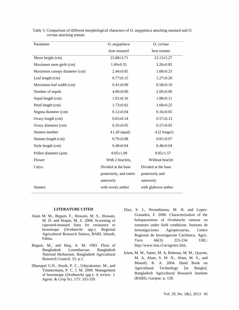
Vol. 29, No. 1&2, 2013 45
Table 3. Comparison of different morphological characters of O. aegyptiaca attacking mustard and O.cernua attacking tomato
Parameter O. aegyptiaca
host mustard
O. cernua
host tomato
Shoot height (cm) 23.88±3.71 23.13±5.27
Maximum stem girth (cm) 1.69±0.35 3.26±0.85
Maximum canopy diameter (cm) 2.44±0.65 1.68±0.25
Leaf length (cm) 0.77±0.15 1.27±0.20
Maximum leaf width (cm) 0.41±0.09 0.58±0.10
Number of sepals 4.00±0.00 2.00±0.00
Sepal length (cm) 1.01±0.16 1.88±0.11
Petal length (cm) 1.73±0.02 1.68±0.25
Stigma diameter (cm) 0.12±0.04 0.16±0.05
Ovary length (cm) 0.65±0.14 0.57±0.13
Ovary diameter (cm) 0.35±0.05 0.27±0.03
Stamen number 4 ( all equal) 4 (2 longer)
Stamen length (cm) 0.70±0.08 0.81±0.07
Style length (cm) 0.48±0.04 0.48±0.04
Pollen diameter (µm) 8.05±1.09 9.05±1.57
Flower With 2 braclets, Without braclet
Calyx Divided at the base
posteriorly, and entire
anteriorly
Divided at the base
posteriorly and
anteriorly
Stamen with wooly anther with glabrous anther
LITERATURE CITED
Alam M. M., Begum, F., Hossain, M. S., Hossain,M. D. and Hoque, M. Z. 2008. Screening ofrapeseed-mustard lines for resistance tobroomrape (Orobanche spp.). RegionalAgricultural Research Station, BARI. Ishurdi,Pabna.
Begum, M., and Huq, A. M. 1983. Flora ofBangladesh. Loranthaceae. BangladeshNational Herbarium. Bangladesh AgriculturalResearch Council. 33: p.1.
Dhanapal G.N., Struik, P. C., Udayakumar, M., andTimmermans, P. C. J. M. 2009. Managementof broomrape (Orobanche spp.): A review. J.Agron. & Crop Sci. 175: 335-359.
Diaz, S. J., Norambuena, M. H. and Lopez-Granados, F. 2006. Characterization of theholoparasitism of Orobanche ramosa ontomatoes under field conditions. Instituto deInvestigaciones Agropecuarias, CentroRegional de Investigacion Carillanca, Agric.Tecn. 66(3): 223-234. URL:http://www.inia.cl/at/agritec.htm.
Islam, M. M., Satter, M. A, Rahman, M. M., Qayum,M. A, Alam, S. M. N., Alam, M. S., andMustafi, R. A. 2004. Hand Book onAgricultural Technology [in Bangla].Bangladesh Agricultural Research Institute(BARI), Gazipur. p. 128.
46 Bangladesh J. Plant Pathol.
Lie Dang Shu. 1998. Flora of China. 18: 231-239.
Linke, K. H., Sauerborn, J., and Saxena, M. C. 1989.Orobanche Field Guide. ICARDA-FLIP,Aleppo, Syria. 42 pp.
Musselman, L. J. 1980. The biology of Striga,Orobanche, and other root-parasitic weeds.Annu. Rev. Phytopathol. 18: 463-489.
Musselman, L.J. 1986. Taxonomy of Orobanche. in:S.J. ter Borg (ed.) Proceedings of a Workshopon the Biology and Control of Orobanche,LH/VPO, Wageningen, The Netherlands. pp.2-10.
Nun, N. B., and Mayer, A. M. 1993. Preconditioningand germination of Orobanche seeds:Respiration and protein synthesis.
Phytochemistry 34(1): 39-45.
Patrick, M. 1994. The Biology and Distribution ofParasitic Plants within the Orobanchaceae: AnOverview of Secondary Plant CompoundInvolvement Colorado State University. FortCollins, Colorado 80523.
Stewart, G. R., and Press, M. C. 1990. Thephysiology and biochemistry of parasiticangiosperms. Annu. Rev. Plant Phys. PlantMol. Biol. 41: 127-151.
Visser, J., and Dorr, I. 1986. The Haustorium (CH
7). In: L. J. Musselman (ed.), Parasitic Weeds
in Agriculture. Vol. I, Striga. CRC Press,
Inc., Boca Raton, FL.