Embed Size (px)
Citation preview
https://doi.org/10.1177/1534735417740630
Integrative Cancer Therapies1 –9© The Author(s) 2017 Reprints and permissions:sagepub.com/journalsPermissions.nav DOI: 10.1177/1534735417740630journals.sagepub.com/home/ict
Creative Commons Non Commercial CC BY-NC: This article is distributed under the terms of the Creative Commons Attribution-NonCommercial 4.0 License (http://www.creativecommons.org/licenses/by-nc/4.0/) which permits non-commercial use,
reproduction and distribution of the work without further permission provided the original work is attributed as specified on the SAGE and Open Access pages (https://us.sagepub.com/en-us/nam/open-access-at-sage).
Original Article
Introduction
Chemotherapy is currently the mainstay of treatment for most types of cancer, with agents exerting their anticancer effect by inducing apoptosis.1,2 One such drug currently in use is paclitaxel. Paclitaxel is most often used for the treat-ment of breast cancer, ovarian cancer, non–small cell lung cancer, and AIDS-related Kaposi’s sarcoma.3 Paclitaxel is able to induce the mitochondrial apoptotic pathway in vari-ous cancer cell types. Paclitaxel induces the mitochondrial permeability transition, triggering the release of prodeath molecules (Bax and Bad, which then inactivate Bcl-2 or Bcl-xL) and activating caspases, which then induce apoptosis of neoplastic cells.4-6 Chemotherapy drugs can be effective in treating cancer; however, many chemotherapeutic agents exhibit dose-limiting toxicities, which can cause congestive heart failure, myelosuppression, neurotoxicity, immune-sup-pression, and mutagenic and carcinogenic effects.7-10 Many attempts have been made to circumvent the toxic effects of chemotherapy, including using chemosensitizers, which
make tumor cells more sensitive to the effects of chemo-therapy and therefore require lower doses of the toxic che-motherapeutic drugs. In the past 40 years, research has focused on the development of potent therapies to circum-vent multidrug resistance (MDR), and several chemosensi-tizing agents have been discovered which enhance the cytotoxic effect of chemotherapy drugs in cancer cells; these include the calcium blockers diltiazem, the biscoclaurine alkaloid cepharanthine, and verapamil11-14; the anti-arrhyth-mic agent quinidine15; and the synthetic isothiocyanate derivative E-4IB.16 However, these agents are also toxic. Calcium antagonist poisoning is well documented, and other
740630 ICTXXX10.1177/1534735417740630Integrative Cancer TherapiesBadr El-Din et alresearch-article20172017
1University of Mansoura, Mansoura, Egypt2Drew University of Medicine and Science, Los Angeles, CA, USA
Corresponding Author:Nariman K. Badr El-Din, Department of Zoology, Faculty of Science, University of Mansoura, Mansoura 35516, Egypt. Email: [email protected]
Baker’s Yeast Sensitizes Metastatic Breast Cancer Cells to Paclitaxel In Vitro
Nariman K. Badr El-Din, PhD1, Ashraf Z. Mahmoud, PhD1, Tahia Ali Hassan, MS1, and Mamdooh Ghoneum, PhD2
AbstractOur earlier studies have demonstrated that phagocytosis of baker’s yeast (Saccharomyces cerevisiae) induces apoptosis in different cancer cell lines in vitro and in vivo. This study aimed to examine how baker’s yeast sensitizes murine and human breast cancer cells (BCC) to paclitaxel in vitro. This sensitizing effect makes lower concentrations of chemotherapy more effective at killing cancer cells, thereby enhancing the capacity of treatment. Three BCC lines were used: the metastatic murine 4T1 line, the murine Ehrlich ascites carcinoma (EAC) line, and the human breast cancer MCF-7 line. Cells were cultured with different concentrations of paclitaxel in the presence or absence of baker’s yeast. Cell survival and the IC50 values were determined by MTT assay and trypan blue exclusion method. Percent of DNA damage, apoptosis, and cell proliferation were examined by flow cytometry. Yeast alone and paclitaxel alone significantly decreased 4T1 cell viability postculture (24 and 48 hours), caused DNA damage, increased apoptosis, and suppressed cell proliferation. Baker’s yeast in the presence of paclitaxel increased the sensitivity of 4T1 cells to chemotherapy and caused effects that were greater than either treatment alone. The chemosensitizing effect of yeast was also observed with murine EAC cells and human MCF-7 cells, but to a lesser extent. These data suggest that dietary baker’s yeast is an effective chemosensitizer and can enhance the apoptotic capacity of paclitaxel against breast cancer cells in vitro. Baker’s yeast may represent a novel adjuvant for chemotherapy treatment.
KeywordsSaccharomyces cerevisiae, apoptosis, paclitaxel, breast cancer, 4T1
Received May 9, 2017; revised September 16, 2017; accepted September 25, 2017

2 Integrative Cancer Therapies
side effects may include dizziness, headache, redness in the face, fluid buildup in the legs and ankles, abnormal heart rate, constipation, and gingival overgrowth.17,18
Recently, several researchers have focused on screening for nontoxic natural modulators to overcome ABC trans-porter-mediated MDR. Two natural products, curcumin and flavonoids, have been extensively studied in the context of modulation of MDR transporter expression.19,20 In addition, work from our laboratory has introduced a novel, natural, dietary product, arabinoxylan rice bran (MGN-3/Biobran), which when combined with chemotherapy allowed for low-ering the drug concentration used during treatment, thereby reducing the toxicity of chemotherapy while maintaining potency against cancer cells in vitro and in vivo.21-23 In this study, we evaluated the ability of another dietary product, baker’s and brewer’s yeast, Saccharomyces cerevisiae, to sensitize cancer cells to chemotherapy in vitro. S. cerevisiae is a potent apoptotic agent against cancer cells. Cancer cells undergo apoptosis upon phagocytosis of S. cerevisiae. Baker’s yeast can induce apoptosis in several human cancer cell lines, including breast, tongue, and colon cells in vitro.24-27 Yeast can also exert anticancer effects in nude mice bearing human breast cancer28,29 and in Swiss albino mice bearing Ehrlich carcinoma.30
Our results reveal that S. cerevisiae induces anticancer effects in murine and human breast cancer cells and, when combined with paclitaxel, induces killing of a greater num-ber of cancer cells than either yeast or paclitaxel used alone. These results suggest that baker’s yeast may be used as an adjuvant during chemotherapy treatment and may have clinical implications for the treatment of breast cancer.
Materials and Methods
Drugs and Chemicals
Paclitaxel was purchased from Bristol-Myers Squibb Inc (Princeton, NJ, USA). It was supplied with initial concen-tration of 100 mg/16.7 mL. Each milliliter of sterile nonpy-rogenic solution contains 6 mg paclitaxel, 527 mg of purified Cremophor EL (polyoxyethylated castor oil), and 49.7% (v/v) dehydrated alcohol, USP. RPMI-1640 supple-mented with 10% fetal calf serum (FCS), 3-(4,5-dimethyl-thiazol-2-yl)-2,5-diphenyltetrazoliumbromide (MTT) from Sigma-Aldrich.
Preparation of Saccharomyces cerevisiae
Commercially available baker’s and brewer’s yeast, S. cere-visiae, was used in suspensions that were washed once with phosphate-buffered saline (PBS). It was then incubated for 1 hour at 90°C to kill the yeast and washed 3 times with PBS. Quantification was carried out using a hemocytome-ter, and cell suspensions were adjusted to 1 × 10⁴, 1 × 10⁵, 1 × 10⁶, 1 × 10⁷, 1 × 10⁸, and 1 × 10⁹ cells/mL.
Breast Cancer Cell Lines and Culture Conditions
Three breast cancer cell (BCC) lines were used in the study: the highly metastatic murine 4T1 line; the murine Ehrlich ascites carcinoma (EAC) cell line, a mammary adenocarci-noma; and the human nonmetastatic breast cancer MCF-7 line. 4T1 and MCF-7 cells were purchased from the American Tissue and Culture Collection (ATCC; Manassas, VA, USA). 4T1 and MCF-7 cells were maintained in our laboratory in a complete medium that consisted of RPMI-1640, supplemented with 10% FCS, 2 mM glutamine, and 100 µg/mL streptomycin and penicillin.
We chose MCF-7 as our human BCC line since MCF-7 cells have been found to be more susceptible to yeast-induced apoptosis as compared with other human BCC lines, including ZR-75 cells and HCC70 cells.24 Furthermore, MCF-7 cells proved to be potent phagocytic cells as exem-plified by their ability to rapidly engulf, digest, and frag-ment yeast cells with lysosomal encirclement of the engulfed yeast cells.25,29
Preparation of Ehrlich Ascites Carcinoma Cells
The transplantable murine tumor cell line, namely EAC cells, was obtained from the National Cancer Institute, Cairo University, Egypt. The EAC cells were maintained in the ascitic form in vivo in Swiss albino mice by means of sequential intraperitoneal transplantation of 2 × 10⁶ cells/mouse after every 10 days. Ascitic fluid was drawn out from EAC-bearing mouse 8 days after transplantation from the peritoneal cavity by aspirating the ascitic fluid into a sterile isotonic saline solution. The freshly drawn fluid was diluted with ice-cold sterile PBS (0.2 M, pH 7.4), and the tumor cell count was adjusted to 2 × 10⁶ cells/mL by sterile PBS immediately before the studies.
Effect of Paclitaxel Plus Yeast on Growth of Breast Cancer Cells
Drug Sensitivity Assay. Drug sensitivity was determined by using a colorimetric MTT assay. Cancer cells (1 × 10⁴ cells/well) were seeded in 96-well plates and cultured in tripli-cate with different concentrations of yeast (1 × 10⁴ to 1 × 10⁹ cells/mL) and in the presence or absence of paclitaxel at different concentrations (1 × 10⁻⁶ to 1 × 10⁻¹ M/L). The final volume of medium in each well after the addition of yeast or paclitaxel was 200 µL. The cultures were incubated at 37°C for 24 and 48 hours, after which 50 µg of MTT were added to each well, and the cultures were incubated for an additional 4 hours. The plates were then centrifuged, the medium was carefully removed, the formazan crystals solu-bilized with acid alcohol, and the plates were read at 590 nm using an ELISA plate reader (Molecular Devices, Menlo Park, CA, USA). The 50% inhibitory concentration (IC50) was determined as the drug concentration resulting in a

Badr El-Din et al 3
50% reduction of cell viability. The IC50 was determined by plotting the logarithm of the drug concentration versus the survival rate of the treated cells.
Trypan Blue Exclusion Method. In sterile test tubes, the cells and chemicals were added with the aforementioned concen-trations of yeast, paclitaxel, and both yeast plus paclitaxel in triplicates. Cells were incubated for 24 and 48 hours at 37°C in a humidified atmosphere of 5% CO2 in sterile medium. Viable cells were counted by trypan blue exclusion using hemocytometer. Then the percentage of live cells was obtained by dividing the viable cells by the total number of cells. All experiments were repeated in triplicates.
Flow Cytometric Analysis for Apoptosis, DNA Damage, and Cell Proliferation
Quantitative detection of apoptosis, DNA damage, and cell proliferation in 4T1 cells treated with yeast with and without paclitaxel was simultaneously determined by multicolor flow cytometric analysis using the Apoptosis, DNA Damage and Cell Proliferation Kit specific for incorporated bromodeoxyuridine (BrdU), phosphorylated H2AX (#H2AX), and cleaved poly ADP ribose poly-merase (PARP) (BD Biosciences Pharmingen, San Diego CA, USA). Following the manufacturer’s instructions, cells were cultured in a CM or with different concentra-tions of yeast (1 × 10⁷ cells/mL) and (1 × 10⁹ cells/mL) with and without paclitaxel (1 × 10³ M) for 24 or 48 hours. Ten microliters of BrdU working solution (1 mM BrdU in 1× [DPBS]) was added to each milliliter of tissue culture medium (the cell culture density was approxi-mately 1 × 10⁶ cells/mL); following this, the cells were incubated for 30 minutes on ice. Cells were washed by adding 1 mL of staining buffer/tube and centrifuged (5 minutes) at 250 × g, and the supernatant was discarded. Cells were fixed with 100 µL of BD Cytofix/Cytoperm Fixation/Permeabilization Solution per tube and incu-bated for 30 minutes at room temperature. Afterward, cells were washed with 1 mL of 1× BD Perm/Wash Buffer, centrifuged, and the supernatant was discarded. Cells were incubated in 100 µL of BD Cytofix/Cytoperm Plus Permeabilization Buffer/ tube for 10 minutes in ice, washed, and then refixed for 5 minutes. One hundred microliters of diluted DNase were added to cells, which were incubated for 1 hour at 37°C and then washed. Cells were resuspended with 20 µL wash buffer plus PerCP-Cy 5.5 mouse anti-BrdU (5 µL/test), Alexa Fluor 647 mouse anti-H2AX (pS139) (5 µL/test), PE anti-cleaved PARP (Asp214) (5 µL/test) for 20 minutes in the dark and then washed. Cells were resuspended in staining buffer for analysis by fluorescence-activated cell sorting (FACSCalibur; BD Biosciences, San Jose, CA, USA) using CellQuest 3.3 software.31,32
Statistical Analysis
Values are reported as the mean ± standard error (mean ± SE), and data were analyzed using 1-way analysis of vari-ance followed by post hoc tests for multiple comparisons. A P value less than .05 was considered statistically significant.
Results
Cytotoxicity of Yeast and Paclitaxel on Breast Cancer Cell Lines
Cytotoxicity of yeast plus paclitaxel was examined against three BCC lines: the highly metastatic murine 4T1 line, the murine EAC cell line, and the human MCF-7 cell line. BCCs were cultured with paclitaxel at different con-centrations (10−6-10−1 M/L) in the presence or absence of yeast at different concentrations (104-109 cells/mL). Results were evaluated with 2 different methods (MTT assay and Trypan blue exclusion method) at 24 and 48 hours incubation time before cell survival and the IC50 values were determined.
4T1 Cells. 4T1 cells were incubated for 48 hours with pacli-taxel and/or yeast, and cell survival was examined by MTT assay and IC50 values were also determined (Figure 1A-D). Paclitaxel treatment alone (10−6-10−1 M/L) caused a decrease in 4T1 cell survival with IC50 (5 × 10⁻⁵ M/L) (Fig-ure 1A). Data depicted in Figure 1B show that yeast treat-ment alone (104-109 cells/mL) resulted in decreasing the cell survival with IC50 (2 × 10⁵ cells/mL). On the other hand, data in Figure 1C show that the cytotoxicity of yeast at low concentration of 107 cells/mL in combination with paclitaxel at different concentrations (10−6-10−1 M/L) resulted in a significant decrease of 4T1 cell survival with IC50 (5 × 10⁻⁶ M/L). The cytotoxic effect of yeast at higher concentration of 109 cells/mL in combination with pacli-taxel became more remarkable with IC50 (2 × 10⁻⁶ M/L) (Figure 1D). Similar results were obtained to a lesser extent at 24 hours. Similar results were noticed when Trypan blue exclusion method was used to determine the levels of toxic-ity by yeast and paclitaxel against 4T1 cells (data not shown).
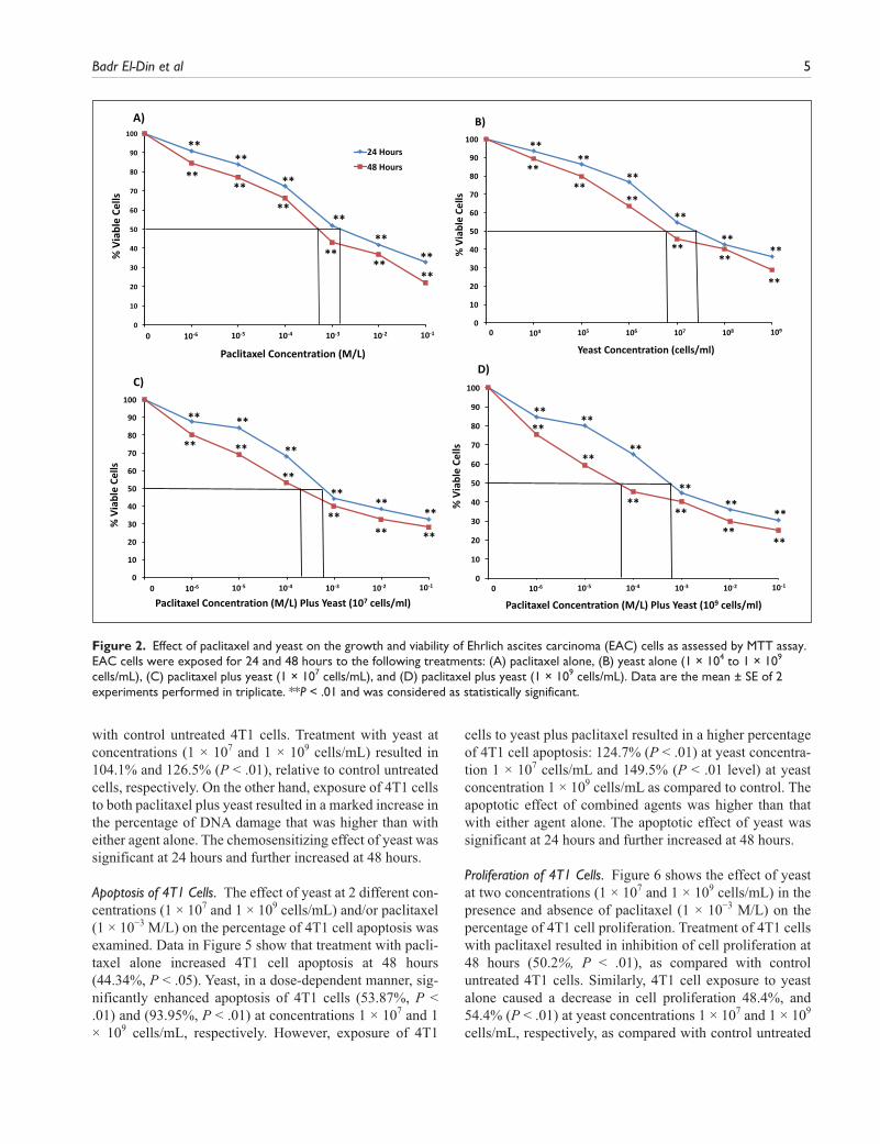
EAC Cell Line. Data in Figure 2A-D show that the combina-tion of yeast with paclitaxel induces higher cytotoxic effects on EAC cells than paclitaxel alone. The decrease in EAC cell survival postexposure to different treatments for 48 hours showed IC50 = 6.86 × 10⁻⁴ M/L for paclitaxel alone (Figure 2A), and IC50 = (7 × 10⁶ cells/mL) for yeast alone (Figure 2B). When paclitaxel was combined with yeast (107 cells/mL), IC50 decreased to 3 × 10⁻⁴ M/L) (Figur 2C) and to 6 × 10⁻⁵ M/L) for 109 cells/mL of yeast (Figure 2D). Sim-ilar results, to a lesser extent, were obtained with yeast

4 Integrative Cancer Therapies
alone at 24 hours. Also, similar results were noticed when the Trypan blue exclusion method was used (data not shown).
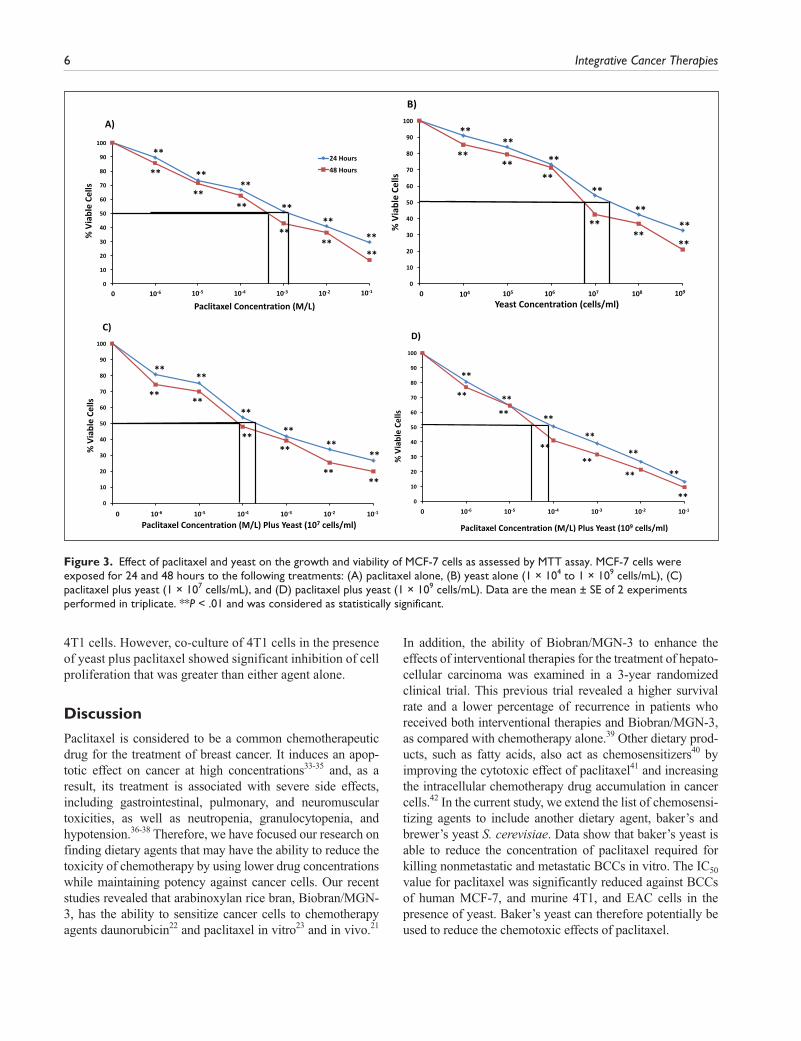
MCF-7 Cell Line. The combined effect of paclitaxel and yeast also yielded a higher cytotoxic effect against human breast MCF-7 cells than either treatment alone. Results in Figure 3A and B show that the decrease in MCF-7 cell survival postexposure to different treatments for 48 hours was IC50 = 6 × 10⁻⁴ M/L for paclitaxel alone, and IC50 = 6.86 × 10⁶ cells/mL for yeast alone, respectively. When the 2 agents were combined, a significant decrease of MCF-7 cell survival was noticed with IC50 = 9 × 10⁻⁵ M/L for 107 cells/mL yeast (Figure 3C), and IC50 = 4 × 10⁻⁵ M/L for 109 cells/mL yeast (Figure 3D). Similar results were obtained to a lesser extent at 24 hours. Also, similar results were noticed when Trypan blue exclusion method was used to determine the levels of toxicity by yeast and paclitaxel against MCF-7 cells (data not shown).
Flow Cytometry Analysis for the Evaluation of Cell Proliferation, DNA Damage, and Apoptosis
Data in Figures 1-3 showed different responses among cell lines toward the cytotoxic effect of paclitaxel, yeast, and yeast plus paclitaxel. We observed the following pattern of sensitivity toward the cytotoxic effect of different treat-ments: 4T1 > MCF-7 > EAC cells, with 4T1 cells proving to be the most responsive. Therefore, the highly metastatic 4T1 cells were used to examine more detailed effects of dif-ferent treatments, including DNA damage, apoptosis, and cell proliferation.
DNA Damage of 4T1 Cells. The effect of yeast at 2 different concentrations (1 × 10⁷ and 1 × 10⁹ cells/mL) and/or pacli-taxel (1 × 10⁻³ M/L) on percentage of DNA damage of 4T1 cells was examined. Data in Figure 4 show that treatment of 4T1 cells with paclitaxel alone significantly increased the percentage of DNA damage (94.3%, P < .01), as compared
Figure 1. Effect of paclitaxel and yeast on the growth and viability of 4T1 cells as assessed by MTT assay. 4T1 cells were exposed for 24 and 48 hours to the following treatments: (A) paclitaxel alone, (B) yeast alone (1 × 10⁴ to 1 × 10⁹ cells/mL), (C) paclitaxel plus yeast (1 × 10⁷ cells/mL), and (D) paclitaxel plus yeast (1 × 10⁹ cells/mL). Data are the mean ± SE of 2 experiments performed in triplicate. *P < .05, **P < .01 and was considered as statistically significant.
Badr El-Din et al 5
with control untreated 4T1 cells. Treatment with yeast at concentrations (1 × 10⁷ and 1 × 10⁹ cells/mL) resulted in 104.1% and 126.5% (P < .01), relative to control untreated cells, respectively. On the other hand, exposure of 4T1 cells to both paclitaxel plus yeast resulted in a marked increase in the percentage of DNA damage that was higher than with either agent alone. The chemosensitizing effect of yeast was significant at 24 hours and further increased at 48 hours.
Apoptosis of 4T1 Cells. The effect of yeast at 2 different con-centrations (1 × 10⁷ and 1 × 10⁹ cells/mL) and/or paclitaxel (1 × 10⁻³ M/L) on the percentage of 4T1 cell apoptosis was examined. Data in Figure 5 show that treatment with pacli-taxel alone increased 4T1 cell apoptosis at 48 hours (44.34%, P < .05). Yeast, in a dose-dependent manner, sig-nificantly enhanced apoptosis of 4T1 cells (53.87%, P < .01) and (93.95%, P < .01) at concentrations 1 × 10⁷ and 1 × 10⁹ cells/mL, respectively. However, exposure of 4T1
cells to yeast plus paclitaxel resulted in a higher percentage of 4T1 cell apoptosis: 124.7% (P < .01) at yeast concentra-tion 1 × 10⁷ cells/mL and 149.5% (P < .01 level) at yeast concentration 1 × 10⁹ cells/mL as compared to control. The apoptotic effect of combined agents was higher than that with either agent alone. The apoptotic effect of yeast was significant at 24 hours and further increased at 48 hours.
Proliferation of 4T1 Cells. Figure 6 shows the effect of yeast at two concentrations (1 × 10⁷ and 1 × 10⁹ cells/mL) in the presence and absence of paclitaxel (1 × 10⁻³ M/L) on the percentage of 4T1 cell proliferation. Treatment of 4T1 cells with paclitaxel resulted in inhibition of cell proliferation at 48 hours (50.2%, P < .01), as compared with control untreated 4T1 cells. Similarly, 4T1 cell exposure to yeast alone caused a decrease in cell proliferation 48.4%, and 54.4% (P < .01) at yeast concentrations 1 × 10⁷ and 1 × 10⁹ cells/mL, respectively, as compared with control untreated
Figure 2. Effect of paclitaxel and yeast on the growth and viability of Ehrlich ascites carcinoma (EAC) cells as assessed by MTT assay. EAC cells were exposed for 24 and 48 hours to the following treatments: (A) paclitaxel alone, (B) yeast alone (1 × 10⁴ to 1 × 10⁹ cells/mL), (C) paclitaxel plus yeast (1 × 10⁷ cells/mL), and (D) paclitaxel plus yeast (1 × 10⁹ cells/mL). Data are the mean ± SE of 2 experiments performed in triplicate. **P < .01 and was considered as statistically significant.
6 Integrative Cancer Therapies
4T1 cells. However, co-culture of 4T1 cells in the presence of yeast plus paclitaxel showed significant inhibition of cell proliferation that was greater than either agent alone.
Discussion
Paclitaxel is considered to be a common chemotherapeutic drug for the treatment of breast cancer. It induces an apop-totic effect on cancer at high concentrations33-35 and, as a result, its treatment is associated with severe side effects, including gastrointestinal, pulmonary, and neuromuscular toxicities, as well as neutropenia, granulocytopenia, and hypotension.36-38 Therefore, we have focused our research on finding dietary agents that may have the ability to reduce the toxicity of chemotherapy by using lower drug concentrations while maintaining potency against cancer cells. Our recent studies revealed that arabinoxylan rice bran, Biobran/MGN-3, has the ability to sensitize cancer cells to chemotherapy agents daunorubicin22 and paclitaxel in vitro23 and in vivo.21
In addition, the ability of Biobran/MGN-3 to enhance the effects of interventional therapies for the treatment of hepato-cellular carcinoma was examined in a 3-year randomized clinical trial. This previous trial revealed a higher survival rate and a lower percentage of recurrence in patients who received both interventional therapies and Biobran/MGN-3, as compared with chemotherapy alone.39 Other dietary prod-ucts, such as fatty acids, also act as chemosensitizers40 by improving the cytotoxic effect of paclitaxel41 and increasing the intracellular chemotherapy drug accumulation in cancer cells.42 In the current study, we extend the list of chemosensi-tizing agents to include another dietary agent, baker’s and brewer’s yeast S. cerevisiae. Data show that baker’s yeast is able to reduce the concentration of paclitaxel required for killing nonmetastatic and metastatic BCCs in vitro. The IC50 value for paclitaxel was significantly reduced against BCCs of human MCF-7, and murine 4T1, and EAC cells in the presence of yeast. Baker’s yeast can therefore potentially be used to reduce the chemotoxic effects of paclitaxel.
Figure 3. Effect of paclitaxel and yeast on the growth and viability of MCF-7 cells as assessed by MTT assay. MCF-7 cells were exposed for 24 and 48 hours to the following treatments: (A) paclitaxel alone, (B) yeast alone (1 × 10⁴ to 1 × 10⁹ cells/mL), (C) paclitaxel plus yeast (1 × 10⁷ cells/mL), and (D) paclitaxel plus yeast (1 × 10⁹ cells/mL). Data are the mean ± SE of 2 experiments performed in triplicate. **P < .01 and was considered as statistically significant.

Badr El-Din et al 7
Many anticancer drugs function by inducing apopto-sis.1,2 Paclitaxel induces apoptosis in different cancer cells, including breast cancer,23 gastric cancer,43 colon cancer,44 and leukemia cells45 by modifying mitochondrial transition permeability, activating caspase-8 and caspase-3,44,46 and Bcl-2 inactivation by a mechanism that may involve the binding of paclitaxel to this antiapoptotic protein.47 As dem-onstrated in this study, heat-killed baker’s yeast also acts as an anticancer agent via induction of apoptosis. These results are in accordance with our earlier studies, which have
shown that human breast, tongue, and colon cancer cells undergo apoptosis on phagocytosis of S. cerevisiae in vitro.24-27 Cancer cells treated with yeast showed clear signs of apoptosis, including nuclear fragmentation and mem-brane blebbing,27 significant decrease in the mitochondrial polarization, and increased activation of caspase-8, -9, and -3 in vitro.24 Furthermore, yeast induced extensive apopto-sis in nude mice bearing human breast cancer and in mice bearing EAC, as determined by histopathological analysis and by flow cytometry.28-30
Earlier studies have also shown that paclitaxel prevents cell proliferation by binding to tubulin in microtubules.48,49 This characteristic may explain the observed inhibition of cell proliferation and DNA damage in 4T1 cells treated with paclitaxel. In the present study, we also noted that exposure of 4T1 cells to paclitaxel plus yeast resulted in a marked inhibition of cell proliferation, significant increase in the percentage of DNA damage, and elevation of apoptotic can-cer cells. The combined effects of both yeast and paclitaxel were more effective than that of either treatment alone. The underlying mechanisms by which yeast sensitizes cancer cells to chemotherapy are not fully understood, but they might be attributed to yeast’s ability to modulate one or more of the various transport proteins of the ABC super-family. These proteins are responsible for MDR by decreas-ing the uptake of the drug or increasing the efflux of the drug from the target organelles. The phytochemical cur-cumin and phytochemical flavonoids have been described as natural modulators of MDR transporter expression.19,20 Curcumin and its metabolite tertrahydrocurcumin were used in restoring drug sensitivity in cancer cells overex-pressing the MDR-linked ABC transporters MRP1,50 Pgp,51
Figure 6. Proliferation of 4T1 cells. The effect of paclitaxel alone (1 × 10⁻³ M/L), and in combination with 2 different concentrations of yeast (1 × 10⁷ and 1 × 10⁹ cells/mL) on the proliferation of 4T1 cells was examined. The percentage proliferation of 4T1 cells was assessed using flow cytometry. Data represent the mean ± SE of 2 experiments performed in triplicate. **P < .01 and was considered as statistically significant.
Figure 4. DNA damage to 4T1 cells. The effect of paclitaxel (1 × 10⁻³ M/L), and 2 different concentrations of yeast (1 × 10⁷ and 1 × 10⁹ cells/mL), and paclitaxel plus yeast on DNA damage to 4T1 cells was examined. DNA damage in 4T1 cells was assessed using flow cytometry. Data represent the mean ± SE of 2 experiments performed in triplicate. **P < .01 and was considered as statistically significant.
Figure 5. Apoptosis of 4T1 cells. The effect of paclitaxel alone (1 × 10⁻³ M/L), and 2 different concentrations of yeast (1 × 10⁷ and 1 × 10⁹ cells/mL), and paclitaxel plus yeast on apoptosis of 4T1 cells at 24 and 48 hours was examined. The percentage of apoptosis of 4T1 cells was assessed using flow cytometry. Data represent the mean ± SE of 2 experiments performed in triplicate. *P < .05, **P < .01 and was considered as statistically significant.

8 Integrative Cancer Therapies
and ABCG252 by directly inhibiting their functions. Similarly, flavonoids have been shown to be potent modula-tors of major ABC drug transporters.53,54
Dietary agents such as Biobran/MGN-3 or the baker’s yeast studied here may provide safe, nontoxic avenues for effective therapies against MDR cancer cells. Studies have shown that yeast is not toxic to nontumorgenic breast epi-thelial (MCF-10A) cells.26 More studies are needed to explore the clinical significance of yeast treatment in differ-ent types of cancer. In our ongoing current studies, intratu-moral injection of yeast, in combination with low doses of paclitaxel, has been found to significantly reduce tumor size in Ehrlich mammary adenocarcinoma bearing mice, as associated with the development of large degenerative necrotic regions in the tumor (unpublished data). In addi-tion, intratumoral injection of yeast has also been found to be effective in inducing apoptotic effects against skin can-cer in rats bearing tumor (unpublished data). We hope that these and other studies will prompt further investments toward studying the effectiveness of yeast treatment for patients with different types of malignancies.
In conclusion, the present study represents the first set of experiments demonstrating that dietary supplementation of baker’s yeast can enhance the apoptotic capacity of pacli-taxel in breast cancer cells in vitro. These data suggest that baker’s yeast may be used as an adjuvant for chemotherapy treatment, which may have clinical implications for the treatment of breast cancer.
Declaration of Conflicting Interests
The author(s) declared no potential conflicts of interest with respect to the research, authorship, and/or publication of this article.
Funding
The author(s) received no financial support for the research, authorship, and/or publication of this article.
References
1. Hickman JA. Apoptosis induced by anticancer drugs. Cancer Metastasis Rev. 1992;11:121-139.
2. Martin SJ, Green DR. Apoptosis as a goal of cancer therapy. Curr Opin Oncol. 1994;6:616-621.
3. Cancer Research UK. General cancer information. Paclitaxel (Taxol). http://www.cancerresearchuk.org/about-cancer/cancers-in-general/treatment/cancer-drugs/paclitaxel. Updated March 19, 2016. Accessed October 17, 2017.
4. Kolfschoten GM, Hulscher TM, Duyndam MC, Pinedo HM, Boven E. Variation in the kinetics of caspase-3 acti-vation, Bcl-2 phosphorylation and apoptotic morphology in unselected human ovarian cancer cell lines as a response to docetaxel. Biochem Pharmacol. 2002;63:733-743.
5. Pienta KJ. Preclinical mechanisms of action of docetaxel and docetaxel combinations in prostate cancer. Semin Oncol. 2001;28(4 suppl 15):3-7.
6. Yamaguchi H, Wang HG. Bcl-XL protects BimEL-induced Bax conformational change and cytochrome C release inde-pendent of interacting with Bax or BimEL. J Biol Chem. 2002;277:41604-41612.
7. Taillibert S, Le Rhun E, Chamberlain MC. Chemotherapy-related neurotoxicity. Curr Neurol Neurosci Rep. 2016;16:81.
8. Singal PK, Iliskovic N. Doxorubicin-induced cardiomyopa-thy. N Engl J Med. 1999;339:900-905.
9. Sanderson BJ, Ferguson LR, Denny WA. Mutagenic and carcinogenic properties of platinum-based anticancer drugs. Mutat Res. 1996;355:59-70.
10. Santin AD, Hermonat PL, Ravaggi A, et al. Effects of con-current cisplatinum administration during radiotherapy vs radiotherapy alone on the immune function of patients with cancer of the uterine cervix. Int J Radiat Oncol Biol Phys. 2000;48:997-1006.
11. Kato T, Suzumura Y. Potentiation of antitumor activity of vincristine by the biscoclaurine alkaloid cepharanthine. J Natl Cancer Inst. 1987;79:527-532.
12. Yamamoto S, Hui PZ, Fukuda Y, Tatsumi K, Mino M. Potentiation of harringtonine cytotoxicity by calcium antago-nist diltiazem and biscoclaurine alkaloid cepharanthine in adriamycin-resistant P388 murine leukemia and K562 human leukemia cells. Biochem Int. 1989;18:1077-1083.
13. Nishikawa K, Asaumi J, Kawasaki S, Kuroda M, Takeda Y, Hiraki Y. Influence of cepharanthin on the intracellular accu-mulation of adriamycin in normal liver cells and spleen cells of mice in vitro and in vivo. Anticancer Res. 1997;17:3617-3621.
14. Tsuruo T, Iida H, Naganuma K, Tsukagoshi S, Sakurai Y. Promotion by verapamil of vincristine responsiveness in tumor cell lines inherently resistant to the drug. Cancer Res. 1983;43:808-813.
15. Tsuruo T, Iida H, Kitatani Y, Yokota K, Tsukagoshi S, Sakurai Y. Effects of quinidine and related compounds on cytotoxic-ity and cellular accumulation of vincristine and adriamycin in drug-resistant tumor cells. Cancer Res. 1984;44:4303-4307.
16. Bodo J, Hunakova L, Kvasnicka P, et al. Sensitisation for cispl-atin-induced apoptosis by isothiocyanate E-4IB leads to signal-ling pathways alterations. Br J Cancer. 2006;95:1348-1353.
17. Pearigen PD, Benowitz NL. Poisoning due to calcium antago-nists. Experience with verapamil, diltiazem, and nifedipine. Drug Saf. 1991;6:408-430.
18. Lip GY, Ferner RE. Poisoning with anti-hypertensive drugs: calcium antagonists. J Hum Hypertens. 1995;9:155-161.
19. Karthikeyan S, Hoti SL. Development of fourth generation ABC inhibitors from natural products: a novel approach to overcome cancer multidrug resistance. Anticancer Agents Med Chem. 2015;15:605-615.
20. Wu CP, Ohnuma S, Ambudkar SV. Discovering natural prod-uct modulators to overcome multidrug resistance in cancer chemotherapy. Curr Pharm Biotechnol. 2011;12:609-620.
21. Badr El-Din NK, Ali DA, Alaa El-Dein M, Ghoneum M. Enhancing the apoptotic effect of a low dose of paclitaxel on tumor cells in mice by arabinoxylan rice bran (MGN-3/Biobran). Nutr Cancer. 2016;68:1010-1020.
22. Gollapudi S, Ghoneum M. MGN-3/Biobran, modified arabi-noxylan from rice bran, sensitizes human breast cancer cells to chemotherapeutic agent, daunorubicin. Cancer Detect Prev. 2008;32:1-6.

Badr El-Din et al 9
23. Ghoneum M, Badr El-Din NK, Ali DA, Alaa El-Dein M. Modified arabinoxylan from rice bran, MGN-3/Biobran, sensitizes metastatic breast cancer cells to paclitaxel in vitro. Anticancer Res. 2014;34:81-87.
24. Ghoneum M, Gollapudi S. Induction of apoptosis in human breast cancer cells by Saccharomyces cerevisiae, the baker’s yeast, in vitro. Anticancer Res. 2004;24:1455-1463.
25. Ghoneum M, Gollapudi S. Modified arabinoxylan rice bran (MGN-3/Biobran) enhances yeast-induced apopto-sis in human breast cancer cells in vitro. Anticancer Res. 2005;25:859-870.
26. Ghoneum M, Gollapudi S. Synergistic role of arabinoxylan rice bran (MGN-3/Biobran) in S. cerevisiae-induced apopto-sis of monolayer breast cancer MCF-7 cells. Anticancer Res. 2005;25:4187-4196.
27. Ghoneum M, Hamilton J, Brown J, Gollapudi S. Human squamous cell carcinoma of the tongue and colon undergoes apoptosis upon phagocytosis of Saccharomyces cerevisiae, the baker’s yeast, in vitro. Anticancer Res. 2005;25:981-989.
28. Ghoneum M, Brown J, Gollapudi S. Yeast therapy for the treatment of cancer and its enhancement by MGN-3/Biobran, an arabinoxylan rice bran. In: Demasi AR, ed. Cellular Signaling and Apoptosis Research. New York, NY: Nova Science; 2007:185-200.
29. Ghoneum M, Wang L, Agrawal S, Gollapudi S. Yeast therapy for the treatment of breast cancer: a nude mice model study. In Vivo. 2007;21:251-258.
30. Ghoneum M, Badr El-Din NK, Noaman E, Tolentino L. Saccharomyces cerevisiae, the baker’s yeast, suppresses the growth of Ehrlich carcinoma-bearing mice. Cancer Immunol Immunother. 2008;57:581-592.
31. Kuo LJ, Yang LX. Gamma-H2AX—a novel biomarker for DNA double-strand breaks. In Vivo. 2008;22:305-309.
32. Krishnakumar R, Kraus WL. The PARP side of the nucleus: molecular actions, physiological outcomes, and clinical tar-gets. Mol Cel. 2010;39:8-24.
33. Gustafson DL, Long ME, Zirrolli JA, et al. Analysis of docetaxel pharmacokinetics in humans with the inclusion of later sampling time-points afforded by the use of a sen-sitive tandem LCMS assay. Cancer Chemother Pharmacol. 2003;52:159-166.
34. Michaud LB, Valero V, Hortobagyi G. Risks and benefits of tax-anes in breast and ovarian cancer. Drug Saf. 2000;23:401-428.
35. Kobayashi D, Kodera Y. Intraperitoneal chemotherapy for gastric cancer with peritoneal metastasis. Gastric Cancer. 2017;20(suppl 1):111-121.
36. Mukai H, Kato K, Esaki T, et al. Phase I study of NK105, a nanomicellar paclitaxel formulation, administered on a weekly schedule in patients with solid tumors. Invest New Drugs. 2016;34:750-759.
37. Ohtsu T, Sasaki Y, Tamura T, et al. Clinical pharmacokinetics and pharmacodynamics of paclitaxel: a 3-hour infusion ver-sus a 24-hour infusion. Clin Cancer Res. 1995;1:599-606.
38. Bielopolski D, Evron E, Moreh-Rahav O, Landes M, Stemmer SM, Salamon F. Paclitaxel-induced pneumonitis in patients with breast cancer: case series and review of the literature. J Chemother. 2017;29:113-117.
39. Bang MH, Van Riep T, Thinh NT, et al. Arabinoxylan rice bran (MGN-3) enhances the effects of interventional therapies
for the treatment of hepatocellular carcinoma: a three-year ran-domized clinical trial. Anticancer Res. 2010;30:5145-5151.
40. Gelsomino G, Corsetto PA, Campia I, et al. Omega 3 fatty acids chemosensitize multidrug resistant colon cancer cells by down-regulating cholesterol synthesis and altering detergent resistant membranes composition. Mol Cancer. 2013;12:137.
41. Kuan CY, Walker TH, Luo PG, Chen CF. Long-chain polyun-saturated fatty acids promote paclitaxel cytotoxicity via inhi-bition of the MDR1 gene in the human colon cancer Caco-2 cell line. J Am Coll Nutr. 2011;30:265-273.
42. Abulrob AN, Mason M, Bryce R, Gumbleton M. The effect of fatty acids and analogues upon intracellular levels of doxo-rubicin in cells displaying P-glycoprotein mediated multidrug resistance. J Drug Target. 2000;8:247-256.
43. Yu XJ, Sun K, Tang XH, et al. Harmine combined with paclitaxel inhibits tumor proliferation and induces apoptosis through down-regulation of cyclooxygenase-2 expression in gastric cancer. Oncol Lett. 2016;12:983-988.
44. Gonçalves A, Braguer D, Carles G, André N, Prevôt C, Briand C. Caspase-8 activation independent of CD95/CD95-L interac-tion during paclitaxel-induced apoptosis in human colon cancer cells (HT29-D4). Biochem Pharmacol. 2000;60:1579-1584.
45. Peng ZG, Liu DC, Yao YB, et al. Paclitaxel induces apoptosis in leukemia cells through a JNK activation-dependent path-way. Genet Mol Res. 2016;15:15013904.
46. Varbiro G, Veres B, Gallyas F Jr, Sumegi B. Direct effect of Taxol on free radical formation and mitochondrial permeabil-ity transition. Free Radic Biol Med. 2001;31:548-558.
47. Rodi DJ, Janes RW, Sanganee HJ, Holton RA, Wallace BA, Makowski L. Screening of a library of phage-displayed pep-tides identifies human Bcl-2 as a Taxol-binding protein. J Mol Biol. 1999;285:197-203.
48. Hernández-Vargas H, Palacios J, Moreno-Bueno G. Molecular profiling of docetaxel cytotoxicity in breast can-cer cells: uncoupling of aberrant mitosis and apoptosis. Oncogene. 2007;26:2902-2913.
49. Morse DL, Gray H, Payne CM, Gillies RJ. Docetaxel induces cell death through mitotic catastrophe in human breast cancer cells. Mol Cancer Ther. 2005;4:1495-1504.
50. Chearwae W, Wu CP, Chu HY, Lee TR, Ambudkar SV, Limtrakul P. Curcuminoids purified from turmeric powder modulate the function of human multidrug resistance protein 1 (ABCC1). Cancer Chemother Pharmacol. 2006;57:376-388.
51. Limtrakul P, Chearwae W, Shukla S, Phisalphong C, Ambudkar SV. Modulation of function of three ABC drug transporters, P-glycoprotein (ABCB1), mitoxantrone resis-tance protein (ABCG2), and multidrug resistance protein 1 (ABCC1) by tetrahydrocurcumin, a major metabolite of cur-cumin. Mol Cell Biochem. 2007;296:85-95.
52. Chearwae W, Shukla S, Limtrakul P, Ambudkar SV. Modulation of the function of the multidrug resistance-linked ATP-binding cassette transporter ABCG2 by the cancer chemopreventive agent curcumin. Mol Cancer Ther. 2006;5:1995-2006.
53. Morris ME, Zhang S. Flavonoid-drug interactions: effects of flavonoids on ABC transporters. Life Sci. 2006;78:2116-2130.
54. Bansal T, Jaggi M, Khar RK, Talegaonkar S. Emerging sig-nificance of flavonoids as P-glycoprotein inhibitors in cancer chemotherapy. J Pharm Pharm Sci. 2009;12: 46-78.