Embed Size (px)
Citation preview

BACTERIOPHAGE T4 LYSIS AND LYSIS INHIBITION:
MOLECULAR BASIS OF AN ANCIENT STORY
A Dissertation
by
TRAM ANH THI TRAN
Submitted to the Office of Graduate Studies of
Texas A&M University in partial fulfillment of the requirements for the degree of
DOCTOR OF PHILOSOPHY
May 2007
Major subject: Biochemistry
BACTERIOPHAGE T4 LYSIS AND LYSIS INHIBITION:
MOLECULAR BASIS OF AN ANCIENT STORY
A Dissertation
by
TRAM ANH THI TRAN
Submitted to the Office of Graduate Studies of
Texas A&M University in partial fulfillment of the requirements for the degree of
DOCTOR OF PHILOSOPHY
Approved by: Chair of Committee, Ryland F. Young Committee Members, Deborah Siegele Paul Fitzpatrick Michael Polymenis Head of Department, Gregory D. Reinhart
May 2007
Major subject: Biochemistry

iii
ABSTRACT
Bacteriophage T4 Lysis and Lysis Inhibition: Molecular Basis of an Ancient Story.
(May 2007)
Tram Anh Thi Tran, B.S., Stephen F. Austin State University;
M.S., Stephen F. Austin State University
Chair of Advisory Committee: Dr. Ryland F. Young
T4 requires two proteins: holin, T (lesion formation and lysis timing) and
endolysin, E (cell wall degradation) to lyse the host at the end of its life cycle. E is a
cytoplasmic protein that sequestered away from its substrate, but the inner membrane
lesion formed by T allows E to gain access to the cell wall. T4 exhibits lysis inhibition
(LIN), a phenomenon in which a second T4 infection occurs ≤ 3 min after primary
infection results a delay in lysis. Mutations that abolish LIN mapped to several genes
but only rV encoding the holin, T, and rI, encoding the antiholin, RI, are required for
LIN in all hosts which support T4 replication. Antiholin RI inhibits T-mediated lysis by
direct interaction with the holin. T has at least one transmembrane domain with its N-
terminus (TNTD) in the cytoplasm and C-terminus in the periplasm (TCTD). In contrast,
the N-terminus of RI (RINTD) is predicted to function as a cleavable signal sequence
allowing the secretion of the RI C-terminal domain (RICTD) into the periplasm. Most of
RI mutations which abolish LIN occur in the RICTD, suggesting RI inhibits T-mediated
lysis by interacting with T via RICTD. Topological analysis of RI and T showed that

iv
fusion of PhoA signal sequence (ssPhoA) to RICTD is necessary and sufficient for LIN
and ssPhoAΦTCTD interferes with RI-mediated LIN, indicating T and RI interact via
periplasmic C-terminal domains.
In T4 infection, LIN is observed only when superinfection takes place, indicating
either the antiholin or the LIN signal must be unstable. Both RI and RINTDΦPhoA are
localized to both the inner membrane and the periplasm suggesting that the RINTD is a
Signal-Anchor-Release (SAR) domain. Protein stability studies indicated that the SAR
domain is the proteolytic determinant of RI, and DegP is the protease that is responsible
for RI degradation.
To date, how TNTD participates in lysis and LIN is not known. Modifications and
deletion of the N-terminus of T change the lysis time, indicating this domain is involved
the in timing of lysis. GFP fusion to holin T allowed microscopic visualization of
fluorescent patches on the membrane at the time of lysis.

v
DEDICATION
This dissertation is dedicated to my dearest grandma and to my parents.
vi
ACKNOWLEDGEMENTS I would like to thank my mom, grandparents and aunts who raised me and gave
me a wonderful childhood in Vietnam. I would like to thank my dad, who drives me
crazy with his expectations, but I guess his strategy worked in some way. I also want to
express my gratitude to Mr. and Mrs. Thornhill (I call them my American parents) who
have helped me a lot, especially when I first started school here in the U. S.
I especially wish to thank Dr. Douglas K. Struck for his almost daily discussions
and supervision. Without his moral support and genius strategies, I do not think I would
get this far for this project. Thanks, Dr. Struck!
I would also like to thank all the members of the Young lab, past and present.
Dr. Min Xu, a friend and a teacher, taught me so much about cloning. Dr. John Deaton
taught me about protein purification, specifically membrane protein purification.
Brenley McIntosh let me stay with her while I wrote my dissertation. It would be
difficult to do this on the street. Carrie Langlais listened to all of my complaints. These
are just some examples. The list goes on. Their friendship made the lab a fun place to
work.
Last, I want to thank my PI, Dr. Ry Young, for giving me the opportunity to
study in his laboratory. The “SWIM” with him every other week was fun, helpful and
fruitful. He is serious about science and also very considerate about each student.
Thanks, Ry!

vii
TABLE OF CONTENTS
Page
ABSTRACT ............................................................................................................ iii
DEDICATION ........................................................................................................ v
ACKNOWLEDGEMENTS .................................................................................... vi
TABLE OF CONTENTS ........................................................................................ vii
LIST OF FIGURES................................................................................................. x
LIST OF TABLES .................................................................................................. xii
CHAPTER I INTRODUCTION........................................................................... 1 The discovery of bacteriophages......................................... 1 Bacteriophage life cycle ...................................................... 5 Strategies of progeny release from infected host ................ 6 Lysis by small phages ............................................. 7 Lysis by large and double stranded DNA phages ... 9 T4 biology ........................................................................... 15 T4 life cycle............................................................. 15 T4 lysis .................................................................... 21 T4 lysis inhibition.................................................... 31 Goals and specific aims....................................................... 47
I. Determination of the interacting domains of antiholin RI and holin T .......................................... 48 II. Characterization of antiholin RI ......................... 48 III.Characterization of holin T................................. 49
II PERIPLASMIC DOMAINS DEFINE HOLIN-ANTIHOLIN
INTERACTIONS IN T4 LYSIS INHIBITION .............................. 50 Introduction ......................................................................... 50 Materials and methods ........................................................ 54 Bacterial strains, bacteriophages, plasmids and culture growth ......................................................... 54

viii
CHAPTER Page Standard DNA manipulations, PCR and DNA sequencing............................................................... 54 Construction of plasmids......................................... 61 Subcellular fractionation ......................................... 66 SDS-PAGE and Western blotting ........................... 66 Immunoprecipitation of RI-T complexes ................ 68 Phage accumulation during LIN ............................. 69 Results ................................................................................. 69 Domain analysis of the RI antiholin........................ 69 RI-dependent LIN requires binding to the
C-terminal domain of T........................................... 72 T and RI form a complex ........................................ 76 Discussion ........................................................................... 77
III THE T4 RI ANTIHOLIN HAS AN N-TERMINAL SAR-DOMAIN THAT TARGETS IT FOR DEGRADATION BY DEGP........................................................................................ 83
Introduction ......................................................................... 83 Materials and methods ........................................................ 85 Bacterial strains, bacteriophages, plasmids and culture growth ......................................................... 85 Standard DNA manipulations, PCR and DNA sequencing............................................................... 86 Subcellular fractionation, SDS-PAGE and Western blotting ...................................................... 86 Construction of plasmids and T4 mutants............... 87 Complementation of T4∆rI with rI alleles expressed from plasmids ......................................... 90
Stability of RI protein.............................................. 92 Phage released assays.............................................. 92 Azide inhibition of secretion ................................... 93
Results ................................................................................. 93 The N-terminal TMD of RI is a SAR domain......... 93 RI is an unstable protein.......................................... 96 The SAR domain is a major determinant of RI’s
instability................................................................. 98 The DegP protease is required for the rapid
turnover of RI .......................................................... 101 Lysis inhibition of ssPhoAΦRICTD
Y42am in T4 infection................................................................... 102
Discussion ........................................................................... 102

ix
CHAPTER Page
IV CHARACTERIZATION OF HOLIN T ......................................... 107 Introduction ......................................................................... 107 Materials and methods ........................................................ 108
Bacterial strains, bacteriophages, plasmids, and culture growth ......................................................... 108 Standard DNA manipulations, PCR and DNA sequencing............................................................... 110 Construction of plasmids......................................... 110 Purification and N-terminal sequencing of This ....... 112
Results ................................................................................. 113 His tag mutagenesis................................................. 113 Purification of T ...................................................... 116 Analysis of N-terminal function.............................. 117 Analysis of rV alleles .............................................. 120 Contribution of N-terminal domain of T in lysis .... 122
Localization of T in the membrane ......................... 123 Discussion ........................................................................... 125 V CONCLUSIONS AND FUTURE DIRECTIONS.......................... 128 T-mediated lysis and interaction with RI ............................ 129 Properties of RI ................................................................... 130 The signal for LIN............................................................... 132 REFERENCES........................................................................................................ 134 VITA ....................................................................................................................... 146
x
LIST OF FIGURES FIGURE Page
1.1 Peptidoglycan growth according to the three-for-one model.......... 8 1.2 Gram negative bacterial peptidoglycan structure and sites of
cleavage by various endolysins ....................................................... 11
1.3 Topology of class I, II and III holins............................................... 13 1.4 Structure of bacteriophage T4, based on chemical cross-linking and
on electron-microscopic structure analysis to a resolution of about 2 to 3 nm.......................................................................................... 16
1.5 Chronology of T4 development ...................................................... 19
1.6 Amino acid sequence of T............................................................... 27
1.7 λ lysis cassette with gene t substituted for S ................................... 29
1.8 Lysis profile of wild type T4 and T4r ............................................. 32
1.9 Amino acid and nucleotide sequence of rI gene ............................. 34
1.10 Amino acid sequence of RII proteins .............................................. 36
1.11 One step growth curves of cells infected with T4∆rII, T4tamA3 or
the double mutant ............................................................................ 39
1.12 Amino acid sequence and topologies of Imm protein..................... 44 2.1 Amino acid sequence of T and RI and their derivatives ................. 52 2.2 Features of plasmid and phage constructs....................................... 62
2.3 Localization of T and RI chimeras .................................................. 71
2.4 The C-terminal domains of T and RI are the determinants of LIN. 73
2.5 Periplasmic TCTD causes T4 to form r-type plaques ....................... 75
2.6 T and RI form a complex ................................................................ 77

xi
FIGURE Page
3.1 Amino acid sequence of RI and RI with leucine substitutions in the predicted signal sequence of RI................................................. 91
3.2 Subcellular localization of RI and its derivatives............................ 95 3.3 Lysis inhibition by RI and its derivatives........................................ 97
3.4 Stability of RI .................................................................................. 99
3.5 Chloramphenicol collapses LIN...................................................... 100
3.6 Plating phenotypes .......................................................................... 103
3.7 Model of how superinfection leads to LIN ..................................... 104
4.1 Amino acid sequence of T, its derivatives and possible topologies
of T .................................................................................................. 109 4.2 Lysis profile and protein levels of different his-tag insertion
alleles of t ........................................................................................ 114
4.3 RI blocked lysis mediated by This.................................................... 115
4.4 Growth curve and Western blot analysis of cells harboring pETduet-RI-This ............................................................................... 116
4.5 Quantification of T in T4D infected cells ....................................... 117
4.6 Lysis profile and protein levels of T mutants with additional
positive charge at the N-terminus.................................................... 119
4.7 Lysis profile and protein levels of cmycThis and AU1This .................... 121
4.8 Lysis, lysis inhibition and protein levels of rV alleles .................... 122
4.9 Lysis, lysis inhibition and protein level of T∆2-28 ............................ 123
4.10 Fluorescence and protein levels of GFPΦT .................................... 124

xii
LIST OF TABLES
TABLE Page
2.1 Phages.............................................................................................. 55 2.2 E. coli strains ................................................................................... 56
2.3 Plasmids .......................................................................................... 57
2.4 Oligonucleotides.............................................................................. 59

1
CHAPTER I
INTRODUCTION
The discovery of bacteriophages
Viruses were first recognized in 1899 by M. W. Beijerinck. He reported that the
tobacco mosaic virus could not be retained by ceramic filters or seen with a light
microscope, or grown on artificial media (129). At that time, the common perception of
viruses was that they were very small and difficult to grow bacteria. However, it was
thought that viruses would grow if a proper novel medium was presented to them (129).
Bacterial viruses were first reported by F. W. Twort in 1915 (129). Twort was
among bacteriologists seeking the “novel” media on which viruses could grow. The
rationale of his approach was that pathogenic viruses were descendants of non-
pathogenic viruses, and non-pathogenic parents would be less difficult to grow in vitro.
The virus he chose to study was smallpox (129). Despite hundred of attempts, Twort
never succeeded in growing smallpox virus. However, when he inoculated the agar with
the unfiltered liquid containing small pox virus, micrococcus, a contaminant in the
solution, grew (32,129). Upon prolonged incubation, these micrococcus colonies
became transparent and watery-looking. Twort called this phenomenon “glassy
transformation” (129). To summarize his observations, (i) the glassy transformed
_______________ This dissertation follows the style of Journal of Bacteriology.
2
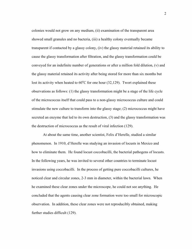
colonies would not grow on any medium, (ii) examination of the transparent area
showed small granules and no bacteria, (iii) a healthy colony eventually became
transparent if contacted by a glassy colony, (iv) the glassy material retained its ability to
cause the glassy transformation after filtration, and the glassy transformation could be
conveyed for an indefinite number of generations or after a million fold dilution, (v) and
the glassy material retained its activity after being stored for more than six months but
lost its activity when heated to 60oC for one hour (32,129). Twort explained these
observations as follows: (1) the glassy transformation might be a stage of the life cycle
of the micrococcus itself that could pass to a non-glassy micrococcus culture and could
stimulate the new culture to transform into the glassy stage; (2) micrococcus might have
secreted an enzyme that led to its own destruction, (3) and the glassy transformation was
the destruction of micrococcus as the result of viral infection (129).
At about the same time, another scientist, Felix d’Herelle, studied a similar
phenomenon. In 1910, d’Herelle was studying an invasion of locusts in Mexico and
how to eliminate them. He found locust coccobacilli, the bacterial pathogens of locusts.
In the following years, he was invited to several other countries to terminate locust
invasions using coccobacilli. In the process of getting pure coccobacilli cultures, he
noticed clear and circular zones, 2-3 mm in diameter, within the bacterial lawn. When
he examined these clear zones under the microscope, he could not see anything. He
concluded that the agents causing clear zone formation were too small for microscopic
observation. In addition, these clear zones were not reproducibly obtained, making
further studies difficult (129).
3
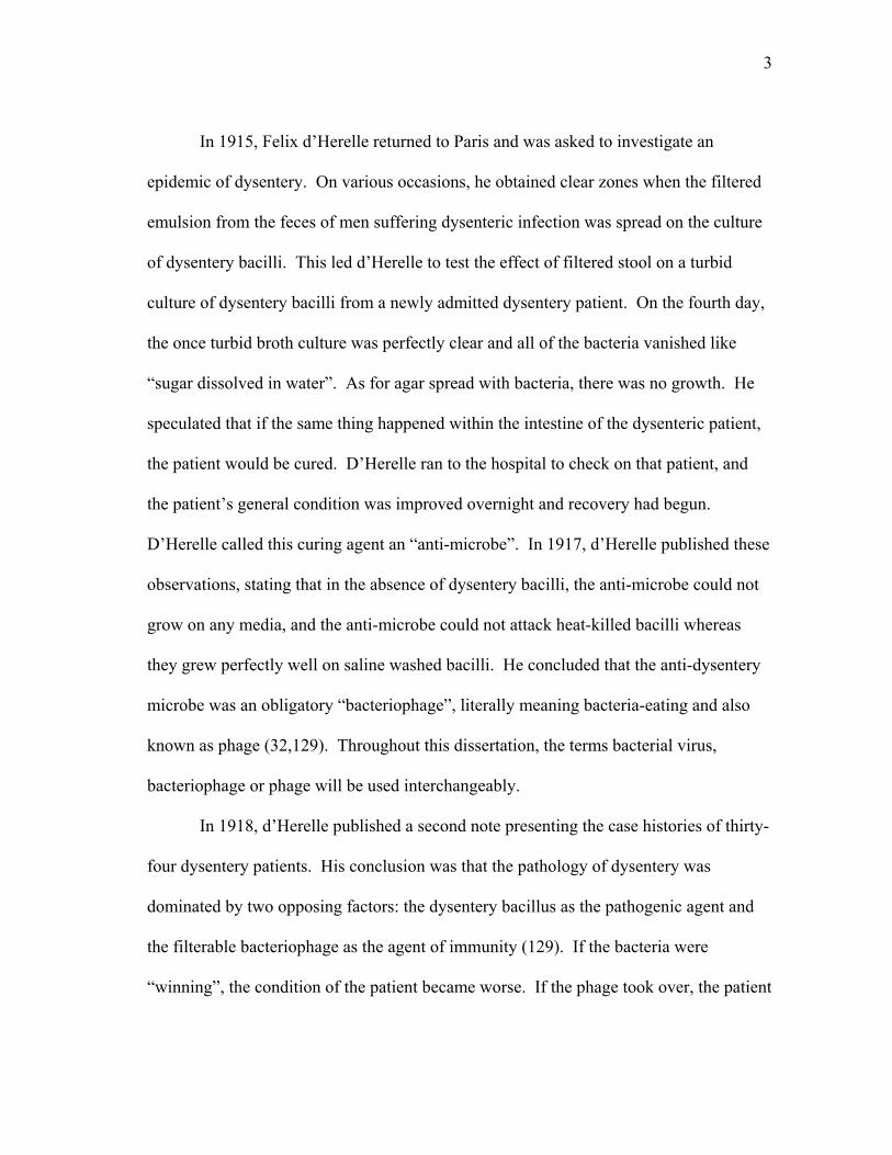
In 1915, Felix d’Herelle returned to Paris and was asked to investigate an
epidemic of dysentery. On various occasions, he obtained clear zones when the filtered
emulsion from the feces of men suffering dysenteric infection was spread on the culture
of dysentery bacilli. This led d’Herelle to test the effect of filtered stool on a turbid
culture of dysentery bacilli from a newly admitted dysentery patient. On the fourth day,
the once turbid broth culture was perfectly clear and all of the bacteria vanished like
“sugar dissolved in water”. As for agar spread with bacteria, there was no growth. He
speculated that if the same thing happened within the intestine of the dysenteric patient,
the patient would be cured. D’Herelle ran to the hospital to check on that patient, and
the patient’s general condition was improved overnight and recovery had begun.
D’Herelle called this curing agent an “anti-microbe”. In 1917, d’Herelle published these
observations, stating that in the absence of dysentery bacilli, the anti-microbe could not
grow on any media, and the anti-microbe could not attack heat-killed bacilli whereas
they grew perfectly well on saline washed bacilli. He concluded that the anti-dysentery
microbe was an obligatory “bacteriophage”, literally meaning bacteria-eating and also
known as phage (32,129). Throughout this dissertation, the terms bacterial virus,
bacteriophage or phage will be used interchangeably.
In 1918, d’Herelle published a second note presenting the case histories of thirty-
four dysentery patients. His conclusion was that the pathology of dysentery was
dominated by two opposing factors: the dysentery bacillus as the pathogenic agent and
the filterable bacteriophage as the agent of immunity (129). If the bacteria were
“winning”, the condition of the patient became worse. If the phage took over, the patient

4
was on the road of recovery. D’Herelle generalized his observations and quickly found
similar phenomena in patients recovered from typhoid fever and in chicken infected with
avian typhosa (129). After a period of disbelief and criticism, numerous medical
bacteriologists joined d’Herelle in finding bacteriophages that could cure bacterial
infections (129). It is clear that d’Herelle gave bacteriophages a glamorous role in
bacteriology in the 1920’s as an agent to control bacteria borne diseases (17). By the
middle of 1930, this glamorous role began to fade because the idea of using
bacteriophage therapeutically failed to materialize. Why bacteriophage, so virulent in
vitro, were impotent in vivo was a mystery at that time. The problems with phage
therapy were mostly due to ignorance, such as a lack of a systematic testing of virulence
of phages against infectious bacteria and of the knowledge of proper dosage used in
patients (53).
D’Herelle was interested in bacteriophage not only as an agent to control
bacterial infections but also as a material for studies of basic science. D’Herelle
developed an assay to determine concentration, or titer, of bacteriophage in a filtrate
(also known as lysate) by limiting dilution in liquid culture. In other word, he identified
the dilution at which the bacteriophage could no longer lyse the bacterial host
consistently. For example, he found only 3 out of 10 tubes of turbid cultures lysed upon
addition of 10-11 mL of the original lysate, meaning the lysate contained between 1010 to
1011 bacteriophage per mL. He also took advantage of the appearance of clear zones on
the dense bacterial lawns, or plaques, to calculate the number of phage in a lysate. The
total number of plaques per mL was inversely proportional to the dilution of the

5
bacteriophage lysate mixed with bacterial culture. Therefore, he proposed that each of
the plaques represented a bacteriophage colony descended from a single parental virus.
He also demonstrated that bacteriophage first fixed, or attached to, the bacteria it later
destroyed, and is now termed phage adsorption. After incubation of bacteriophage with
its host for a few minutes, the bacteria were pelleted and the supernatant was titered for
phage concentration. The results showed that the titer in the supernatant was much less
than the titer of input lysate suggesting that the phage loss was due to the adsorption of
phage to the host cells. However, this observation was only true when bacteriophage was
incubated with its respective host, indicating the adsorption was specific (129).
Bacteriophage life cycle
Like all other viruses, phages are not living organisms. They are pieces of
genetic material protected by protein or protein-lipid shells called capsids, and they are
able to utilize host machinery to replicate. There are five major stages in the
bacteriophage life cycle or phage vegetative growth:
1. Infection of a new host (adsorption and penetration of its genetic material)
2. Expression of the bacteriophage genes (“early genes”)
3. Replication of bacteriophage DNA or RNA
4. Expression of late genes and assembly of new virions
5. Escape from the host cell by lysis
6
Phages that replicate exclusively by vegetative growth, like T4, are termed “virulent” or
“lytic” (26,49,79,87). Some bacteriophages, such as lambda (λ)-like phages, can alter
this life cycle by becoming a prophage. This involves integrating the phage genomic
DNA into the host genome and replicating along with the host genome or alternatively,
becoming a stably replicating plasmid (18). For both cases, this state is known as
lysogeny, and the phage genes are generally not expressed, except for the lysogenic
repressor. Phages that can undergo lysogeny like λ are known as “temperate” or
“lysogenic” phage.
Strategies of progeny release from infected host
Once an infection is successful and virus particles are assembled, phages must
escape from the host to find new prey. Progeny release into the medium is prevented by
the cell wall. The cell wall, or peptidoglycan, is a continuous mesh of glycan strands
that are cross-linked together by di-isopeptides (55). This layer protects bacteria from
osmotic lysis and also is the shape-determinant of bacterial cells. Phages use several
strategies to release the phage progeny from the infected host. Due to their unique
morphogenesis, filamentous phages, such as M13, can extrude themselves through the
host envelope without fatal consequences to the host (93,137). All other known phages
release progeny by lysis of the host (146).
7
Lysis by small phages
To lyse the host, phages have to compromise the peptidoglycan layer. There are
two ways to accomplish this: actively degrade the cell wall or inhibit peptidoglycan
synthesis. The cell wall is not a static, rigid structure but a highly dynamic structure that
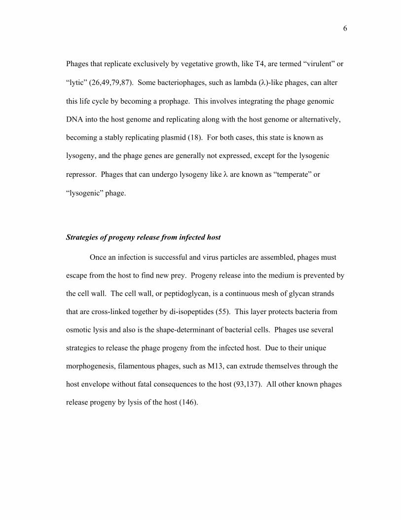
constantly undergoes biogenesis when the cell is growing (101). The most accepted
current model of peptidoglycan growth is the “three-for-one” model proposed by Höltje
as shown in Fig 1.1 (55). During elongation, three new glycan strands replace one old
glycan strand in the peptidoglycan layer (Fig 1.1A). At the constriction site (developing
septum), three new glycan strands also replace one old glycan chain in the peptidoglycan
layer, but the formation of constriction is the result of repeated additions of new glycan
strands at the same site (Fig 1.1B)(55). Insertion of new glycan strands requires
removing the cross-linker peptide and then re-establishing new cross-links. Inhibition of
peptidoglycan biogenesis, coupled with turn over and/or recycling, during active growth,
weakens the cell wall and eventually results in cell rupture from the osmotic pressure
difference between the internal and external environment of the bacterium. This is the
strategy that some small single stranded (ss) DNA and ssRNA phages utilize to elicit
host cell lysis. For example, the A2 protein of ssRNA phage Qβ and the E protein of
ssDNA phage φX174 inhibit cell wall synthesis at different steps but with the same
terminal phenotype, lysis when the cell is growing. A2 inhibits MurA, the first enzyme
in the dedicated peptidoglycan precursor biosynthesis pathway, and E inhibits MraY, the
only membrane-embedded enzyme in the same pathway (7-10).

8
FIG. 1.1. Peptidoglycan growth according to the three-for-one model. (A) Peptidoglycan synthesis during cell elongation. Free preexisting glycan strands form a murein triplet. The triplet is then attached to the peptide bridges on the right and the left of the docking strand in the peptidoglycan layer. Removal of the docking strand by murein hydrolase results in the insertion of 3 glycan strands in place of 1 strand. This is proposed to occur all over and at different sites of an elongated cell. (B) Peptidoglycan synthesis at the constriction site. Similar proposed mechanism as in peptidoglycan growth of the elongated cell, but instead of uniform insertion at different sites of the cell wall, the repeated addition of glycan strands at one site is proposed to result in the constriction of the cell (55).

9
Lysis by large and double stranded DNA phages
According to the classical lysis paradigm, large double stranded (ds) DNA
phages require two proteins for lysis, the endolysin and the holin (146). The endolysin is
an enzyme that degrades the cell wall, but it is sequestered in the cytoplasm. This is why
the holin is required for lysis. The holin is a membrane protein that accumulates in the
cytoplasmic membrane, without harming the cells, until a genetically programmed time,
when they trigger to form lesions in the membrane. The hole needs to be large enough
to allow the endolysin to pass through the membrane and into the periplasm where it has
access to the cell wall. At this point, the endolysin degrades the cell wall and lysis
occurs (146). Hole formation also causes the collapse of membrane potential. However,
recent findings suggest a more general role of holin in lysis. The endolysin of phage P1
and phage 21 were found to have an N-terminal SAR (signal anchor release) domain
(145). The SAR domain allows the endolysin to be translocated and anchored in the
membrane in an inactive form. Hole formation by holin collapses the membrane
potential which facilitates the release and activation of the SAR endolysin. The
endolysin is then free in the periplasm to degrade the cell wall and cause lysis (145). In
both systems, hole formation by the holin frees the endolysin from an inactive state, but
the mechanisms are different. In the classical paradigm, hole formation by the holin
opens a path for active, sequestered endolysin and allows it to gain access to its
substrate. In the new paradigm, hole formation allows the release of the endolysin from
the membrane, resulting in activation of the enzyme. In either case, the holin controls
the timing of lysis. It should be noted that SAR endolysins can spontaneously be

10
released from the membrane at a slow rate. Therefore, holins are not essential for lysis
or plaque formation in phages with a SAR endolysin (145).
Endolysins
To be classified as a lysozyme, Salton (117) stated that a protein should catalyze
an enzymatic reaction that results in: (1) the liberation of glucosamine and muramic acid
from bacterial cell wall, (2) the generation of reducing groups and (3) the lysis of
bacteria under proper conditions (69). An enzymatic activity was found in the phage-
free T2 and T6 lysates that was able to lyse chloroform-killed bacteria. The material
released from the cell wall was similar to that from a reaction using lysozyme. Thus, the
conclusion was that the enzyme mediating this reaction was a phage lysozyme (68).
Later, the word endolysin was used to described the lytic substance in the phage λ lysate
(59). This term was later broadened and used for any phage-encoded enzyme that was
required for host lysis (146). An endolysin can possess one or more of the four
following enzymatic activities: glycosidase, transglycosylase, endopeptidase and
amidase, as illustrated in Fig 1.2. A glycosidase, or true phage lysozyme, such as T4E,
hydrolyzes β(1, 4)-glycosidic bonds (134). A transglycosylase, such as λR, also attacks
glycosidic bonds, but instead of generating reducing ends, it forms a cyclic 1,6-
disaccharide product (11). Endopeptidases, such as PLY118 and PLY500 of Listeria
monocytogenes phages A118 and A500, respectively, cleave between amino acid
residues in the cross-linking oligopeptide (81). Finally, an amidase, such as the
bacteriophage T7 amidase, cleaves the amide bond between aminoglycosidic subunits

11
FIG. 1.2. Gram-negative bacterial peptidoglycan structure and sites of cleavage by various endolysins. Abbreviation: GlcNac, N-acetylglucosamine; MurNac, N-acetylmuramic acid; L/D-Ala, L/D alanine, D-Glu, D-glutamic acid; m-DAP, meso-diaminopimelic acid; Lys, lysine; LPP, Braun’s lipoprotein (107)

12
and the tetrapeptide chain (56). The crystal structures of T4E glycosidase, λR
transglycosylase and T7 amidase have been determined. T4E glycosidase (142) and λR
transglycosylase (38) have similar folds with their active sites located in a crevice
between two lobes connected by a helix (39). The structure of T7 amidase is very
different compared to the structure of T4E and λR, as it has only one lobe (20). While
T4E and λR have critical acidic residues on the opposite sides of the catalytic cleft
(E11/D20 of T4E and E19/D34 for λR), no acidic residues are found in the catalytic cleft
of T7 amidase (20,39). Instead, the residues important for catalytic activity of T7
amidase are lysine 128 at the top of the catalytic cleft and tyrosine 46 and histidine 17 at
the base of the catalytic cleft (20).
Holins
The best studied holin is the holin of bacteriophage λ, S105, a product of the S
gene. S105 has 105 amino acids with three transmembrane domains (TMD) arranged
with the N-terminus out and C-terminus in (Fig 1.3) (46). The formyl group at the N-
terminus of S105 is retained, indicating that TMD1 is integrated into the membrane
rapidly and before cytoplasmic deformylase can act (R. White et al., manuscript in
preparation). The wild type S105 holin mediates lysis at ~ 50 min after infection. Raab
(106) isolated a number of S mutants with single amino acid changes that delayed the
lysis time. In addition, there is another set of mutants that were completely defective in
lysis (105) even though the proteins were expressed at the wild-type level (47). In cross-

13
linking experiments using cells expressing wild type S gene, S protein oligomers were
detected up to a hexamer (149). Additional cross-linking experiments with lysis
defective mutants showed that S105A48V could not form dimers while several others,
S105A52V, S105A23V, S105D60N or S105R33L, could not form higher order oligomers.
Another mutant, S105R59C, could form oligomers but could not lyse the host, indicating
lesion formation was defective (47,105). Together, these results suggested that to form a
hole, S105 has to dimerize, oligomerize and then undergo a concerted conformational
change at the tertiary or quaternary level (47).
The holes formed by S105 were big enough for cmycRΦLacZ to cross the
membrane and degrade the cell wall (139). The cmycRΦLacZ was a fusion of N-
terminal cmyc (EQKLISEEDL) tagged λR endolysin to the N-terminus of the entire
LacZ (β-galactosidase) protein (139). The molecular weight of this fusion protein is ~
119 kDa (139). When LacZ is folded properly, it exists as a tetramer in solution (60)
with ~ 500 kDa molecular weight. The cmycRΦLacZ chimera was found to be fully
I : Sλ II : S21 III : T4 T
N
N NCC
C
I : Sλ II : S21 III : T4 T
N
N NCC
C
FIG. 1.3. Topology of class I, II and III holins (133).

14
active and had a ~ 0.5 MDa molecular mass as judged by size exclusion chromatography
(139). From the crystal structure, the dimension of a β-gal tetramer is 17.5 x 13.5 x 9.0
nm (60). Lysis mediated by holin S105 and cmycRφLacZ was almost identical, in term
of kinetic, to the lysis mediated by S105 with cmycR. Thus, the hole formed must be
larger than this dimension. Therefore, the hole formed by holin S105 should be
significantly larger than 20 nm (139).
There are at least three classes of holin proteins, as shown in Fig 1.3. The
prototype of class I is the bacteriophage λ holin with three TMDs (46). Class II, with
two TMDs, can be divided in to two subclasses, IIA and IIB. An example of class IIA is
S68 of phage 21. Although S68 has two predicted TMDs, TMD1 has been shown to be
a SAR domain and exits the membrane, leaving TMD2 in the membrane and the C-
terminus in the cytoplasm (102). Even though there is no well-studied example for class
IIB, the speculation is that class IIB members are S68 holin relatives that possess a
TMD1 that remains membrane embedded (102). Most holins identified in phage
genomics studies were of class I or II, based on the predicted transmembrane topology.
Also, these holins are very diverse; more than 50 unrelated families of class I and II
holins have been identified. Class III consist of a single family, the prototype of which
is bacteriophage T4 holin T. T, encoded by T4t, is predicted to have the N-terminus
inside and the C-terminus outside of the membrane with a single TMD (108).

15
T4 biology
T4 life cycle
Bacteriophage T4 is a large and complex bacterial virus as shown in Fig 1.4. The
T4 particle consists of an icosahedral head that is attached to a contractile tail via a neck
structure where six whiskers (gene product (gp) wac) are attached. The contractile tail is
composed of a contractile tail sheath of gp18 monomer units surrounding a non-
contractile tail tube comprised of gp19 monomer units (34). The end of the tail is
attached to a hexagonal baseplate where the long and the short tail fibers are attached.
The adsorption apparatus, composed of the contractile tail, long tail fibers, short tail
fibers and the baseplate, makes T4 infection (i.e. absorption to the host and injection of
its DNA) exceptionally efficient, 99% after 5 min of incubation with the host (22).
The T4 head contains a linear dsDNA of about 172 kb. The genome of T4 is
168,903 bp, encoding nearly 300 gene products, almost half of which have not been
characterized (92). Only 63 genes are essential for phage growth (92). Over 65 kb of T4
genome is occupied by structural genes of the phage head, tail and tail fibers and genes
necessary for phage particle assembly. More than 40 polypeptides are present in the
phage particle, a large number compared to simple viruses where the capsid can be made
from a single protein, such as tobacco mosaic virus or ΦX174 (33,34). T4 DNA
replication generates concatemeric DNA molecules of multiple unit genomes. T4
packaging begins at a random site on the concatemer and proceeds by headful
packaging. The T4 head can accept 102% of a unit genome length, so every T4 particle

16
contains a dsDNA with a 3.4 kb terminal redundancy (89). T4 DNA is circularly
permuted in a population of particles, so the T4 genetic map is circular even though the
genomic DNA in each virion head is linear.
The T4 virion is more than just a container filled with viral genomic DNA. It
undergoes a number of structural changes during infection. When a T4 encounters an E.
coli cell, one or more long tail fiber binds reversibly to receptors on the surface of the
host (either the diglycosyl residue of lipopolysaccharide (LPS) (43) or OmpC protein in
FIG. 1.4. Structure of bacteriophage T4, based on chemical cross-linking and on electron-microscopic structure analysis to a resolution of about 2 to 3 nm (43). Locations of known viral proteins are shown.

17
the exterior surface of the outer membrane (41)). Reversible binding of the long tail
fibers allows T4 to “walk” on the surface of the host and eventually bring the baseplate
closer to the cell surface. This event allows the short tail fibers to be released from their
stored position and bind irreversibly to the surface of the host (114). This irreversible
binding creates a tension that is transmitted to the closed conformation of the hexagonal
baseplate and causes it to expand into the open hexagram (star) conformation (123). The
star conformation induces a conformational change of gp18, which propagates in the
manner of “falling dominos”, and results in the contraction of the tail sheath (33,94).
The sheath is contracted to approximately 37% of its length and drags the base-plate
along with it. Because the baseplate is fixed to the cell surface, this process drives the
tail tube downward into the bacterial cell (123). The tube penetrates through the cell
wall with the help of gp5, a lysozyme and a part of the baseplate hub, and reaches the
outer surface of the cytoplasmic membrane (64). Through an unknown process, a
channel is formed in the cytoplasmic membrane and, subsequently, T4 DNA is released
into the host cytoplasm through the tail tube (123).
The detailed mechanism of how DNA is released from the head into the host
cytoplasm is not known, but DNA uptake requires the proton motive force, specifically
the membrane potential and not the pH gradient (42). DNA ejection takes place only
when the tube interacts with the cytoplasmic membrane (42). Although the tail sheath
contraction can be induced by treatment with 6 M urea, formaldehyde or exposure to
LPS, DNA ejection does not occur (42,43). In addition, ion leakage occurs during DNA
injection, but stops when injection is complete (122). This suggests that the channel that

18
is formed by either host proteins, phage proteins or both is sealed after injection.
Alternatively, Furakawa and Tarahovsky (41,42,95) proposed that the T4 tail tube
protrusion penetrates the outer membrane by the mechanical force generated by the tail
sheath contraction, and somehow induces a local fusion zone of inner and outer
membranes. Then, the tail tube acts as a channel to deliver the DNA into the host
through the fusion zone (95). In either case, ion leakage stops within a few minutes after
infection, indicating that membrane lesion is sealed (122).
Injection of T4 DNA into the host starts the T4 vegetative cycle. The chronology
of major events of T4 development is shown in Fig 1.5. T4 transcription utilizes three
major classes of promoters, early (Pe), middle (Pm) and late (Pl), which broadly define
the T4 developmental cycle. Many genes are served by multiple classes of promoters,
and T4 relies entirely on host RNA polymerase for its transcription (92). Host
replication, transcription and translation are blocked almost immediately after T4
infection (77). Gpalt is a protein that comes with the T4 DNA into the host cytoplasm
and is responsible for host RNA polymerase (RNAP) modification. Gpalt ADP-
ribosylates the α subunit of host RNAP to diverge it to transcribe T4 DNA. Genes
involved in shutting down host metabolism are transcribed and translated to perform a
number of functions. First, Gpndd morphologically disrupts the host nucleoid while
other phage encoded endonucleases, such as gpdenA, gpdenB and gp46/47, degrade host
DNA to mononucleotides (95). T4 genomic DNA contains glucosylated hydroxymethyl
cytosine, instead of cytosine, which allows the T4 nucleases to distinguish the difference
between T4 genomic DNA versus host genomic DNA (95). Second, Gpalc is an RNAP

19
binding protein that diverts E. coli RNAP from transcribing the cytosine-containing host
DNA to transcribe the glucosylated hydroxymethyl cytosine-containing phage DNA
(95). Also, ModA and ModB ADP-ribosylate the α subunit of RNA polymerase so that
transcription from the T4 middle promoter can take place later (92). Another protein,
MotA, is a transcription factor that binds the Mot box at about the -30 position in the
middle promoters and allows transcription of genes under control of these promoters to
occur (92). Lastly, AsiA is an anti-sigma factor that activates middle mode transcription
by forming an AsiA-σ70 dimer. The interaction of AsiA and σ70 interferes with
recognition of early promoters, and stimulates T4 transcription from the middle
promoters (92).
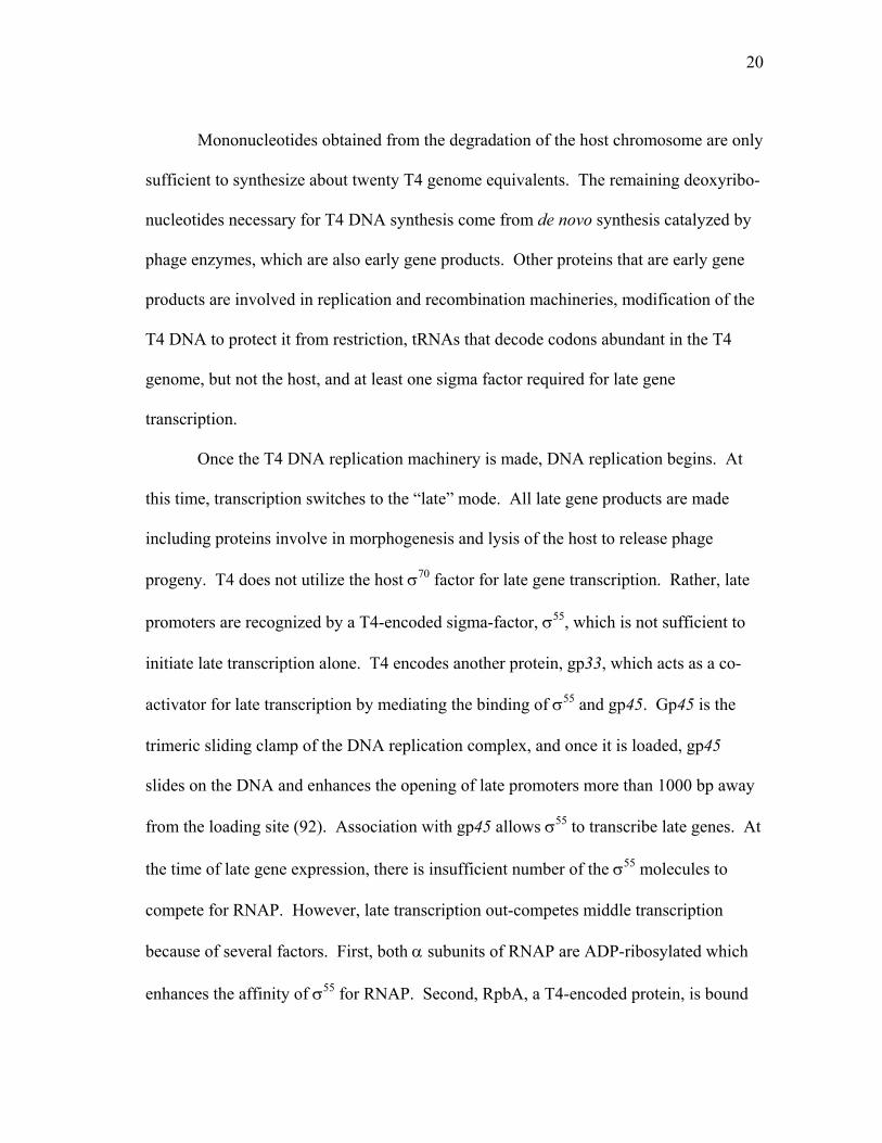
FIG. 1.5. Chronology of T4 development (89).
20
Mononucleotides obtained from the degradation of the host chromosome are only
sufficient to synthesize about twenty T4 genome equivalents. The remaining deoxyribo-
nucleotides necessary for T4 DNA synthesis come from de novo synthesis catalyzed by
phage enzymes, which are also early gene products. Other proteins that are early gene
products are involved in replication and recombination machineries, modification of the
T4 DNA to protect it from restriction, tRNAs that decode codons abundant in the T4
genome, but not the host, and at least one sigma factor required for late gene
transcription.
Once the T4 DNA replication machinery is made, DNA replication begins. At
this time, transcription switches to the “late” mode. All late gene products are made
including proteins involve in morphogenesis and lysis of the host to release phage
progeny. T4 does not utilize the host σ70 factor for late gene transcription. Rather, late
promoters are recognized by a T4-encoded sigma-factor, σ55, which is not sufficient to
initiate late transcription alone. T4 encodes another protein, gp33, which acts as a co-
activator for late transcription by mediating the binding of σ55 and gp45. Gp45 is the
trimeric sliding clamp of the DNA replication complex, and once it is loaded, gp45
slides on the DNA and enhances the opening of late promoters more than 1000 bp away
from the loading site (92). Association with gp45 allows σ55 to transcribe late genes. At
the time of late gene expression, there is insufficient number of the σ55 molecules to
compete for RNAP. However, late transcription out-competes middle transcription
because of several factors. First, both α subunits of RNAP are ADP-ribosylated which
enhances the affinity of σ55 for RNAP. Second, RpbA, a T4-encoded protein, is bound

21
to the RNAP core (70) and acts as a decoy to help σ55 compete for RNAP. Lastly, the
T4 Srd protein resembles a segment of RNAP that interacts with σ70 and σ38 which helps
σ55 compete for RNAP (92). Twenty-five minutes after infection, ~100 phage progeny
are assembled inside the host, and the vegetative cycle of the T4 is terminated by lysis of
the host cell.
T4 lysis
T4 utilizes the holin-endolysin strategy to lyse the host at the end of the
infectious cycle (61,132,146). The holin of T4 is T, or gpt, and the endolysin is E, or
gpe. Both of these genes were identified by genetics as mutants that failed to lyse the
host but retained the ability to produce virions. T4t+e- does not lyse the host, but the cell
stops respiring at 25 min after infection, and there is no phage accumulation after the cell
ceases to respire (96). T4t-e+ does not lyse the host; the cell continues to grow, and
phage particles continue to accumulate indefinitely. Lysis of these mutants can be
achieved by adding chloroform, which dissolves the membrane and releases the
endolysin (61,62). Lysis mediated by wild type T4 at the end of the vegetative cycle is
called lysis from within or LI.
Lysis mediated by the T4 holin and endolysin is rapid; the culture changes from
turbid to clear in a few minutes. Premature lysis by T4 can be induced by the addition of
chemicals that compromise the membrane potential or by switching to anaerobic growth
late (~17.5 min after infection) in the latent period (28,62). However, compromising the

22
membrane potential early (~7.5 min after infection) in the latent period does not induce
premature lysis (28), presumably because late gene expression has barely started and so
little holin or endolysin is present.
Endolysin E
In 1961, Streisinger mutagenized T4 in order to isolate temperature-sensitive
mutations that affected the lysozyme activity of T4 phage, and he called these mutations
T4e (132). Gene e was mapped between 66.4 and 67.0 kb on the T4 map (78). Gene e
is served by both early and late promoters as shown in 1986 by McPheeters and
colleagues (90). Endolysin E is one of the most well-studied enzymes in terms of
structure, mechanism and stability (88). E activity can be detected as soon as 8 min after
infection, and accumulates in a fully folded form in the cytoplasm throughout
morphogenesis (19). Because E lacks a secretory signal sequence, it cannot cross the
cytoplasmic membrane to degrade the cell wall (146). Therefore, E-mediated lysis is
subjected to the timing program of the holin T (141).
Gp5 as another muralytic enzyme
Infection of E. coli at a high multiplicity of infection (m.o.i.) (≥ 50 phages per
cell) results in lysis and release of cell wall components. In addition, lysis also can be
observed when E. coli, resuspended in PBS (phosphate saline buffer), are infected at a
low m.o.i. with either T4 wild type or T4 “ghosts”, which are osmotically shocked T4

23
with empty heads (58). This kind of lysis is called lysis from without or LO. For some
time, both of the above muralytic activities were believed to originate from T4 lysozyme
E. However, an experiment by Loeb (1974) showed that cell wall fragments were
released during the adsorption process, even when E. coli was infected with a T4e null
mutant. This suggested that the cell wall degradation at the time of adsorption was not
the result of E activity. In 1980, Kao and McClain isolated T4 mutants that allowed
lysis in the absence of gene e. These mutations were mapped to gene 5. Gene 5 is in the
first cluster of tail genes between 75.3 to 92.1 kb in the T4 map (22), and it is served by
the late promoter (78). The lysozyme activity of gp5 was confirmed in vitro by
Nakagawa et al. (98). In addition, Lin et al. (1989) found significant sequence
homology between gpe and the middle region of gp5. Expression of gp5 from a
plasmid gave rise to a protein with an apparent molecular weight of 63 kDa, but the gp5
isolated from T4 particles had an apparent molecular weight of 42 kDa, indicating the
gp5 incorporated into the phage particle is processed during morphogenesis (22).
Among the revertants that suppressed the E defective phenotype, there were alleles that
were temperature-sensitive for assembly of gp5 into the baseplate hub (34,35,66). Cell
wall degradation appeared to be better when gp5 was free as compared to when it was in
the hub complex (65). The simplest interpretation of these data was that the suppression
of the lysis defect due to the absence of endolysin E could be accomplished by mutations
in gp5 that had conditionally lost the ability to incorporate into the baseplate hub
complex. This way, there are some gp5 that are free and some that are incorporated into
the hub complex. Gp5 that is incorporated into the hub complex allows the maturation

24
of infectious particles, and the free gp5 with cell wall degrading activity replaces E to
become the endolysin to restore lysis.
Spackle involved in lysis
Spackle (sp) mutations were isolated in 1968 by Emrich (35) as pseudorevertants
that suppressed the loss of function of E. This suppression is temperature-sensitive, as
T4 e sp gives plaques at 43oC, but not at 30oC (35). T4sp gave small wt (wild type)
plaques at 30oC, large r plaques at 43oC, and at 37oC, the plaque size was variable and
intermediate between wt and r plaques (35). Even though the intracellular phage
accumulation was about the same for T4 wt, T4 e and T4 e sp, phage liberation of T4 e
sp was only 10% compared to the wt T4 (35). Because of the incomplete suppression of
the loss of function of E, this gene was named spackle. Since E activity is required for
lysis and spackle mutations restored lysis in T4e mutants, the speculation was that
spackle mutations somehow increased lysozyme activity. However, the lysozyme
activities of T4e and T4e sp were not different from each other (35). As mentioned in
the gp5 section, mutations in gene 5 also suppress T4e defective mutants, and free gp5
seems to participate better in lysis compared to gp5 in the hub complex (66). This led to
the speculation that spackle inhibited the lysozyme function of gp5. In the absence of
the spackle protein, the lysozyme function of gp5 is no longer inhibited; thus, gp5 can
replace gpe to cause lysis from within (66).
As mentioned earlier, LO occurs when E. coli cells are infected with a high m.o.i.
of T4 because of multiple punctures of the cell wall by gp5 located on the base plate hub

25
complex. However, when the cells are infected by low m.o.i. and then superinfected
with a high m.o.i., LO does not occur (138). This phenotype is possible because an
infected cell produces proteins that change the cell envelope such that it becomes
resistant to LO. One of these proteins is spackle. Cells infected with T4sp are more
sensitive to LO than cells infected wild type T4 (35).
Spackle phenotypes are recessive based on the following evidence: (1) T4e does
not lyse the host, but T4e sp does. In mixed infections of T4e and T4 e sp, no lysis is
observed. (2) Cells pre-infected with T4 wild type are LO resistant while cells pre-
infected with T4 sp are LO sensitive. Pre-infecting cells with a mixture of T4 sp and
wild type T4 gives a culture that is LO resistant (35).
In 1988, the spackle gene was mapped to gene 40, but Abedon invalidated this
result in 1994 (63). In 1999, the spackle mutations were mapped to gene 61.3.
Mutations generated in gene 61.3 gave the same phenotype as the spackle mutants that
Emrich isolated in 1968. In addition, the complementation experiment using gene 61.3
expressed from a plasmid in T4sp infected cells had a similar phenotype as coinfection
of E. coli with T4 wild type and T4sp (63).
Holin T
Streisinger observed that T4e mutants failed to lyse the host at the normal lysis
time, but the cells also stopped growing (96). T4e mutants failed to make plaques, but
plates that were supplemented with 5 mM Tris pH 8, 0.25% citrate and 500 µg of egg
white lysozyme complemented this defect (36). Based on this observation, Josslin

26
(1970) hypothesized that there were at least two components of T4 lysis, one which
subverted the integrity of the cytoplasmic membrane, resulting in the cessation of host
respiration of the infected cells and liberated the second, which was the phage lysozyme.
Phage lysozyme then degraded the cell wall, resulting in lysis and release of progeny
phage. Using 2-aminopurine (2-AP) and 5-bromodeoxyurindine (5-BUdR) as mutagens,
he isolated two mutants, amA3 and amB5, that plated on the permissive amber
suppressor host, but not on the restrictive non-suppressor host. In both mutants, phage
lysozyme was synthesized; however, lysis was not observed, and phage accumulated
beyond the normal lysis time. Josslin called the gene responsible for the lysis defective
phenotype of T4 “t” for Tithonus. (Tithonus was the son of the dawn goddess Aurora
and a Trojan prince. Since Tithonus was an “underbred” god, he was awarded a
diminished sort of immortality, one that did not protect him from aging (61).) One
possible explanation of T function was to inhibit cellular respiration, which somehow
allowed E to degrade the cell wall. This interpretation led to another question, could
chemicals or agents that inhibited cellular energy metabolism complement the lysis
defect of T4tam? Addition of cyanide or switching to anaerobic growth condition
induced premature lysis of cultures infected with wild type T4. However, neither of the
above restored lysis in t-defective T4 (62). The simplest interpretation was that
inhibition of cellular respiration was not the function of T, but rather, was the result of
the function of T. Josslin proposed a model in which T disrupted the cytoplasmic
membrane, which then destroyed the electron transport chain (62).

27
Gene t was mapped at about ~ 160 kb in the T4 genome, between gene 38,
encoding a long tail fiber modifying protein, and gene motA, encoding the middle gene
activator (113), and is served by a late promoter (78). The T amino acid sequence is
shown in Fig 1.6. T is unusually large for a holin with 218 amino acids (a a). Most
holins are about 60-120 a a; for example, the class I S105 holin of bacteriophage λ is105
a a and the class II holin of phage 21 is 68 a a.
+ - -+ +- + + + MAAPRISFSPSDILFGVLDRLFKDNATGKVLASRVAVVILLFIMAIVWYR 10 20 30 40 50 K V - - + + - - -+-+ + - - - - GDSFFEYYKQSKYETYSEIIEKERTARFESVALEQLQIVHISSEADFSAV 60 70 80 90 100 I + + - - + -+ -+ -- + YSFRPKNLNYFVDIIAYEGKLPSTISEKSLGGYPVDKTMDEYTVHLNGRH 110 120 130 140 150 + ++ - - + - YYSNSKFAFLPTKKPTPEINYMYSCPYFNLDNIYAGTITMYWYRNDHISN 160 170 180 190 200 -+ - + + + DRLESICAQAARILGRAK 210
FIG. 1.6. Amino acid sequence of T. Underlined is the predicted TMD of T. Bold residues are mutations in T that makes it LIN insensitive (i.e., rV alleles of t).

28
Expression of the t gene from a high copy number pUC19 plasmid (500-700
copies/cell) under the lac repressor control was lethal (84). Induction of the t gene from
the same plasmid with IPTG complemented the lysis defect of a λSam7 lysogen
although the complemented lysis was rather gradual (84) in contrast to lysis mediated by
the S105 holin which was sharp and rapid. A chimera with mature OmpA replacing
residues 1-35 of T was not toxic to cells. This chimera could be expressed abundantly
for purification, and purified OmpA-T was used to raise antibodies against T. Detection
of T using these antibodies was possible only when T protein was labeled with [35S]
methionine followed by immunoprecipitation prior to SDS-PAGE and autoradiography.
Cellular fractionation of cells expressing T indicated that T was associated with the
cytoplasmic membrane. This result was confirmed with the sodium hydroxide
extraction of membrane from cells expressing T, where T protein ended up in the pellet
of a sodium hydroxide wash (84). In addition, cross-linking experiments using DSP
(dithiobis[succinimidyl propionate]), showed that T formed a dimer, trimer and tetramer
with increasing concentration of cross-linker (84). These results suggested that T acted
in an oligomeric form in the membrane.
The T-mediated lysis observed by Lu et al. in 1992 was very gradual making it
unclear whether T really was functioning as a holin. However, a series of experiments
were performed by Ramanculov to confirm that T was the holin of T4. In these
experiments, S105 was replaced by t in the λ lysis cassette, as shown in Fig 1.7, and this
cassette was cloned into a pBR322-derivative plasmid called pER-T. This plasmid was
used to recombine t onto λ to make the hybrid phage λ-t. Induction of the λ-t lysogen

29
resulted in the saltatory lysis at approximately 23 min (108,109). The extremely short
latent period of λ-t reduced the burst size almost two orders of magnitude compared to
the parental λ phage, and the plaque morphology was also reduced to pin-point size
(108). The defect in plaque morphology of λ-t was exploited to isolate mutants that
produced bigger plaques and had higher plating efficiencies, that is, plaque size
increased as a result of mutations in t gene that delayed lysis (108). All mutants
obtained, either from spontaneous or UV mutagenesis, had longer lysis times than wild
type λ-t; they had point mutations that changed a single amino acid in T. Among these
mutants, increasing plaque size correlated with increasing lysis time up to about 50 min,
the normal lysis time of λ (108). The plaque size stayed constant with lysis time
between 50 and 60 min but decreased rapidly with further increases in lysis time. Phage
λ-t failed to make plaques when the lysis time was longer than 70 min (108).
The membrane topology of holin T was investigated by Ramanculov using the
periplasmic protein reporter alkaline phosphatase (PhoA) and cytoplasmic protein
reporter β-galactosidase (LacZ) (108). In this study, PhoA-LacZ was inserted between
residues in T creating a sandwich fusion (2). This strategy was used to avoid the loss of
the downstream topology determinants and to ensure the essence of the results. A result
FIG. 1.7. λ lysis cassette with gene t substituted for S (133).

30
was only considered meaningful when it was Lac+ and PhoA-, indicating cytoplasmic
localization, or Lac- and PhoA+, indicating periplasmic localization. Five fusions were
made between codons 1 and 2 (t1::pho-lac), 26 and 27 (t26::pho-lac), 70 and 71
(t70::pho-lac), 146 and 147 (t146::pho-lac) and 218 and the stop codon (t218::pho-lac).
Only t1::pho-lac and t218::pho-lac alleles retained holin activity. PhoA activity assays
indicated that t1::pho-lac and t26::pho-lac were PhoA- while fusions at 70, 146 and 218
were PhoA+. Weak β-gal activity was detected in fusions 1 and 26, but not for the other
constructs. Based on the above criteria, fusions at 70, 146 and 218 gave meaningful
results which implied that T had at least one TMD with the C-terminal domain from at
least residue 70 to the end of the protein in the periplasm (108).
The domain of T responsible for lysis was also assessed using truncations and
one deletion. All truncations, T148+R+27, T148+P+27, T134+23, T96+15 and T96+7, except T96+18,
(the bold and italicized number following T represents the last parental residue and the
number and following it was the number of vector-encoded amino acids added to T due
to the deletion method) delayed lysis. The only internal deletion, T∆(97-164), also delayed
lysis. Although the lysis times of these mutants were prolonged, they were not in a
predictable trend (108). Thus, the only conclusion from this experiment was that T
required at least the first 96 a a including the TMD to lyse the host (108).
Like S105, membrane lesions by holin T are large enough to allow cmycRφLacZ
to pass through the membrane and to enter into the periplasm (139). The lysis profile of
the holin T with the fusion endolysin cmycRφLacZ was almost identical to the lysis

31
profile of the holin T and cmycR (139). Therefore, the size of the hole formed by T is
comparable to the hole formed by S105 (139).
T4 lysis inhibition
Many basic molecular biology concepts, such as the definition of a gene, the
nature of the codon and recombination, were elucidated because of an interesting
phenomenon exhibited in T4, called lysis inhibition or LIN. In the scenario of one T4
infecting one E. coli cell as shown by Doermann in 1948, lysis occurs at approximately
25 min after infection (27). This situation can be achieved by infecting a culture at a low
m.o.i. (from 0.01 to 0.1), followed by a thousand-fold dilution, to create an environment
in which there is no significant chance of physical contact between the infected cells and
phages liberated during the infection cycle. However, following the lysis profile of a
turbid culture (A550 ~ 0.4) infected with wild type T4 at m.o.i. ~ 5, a sudden decrease in
turbidity is not observed (27). This is illustrated in Fig 1.8A, which shows the lysis
profile of a turbid, T4-infected culture. There are three successive decreases in optical
density (OD). The first decrease results from culture dilution when phages were added.
The second drop corresponds to the lysis of the earliest infected cells, due to the non-
synchronized nature of the infection process, and release of ~ 100 phages per lysed cell
into the medium. These newly released T4 from the minority of lysing cells
subsequently infected the surrounding infected cells, which caused the bulk of the
culture to become lysis inhibited. This state of lysis inhibition is called LIN. These
ideas were supported when the plaque forming unit (pfu) titer was determined from the

32
.
.
FIG. 1.8. Lysis profile of wild type T4 and T4r. (A) Lysis profile of wild type T4, curve A and T4r, curve B. (B) Lysis profile of wild type T4, curve A and the phage titer of cells in the same culture at the corresponding time.
33
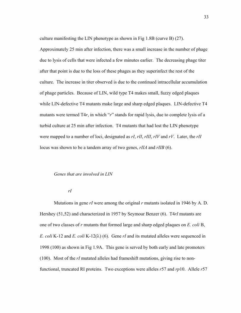
culture manifesting the LIN phenotype as shown in Fig 1.8B (curve B) (27).
Approximately 25 min after infection, there was a small increase in the number of phage
due to lysis of cells that were infected a few minutes earlier. The decreasing phage titer
after that point is due to the loss of these phages as they superinfect the rest of the
culture. The increase in titer observed is due to the continued intracellular accumulation
of phage particles. Because of LIN, wild type T4 makes small, fuzzy edged plaques
while LIN-defective T4 mutants make large and sharp edged plaques. LIN-defective T4
mutants were termed T4r, in which “r” stands for rapid lysis, due to complete lysis of a
turbid culture at 25 min after infection. T4 mutants that had lost the LIN phenotype
were mapped to a number of loci, designated as rI, rII, rIII, rIV and rV. Later, the rII
locus was shown to be a tandem array of two genes, rIIA and rIIB (6).
Genes that are involved in LIN
rI
Mutations in gene rI were among the original r mutants isolated in 1946 by A. D.
Hershey (51,52) and characterized in 1957 by Seymour Benzer (6). T4rI mutants are
one of two classes of r mutants that formed large and sharp edged plaques on E. coli B,
E. coli K-12 and E. coli K-12(λ) (6). Gene rI and its mutated alleles were sequenced in
1998 (100) as shown in Fig 1.9A. This gene is served by both early and late promoters
(100). Most of the rI mutated alleles had frameshift mutations, giving rise to non-
functional, truncated RI proteins. Two exceptions were alleles r57 and rp10. Allele r57

34
has an R78P missense mutation, and allele rp10 is a deletion of codon 92 (100). It was
not until 2001 that Ramanculov (110) cloned and studied the function of RI,
demonstrating that RI functions as an antiholin that specifically inhibits T-holin
A
B
FIG 1.9. Amino acid and nucleotide sequence of rI gene. (A) Mutations in rI genes that result in the lost of LIN (100) are indicated on top of the nucleotide sequence. (B) Amino acid sequence of RI with annotated charges, predicted signal sequence (bold) and the predicted cleavage site (arrow).
+ -↓ - -+ - + - +
MALKATALFAMLGLSFVLSPSIEANVDPHFDKFMESGIRHVYMLFENKSV
10 20 30 40 50
- - + + - - - -+ - + + - -
ESSEQFYSFMRTTYKNDPCSSDFECIERGAEMAQSYARIMNIKLETE
60 70 80 90

35
mediated lysis. The inhibition was shown to occur via a direct interaction with the holin
T (110). In addition, it appears that RI is needed on all hosts to make wild type plaques.
rIIA and rIIB
T4rII mutants were also originally isolated by A. D. Hershey in 1946 (51,52).
Because of the unique plating phenotype on different hosts (r plaques on E. coli B, wild
type plaques on E. coli K-12 but no plaques on E. coli K-12(λ) (6), rII mutants were
utilized for analysis of gene and codon structure, complementation, recombination,
mutagenesis, transcriptional regulation and translational control (124). T4rII mutants
were further grouped into two different classes, A and B (6), which later were shown to
be two independent complementation groups. Currently, it is still unclear what the
functions of RIIA and RIIB are (124), or, specifically, what is the contribution of rII
gene products to LIN.
Gene rIIA and rIIB are under the control of a middle promoter (124). Both RIIA
and RIIB are membrane-associated proteins (37,103,143), but the amino acid sequences
of RIIA and RIIB are rich in charged residues (25,104), as shown in Fig 1.10, indicating
they may associate with other membrane proteins, instead of being integral membrane
proteins. RIIA and RIIB seem to be involved in multiple functions of T4 physiology
(124), including:

36
+ + - - + - - - ++ + -- ++ + - + -MYNIKCLTKNEQAEIVKLYSSGNYTQQELADWQGVSVDTIRRVLKNAEEAKRPKVTISGDITVKVNSDAV
+ - ++ - - -+ -- + ++ -+ -IAPVAKSDIIWNASKKFISITVDGVTYNATPNTHSNFQEILNLLVADKLEEAAQKINVRRAVEKYISGDV
+ - - + -+ - -+ - - - - + + - - - --RIEGGSLFYQNIELRSGLVDRILDSMEKGENFEFYFPFLENLLENPSQKAVSRLFDFLVANDIEITEDGY
+ + - - + + + + --- + + + + + + FYAWKVVRSNYFDCHSNTFDNSPGKVVKMPRTRVNDDDTQTCSRGLHVCSKSYIRHFGSSTSRVVKVKVH
+- - - + + - -- - +PRDVVSIPIDYNDAKMRTCQYEVVEDVTEQFK
-+- + + + + - + + +- - -+ MIITTEKETILGNGSKSKAFSITASPKVFKILSSDLYTNKIRAVVRELITNMIDAHALNGNPEKFIIQVP
+ - + +- - - -- + - - + - +GRLDPRFVCRDFGPGMSDFDIQGDDNSPGLYNSYFSSSKAESNDFIGGFGLGSKSPFSYTDTFSITSYHK
- + - - + +- --+ - --+- + - + +- -+-GEIRGYVAYMDGDGPQIKPTFVKEMGPDDKTGIEIVVPVEEKDFRNFAYEVSYIMRPFKDLAIINGLDRE
- - -- -+ -+ - +-+ + - + - +-IDYFPDFDDYYGVNPERYWPDRGGLYAIYGGIVYPIDGVIRDRNWLSIRNEVNYIKFPMGSLDIAPSREA
--+ ++ -+ +- -+ -- ++ +- + +- + +- + + ++LSLDDRTRKNIIERVKELSEKAFNEDVKRFKESTSPRHTYRELMKMGYSARDYMISNSVKFTTKNLSYKK
- - + - - + ++ + - + + - + + + MQSMFEPDSKLCNAGVVYEVNLDPRLKRIKQSHETSAVASSYRLFGINTTKINIVIDNIKNRVNIVRGLA
+ -- - -+ - - - - - -- - - + + + + RALDDSEFNNTLNIHHNERLLFINPEVESQIDLLPDIMAMFESDEVNIHYLSEIEALVKSYIPKVVKSKA
+ + + - +- + -++ + - -- + - - + PRPKAATAFKFEIKDGRWEKRNYLRLTSEADEITGYVAYMHRSDIFSMDGTTSLCHPSMNILIRMANLIG
- + ++ +- - +- - -- - -+ ++ -+ + - - + INEFYVIRPLLQKKVKELGQCQCIFEALRDLYVDAFDDVDYDKYVGYSSSAKRYIDKIIKYPELDFMMKY
-- -- + +- - - + -+ - ++ + -FSIDEVSEEYTRLANMVSSLQGVYFNGGKDTIGHDIWTVTNLFDVLSNNASKNSDKMVAEFTKKFRIVSD
+ --- + + FIGYRNSLSDDEVSQIAKTMKALAA
A
B+ + - - + - - - ++ + -- ++ + - + -
MYNIKCLTKNEQAEIVKLYSSGNYTQQELADWQGVSVDTIRRVLKNAEEAKRPKVTISGDITVKVNSDAV
+ - ++ - - -+ -- + ++ -+ -IAPVAKSDIIWNASKKFISITVDGVTYNATPNTHSNFQEILNLLVADKLEEAAQKINVRRAVEKYISGDV
+ - - + -+ - -+ - - - - + + - - - --RIEGGSLFYQNIELRSGLVDRILDSMEKGENFEFYFPFLENLLENPSQKAVSRLFDFLVANDIEITEDGY
+ + - - + + + + --- + + + + + + FYAWKVVRSNYFDCHSNTFDNSPGKVVKMPRTRVNDDDTQTCSRGLHVCSKSYIRHFGSSTSRVVKVKVH
+- - - + + - -- - +PRDVVSIPIDYNDAKMRTCQYEVVEDVTEQFK
-+- + + + + - + + +- - -+ MIITTEKETILGNGSKSKAFSITASPKVFKILSSDLYTNKIRAVVRELITNMIDAHALNGNPEKFIIQVP
+ - + +- - - -- + - - + - +GRLDPRFVCRDFGPGMSDFDIQGDDNSPGLYNSYFSSSKAESNDFIGGFGLGSKSPFSYTDTFSITSYHK
- + - - + +- --+ - --+- + - + +- -+-GEIRGYVAYMDGDGPQIKPTFVKEMGPDDKTGIEIVVPVEEKDFRNFAYEVSYIMRPFKDLAIINGLDRE
- - -- -+ -+ - +-+ + - + - +-IDYFPDFDDYYGVNPERYWPDRGGLYAIYGGIVYPIDGVIRDRNWLSIRNEVNYIKFPMGSLDIAPSREA
--+ ++ -+ +- -+ -- ++ +- + +- + +- + + ++LSLDDRTRKNIIERVKELSEKAFNEDVKRFKESTSPRHTYRELMKMGYSARDYMISNSVKFTTKNLSYKK
- - + - - + ++ + - + + - + + + MQSMFEPDSKLCNAGVVYEVNLDPRLKRIKQSHETSAVASSYRLFGINTTKINIVIDNIKNRVNIVRGLA
+ -- - -+ - - - - - -- - - + + + + RALDDSEFNNTLNIHHNERLLFINPEVESQIDLLPDIMAMFESDEVNIHYLSEIEALVKSYIPKVVKSKA
+ + + - +- + -++ + - -- + - - + PRPKAATAFKFEIKDGRWEKRNYLRLTSEADEITGYVAYMHRSDIFSMDGTTSLCHPSMNILIRMANLIG
- + ++ +- - +- - -- - -+ ++ -+ + - - + INEFYVIRPLLQKKVKELGQCQCIFEALRDLYVDAFDDVDYDKYVGYSSSAKRYIDKIIKYPELDFMMKY
-- -- + +- - - + -+ - ++ + -FSIDEVSEEYTRLANMVSSLQGVYFNGGKDTIGHDIWTVTNLFDVLSNNASKNSDKMVAEFTKKFRIVSD
+ --- + + FIGYRNSLSDDEVSQIAKTMKALAA
A
B
FIG 1.10. Amino acid sequence of RII proteins. (A) RIIA. (B) RIIB

37
- Mutations in T4rII suppress a mutation in T4DNA ligase as long as functional
host ligase is available.
- Replication complexes in T4 infection are enriched for RIIA and RIIB
- They are essential for recombination in a λ-lysogenic host and may be
involved in anchoring the replication/recombination complex to the membrane.
- Mutations in rII allow the maturation of unit size T4 genomes or even parental
unreplicative DNA for packaging.
The most prominent phenotype of T4rII mutants is the Rex (rII exclusion)
phenotype. The genes rII are absolutely required for T4 to have a successful infection in
E. coli K-12 (λ) (5). T4rII mutants do not make plaques on E. coli K-12 (λ) lawns, but
they do on E. coli K-12 lawns. While E. coli K-12 in liquid culture infected with T4rII
produces progeny, no progeny is obtained when T4rII infects E. coli K-12 (λ) in liquid
culture. Phage infection that does not produce plaques and progeny is called abortive
infection. In an abortive infection, expression of phage genes occurs normally.
However, about the time DNA replication starts (in T4 infection, this is ~ 5 min after
phage DNA injection takes place), all gene expression is stopped (127). The abortive
infection of T4rII on E. coli K-12 (λ) results of the activity of the RexA and RexB
proteins, which are expressed from the rexA and rexB genes adjacent to the cI gene in
the immunity region of λ (124). Rex- lysogens do not restrict T4rII mutants (124). In
addition, over-expression of RexA and RexB from a multicopy plasmid extends the Rex
phenotype toward both T4 wild type and T4rII infections (119). Snyder and William
(128) noted that over-expression of RexA relative to RexB gave a similar phenotype as

38
when a λ lysogen was infected with T4rII mutants. Together, it was hypothesized that
the infection of a λ lysogen with T4rII mutants somehow altered the ratio of RexA and
RexB in the cells and therefore resulted in the Rex phenotype (125,128). E. coli B (λ)
also gives a similar restrictive phenotype as E. coli K-12(λ) (124).
T4rII makes r plaques on an E. coli B lawn but wild-type plaques on E. coli K-12
(6), on a Bc variant or BB (the Berkley) variant of E. coli B and E. coli K-12(P2) (116).
Josslin (61) examined the release of phage from cells infected with T4∆rII, T4tamA3 and
T4tamA3 ∆rII mutants. He observed that cells infected with T4∆rII or T4tamA3 ∆rII
released phage while cells infected with T4tamA3 did not. This indicated that the deletion
of both rIIA and rIIB suppressed t amber mutations and allowed phages to be released
from one-step-growth experiments as shown in Fig 1.11 (61). Upon further examination
of these data, phage released from T4∆rII infected cells (~ 200 phage/cell) was
completed at ~ 50 min after infection. However, at 50 min after infection, there were ~
200 phage particles assembled in one T4tamA3 ∆rII infected cell, but only ~ 3 phage
particles were released. At 90 min after infection, there were ~ 1000 phage particle in
one T4tamA3 ∆rII infected cell, but only ~ 200 phage particles were released. Thus, the
suppression was only ~ 1.5% at 50 min after infection and went up to ~ 20% at 90 min
after infection. Together, it can be stated that this phenotype is not true suppression, but
either a direct or indirect effect of RII proteins on T-mediated lysis.

39
rIII
Like T4rI and T4rII, T4rIII mutants were also isolated by A. D. Hershey in 1946
(51,52) and characterized by Seymour Benzer in 1957 (6). They make r plaques on E.
FIG 1.11. One step growth curves of cells infected with T4∆rII (circle), T4tamA3 (triangle) or the double mutant (square). Open symbol was before and solid symbol was after the addition of CHCl3. Samples collected before CHCl3 addition represented phage released due to lysis, and samples collected after CHCl3 addition represented total phages assembled intracellularly (62).
T4tamA3
T4tamA3∆rII
T4∆rII
T4∆rII
T4tamA3∆rII T4tamA3
T4tamA3
T4tamA3∆rII
T4∆rII
T4∆rII
T4tamA3∆rII T4tamA3

40
coli B lawns but wild type plaques on E. coli K-12 and E. coli K-12(λ) (6). Gene rIII is
the nearest downstream neighbor of gene 31 (111), and the rIII nucleotide sequence and
its mutated alleles were published in 1993 (112). GprIII is predicted to be an 82 a a
cytoplasmic protein (112). All of the rIII mutant alleles carry missense changes except
rBB9, rCR28 and rCR24. Allele rBB9 harbors a nonsense mutation as a result of
altering codon 16 to an opal stop codon. Allele rCR28 possesses a non-sense mutation
that changes codon 12 to an ochre stop codon. Allele rCP24 is allele rCR28 with a
second missense substitution, A70V (112). Using a rIII::lacZ fusion, β-galactosidase
activity was detected as early as 5-6 min after infection until the end of the T4 vegetative
cycle (30). The interpretation of this result was that rIII is under control of both early
and late promoters (30).
rIV and rV
Another set of T4r mutants, identified as rIV and rV, were isolated later (76).
T4rV mutants had an identical phenotype to T4rI mutants, that is, they make r plaques
on E. coli B, E. coli K-12 and E. coli K-12(λ). However, they were mapped to a
different location in the T4 genome, close to genes 38 and t (75). In 1999 (29), several
rV mutants were sequenced and shown to be allelic to t; the mutations are shown in Fig
1.6. T4 ts64 is an R5K substitution, r2 is I39V and r3 is T75I. Two of these mutations
are rather conservative, yet somehow these mutations make T insensitive to inhibition by
antiholin.

41
Gene rIV has been suggested to be equivalent to the sp gene. As mentioned
above, sp mutations suppress lysozyme mutations and allow release of progeny phages
(35). The product of spackle plays an important role in resistance to lysis from without
(66). T4sp mutations also have different plaque morphologies at different temperatures,
wild type at 30oC and r at 43oC (35). The different morphologies indicated the
temperature-sensitivity of the spackle gene towards LIN. Ramanculov (107) suggested
that spreading of phages from lysed cells inside a plaque is limited by the rate of
diffusion; thus, the local concentration of phage inside the plaque is high. In this
condition, infected cells at the edge of a growing plaque are subjected to high
multiplicity of superinfection. Therefore, mutations in the gene essential for LO
resistance resulted in lysis from without, instead of a lysis inhibited zone. This gives rise
to big and sharp edged plaques similar to T4r plaques. How spackle mutations give
different plaque morphologies at different temperatures is not known, but the loss of LO
resistance in T4sp is not temperature sensitive (66).
Factors that trigger LIN
If the expression of RI alone is sufficient for lysis inhibition, in principle, LIN
should be observed all of the time. However, this is not what happens during T4
infection. LIN can only be observed when superinfection takes place; therefore, LIN is
the result of superinfection. How superinfection causes LIN at the molecular level is not
clear, but it occurs when there is a high concentration of free phage and a low
concentration of uninfected cells. At this point, T4 has two choices: either undergoes

42
lysis, which produces more free phages, but free phage concentration does not increase
after lysis occurs; or undergo lysis inhibition, which does not produce free phages, but
accumulates phages intracellularly. From the standpoint of the phage, it is better to
increase the number of progeny linearly within the cell rather than releasing progeny in a
host-poor environment. Thus, high free phage concentration and low uninfected cell
concentration provide a quorum sensing mechanism so that T4 can decide whether it is
the optimal time to lyse the host. Proteins and phenotypes involved in superinfection are
described below.
Immunity protein
In the early 1950s, it was reported that T2 ghosts, osmotically shocked T2
particles in with empty heads, had certain biological properties of intact T2 phage, such
as (i) host range specificity, (ii) killing action, (iii) inhibition of host nucleic acid
synthesis, (iv) induction of lysis and (v) induction of leakage of host phosphorus
containing compounds (50). Later, it was shown that preinfection with ghosts inhibited
infective center formation by T4, but preinfection with wild type T4 followed by T4
ghost infection had no or little effect on infective center formation as long as the
preinfection with wild type T4 was 5 min or longer (31). T4 mutants that lost the ability
to protect the host from the killing action of T4 ghosts were isolated in 1972 (136). This
gene was named “imm”, as in immunity to ghost superinfection (135). Gene imm was
mapped between genes 42 and 43 in the T4 genome (21,24). The imm gene is under
control of an early promoter, and the action of immunity protein can be observed as

43
early as 1 to 2 min after primary infection. In 1989, Lu and Henning (83) cloned T4imm
and found that expression of the Imm protein from a plasmid protects 60% of a
population of E. coli K-12 and 25% of E. coli B from wild type T4 infection at an m.o.i.
of 6. Using isopycnic sucrose gradient fractionation and western blotting, Imm protein
was found to associate with the plasma membrane (85). The amino acid sequence of
Imm is shown in Fig 1.12A. Membrane topology analysis of the Imm protein using the
positive inside rule predicts that Imm has two TMDs with both the N- and C-termini
outside of the cell. These two TMDs would be unusually long, about 29 amino acid
(85). TMpred (http://www.ch.embnet.org/software/TMPRED_form.html) supports this
prediction, but TMHMM (http://www.cbs.dtu.dk/services/TMHMM-2.0/) predicts that
Imm has two TMDs with both N- and C-termini inside the cell. Topological analysis of
Imm using either PhoA or LacZ fusion was performed by Lu et al. in 1993. All Imm-
PhoA fusions, starting from residue 5, gave about 4 fold lower activities than when
PhoA is in the periplasm. In addition, fusion of the first 74 residues of Imm to mature
PhoA allowed the detection of Imm in the inner membrane by immuno-labeled electron
microscopy, and this fusion was protease resistant unless the cell was permeabilized with
the detergent Triton X-100. In both of the above experiments, fusion protein was
detected using PhoA antibodies. These results indicate that the PhoA fusions are
cytoplasmic. In contrast, all Imm-LacZ fusions, starting at residue 32, had high β-gal
activity indicating the proper localization of LacZ, or this region of Imm, in the
cytoplasm. Together, these results indicated that Imm has a single transmembrane
domain with N-terminus out and C-terminus inside the cell (85) as shown in Fig 1.12.

44
Host proteins involved in LIN
EndA
LIN takes place only when the superinfecting phage is intact; i. e. T4 ghosts do
not induce LIN. Thus, the signal for LIN is either the T4 genomic DNA or the internal
FIG. 1.12. Amino acid sequence and topologies of Imm protein. (A) Amino acid sequence of Imm protein. Gray shade, predicted TMDs by positive inside rule. Solid arrow, fusion join with LacZ. Open arrow, fusion join with PhoA. (B) Predicted topology and topology obtained from fusion studies.
- + + METLVAGSIFMVLVSGVLAIIIYMLPWFIALMRGSKSTVGIFFASLLFNW
10 20 30 40 50
- ++ +-+-SIIGWFITFIWSIAGETKKSAQPNQVIIIREKE
60 70 80
NNC
CCytoplasm
Periplasm
Predicted Experimental
A
B
- + + METLVAGSIFMVLVSGVLAIIIYMLPWFIALMRGSKSTVGIFFASLLFNW
10 20 30 40 50
- ++ +-+-SIIGWFITFIWSIAGETKKSAQPNQVIIIREKE
60 70 80
NNC
C
NNC
CCytoplasm
Periplasm
Predicted Experimental
A
B

45
proteins that are carried in the head of superinfecting phages but missing from ghosts.
During the primary infection, the phage DNA is injected into the cytoplasm and genetic
markers from the primary phage are inherited in the progeny. However, as shown by
Anderson et al. (4), DNA of the superinfecting phage ended up in the periplasm of the
host, and thus cannot participate in the genetic make-up of the progeny. Gene endA of
E. coli encodes periplasmic endonuclease I (23) and degradades DNA that ends up in the
periplasm from the injection of superinfecting phage (3). The fact that genetic markers
of superinfecting phage are not inherited in the progeny is called superinfection
exclusion. This superinfecting phage DNA can be visualized by electron microscopy
coupled with 32P labeling and radiography when the host is endA- (4). The degradation
of the DNA injected from the superinfecting phage by EndA is called superinfection
breakdown. The length of LIN in endA+ or endA- strains is not known, but both strains
allow genetic exclusion from the superinfecting phage. It is thought that the presence of
Imm protein from the primary infection or expressed from a plasmid alters the E. coli
envelope so that the DNA of the superinfecting phage is diverted to the periplasm (83).
DegP
Internal proteins (IP) are proteins that are packaged into the T4 head along with
T4 genomic DNA. In 1971, Black et al. determined the species and number of each
species of the internal proteins from an osmotically shocked 14C labeled T4 lysate by
analytical chromatography. The results from this experiment showed that there were
three different IPs: IPI, IPII and IPIII. In a single T4 particle, there are approximately

46
180, 90 and 30 molecules of IPI, II and III, respectively (12). In primary infection, both
T4 genomic DNA and the internal proteins are injected into the cytoplasm of the host.
In superinfection, since the T4 genomic DNA is injected into the periplasm, the internal
proteins are presumably also injected into the periplasm, and serve as the LIN signal.
The temporary nature of LIN, i.e. under controlled circumstances where only one round
of secondary infection occurs, LIN lasts only for minutes instead of hours (14,27,115),
gives rise to the hypothesis that the LIN signal should also be temporary. The easiest
way to make the LIN signal temporary is to degrade it. The speculation is that the LIN
signal ends up in the periplasm; thus, to degrade the LIN signal, a periplasmic protease is
required. One obvious periplasmic protease is DegP. DegP, a member of the HtrA
protease family, is a well-characterized periplasmic protease in E. coli. E. coli DegP
was identified from a mutant hunt that isolated mutants lacking proteolytic enzyme
activity. Analysis of the degP- strain showed that it grew poorly at high temperature
(130,131). Later, Lipinska et al. (80) showed that DegP has endopeptidase activity using
α and β casein. The endopeptidase activity of DegP can be inhibited by
diisopropylfluorophosphate, a serine protease inhibitor (80), suggesting that DegP is a
serine protease. In 1996, Kolmar et al. showed that the cleavage site for DegP is at V/A,
V/S (Val/Xaa) and I/Y (Ile/Xaa) residues, and the activity of DegP does not require
ATP, reducing agents or divalent cations (72). A number of natural substrates of DegP
were described including colicin A lysis protein, K88 and K99 pilin subunits, MalS, and
PapA pilin (67). DegP does not degrade folded proteins, but appears to cleave partially
unfolded proteins. This finding is supported by the sequence of the cleavage site of

47
DegP, Val/Xaa and Ile/Xaa. These amino acids are hydrophobic and usually not
exposed on the surface of a protein. However, these sites are often exposed when a
protein is partially unfolded (72).
DegP also possesses a chaperone activity at low temperature (67). The crystal
structure of DegP helped to explain the different activities of DegP at different
temperatures. DegP possesses a protease domain at the N-terminus and two PDZ
domains at the C-terminus (67,73). PDZ domains are involved in protein-protein
interactions, specifically in interactions with a short peptide at the extreme C-terminus of
the target protein. Within the protease domain, there is a Q-linker, which is either absent
or shorter in other members of the HtrA family (67). The hexameric structure of DegP
suggests DegP may have limited access to its substrate (73). PDZ domains of DegP
have two orientations, a closed state that denotes the chaperone activity and an open
state that denotes protease activity (73). It is thought that the PDZ domains function as a
proteolytic regulator, much like a gatekeeper, to either restrict or allow substrate access
to the proteolytic chamber (67).
Goals and specific aims
Lysis inhibition is the only known example of real time control, signal
transduction in holin regulation. In addition, this phenomenon is historically significant
in that it was used in the founding experiments of modern molecular genetics. The long

48
term goal of this project is to determine the detailed mechanism of LIN at the molecular
level.
I. Determination of the interacting domains of antiholin RI and holin T
Ramanculov established that the antiholin RI inhibits T-mediated lysis by direct
interaction using formaldehyde cross-linking. However, the specific interaction domains
are not known. Thus, the first step in understanding the mechanism of LIN is to
determine which domains of T and RI interact.
II. Characterization of antiholin RI
To date, the only thing known about RI is its predicted amino acid sequence and
the sequence of the mutant rI alleles with LIN defects. Nothing is known about the
physical characteristics of RI. Even though it was predicted to be a secreted protein by
Paddison in 1991, no experimental data are available to support this prediction. In
addition, Ramanculov had difficulty in detecting RI tagged with a c-myc tag, suggesting
the protein is unstable, a very small amount of protein is made, or the tag is removed by
proteolytic cleavage. The only time tagged-RI was detected was when it was cross-
linked to T. Since RI is one of the two major players of LIN, understanding its physical
characteristics may help to elucidate the molecular mechanism of LIN.

49
III. Characterization of holin T
In addition to its role in LIN, T is a large protein compared to other known holins
and has a unique topology. In this specific aim, I propose to determine the topology of
T, the domain important for oligomerization, and the localization of T in the E. coli.
membrane when lysis occurs.
50
CHAPTER II
PERIPLASMIC DOMAINS DEFINE HOLIN-ANTIHOLIN INTERACTIONS IN
T4 LYSIS INHIBITION*
Introduction
An E. coli cell infected at 37ºC by a wt T4 phage undergoes lysis at about 25 min
after infection and releases ~200 progeny virions. Lysis requires the muralytic activity
of the T4 lysozyme, E, one of the best characterized soluble enzymes in terms of its
structure, enzymatic mechanism, and thermodynamic stability (88). The precise timing
of lysis, however, is not determined by E, which accumulates fully folded and active in
the cytoplasm throughout the morphogenesis period. Instead, like all dsDNA phages,
the timing of T4 lysis is controlled by its holin, T, an integral membrane protein that
suddenly triggers to disrupt the bilayer at an allele-specific time (141,147). Membrane
disruption allows the T4 lysozyme to attack the cell wall, after which the infected cell
bursts and releases the progeny virions. T4 t, like the λ holin gene S, is genetically
malleable, in that many missense alleles have been isolated, with lysis times either
advanced or delayed relative to the wt allele (47,106,108,141,146). This malleability is
significant, because it is thought that investing lysis timing exclusively in the holin gene
_________________ * Reprinted with permission from “Periplasmic domains define holin-antiholin interaction in T4 lysis inhibition” by Tran, T. A., D. K. Struck, and R. Young, 2005. Journal of Bacteriology, 187, 6631-6640. Copyright 2005 by ASM Press.

51
allows dsDNA phages to evolve rapidly in response to changed conditions. For
example, an environment with reduced host numbers should favor phages with an
extended latent period, allowing the intracellular accumulation of more progeny virions
before they are released into the host-poor medium (140). Holin genes have enormous
diversity, with more than 50 unrelated gene families having been described (141).
However, compared to other known holins, the T4 holin has an unusual topology. All
other characterized holins have either three (class I; e.g., the S105 product of phage λ
gene S) or two (class II; e.g., the S2168 product of S21, the holin gene of lambdoid phage
21) TMDs (Fig. 1.3). In contrast, T and its orthologs in T4-like phages constitute a
single protein family with bitopic topology and a single TMD (Figs. 1.3). T, at 218
residues, is substantially larger than other holins (e.g., the λ holin S105 is only 105
residues and the S2168 is only 68 residues). The extra mass comes principally from its
large C-terminal periplasmic domain of 163 residues (Fig. 2.1A).
In addition to its unusual topology and size, the function of the T4 holin is
subject to a type of control not seen with the prototypical class I and II holins. Almost
60 years ago, in publications now considered landmarks in the history of molecular
genetics, it was reported that T4-infected cells are subject to “lysis-inhibition”, or LIN
(27,52). The LIN state, in which the normal lysis timing of the holin is over-ridden, is
established if a T4-infected cell undergoes super-infection by another T4 particle. The
LIN state is unstable, requires continued super-infection to be maintained, and can be
subverted by addition of energy poisons that collapse the membrane potential (1). In
infected cultures at visible optical densities, the lysis of a small fraction of the cells

52
+ - -+ +- + + + - - + + - - -+-+ + - - - -MAAPRISFSPSDILFGVLDRLFKDNATGKVLASRVAVVILLFIMAIVWYRGDSFFEYYKQSKYETYSEIIEKERTARFESVALEQLQIVHISSEADFSAV1 10 20 30 40 50 60 70 80 90 100
MKQSTIALALLPLLFTPVTKARTPEMΦMFALLCGCILLALAFLLGΦ
ΦPVLE.......+ + - - + -+ -+ -- + + ++ - - + -
YSFRPKNLNYFVDIIAYEGKLPSTISEKSLGGYPVDKTMDEYTVHLNGRHYYSNSKFAFLPTKKPTPEINYMYSCPYFNLDNIYAGTITMYWYRNDHISN110 120 130 140 150 160 170 180 190 200
-+ - + + +DRLESICAQAARILGRAK
210 218
(TTMDφPhoA)
(ssPhoAφTCTD) (FtsITMDφ70T )
T
r2V
r3I
ΦPVLENRAA.........+ - - -+ - + - + - - + + - - - -+ - + + - -
MALKATALFAMLGLSFVLSPSIEANVDPHFDKFMESGIRHVYMLFENKSVESSEQFYSFMRTTYKNDPCSSDFECIERGAEMAQSYARIMNIKLETE1 10 20 30 40 50 60 70 80 90
MKQSTIALALLPLLFTPVTKARTPEMΦ.......
RI
(ssPhoAφRICTD)
(RITMDφPhoA)
TCTD
RICTD
P
A
B
^
+ - -+ +- + + + - - + + - - -+-+ + - - - -MAAPRISFSPSDILFGVLDRLFKDNATGKVLASRVAVVILLFIMAIVWYRGDSFFEYYKQSKYETYSEIIEKERTARFESVALEQLQIVHISSEADFSAV1 10 20 30 40 50 60 70 80 90 100
MKQSTIALALLPLLFTPVTKARTPEMΦMFALLCGCILLALAFLLGΦ
ΦPVLE.......+ + - - + -+ -+ -- + + ++ - - + -
YSFRPKNLNYFVDIIAYEGKLPSTISEKSLGGYPVDKTMDEYTVHLNGRHYYSNSKFAFLPTKKPTPEINYMYSCPYFNLDNIYAGTITMYWYRNDHISN110 120 130 140 150 160 170 180 190 200
-+ - + + +DRLESICAQAARILGRAK
210 218
(TTMDφPhoA)
(ssPhoAφTCTD) (FtsITMDφ70T )
T
r2V
r3I
ΦPVLENRAA.........+ - - -+ - + - + - - + + - - - -+ - + + - -
MALKATALFAMLGLSFVLSPSIEANVDPHFDKFMESGIRHVYMLFENKSVESSEQFYSFMRTTYKNDPCSSDFECIERGAEMAQSYARIMNIKLETE1 10 20 30 40 50 60 70 80 90
MKQSTIALALLPLLFTPVTKARTPEMΦ.......
RI
(ssPhoAφRICTD)
(RITMDφPhoA)
TCTD
RICTD
P
A
B
^
FIG. 2.1. Amino acid sequence of T and RI and their derivatives. (A) Amino acid sequence of T and its derivatives. The predicted TMD of T is boxed and shaded. The rV mutants r2(I39V) and r3(T75I) are indicated above the sequence. In This, a hexa-histidine tag (HHHHHHGG) was inserted between the residues G132 and Y133 (underlined). For the ssPhoAΦTCTD construct, the signal sequence of PhoA, shown below the T sequence, was substituted for residues 1-55 of T. For the FtsITMDΦT construct, the transmembrane domain of FtsI, shown in a shaded box below the T sequence, was substituted for residues 2-69 of T. For the TTMDφPhoA construct, residues 1-70 of T was substituted for residues 1-26 in the full-length primary gene product of phoA; the PhoA sequence begins with PVLE (PhoA residues 27 on, as indicated). (B) Amino acid sequence of RI and its derivatives. The predicted signal sequence of RI is boxed, with the leader peptidase I cleavage site predicted by the SignalP program (http://www.cbs.dtu.dk/services/SignalP/) {Bendtsen, 2004 133 /id} indicated by a carat. In the cmyc-tagged version of RI, the cmyc tag (QKLISEEDL) was inserted after the terminal glutamate at position 97, while in the GFPΦRIcmyc constructs, the entire GFP sequence was inserted after the initial Met residue. For the ssPhoAφRICTD construct, the signal sequence of PhoA, shown below the RI sequence, was substituted for residues 1-24 of RI. For the RINTDφPhoA construct, the predicted signal sequence of RI replaced residues 1-26 in the uncleaved precursor to mature PhoA. The sole known rI missense mutation, R78P, is shown by a down arrow (133).

53
generates sufficient free virions in the medium to establish and maintain LIN throughout
the bulk culture, allowing progeny to accumulate to >103 virions per cell over a period of
hours. On agar lawns, LIN causes T4 to make small, indistinct plaques, and it is easy to
isolate T4 mutants defective in LIN by virtue of their large, clearly defined plaque
morphology (51). These mutants were called r mutants for “rapid lysis” and were
mapped to multiple T4 loci, including rI, rIIAB, and rV depending on the host used
(146). The r genetic system was exclusively exploited to establish many of the
fundamental principles of molecular genetics (17). Ultimately, only two genes, rI (6)
(100) and rV (74,75), later shown to be allelic to t (29), are required to maintain the
wild-type plaque phenotype and to establish LIN with E. coli K-12. Nevertheless,
despite the central importance of the r genetic system in the history of molecular
biology, the molecular basis of LIN has remained obscure.
Recently, we have undertaken a molecular analysis of T4 lysis and the LIN
phenomenon, as part of our study of the mechanisms of phage lysis and its regulation.
We reported here the evidence that RI (Fig. 2.1B) is an antiholin that specifically binds
to and inhibits the T holin. This clearly distinguishes T4 from bacteriophage λ whose
antiholin, S107, is the product of an alternative translational start in its holin gene, S,
which also encodes S105, the λ holin. Given its near identity with S105 and the fact that
holins oligomerize in the process of forming membrane lesions, it is not surprising that
S107 dimerizes with S105. The formation of these dimers is responsible for the ability
of S107 to prevent the spontaneous triggering of S105. In contrast, RI has no sequence
similarity to T (Fig. 2.1) that might support homotypic interactions of the type observed
54
in the λ S105/S107 system. Here we report experiments designed to identify the
topological determinants of RI and T that lead to the LIN state. The results are discussed
in terms of the unique ability of RI to respond to an environmental signal (i.e., a super-
infecting T4 phage).
Materials and methods
Bacterial strains, bacteriophages, plasmids, and culture growth
The bacterial strains, bacteriophages, and plasmids used in this work are
described in Tables 2.1, 2.2 and 2.3. T4 phage stocks were prepared as described
previously (109). Bacterial cultures were grown in standard LB medium supplemented
with ampicillin (100 µg/ml), kanamycin (40 µg/ml), chloramphenicol (10 µg/ml) when
appropriate. Growth and lysis of cultures were monitored as A550 as previously
described (109). When indicated, isopropyl β-D-thiogalactoside (IPTG), KCN, or
CHCl3 was added to give final concentrations of 1 mM, 10 mM, or 1%, respectively.
Standard DNA manipulations, PCR, and DNA sequencing
Isolation of plasmid DNA, DNA amplification by PCR, DNA transformation,
and DNA sequencing were performed as previously described (145). Oligonucleotides
listed in Table 2.4 were obtained from Integrated DNA Technologies, Coralville, IA, and
were used without further purification. The Rapid DNA ligation kit from Roche

55
Molecular Biochemicals was used for ligation reactions. All other enzymes were
purchased from New England Biolabs except for Pfu polymerase which was from
Stratagene. Automated fluorescent sequencing was performed at the Laboratory for
Plant Genome Technology at the Texas Agricultural Experiment Station.
Single base changes and small insertions were introduced using commercially
synthesized primer in conjunction with the Quikchange kit from Stratagene. Larger
insertions, replacements, and gene fusions were generated using a modification of the
basic QuikChange site-directed mutagenesis protocol. Here, a donor sequence is PCR
amplified using primers that have 5’ ends that anneal to appropriate sequences in a target
plasmid. The first PCR product is then used as the primer for a second PCR reaction
using the target plasmid as a template. All subsequent steps are identical to those in the
basic QuikChange protocol.
TABLE 2.1. Phages
Genotypes/features Sources, references
T4 wt T4D I. Molineux T4rI rI48, single base deletion at position 195; N66-E97
replaced with MTRALLILNV D. Hall
λkan∆(SR) λb515 b519 att::Tn903 cI857 nin5 ∆(SR) (105) λkan this λb515 b519 att::Tn903 cI857 nin5 ∆S::this This study T4∆rI Deletion of rI gene from nt 59204 to nt 59496 in T4
genome This study
T4eam M41, M41T4 with an amber mutation in e gene Drake, J. T4ssphoAΦrICTD
y42am rI gene, from nt 59214 to nt 59496 in T4, replaced with ssphoAΦrICTD
y42am This study
λ-t λb515 b519 att::Tn903 cI857 nin5 ∆S::t (109) λcam∆(SR) λstf::cat::tfa cI857 ∆(SR); loss of l nt 45135-45815 (126)

56
TABLE 2.2. E. coli strains
Genotypes/features Sources, references
CQ21 E. coli K-12 ara leu lacIq1 purE gal his argG rpsL xyl mtl ilv
(105)
CQ21λkan∆(SR) Lysogen carrying λkan∆(SR) prophage (105) CQ21[λkan∆(SR)] recA
srl::Tn10 Lysogen carrying λkan∆(SR) prophage This study
CQ21(λkan this) Lysogen carrying λkan this prophage This study MG1655 F- ilvG rfb50 rph1 E. coli
Genetic Stock Center
MDS12 MG1655 with 12 deletions, totaling 376,180 nt including cryptic prophages
(71)
MDS12 tonA::Tn10 lacIq1 This study XL-1Blue E. coli K-12 recA endA1 gyrA96 thi1 hsdR17 supE44
relA1 lac[F’ proAB lacZ∆M15::Tn10] Stratagene
BL21(DE3) F- ompT hsdSB (rB- mB
-) gal dcm (DE3) Novagen BL21(DE3) tonA::Tn5 slyD::TetR
This study
CQ21(λ-t) Lysogen carrying (λ-t) prophage This study MG1655 tonA::Tn10 lacIq1
(102)
KS474 F- ∆lacX74 galE galK thi rpsL(strA) ∆phoA(PvuII) degP (91) NJH110 F- araD139 ∆(argF-lac) U169 rpsL150 relA1 flbB5301
deoC1 ptsF25 rbsR degP::Cm (57)
MDS12 tonA::Tn10 lacIq1degP::Knr
This study
MDS12 tonA::Tn10 lacIq1degP::Cmr
This study
LE392 E. coli K-12 hsd R574, (rk-, mk
+), supE44, supF58, ∆ (lac1ZY)6, gal K2, gal T22, metB1, trpR55
(97)
LE392 tonA::Tn10 This study MC4100 E. coli K-12 F- araD139 ∆(argF-lac)U169 rpsL15
relA1flbB3501 deo pstF25 rbsR (121)
MC4100λcam∆(SR) Lysogen carrying λkan∆(SR) prophage Laboratory stock
C43(DE3) F- ompT hsdSB (rB- mB
-) gal dcm (DE3) with uncharacterized mutation that allows for toxic proteins to be induced at higher levels than BL21(DE3)
Avidis
C43(DE3)tonA::Tn10 slyD::knR
This study

57
TABLE 2.3. Plasmids
Genotypes/features Sources, references
pT4T pBR322 derivative carrying late promoter and lysis cassette of λ with the S gene replaced by T4 t
I. N. Wang
pT4T132his (pT4This)
pT4T with a HHHHHHGG inserted between residues 132 and 133 of t This study
pT4TRI pT4T with rI gene inserted distal to Rz1 gene This study pZA32-luc p15A origin, PLlacO-1 promoter, camR (86) pZA-RI pZA32∆luc::rI This study pZA-RICTD pZA32∆luc::rICTD, codons 25 to 97 of rI This study pZA-PhoA pZA32∆luc::phoA, codons 27 to 471 of phoA, encoding the mature PhoA This study pZA-ssPhoAΦRICTD
Codons 1-24 of rI in pZA-RI replaced by codons 1-26 of phoA, encoding the signal sequence.
This study
pZA-ssPhoAΦRICTD
cmyc pZA-ssPhoAΦRICTD with 10 codons encoding QKLISEEDL inserted after codon 97 of rI
This study
pZA-RITMDΦPhoA The first 24 codons of rI inserted in front of codon 27 of phoA in pZA-PhoA
This study
pZA-T pZA32∆luc::t This study pZA-TCTD pZA32∆luc::tCTD, codons 56 to 218 of t This study pZA-ssPhoAΦTCTD Codons 1-55 of t in pZA-T replaced by codons 1-26 codons of phoA,
encoding the signal sequence This study
pZA-ssPhoAΦTCTD
T75I pZA-ssPhoAΦTCTD carrying the T75I rV mutation This study
pZA-TTMDΦPhoA First 70 codons of t inserted before codon 27 of phoA in pZA-PhoA This study pZA-T 70-218 pZA32∆luc::t70-218, codons 70 to 218 of t This study pZA-FtsITMDΦ70T Codons 24 to 40 of ftsI, encoding the TMD, inserted before codon 70 of t
in pZA-T70-218. This study
pZE12-luc ColE origin, PLlacO-1 promoter, ampR (86) pZE-RI pZE12 ∆luc::rI This study pZE-RIcmyc pZE12 ∆luc::rIcmyc with codons encoding QKLISEEDL inserted after
codon 97 of rI This study
pZE-GFPΦRIcmyc DNA fragment encoding GFP inserted between codons 1 and 2 of rIcmyc in pZE12-luc
This study
pDS439 pBR322 origin, ParaBAD promoter ampR, carrying gfpmut2 (120) pET11a-TCTD
his pBR322 origin, T7 promoter, ampR, carrying DNA fragment encoding a Met residue, then residues 70 to 218 of T with the HHHHHHGG insertion between residues 132 and 133
This study
pER-T Carrying lysis cassette of λ except S is replaced by T4 t gene, ampR, kanR (109) pPRI pBR322 origin, λ PR’ promoter, ampR, carrying DNA fragment encoding
RI This study
pPRIhis pPRI with 8 codons encoding GGHHHHHH inserted after codon 97 of rI This study pET11a-RICTD
his pBR322 origin, T7 promoter, ampR, carrying DNA fragment carrying a Met codon, then codons 25 to 97 of RI, followed by codons for the sequence GGHHHHHH.
This study
pZA-RIL pZA32 ∆luc::rIA7L This study pZA-RI2L pZA32 ∆luc::rIA7L T6L This study pZA-RI3L pZA32 ∆luc::rIA7L T6L A5L This study pZE-∆luc pZE12 ∆luc This study pZE-RIL pZE12 ∆luc::rIA7L This study pZE-RI2L pZE12 ∆luc::rIA7L T6L This study pZE-RI3L pZE12 ∆luc::rIA7L T6L A5L This study pZE-500RI pZE12 ∆luc::500rI This study
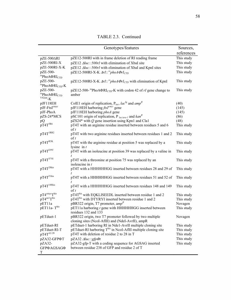
58
TABLE 2.3. Continued
Genotypes/features Sources, references
pZE-500∆RI pZE12-500RI with in frame deletion of RI reading frame This study pZE-500RI-X pZE12 ∆luc::500rI with elimination of XbaI site This study pZE-500RI-X-K pZE12 ∆luc::500rI with elimination of XbaI and KpnI sites This study pZE-500-
ssPhoAΦRICTD pZE12-500RI-X-K ∆rI:: ssphoAΦrICTD This study
pZE-500-
ssPhoAΦRICTD-K pZE12-500RI-X-K ∆rI:: ssphoAΦrICTD with elimination of KpnI This study
pZE-500-
ssPhoAΦRICTD Y42am-K
pZE12-500- ssPhoAΦRICTD-K with codon 42 of rI gene change to amber
This study
pJF118EH ColE1 origin of replication, Ptac, lacIq and ampR (40) pJF-FtsIcmyc pJF118EH harboring ftsIcmyc gene (145) pJF-PhoA pJF118EH harboring phoA gene (145) pZS-24*MCS pSC101 origin of replication, P lac/ara-1 and kanR (86) pQ pZS24* with Q gene insertion using Kpn1 and Cla1 (48) pT4T5R6 pT4T with an arginine residue inserted between residues 5 and 6
of t This study
pT4T1RR2 pT4T with two arginine residues inserted between residues 1 and 2 of t
This study
pT4TR5K pT4T with the arginine residue at position 5 was replaced by a lysine in t
This study
pT4TI39V pT4T with an isoleucine at position 39 was replaced by a valine in t
This study
pT4TT75I pT4T with a threonine at position 75 was replaced by an isoleucine in t
This study
pT4T28his pT4T with a HHHHHHGG inserted between residues 28 and 29 of t
This study
pT4T51his pT4T with a HHHHHHGG inserted between residues 51 and 52 of t
This study
pT4T148his pT4T with a HHHHHHGG inserted between residues 148 and 149 of t
This study
pT4cmycThis pT4This with EQKLISEEDL inserted between residue 1 and 2 This study pT4au1This pT4This with DTYRYI inserted between residue 1 and 2 This study pET11a pBR322 origin, T7 promoter, ampR Novagen pET11a- This pET11a harboring t gene with HHHHHHGG inserted between
residues 132 and 133 This study
pETduet-1 pBR322 origin, two T7 promoter followed by two multiple cloning sites (NcoI-AflII) and (NdeI-AvrII), ampR
Novagen
pETduet-RI pETduet-1 harboring RI in Nde1-AvrII multiple cloning site This study pETduet-RI-T pETduet-RI harboring This in NcoI-AflII multiple cloning site This study pT4T∆2-28 pT4T with deletion of residue 2 to 28 in T This study pZA32-GFPΦT pZA32 ∆luc::gfpΦt This study pZA32-GFPΦAGSAGΦT
pZA32-gfp-T with a coding sequence for AGSAG inserted between residue 238 of GFP and residue 2 of T
This study

59
TABLE 2.4. Oligonucleotides
Oligo name Sequence HindIIIpR’for……… CATTAAAGCTTGAAGGAAATA CRzNRIrev………… TGTATTTACTTTGTGCCGATGATGGGCAACTCTATCTGCACT CRzNRIfor………… AGTGCAGATAGAGTTGCCCATCATCGGCACAAAGTAAATACA ClaIRIrev………….. ATATATATCGATCTAGACCACTTTGTGAAAAGT 132-H6G2-133for…. GAAAAATCACTTGGAGGACACCACCACCACCACCACGGAGGAT
ATCCTGTTGATAAAACT 132-H6G2-133rev…. AGTTTTATCAACAGGATATCCTCCGTGGTGGTGGTGGTGGTGTCC
TCCAAGTGATTTTTC Kpn1RIfor ………… GCGCGCGGTACCATGGCCTTAAAAGCAACAGCA RIAvrIIrev…………. ATATATCCTAGGCTCTAGACCACTTTGTGA 25RIKpnfor ……….. ATATATGGTACCAATGTCGATCCTCATTTTGAT ss25RIfor ………….. ATTAAAGAGGAGAAAGGTACCATGAAACAAAGCACTATTGCA ss25RIrev………….. ATCAAAATGAGGATCGACATTCATTTCTGGTGTCCGGGCTTT KpnItfor……………. ATATATGGTACCATGGCAGCACCTAGAATA AvrIIt rev…………... ATATATCCTAGGAGCAGCGAACAAATAATTATT 56tKpnIfor…………. ATATATGGTACCGAGTACTATAAGCAATCAAAG PhoAss70tfor………. ATTAAAGAGGAGAAAGGTACCATGAAACAAAGCACTATTGCAC 56tssrev……………. CTTTAGTTGCTTATAGTACTCCATTTCTGGTGTCCGGGCTTT KpnIphoAfor ……… ATATATGGTACCCCTGTTCTGGAAAACCGGGCT AvrIIphoArev……… ATATATCCTAGGTTATTTCAGCCCCAGGGC RItmphoAfor ……… ATTAAAGAGGAGAAAGGTACCATGGCCTTAAAAGCAACAGCA RItmphoArev……..... AGCCCGGTTTTCCAGAACAGGCGCTTCAATCGATGGAGATAA KpnI70tfor ……........ ATATATGGTACCATTGAAAAGGAAAGAACTGCA FtsITM70tfor ……… ATTAAAGAGGAGAAAGGTACCATGTTTGCGTTGTTATGCGGC
TGT FtsITM70trev……… TGCAGTTCT TTCCTTTTCAATTCCGAGCAGAAAAGCCAGCGC T4TtmphoAfor…….. ATTAAAGAGGAGAAAGGTACCATGGCAGCACCTAGAATA TCA T4TtmphoArev…….. AGCCCGGTTTTCCAGAACAGGAATTTCACTGTATGTTTCATA KpnI-gfp-for ………. ATATATGTTACCATGAGTAAAGGAGAAGAACTT N-RIC-gfprev……… AAGTGCTGTTGCTTTTAAGGCTTTTAAGGCTTTGTATAGTTCATC
CATGCC C-gfpN-RIfor………. GGCATGGATGAACTATACAAAGCCTTAAAAGCAACAGCACTT RI-XbaIrev………… GTCGACTCTAGACCACTTTGTGAA NdeI70tfor…………. ATATATCATATGATTGAAAAGGAAAGAACTGCA BamH1trev………… ATATATGGATCCAGCAGCGAACAAATAATTATT 25RINdeIfor……….. ATATATCATATGAATGTCGATCCTCATTTT RIhisBamrev………. ATATATGGATCCTCAGTGGTGGTGGTGGTGGTG RIG2H6for……….... AACATTAAATTGGAGACTGAAGGAGGACACCACCACCACCACC
ACTGAAATTCAGCGACTTTTCAC RIG2H6rev………… GTGAAAAGTCGCTGAATTTCAGTGGTGGTGGTGGTGGTGTCCTC
CTTCAGTCTCCAATTTAATGTT -500RIKpn1for…….. ATATATACCGACGTAGATATTTCCTTC +500RIXba1rev……. ATATATTCTAGAGCTAAGGACTGATTTCGC RIA7Lfor…………... GCCTTAAAAGCAACACTACTTTTTGCCATGCTA RIA7Lrev………….. TAGCATGGCAAAAAGTAGTGTTGCTTTTAAGGC 500delRIfor………... AAAGGCCTCCTATCATTT 500delRIrev………... TGAAATTCAGCGACTTTT RIT6Lfor…………... ATGGCCTTAAAAGCACTACTACTTTTTGCCATG
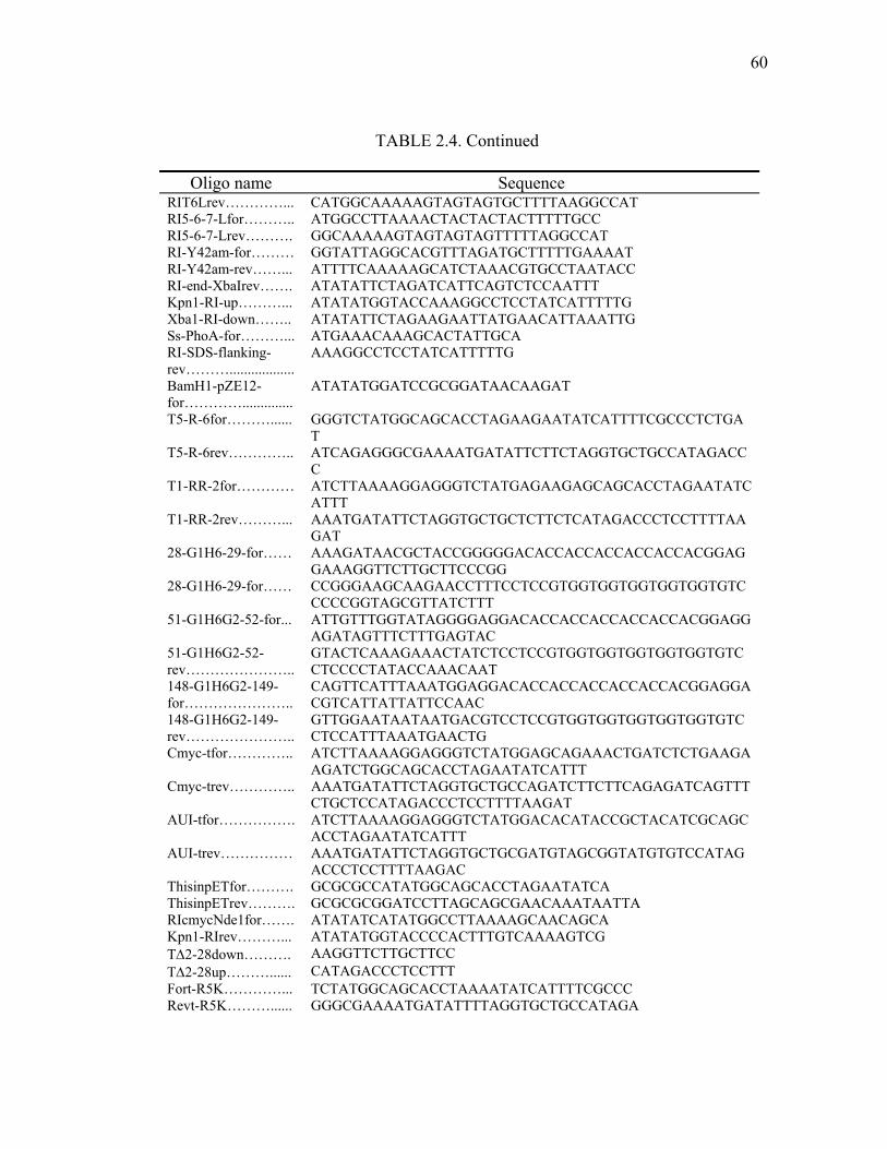
60
TABLE 2.4. Continued
Oligo name Sequence RIT6Lrev…………... CATGGCAAAAAGTAGTAGTGCTTTTAAGGCCAT RI5-6-7-Lfor……….. ATGGCCTTAAAACTACTACTACTTTTTGCC RI5-6-7-Lrev………. GGCAAAAAGTAGTAGTAGTTTTTAGGCCAT RI-Y42am-for……… GGTATTAGGCACGTTTAGATGCTTTTTGAAAAT RI-Y42am-rev……... ATTTTCAAAAAGCATCTAAACGTGCCTAATACC RI-end-XbaIrev……. ATATATTCTAGATCATTCAGTCTCCAATTT Kpn1-RI-up………... ATATATGGTACCAAAGGCCTCCTATCATTTTTG Xba1-RI-down…….. ATATATTCTAGAAGAATTATGAACATTAAATTG Ss-PhoA-for………... ATGAAACAAAGCACTATTGCA RI-SDS-flanking-rev………..................
AAAGGCCTCCTATCATTTTTG
BamH1-pZE12-for…………..............
ATATATGGATCCGCGGATAACAAGAT
T5-R-6for………...... GGGTCTATGGCAGCACCTAGAAGAATATCATTTTCGCCCTCTGAT
T5-R-6rev………….. ATCAGAGGGCGAAAATGATATTCTTCTAGGTGCTGCCATAGACCC
T1-RR-2for………… ATCTTAAAAGGAGGGTCTATGAGAAGAGCAGCACCTAGAATATCATTT
T1-RR-2rev………... AAATGATATTCTAGGTGCTGCTCTTCTCATAGACCCTCCTTTTAAGAT
28-G1H6-29-for…… AAAGATAACGCTACCGGGGGACACCACCACCACCACCACGGAGGAAAGGTTCTTGCTTCCCGG
28-G1H6-29-for…… CCGGGAAGCAAGAACCTTTCCTCCGTGGTGGTGGTGGTGGTGTCCCCCGGTAGCGTTATCTTT
51-G1H6G2-52-for... ATTGTTTGGTATAGGGGAGGACACCACCACCACCACCACGGAGGAGATAGTTTCTTTGAGTAC
51-G1H6G2-52- rev…………………..
GTACTCAAAGAAACTATCTCCTCCGTGGTGGTGGTGGTGGTGTCCTCCCCTATACCAAACAAT
148-G1H6G2-149-for…………………..
CAGTTCATTTAAATGGAGGACACCACCACCACCACCACGGAGGACGTCATTATTATTCCAAC
148-G1H6G2-149-rev…………………..
GTTGGAATAATAATGACGTCCTCCGTGGTGGTGGTGGTGGTGTCCTCCATTTAAATGAACTG
Cmyc-tfor………….. ATCTTAAAAGGAGGGTCTATGGAGCAGAAACTGATCTCTGAAGA AGATCTGGCAGCACCTAGAATATCATTT
Cmyc-trev………….. AAATGATATTCTAGGTGCTGCCAGATCTTCTTCAGAGATCAGTTTCTGCTCCATAGACCCTCCTTTTAAGAT
AUI-tfor……………. ATCTTAAAAGGAGGGTCTATGGACACATACCGCTACATCGCAGCACCTAGAATATCATTT
AUI-trev…………… AAATGATATTCTAGGTGCTGCGATGTAGCGGTATGTGTCCATAGACCCTCCTTTTAAGAC
ThisinpETfor………. GCGCGCCATATGGCAGCACCTAGAATATCA ThisinpETrev………. GCGCGCGGATCCTTAGCAGCGAACAAATAATTA RIcmycNde1for……. ATATATCATATGGCCTTAAAAGCAACAGCA Kpn1-RIrev………... ATATATGGTACCCCACTTTGTCAAAAGTCG T∆2-28down………. AAGGTTCTTGCTTCC T∆2-28up………...... CATAGACCCTCCTTT Fort-R5K…………... TCTATGGCAGCACCTAAAATATCATTTTCGCCC Revt-R5K………...... GGGCGAAAATGATATTTTAGGTGCTGCCATAGA

61
TABLE 2.4. Continued
Oligo name Sequence Fort-I39V………....... CGGGTAGCTGCTGTAGTTCTTTTGTTTATAATG Revt-I39V………….. CATTATAAACAAAAGAACTACGACAGCTACCCG Fort-T75I…………... ATTGAAAAGGAAAGAATTGCACGCTTTGAATCT Revt-T75I………….. AGATTCAAAGCGTGCAATTCTTTCCTTTTCAAT CgfpNt-rev………… TGATATTCTAGGTGCTGCTTTGTATAGTTCATCCAT CgfpNt-for…………. ATGGATGAACTATACAAAGCAGCACCTAGAATATCA AG-T-for…………... GCAGGTGCAGCACCTAGAATATCA Gfp-AGS-rev………. ACTTCCAGCTTTGTATAGTTCATCCAT
Construction of plasmids
Plasmid pT4T was derived by removing the aphI (kanamycin-resistance) gene
from pER-t (109) and was a gift from I.-N. Wang. It carries a hybrid lysis cassette in
which the T4 t gene (Fig. 2.2; nt160204 to 160884 of the T4 genome) replaces the λ S
gene (nt 45157 to 45465 of the λ genome) in a DNA segment comprising pR’, the λ late
promoter, the downstream genes SRRzRz1, and a deletion of the bor gene (Fig. 2.2).
This lysis cassette is flanked by unique HindIII and ClaI sites (not shown). The plasmid,
pT4TRI, was constructed by PCR amplifying the lysis cassette from pT4T using the
forward and reverse primers HindIIIpR’for and CRzNRIrev. In a separate PCR reaction
the rI gene was amplified using the forward and reverse primers CRzNRIfor and
ClaIRIrev. The rI gene in the template used for this reaction had its internal ClaI site
destroyed by the introduction of the silent mutation G63A by site-directed mutagenesis.
Since the primers CRzNRIrev and CRzNRIfor were complementary, it was possible to
fuse the rI gene sequence (nt 59540 to 59177 of the T4 genome) to the 3’ end of the
hybrid lysis cassette (after the base corresponding to 46437 of the λ genome, beyond the

62
end of the Rz gene; Fig. 2.2) by using the two PCR products as templates in an SOE
(splicing by overlapping extension) reaction (54) using the HindIIIpR’for and ClaIRIrev
primers. The product from this reaction was digested with HindIII and ClaI and ligated
into the vector backbone produced by digesting pT4T with the same enzymes. The
plasmid pT4This was generated by introducing a hexahistidine tag between codons 132
and 133 of the t gene (Fig. 2.1A, underlined residues) in pT4T, using a pair of
oligonucleotides, 132-H6G2for/rev, encoding His6Gly2.
Two plasmids were constructed for overexpression of the his-tagged C-terminal
domain of RI (RICTDhis) and T (TCTD
his). For the TCTD plasmid, the DNA fragment
encoding TCTDhis was PCR amplified from pT4This using the primer pair NdeI70 for and
FIG. 2.2. Features of plasmid and phage constructs. All plasmids used for inductions are based on the structure of pS105 (109)(upper structure), with the λ late promoter and the promoter-proximal genes of the late transcriptional unit (lower structure), except that in the plasmid pT4T, the S gene in the λ SRRzRz1 lysis cassette of pER157 (47) is replaced by the phage T4 t gene. In the plasmid pT4TRI, the rI gene is inserted 13 nucleotides downstream of the Rz gene of pT4T. “X” designates areas where homologous recombination can occur for generation of λt recombinants (133).
Q
45
PR'
bor
Pbor46
S R Rz/Rz1
t∆
Q
45
PR'
bor
Pbor46 47
S R Rz/Rz1
44
∆SR
x x
rI

63
BamH1trev. The doubly-digested PCR product was inserted into the multiple cloning
site of plasmid pET11a to generate the plasmid pET11a-TCTDhis. To construct the RICTD
plasmid, a fragment carrying gene rI was produced by cleaving pZE-RI with EcoRI and
XbaI. The plasmid pER-t was digested using the same enzymes to generate the
backbone for the plasmid pPRI, and this backbone fragment was ligated to the EcoRI-
XbaI fragment carrying rI to produce pPRI. The plasmid pPRIhis was made by inserting
hexahistidine tag after codon 97 of the rI gene (Fig. 2.1B) in pPRI, using the primer pair,
RIG2H6for/rev, encoding Gly2His6. The DNA fragment encoding the RICTDhis PCR was
amplified from pPRIhis using 25RINdeIfor and RIhisBamH1rev with NdeI and BamH1
restriction sites at their 5’ ends, respectively. The doubly-digested PCR product was
inserted into the multiple cloning site of pET11a to yield pET11a-RICTDhis.
Phage λthis was generated by homologous recombination between pT4This and
the lysis-defective phage λkan∆(SR) (formerly designated as λ∆SR (105); Fig. 2.2).
Recombinants were identified by their plaque-forming ability and the presence of the
hybrid lysis cassette was verified by DNA sequencing. Lysogens were prepared by
infecting cells with λthis and plating at 30ºC for survivors on media containing
kanamycin.
The T4 t and rI genes and their derivatives were also expressed from the lac
promoter of the pZA and pZE plasmids from the family of modular pZ vectors (86). To
construct pZA-RI and pZA-RIcmyc, the primer pair, Kpn1RIfor and RIAvrIIrev was used
to PCR amplify the rI gene from pZE-RI or pZE-RIcmyc, respectively. After digestion
with KpnI and AvrII, these PCR products were used to replace the luc gene in pZA32-

64
luc. The plasmid, pZA-RICTD, carrying a DNA fragment encoding the C-terminal
domain (residues 25-97; Fig. 2.1B) of the RI protein was similarly constructed using the
PCR product from a PCR reaction with primers 25RIKpnIfor and RIAvrIIrev and pZE-
RI as the template. The signal sequence of alkaline phosphatase (PhoA) was fused to the
RI fragment in pZA-RICTD by the modified site-directed mutagenesis procedure
described above. In the first PCR reaction, the PhoA signal sequence was amplified
using the ss25RIfor and ss25RIrev primers. The second PCR reaction used the above
PCR product as the primer and pZA-RICTD as the template yielding pZA-ssPhoAΦRICTD.
The identical reactions were used to generate pZA-RICTDcmyc and pZA-ssPhoAΦRICTD
cmyc
from pZE-RIcmyc. A similar strategy was used to generate the complementary series of
plasmids, pZA-T, pZA-TCTD and pZA-ssPhoAΦTCTD, using the primer pairs,
KpnItfor/AvrIItrev, 56tKpnIfor/AvrIItrev, and ss25RIfor/56tssrev, respectively. Here,
the TCTD consists of residues 56 to 218 of T (Fig. 2.1A).
The sequence encoding residues 27-471 of the phoA gene was PCR amplified
using the forward and reverse primers KpnIphoAfor and AvrIIphoArev, with KpnI and
AvrII restriction sites at their 5’ ends, respectively. The doubly-digested PCR product
was used to replace the luc gene in pZA32-luc to yield pZA-PhoA. To fuse the N-
terminal domain (residues 1 to 24, RINTD) of RI to the mature form of PhoA, the DNA
encoding the RINTD was PCR amplified using the primers RItmphoAfor and
RItmphoArev. The PCR product was then used to conduct a modified site-directed
mutagenesis reaction using pZA-PhoA as the template to generate pZA-RINTDΦPhoA.
The plasmid pZA-TTMDΦPhoA in which residues 1 to 70 of T are fused to the mature

65
sequence of PhoA was constructed in a similar fashion using the primers T4TtmphoA
for and T4TtmphoArev.
The ftsItmdφt chimera, encoding a protein with the TMD of FtsI (Fig. 2.1A)
replacing the TMD of T, was constructed in two steps. First, a DNA fragment encoding
residues 70 to 218 of T was PCR amplified using the forward and reverse primers
AvrIItrev and KpnI70tfor, with AvrII and KpnI restriction sites at their 5’ ends,
respectively. The doubly digested PCR product was used to replace the luc gene in
pZA32-luc generating the intermediate plasmid, pZA-T70-218. Then, a DNA fragment
encoding a methionine codon followed by the transmembrane segment of FtsI (residues
24 to 40) was PCR amplified using the forward and reverse primers, FtsITM70tfor and
FtsITM70trev The PCR product was then used to conduct a modified site-directed
mutagenesis reaction by using the with pZA-T70-218 as the template to generate pZA-
FtsITMDΦT.
GFP was fused to cmyc-tagged RI (Fig. 2.1B) by another SOE reaction. First, a
DNA fragment encoding GFP was PCR amplified from pDS439 (120) using the primers
KpnIgfpfor and N-RIC-gfprev. Separately, a DNA fragment encoding residues 2-106 of
the cmyc-tagged RI protein was PCR amplified from pZE-RIcmyc using the primers C-
gfpN-RIfor and RI-Xbarev. The PCR products from the two reactions were combined
and amplified using the primers KpnIgfpfor and RI-Xbarev. The fusion product was
digested with KpnI and XbaI and ligated into pZE12 digested with the same enzymes,
yielding pZE-GFPΦRIcmyc.
66
Subcellular fractionation
To prepare total membrane and soluble fractions, cell pellets from 150 ml
cultures were resuspended in 1 ml of French press buffer (100 mM Na2HPO4, 100 mM
KCl, 5 mM EDTA, pH 8.0, 1 mM PMSF, 25 mM MgCl2, 50 µg/ml DNAse, 50 µg/ml
RNAse) and 2.5 µl of Protease Inhibitor Cocktail (4-(2-aminoethyl)benzenesulfonyl
fluoride, bestatin, pepstatin, E-64 and phosphoramidon; Sigma). The cells were
disrupted by passage through a French pressure cell (Spectronic Instruments, Rochester,
N.Y.) at 16,000 psi (1 psi = 6.89 kPa). The unbroken cells were removed by
centrifugation in a Damon-IEC Spinette clinical centrifuge at 1000 x g for 10 min. The
membrane and soluble fractions were separated by centrifugation at 100,000 x g for 60
min at 4ºC. To identify periplasmic proteins, cells from 30 ml cultures were collected by
centrifugation and the pellets were resuspended in 250 µl of 25% sucrose, 30 mM Tris-
HCl, pH 8.0. Then, 10 µl of 0.25 M EDTA, 10 µl of lysozyme (20 mg/ml in water), and
250 µl of distilled water were added, sequentially (16). When microscopic examination
showed that ~95% of the cells had formed spheroplasts, the samples were centrifuged at
9000 x g for 10 min to separate the periplasm from the spheroplasts (membrane and
cytosol).
SDS-PAGE and Western blotting
SDS-PAGE and Western blotting were performed as previously described (46).
To generate antibodies against RI and T, BL21(DE3) tonA::Tn5 slyD::Tetr cells
67
harboring either pET11a-TCTDhis or pET11a-RICTD
his were grown in 1 liter of LB-
ampicillin to A550 ~ 0.5; then the cultures were induced with IPTG for 3 hrs. The cell
pellets was resuspended in 20 ml of 20 mM BES (N,N-bis[2-hydroxyethyl]-2-
aminoethanesulfonic acid; Sigma), 0.5 M NaCl pH 7.5 supplemented with 20 µl
Protease Inhibitor Cocktail, 700 µl MgCl2 (1M), 100 µl RNase (10 mg/ml), 100 µl
DNase (10 mg/ml), and 20 µl DTT (1 M). The cells were lysed by passage through a
French pressure cell (Spectronic Instruments, Rochester, N.Y.) at 16,000 psi (1 psi =
6.89 kPa). Inclusion bodies were collected by centrifugation of the French press lysates
at 17,500 x g for 30 min. The pellets were extracted with 20 ml of 20 mM BES, 6 M
guanidine hydrochloride pH 7.5 for 3 hrs. The extracts were cleared by centrifugation at
17,500 x g for 10 min. The supernatant was used as the starting material for purifying
TCTDhis and RICTD
his using Talon metal affinity resin (Clontech). The 5 ml resin bed is
equilibrated with 20 mM BES, 6 M guanidine hydrochloride (United States
Biochemical) pH 7.5 and the bound proteins were eluted with 20 mM BES, 6 M
guanidine hydrochloride, 0.5 M imidazole (Sigma), pH 7.5.
Antibodies against the purified, C-terminal domains of This and RIhis were
prepared in rabbits by ProSci Incorporated, Poway, CA. Antibodies against the cmyc
epitope were purchased from Babco (Richmond, CA). Reagents and methods for
immunodetection have been described (145). Equivalent sample loadings were used
whenever multiple fractions from the same culture were analyzed.

68
Immunoprecipitation of RI - T complexes
MDS12 tonA::Tn10 lacIq1 cells harboring the indicated allele of pZA-
ssPhoAΦTCTD and pZE-GFPΦRIcmyc either alone or in combination were grown to an
A550 ~ 0.4 and then induced with 1 mM IPTG for 30 min. A 30 ml volume of each
culture was taken through the EDTA-lysozyme treatment used to prepare spheroplasts
(16). Instead of centrifuging the samples after the addition of water, the spheroplasts
were lysed by adding 5 µl of Protease Inhibitor Cocktail and 500 µl lysis buffer (100
mM Tris-HCl pH 8.0, 150 mM NaCl, 1% NP40). Next, 100 µl of 1M MgCl2, 10 µl of
10 mg/ml DNase I, and 10 µl of 10 mg/ml pancreatic RNase were added and the samples
incubated at room temperature for 15 min with occasional mixing.
To collect complexes containing the TCTD and GFPΦRIcmyc proteins, 200 µl of
the indicated lysate was diluted with an equal volume of wash buffer (lysis buffer
containing 1 mg/ml bovine serum albumin). Next, 2 µl of a pre-immune rabbit serum,
rabbit anti-T, or mouse anti-GFP (Stressgen) were added and the samples were incubated
for 2 hrs at 4oC with slow agitation. Then, 50 µl of the appropriate iron-conjugated
secondary antibody (Pierce) was added and the mixture incubated for an additional for 2
hrs at 4oC. The immune complexes were collected magnetically and washed three times
with 0.5 ml of wash buffer. The complexes were dissociated by boiling in SDS-PAGE
sample buffer and analyzed by SDS-PAGE and Western blotting followed by
immunodetection using anti-T or anti-cmyc antibodies, as indicated.

69
Phage accumulation during LIN
For the assessment of the LIN state, CQ21 cells lysogenic for λkan this and
harboring either no plasmid, pZA-RI or pZA-ssPhoAΦRICTD, were grown at 30oC and
induced both by adding IPTG and simultaneously shifting the growth temperature to
42oC for 15 min then to 37oC. At 30, 60, 90, 120, and 150 min after induction, 1 ml was
taken from each culture and the cells lysed by the addition of CHCl3. Debris was
removed by centrifugation, and the plaque-forming titers in the cleared lysates
determined in triplicate on MDS12 tonA::Tn10 lacIq1.
Results
Domain analysis of the RI antiholin
Paddison et al. (100) used primary structure analysis algorithms to predict that RI
has an N-terminal secretory signal and was thus a secreted periplasmic protein.
Although RI has been visualized as a cross-linked complex with T, it had previously
escaped detection as an independent polypeptide, which presumably was due to
proteolytic lability (110). Thus, whether this N-terminal sequence serves as a signal
anchor, leaving its periplasmic C-terminal domain (RICTD) tethered to the cytoplasmic
membrane, or a cleavable signal allowing release of the RICTD into the periplasm was
unclear. Sequence analysis of the classical rI alleles of Doermann (27) revealed that
most were frame-shifts distal to the predicted secretory signal. Moreover, the single
70
missense allele, (R78P; listed as R78G in Paddison et al. (100); Fig 2.1B), also mapped
there, implicating the RICTD as critical to the antiholin function of RI.
To resolve RI into topological components, we constructed two chimeric genes:
one, ssphoAφrICTD, with RICTD fused to the signal sequence of alkaline phosphatase
(PhoA); the other, rINTDφphoA, with the N-terminal domain of RI (RINTD) fused to the
periplasmic domain of PhoA (Fig. 2.1B). Using antibodies raised against RICTD, the
ssPhoAΦRICTDcmyc protein could be detected in whole cells (Fig. 2.3A). However,
efforts to localize it using conventional subcellular fractionation were unsuccessful,
again presumably due to rapid proteolysis after cell disruption. To provide evidence that
ssPhoAφRICTDcmyc was present in the periplasm, the ssphoAφrICTD gene was expressed in
cells grown in the presence or absence of azide to inhibit SecA. A slower migrating
species accumulated in the presence of azide, indicating that, when SecA is not
inhibited, ssPhoAΦRICTDcmyc is processed and localized to the periplasm (Fig. 2.3A). By
contrast, the product of the rINTDφphoA construct was stable, unprocessed and was found
in both the membrane and soluble fractions (Fig. 2.3B). The dual localization of the
unprocessed RINTDΦPhoA protein will be considered in the Chapter III. We next
compared the ability of these chimeras to support LIN with that of wt RI. While the
rINTDΦphoA fusion had no biological function, the ssphoAφrICTD chimera blocked t-
mediated lysis, as assessed by monitoring the turbidity of the induced culture (Fig.
2.4A). Moreover, in addition to preventing the loss of optical density, both rI and
ssphoAφrICTD allowed the extended intracellular accumulation of virions and suppressed
their release to the medium (Fig. 2.4B). Finally, the LIN state supported by both rI and

71
1A
2 3 4 516
64
B1
αPhoA64
2 3 4 5 6 7 8 9 10 1112
50
36
22
1 2 3 4 5 6 7 8 9 10 11
αT
C
αRI
1A
2 3 4 516
64
B1
αPhoA64
2 3 4 5 6 7 8 9 10 1112
50
36
22
1 2 3 4 5 6 7 8 9 10 11
αT
C
αRI
FIG. 2.3. Localization of T and RI chimeras. Subcellular fractions were prepared and analyzed by SDS-PAGE and Western blotting as described in Materials and Method. (A) Cells carrying pZA-ssPhoAΦRICTD
cmyc were grown in the absence (lanes 2 and 3) or the presence (lanes 4 and 5) of 1 mM azide for 10 min in advance of induction. Cells from these cultures were collected by TCA precipitation and centrifugation, resuspended in SDS-PAGE sample buffer and subjected to SDS-PAGE and Western blotting using anti-RI antisera as the primary antibody. Lane 1, molecular mass standards; lanes 2 and 4, samples from uninduced cultures; lanes 3 and 5, samples from induced cultures. (B) Cells carrying the pZA-RINTDΦPhoA were induced, fractionated, and analyzed by SDS-PAGE and Western blotting using anti-PhoA as the primary antibody. Lane 1, molecular mass standards; lane 2, mature form of PhoA; lane 3, blank; lane 4, cells from an uninduced culture; lane 5, cells from an induced culture; lane 6, total cell lysate; lane 7, 1000 x g pellet; lane 8, 1,000 x g supernatant; lane 9, 100,000 x g supernatant (soluble fraction); lane 10, 100,000 x g pellet (membrane fraction); lane 11, detergent extractable (1% NP40) membrane fraction; lane 12, detergent insoluble fraction. (C) Cells carrying pZA-ssPhoAΦTCTD were grown in the absence (lanes 2-6) or the presence (lanes 7-11) of 1 mM azide for 10 min in advance of induction, harvested, fractionated and analyzed by SDS-PAGE and Western blotting using anti-T antisera as the primary antibody. Lane 1, molecular mass standards; lanes 2 and 7, uninduced cells; lanes 3 and 8, induced cells; lanes 4 and 9, cells after spheroplasting; lanes 5 and 10, spheroplasts; lanes 6 and 11, periplasm (133).

72
ssphoAφrICTD could be subverted by the addition of energy poisons (Fig. 2.4A). We
conclude that the periplasmic domain of RI is necessary and sufficient for authentic LIN.
Indeed, the ssphoAφrICTD allele is more effective than the parental rI gene, as judged by
the stability of the LIN phenotype (Fig. 2.4A).
RI-dependent LIN requires binding to the C-terminal domain of T
The unusually large C-terminal periplasmic domain of T (TCTD) is a feature that
distinguishes it from the numerous class I and class II holins (141). To determine
whether this domain is the target for RICTD, we constructed an allele of t in which the
sequences encoding the predicted cytoplasmic and transmembrane domains were
replaced by the segment of phoA encoding its secretory signal sequence (ssphoAφtCTD;
Fig. 2.1A). Like the RICTD, the TCTD was also efficiently secreted by the PhoA signal
sequence (Fig. 2.3C). This allele was lytically incompetent but exerted a weak
dominant-negative phenotype in that it caused a short delay in T-mediated lysis (Fig.
2.4C inset), suggesting that homotypic interactions in the TCTD are involved in the lytic
function of T. The biological function of this chimera was assessed in a system in which
t and rI are both expressed from the λ late promoter, shown in previous work to support
physiologically meaningful lysis timing with the λ lysis cassette (47). The results
clearly showed that supplying periplasmic TCTD partially blocked the imposition of LIN
(Fig. 2.4C). More dramatic results were obtained when cells producing the
ssPhoAΦTCTD were infected with T4 phage; LIN was completely subverted by the
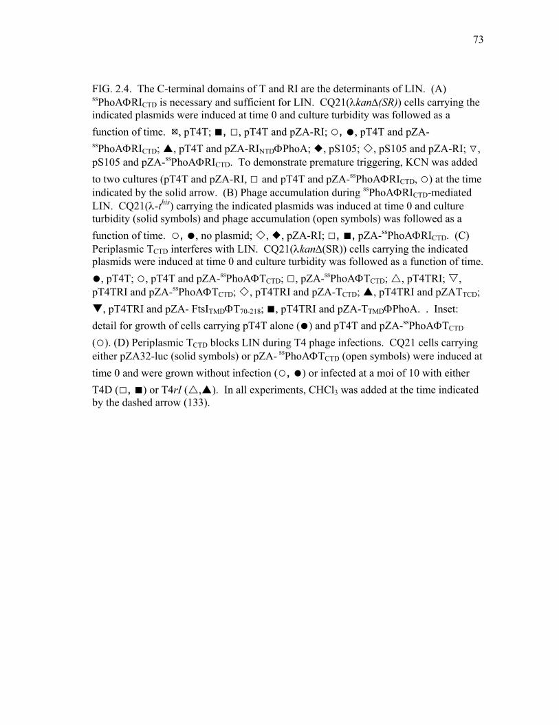
73
FIG. 2.4. The C-terminal domains of T and RI are the determinants of LIN. (A) ssPhoAΦRICTD is necessary and sufficient for LIN. CQ21(λkan∆(SR)) cells carrying the indicated plasmids were induced at time 0 and culture turbidity was followed as a function of time. :, pT4T; ■, □, pT4T and pZA-RI; ○, ●, pT4T and pZA-ssPhoAΦRICTD; , pT4T and pZA-RINTDΦPhoA; , pS105; , pS105 and pZA-RI; ▽, pS105 and pZA-ssPhoAΦRICTD. To demonstrate premature triggering, KCN was added to two cultures (pT4T and pZA-RI, □ and pT4T and pZA-ssPhoAΦRICTD, ○) at the time indicated by the solid arrow. (B) Phage accumulation during ssPhoAΦRICTD-mediated LIN. CQ21(λ-this) carrying the indicated plasmids was induced at time 0 and culture turbidity (solid symbols) and phage accumulation (open symbols) was followed as a function of time. ○, ●, no plasmid; , , pZA-RI; □, ■, pZA-ssPhoAΦRICTD. (C) Periplasmic TCTD interferes with LIN. CQ21(λkan∆(SR)) cells carrying the indicated plasmids were induced at time 0 and culture turbidity was followed as a function of time. ●, pT4T; ○, pT4T and pZA-ssPhoAΦTCTD; □, pZA-ssPhoAΦTCTD; , pT4TRI; , pT4TRI and pZA-ssPhoAΦTCTD; , pT4TRI and pZA-TCTD; , pT4TRI and pZATTCD;
, pT4TRI and pZA- FtsITMDΦT70-218; ■, pT4TRI and pZA-TTMDΦPhoA. . Inset: detail for growth of cells carrying pT4T alone (●) and pT4T and pZA-ssPhoAΦTCTD (○). (D) Periplasmic TCTD blocks LIN during T4 phage infections. CQ21 cells carrying either pZA32-luc (solid symbols) or pZA- ssPhoAΦTCTD (open symbols) were induced at time 0 and were grown without infection (○, ●) or infected at a moi of 10 with either T4D (□, ■) or T4rI ( , ). In all experiments, CHCl3 was added at the time indicated by the dashed arrow (133).
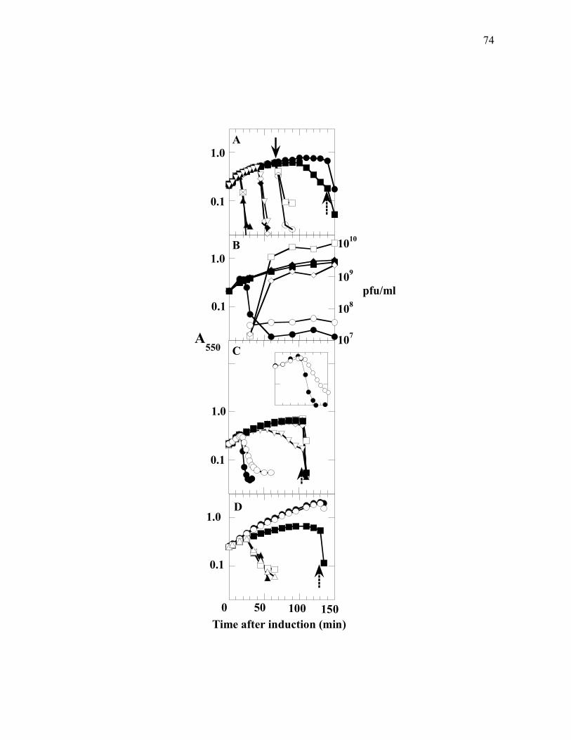
74
0 50 100 150Time after induction (min)
A550
0.1
0.1
0.1
0.1
1.0
1.0
1.0
1.0
107
1010
109
108
pfu/ml
A
D
C
B
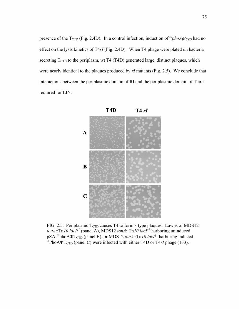
75
presence of the TCTD (Fig. 2.4D). In a control infection, induction of ssphoAφtCTD had no
effect on the lysis kinetics of T4rI (Fig. 2.4D). When T4 phage were plated on bacteria
secreting TCTD to the periplasm, wt T4 (T4D) generated large, distinct plaques, which
were nearly identical to the plaques produced by rI mutants (Fig. 2.5). We conclude that
interactions between the periplasmic domain of RI and the periplasmic domain of T are
required for LIN.
T4D T4 rI
A
B
C
T4D T4 rI
A
B
C
FIG. 2.5. Periplasmic TCTD causes T4 to form r-type plaques. Lawns of MDS12 tonA::Tn10 lacIq1 (panel A), MDS12 tonA::Tn10 lacIq1 harboring uninduced pZA-ssphoAΦTCTD (panel B), or MDS12 tonA::Tn10 lacIq1 harboring induced ssPhoAΦTCTD (panel C) were infected with either T4D or T4rI phage (133).

76
T and RI form a complex
To provide further evidence that the T and RI proteins interact, we performed co-
immunoprecipitation experiments using lysates prepared from cells expressing
ssPhoAΦTCTD and a GFPΦRIcmyc chimera. The GFPΦRIcmyc chimera was used since it
was readily visualized by immunoblot, presumably because of decreased lability
compared to either RI or ssPhoAφRICTD, both of which were undetectable in
immunoprecipitations. Using either anti-T or anti-GFP as the first antibody, the two
proteins were found to co-precipitate (Fig. 2.6A). These complexes were also formed
when detergent-solubilized extracts prepared from cells expressing T or GFPΦRIcmyc
separately were mixed and then subjected to immunoprecipitation (not shown). Identical
results were obtained when the T75I mutation found in a t allele known to be insensitive
to RI-mediated LIN was introduced into the ssPhoAφTCTD protein (Fig. 2.6B). Since the
R78P allele of rI is defective for LIN, we attempted to test the effect of this mutation on
the ability of RI to form complexes with T. Unfortunately, the product of the R78P
allele of gfpφrIcmyc does not accumulate in whole cells to levels detectable by Western
blot.
Interestingly, the ratio of unprocessed ssPhoAΦTCTD to mature periplasmic TCTD
was reproducibly higher when GFPΦRIcmyc was present (Fig. 2.6AB, lanes 5 -7). This
suggests that the binding of RI to the periplasmic domain can occur while TCTD is
nascent and that this binding interferes with leader peptidase cleavage of the signal
sequence.

77
Discussion
Infections of T4 and the other T-even phages are the only examples where lysis
timing has been demonstrated to be directly affected by the environmental conditions
surrounding the infected cell. The classic LIN phenotype can be thought of as a “no
quorum” signal, in the sense that an effector molecule (i.e., a super-infecting T4 phage)
indicating the lack of available hosts binds to an infected cell and initiates an as yet
unknown signal transduction pathway leading to activation of an effector, RI. The
50
αcmyc
αT22
36
36
A
22
36
1 2 3 4 5 6 7 8 9 10
αcmyc
αT
B
blot:50
αcmyc
αT22
36
36
A
22
36
1 2 3 4 5 6 7 8 9 101 2 3 4 5 6 7 8 9 10
αcmyc
αT
B
blot:
FIG. 2.6. T and RI form a complex. (A) Immunoprecipitations were performed with samples containing either ssPhoAΦTCTD (lanes 2 – 4), both ssPhoAΦTCTD and GFPΦRIcmyc (lanes 5 – 7), or GFPΦRIcmyc only (lanes 8 – 10), prepared from induced cells carrying either of the plasmids pZA- ssPhoAΦTCTD or pZE-GFPΦRIcmyc, or both. Primary antibodies for immunoprecipitations: lanes 2, 5 and 8, rabbit preimmune serum; lanes 3, 6, and 9, anti-T rabbit antibody; lanes 4, 7 and 10, anti-GFP monoclonal antibody. Samples were analyzed by SDS-PAGE and immunoblotted with either anti-T or polyclonal anti-cmyc, as indicated to the right. (B) Same as (A) except that the rV variant ssPhoAΦTCTD
T75I, produced from the plasmid pZA- ssPhoAΦTCTDT75I was used.
Molecular mass appears in lane 1 for each blot (133).
78
simplest interpretation of our data is that activated RI directly binds to and inhibits the
target molecule, T, so that lysis is blocked. Both RI and T have hydrophobic domains
near their N-termini, but the results presented here demonstrate conclusively that the
interactions essential for the transmission of the signal are entirely between the
periplasmic domains of the two proteins. Moreover, we have shown that RI and T are
present in a complex that can be recovered by immunoprecipitation without prior
covalent cross-linking. Unlike class I and II holins which consist of two or three
transmembrane domains, short interconnecting loops, and a 10-25 residue C-terminal
cytoplasmic domain, T and its orthologs in T4-like phage have a single transmembrane
domain and a relatively large C-terminal periplasmic domain (108). Our findings
suggest that this periplasmic domain has evolved to serve as a receptor for the LIN
signal provided by “activated” RI. This idea is consistent with the observation that large
deletions within the TCTD can be tolerated without entirely abolishing holin function
(108). In addition, the co-immunoprecipitation experiments also suggest that RI binds to
nascent T, since the presence of GFPΦRIcmyc appears to reduce the efficiency of the
maturation of ssPhoAΦTCTD (Fig. 2.6 A, lanes 5-7). This idea that RI bind to nascent T
is consistent with the finding that in T4 infections, T4 superinfection can confer RI-
mediated LIN on a pre-existing pool of wt LIN-sensitive T molecules if the incoming
phage has a wt, LIN-sensitive t allele, but not if it has an rV, LIN-insensitive t allele
(109). The interference with signal sequence processing also suggests that
conformational changes derived from binding of RI to the TCTD can be transmitted to the

79
membrane, which is consistent with the fact that one of the classic rV, LIN-defective
mutations is a subtle missense change, I39V, in the hole-forming TMD of T.
The classic LIN phenotype appears to involve a direct interaction between the
periplasmic domains of RI and T and can be subverted by the collapse of the membrane
potential by energy poisons. Thus, at a superficial level, the antiholin function of RI
resembles that of S107 and S2171, the antiholins of λ and lambdoid phage 21,
respectively. The latter two proteins are identical to their cognate holins except for a
short N-terminal extension. In both cases, antiholin-holin interactions are thought to
prevent quaternary rearrangements that are required to convert holin oligomers into
“holes”. We suggest that in the energized membrane, the single TMD of T is unable to
oligomerize into a functional “hole” in the absence of interactions between TCTDs in the
periplasm. The antiholin activity of RI would then be due to its ability to bind to the
TCTD preventing these interactions. Mutations that alter the periplasmic interactions
between TCTD molecules and between TCTD and RICTD would allow for the genetic
malleability of lysis timing for T4. For bacteriophages λ (13,44,45,106) and the
lambdoid phage 21 (102), collapse of the membrane potential allows premature
triggering of the holin by causing topological changes in the antiholin that lead to its
inactivation. Moreover, in these cases, representing canonical class I and II holins
respectively, these topological changes in the antiholin effectively convert it into the
functional equivalent of its cognate holin. It seems unlikely that T and RI have the same
relationship as is seen with the antiholin/holin pairs of phage λ and 21 for several
reasons. First, the homotypic interactions that presumably characterize the S105/S107

80
and S2171/ S2168 systems are not possible since T and RI do not share amino acid
sequence homology. Second, RINTD is not essential for its antiholin activity, indicating
that RI does not directly interact with the hole-forming TMD of the T holin. Finally, in
contrast to S107 or S2171, RI appears to be a labile protein whose antiholin activity is
only realized physiologically under conditions of superinfection.
While the mechanism of action of RI may be fundamentally different than that
of S107 or S2171, a feature common to the antiholin function of all three proteins is its
abrogation by collapse of the membrane potential. Although the reason for this behavior
is not obvious, we propose that it endows bacteriophage lysis systems with a “sentinel”
function. Here, the injection of a heterologous phage DNA into a previously infected
cell is detected by the resident holin as a transitory depolarization of the membrane,
associated with the channel formed in the bilayer through which the DNA passes. The
resident holin is thus triggered prematurely, aborting the new infection and allowing
release of progeny from the initial infection.
The results presented here indicate that the large periplasmic domain of the T4
holin is fundamentally involved in real-time regulation by RI. In the T4 infection cycle,
there is evidence that rI is transcribed from both early and late promoters (100).
Moreover, the RI-dependent LIN phenotype is imposed by superinfections at 3 minutes
after infection and beyond, before the first molecule of T, as a late gene product, is
made. Given the temporal relationship between the expression of the rI and t genes, why
does RI not inhibit T-mediated lysis in the absence of superinfection? The answer to this
question may lie with the stability of RI. LIN is a transient phenomenon which requires

81
continual reinfection to significantly prolong the latent period of the initial T4 infection
(1). This, in itself, suggests that the effector molecule that transmits the LIN signal to
the T protein is unstable. In fact, we can detect RI in whole cells collected by TCA
precipitation but not if cells are fractionated, which suggests that it is extremely labile.
This leads to the parsimonious model that LIN is imposed only if RI reaches a certain
level, which can be attained either by virtue of a stabilization signal provided by
superinfection or by over-expression from induction of a multicopy plasmid. Our
hypothesis is thus that RI function is regulated by its proteolytic instability in the
periplasm. The nature of the stabilization signal is unknown but consideration of the
molecular events during super-infection may provide a clue. It is thought that Imm, a
small cytoplasmic membrane protein produced in quantity early in infection, causes
secondary infections to fail, resulting in ectopic periplasmic localization of the capsid
contents, which includes the 170 kb T4 chromosome and the more than 1000 molecules
of internal head proteins (1,83,85,118). T4 “ghosts”, emptied of DNA and internal
proteins, do not cause LIN, although the ability to undergo tail contraction and induce
lethal channels in the cytoplasmic membrane is unaffected (1,42). The simplest model is
that either the T4 DNA or the internal head proteins interfere in proteolysis of RI, thus
indirectly activating the r system to block T holin. Recently Los et al. (82) reported that
T4 wt, but not T4rI, exhibited delayed lysis in slow-growing cells cultured in
chemostats, even when there were insufficient free phage to effect LIN by super-
infection. This may reflect a significant stabilization of RI due to cellular responses to
slow chemostat growth conditions rather than to the ectopic localization of the contents

82
of superinfecting phage. In any case, experiments to test our model and address other
unanswered questions about T4 lysis and LIN that arose decades ago during the classical
era of the Delbrück “Phage Church” (15,17), including the roles of the intensively
studied rIIAB genes and also rIII, will be presented elsewhere.

83
CHAPTER III
THE T4 RI ANTIHOLIN HAS AN N-TERMINAL SAR-DOMAIN THAT
TARGETS IT FOR DEGRADATION BY DEGP
Introduction
For dsDNA bacteriophages, the length of the infection cycle is controlled by a
holin, which triggers to disrupt the membrane at an allele-specific time (141,146,148).
This allows a phage-encoded endolysin to attack the cell wall, leading to the rupture of
the cell and release of the progeny virions. Many phages also encode an antiholin which
contributes to the timing of host lysis by inhibiting the holin. In phage T4, the holin,
endolysin and antiholin are the products of genes t, e, and rI, respectively (61,84,107-
110,132). Normally, E. coli cells lyse ~25 min after infection by T4 with a burst size of
~ 100. If the infected cell undergoes a secondary infection beginning at 3-5 min after the
primary infection, lysis is inhibited and the infection cycle is extended. The lysis-
inhibited (LIN) state is unstable; repeated superinfections are required to continue
extension of the LIN state (1). In practice, this leads to a greatly prolonged phase of
intracellular virus accumulation with each cell containing thousands of progeny virions.
The LIN state can be terminated by depolarization of the membrane, after which lysis is
essentially instantaneous (1). The LIN phenomenon, the T4 genes associated with it,
and the interaction of these genes with host genes were the basis of the classic studies by
which the fundamentals of genetic structure were defined (17). Nevertheless, nearly

84
sixty years after Hershey (51) first reported T4 “r” mutants (“r” for rapid lysis, or LIN-
defective), the molecular basis of LIN is still obscure.
The mechanism of LIN began to be clarified in the past few years, when it was
established that RI and T are the only phage proteins required for LIN in all E. coli hosts
(B, K-12 and K-12(λ)) (29,100,107-110). T is unique among holins in that it has a large
periplasmic domain and only a single transmembrane domain (TMD) (Fig. 2.1A),
whereas other holins have two or more TMDs and no significant periplasmic component
(141). RI is a small protein with a predicted signal sequence and a periplasmic domain
of about 75 residues (Fig. 2.1B). As discussed in the previous chapter, LIN requires the
interaction of the periplasmic domains of holin T and antiholin RI (133). A chimera in
which the N-terminal domain (NTD) of RI was replaced by the cleavable signal
sequence of periplasmic alkaline phosphatase, PhoA, (ssPhoAΦRICTD) was found to be
more effective at LIN than the wt RI. By contrast, a complementary chimera
constructed of RINTD and the periplasmic domain of PhoA (RINTDΦPhoA) had no effect
on T-mediated lysis (133).
Strikingly, the unprocessed RINTDΦPhoA protein was found to be present in both
the membrane and periplasmic compartments leading us to suspect that the RINTD might
be a SAR (signal-anchor release) domain. The first SAR domain to be characterized was
the predicted N-terminal TMD of the bacteriophage P1 endolysin, Lyz (145). Xu et al.
provided evidence that the N-terminal TMD of Lyz is initially retained in the bilayer and
that this membrane-inserted form is enzymatically inactive (144,145). At a slow rate,
the Lyz TMD exits the bilayer spontaneously, which activates the enzyme (145).

85
Although the mechanism by which the SAR domain of Lyz moves from the membrane
to the periplasm is unknown, this process is accelerated by depolarization of the
membrane, either artificially by the addition of exogenous energy poisons or
physiologically by the action of the P1 holin (145). Similar SAR domains are found in
many bacteriophage endolysins and are characterized by a high content of residues of
reduced hydrophobicity. More recent studies have shown that SAR domains are not
restricted to phage-encoded endolysins. The N-terminal TMD of the type II holin, S21, is
also a SAR domain and must exit the bilayer in order for the holin to trigger and form its
lethal membrane holes (102). Here we report the results of experiments to determine
whether the NTD of RI is also a SAR domain and incorporate them into a model for the
molecular basis of LIN.
Materials and methods
Bacterial strains, bacteriophages, plasmids, and culture growth
The bacterial strains, bacteriophages, and plasmids used in this work are
described in Tables 2.1, 2.2 and 2.3. T4 phage stocks were prepared as described
previously (109). Bacterial cultures were grown in standard LB medium supplemented
with ampicillin (100 µg/ml), kanamycin (40 µg/ml) or chloramphenicol (10 µg/ml) when
appropriate. Growth and lysis of cultures were monitored as A550 as previously

86
described (109). When indicated, isopropyl β-D-thiogalactoside (IPTG), NaN3, or
CHCl3 was added to give final concentrations of 1 mM, 1 mM, or 1%, respectively.
Standard DNA manipulations, PCR and DNA sequencing
Isolation of plasmid DNA, DNA amplification by PCR, DNA transformation,
and DNA sequencing were performed as previously described (133). Oligonucleotides,
listed in Table 2.4, were obtained from Integrated DNA Technologies, Coralville, IA,
and were used without further purification. Enzymes were purchased from New
England Biolabs except for Pfu polymerase which was from Stratagene. Automated
fluorescent sequencing was performed at the Laboratory for Plant Genome Technology
at the Texas Agricultural Experiment Station. Site-directed mutagenesis or modified
site-directed mutagenesis on plasmids was performed as described (133).
Subcellular fractionation, SDS-PAGE and Western blotting
Subcellular fractionation, sodium dodecyl sulfate-polyacrylamide gel
electrophoresis (SDS-PAGE) and Western blotting were performed as described in
Chapter II. Rabbit polyclonal antibodies against polypeptide AQSYARIMNIKLETE of
RI were obtained from Sigma-Genosys. The blocking solution for western blotting was
a 1:16 dilution of 1% gelatin (Difco) and 1% BSA (ICN) in TBS (150 mM NaCl and 10
mM Tris pH 7.7).

87
Construction of plasmids and T4 mutants
In these constructions, vector backbone fragments were prepared by digestion
with appropriate enzymes then isolated by gel purification. To generate pZE-500RI, the
primers -500RIKpn1for and +500RIXba1rev were used to amplify the T4 genome from
nucleotide 58681 to nucleotide 60000. The PCR product was digested with KpnI and
XbaI then ligated into the pZE12-luc (86) backbone, which had been digested with the
same enzymes. The DNA sequence coding for RI was deleted from pZE-500RI by PCR
amplification using the primers 500delRIfor/ 500delRIrev. The PCR product was
phosphorylated and ligated to make pZE-500∆RI.
T4∆rI phage was made by homologous recombination between pZE-500∆RI and
T4D. Plasmid pZE-500∆RI was transformed into MDS12 tonA::Tn10 lacIq1, and the
transformants were grown to A550 of ~ 0.4. The culture was infected with T4D at an
m.o.i. of ~ 10 for 3 hrs. The infected cells were lysed using CHCl3 to make the
recombinant lysate. T4∆rI in the recombinant lysate was enriched three times as
follows: a culture of MDS12 tonA::Tn10 lacIq1 at A550 ~ 0.3 was infected with the
recombinant lysate at an m.o.i. of ~ 0.1 for 5 min. The infected culture was then
superinfected with T4Eam at an m.o.i. of ~ 10 for 30 min. The infected culture was
harvested, rapidly filtered through a 0.22µm syringe filter, and the filtrate was used for
the next round of enrichment. At the end of the third enrichment, the filtrate was titered,
plated and screened for large plaques. Phages were recovered from the large plaques,
and the rI deletion was confirmed by PCR using primer pair -500RIKpn1for
88
/+500RIXba1rev. The PCR product from T4D should be 292 bp longer than the PCR
product from T4∆rI.
We wanted to replace the rI gene in pZE-500RI with ssphoAΦrICTD. This process
involved the use of XbaI and KpnI restriction enzymes; thus the XbaI and KpnI sites in
pZE-500RI had to be removed. The XbaI site in pZE-500RI was eliminated by digestion
of the plasmid with XbaI followed by end repair (Lucigen Corporation) then ligation.
This step produced pZE-500RI-X. The integrity of this plasmid was confirmed by a
diagnostic digest screen of candidates that had lost the XbaI site but retained the same
molecular weight. Subsequently, plasmid pZE-500RI-X was treated with KpnI, end-
repaired and religated to obtain pZE-500RI-X-K. This plasmid was confirmed by
screening candidates that had lost the KpnI site but retained the same molecular weight.
To obtain pZE-500ssPhoAΦRICTD, two different PCRs were performed. First, KpnI and
XbaI sites were introduced into this plasmid at the 5’ and 3’ ends of the rI gene in pZE-
500RI-X-K using primer pair Kpn1-RI-up and Xba1-RI-down. This reaction also
excluded the rI gene from the plasmid. Second, the DNA fragment coding for
ssPhoAΦRICTD was cloned by PCR using pZA- ssPhoAΦRICTD as the template and the
primer pair BamH1-pZE12-for and RI-end-XbaI-rev. The PCR products obtained from
these two reactions were digested with KpnI and XbaI then ligated resulting in pZE-
500ssPhoAΦRICTD. We wanted to compare the function of RI and ssPhoAΦRICTD in
T4D and T4ssphoAΦrICTD respectively; thus, we wanted to make these two phages as
isogenic as possible which started at the level of materials used for the recombination.
The KpnI site at the 5’ end of ssphoAΦrICTD in pZE-500ssPhoAΦRICTD inserted four

89
nucleotides between the start codon and the Shine Dalgarno sequence, which might lead
to a different level of translation once we recombined ssphoAΦrICTD into T4. Thus, the
KpnI site in pZE-500ssPhoAΦRICTD had to be eliminated. The KpnI site was excluded
by PCR using primer pair Ss-PhoA-for and RI-SDS-flanking-rev followed by
phosphorylation, ligation and DpnI digestion. The resulting plasmid was pZE-
500ssPhoAΦRICTD-K. An amber codon was introduced into this plasmid using primer
pair RI-Y42am-for and RI-Y42am-rev resulting in pZE-500ssPhoAΦRICTDY42am-K.
The plasmid pZE-500ssPhoAΦRICTDY42am-K was transformed into MDS12
tonA::Tn10 lacIq1. The transformants were grown to A550 of ~ 0.4 and infected with
T4D at an m.o.i. of ~ 10. The culture was allowed to grow for 3hrs, CHCl3 was added to
terminate the infection, and the resulting recombinant lysate was collected. To enrich
for T4ssphoAΦrICTDy42am, the non-suppressor strain MDS12 tonA::Tn10 lacIq1 was used
as the host for infection by the recombinant lysate at an m.o.i. of ~ 0.1 for 30 min. The
culture was quickly filtered with a 0.22 µm filter, and the filtrate was used for the next
round of enrichment. The enrichment was repeated 3 times then plated on the non-
suppressor host MDS12 tonA::Tn10 lacIq1. Plaques with r morphology were picked and
plated on both suppressor LE392 tonA::Tn10 and non-suppressor MDS12 tonA::Tn10
lacIq1 strains. The presence of ssphoAΦrICTDY42am in the T4 genome of the enriched r
plaque candidates was confirmed by PCR using the primer pair Ss-PhoA-for and
RIend+874-rev.
The deletion of the luc gene in pZE12-luc was constructed by digesting the
plasmid with KpnI and XbaI. The plasmid backbone was gel purified, and the overhang

90
was filled in with Klenow and then ligated. The deletion of the luc gene was confirmed
by comparing single digest of the parental and candidate plasmids with AatII, which has
a single site. The pZE-∆luc candidates were 1659 bp shorter than the parental plasmid.
Leucines were introduced into RI, as shown in Fig 3.1, via QuikChange using
pZA-RI (133) as the template and RIA7Lfor and RIA7Lrev primers. This QuikChange
reaction generated pZA-RIL. Then, pZA-RIL was used as the template to make pZA-
RI2L using RIT6Lfor and RIT6Lrev primers. Plasmid pZA-RI2L was used as the
template to make pZA-RI3L using RI5-6-7-Lfor and RI5-6-7-Lrev primers. Plasmids
pZE-RIL, pZE-RI2L and pZE-RI3L were generated by digesting pZA-RIL, pZA-RI2L and
pZA-RI3L with KpnI and AvrII followed by ligation into the pZE12-luc (86) backbone
digested with same enzymes.
The DNA fragment encoding RITMDΦPhoA was cut out of plasmid pZA-
RITMDΦPhoA (133) by digestion with KpnI and AvrII. To make plasmid pZE-
RITMDΦPhoA, this DNA fragment was then ligated into the pZE12-luc (86) backbone
digested with the same enzymes.
Complementation of T4∆rI with rI alleles expressed from plasmids
Fresh transformants of the desired strains harboring either pZE-RI, pZE-
ssPhoAΦRICTD or pZE-∆luc were grown in 25 ml of LB-Amp to A550 ~ 0.2 and split into
three 7 ml aliquots. Each aliquot was diluted with LB-Amp to make final volume of 25
ml; the cultures were grown until A550 ~ 0.3, and induced with IPTG for 10 min. One of

91
MALKATLLFAMLGLSFVLSPSIEANVDPHFDKFMESGIRHVYMLFENKSVESSEQFYSFMRTTYKNDPCSSDFECIERGAEMAQSYARIMNIKLETE
MALKALLLFAMLGLSFVLSPSIEANVDPHFDKFMESGIRHVYMLFENKSVESSEQFYSFMRTTYKNDPCSSDFECIERGAEMAQSYARIMNIKLETE
MALKLLLLFAMLGLSFVLSPSIEANVDPHFDKFMESGIRHVYMLFENKSVESSEQFYSFMRTTYKNDPCSSDFECIERGAEMAQSYARIMNIKLETE
RIL
RI2L
RI3L
MALKATALFAMLGLSFVLSPSIEANVDPHFDKFMESGIRHVYMLFENKSVESSEQFYSFMRTTYKNDPCSSDFECIERGAEMAQSYARIMNIKLETE
RI FI
G 3
.1.
Am
ino
acid
sequ
ence
of R
I and
RI w
ith le
ucin
esu
bstit
utio
ns in
the
pred
icte
d si
gnal
se
quen
ce o
f RI.
Bol
d: p
redi
cted
sign
al se
quen
ce o
f RI.
Und
erlin
e: le
ucin
esu
bstit
utio
ns.

92
these cultures was left uninfected while the other two were infected with either T4D or
T4∆rI at an m.o.i. of ~ 10, and the culture growth was monitored by A550.
Stability of RI protein
The desired host strain harboring a plasmid with the indicated allele of rI was
grown in 30 ml of LB-Amp to A550 ~ 0.4. The culture was then split into two 15 ml
aliquots, and each aliquot was diluted to 60 ml with LB-Amp. These cultures were
grown until A550 ~ 0.4 and induced with IPTG for 1 hr. Protein synthesis from one of
the cultures was inhibited using chloramphenicol (Cam) (300 µg/ml final concentration).
At ~ 1.5 min intervals for 10 min after addition of Cam, 5 ml from each culture was
taken and added to 5 ml of 10% tri-chloroacetic acid (TCA). The TCA precipitates were
collected by centrifugation and subjected to SDS-PAGE and Western blotting using anti
RI antibodies. The intensity of the bands on the Western blot corresponding to RI was
analyzed using the Image J program downloaded from NIH website
(http://rsb.info.nih.gov/ij/).
Phage released assays
MDS12 tonA::Tn10 lacIq1 was grown to A550 ~ 0.4 and infected with the
indicated T4 phage at an m.o.i. of ~ 10. To cultures infected with T4∆rI, chemicals
(chloramphenicol or the same volume of 95% ethanol) were added at 15 min after
infection. To cultures infected with T4D, the same reagents were added at 30 min after

93
infection. The A550 of these cultures was followed for ~ 2hrs, and samples were
collected at 1 hr after infection from T4D-infected cells. Total phage was assessed by
titering a CHCl3 treated aliquot of the infected culture. Free phage was determined in
the filtrate collected through a 0.22 µm filter. The ratio of plaque forming units (pfu) in
the filtrate to that in the total phage sample was used as percentage of phage released.
Azide inhibition of secretion
Fresh transformants of MG1655 tonA::Tn10 lacIq1 harboring pQ, pT4T with or
without pZA-ssPhoAΦRICTD were grown at 37oC until A550 ~ 0.2. The cultures were
split into three 7 ml aliquots; then each aliquot was diluted with LB containing
appropriate antibiotics to a final volume of 25 ml. The cultures were grown to A550 ~
0.2, then NaN3 at a final concentration of 1mM was added to one aliquot. Ten minutes
after the addition of NaN3, IPTG was added, and the A550 was followed until lysis
occurred.
Results
The N-terminal TMD of RI is a SAR domain
Previously, we reported that the C-terminal periplasmic domain of RI, the T4
antiholin, was both necessary and sufficient to block lysis mediated by the T4 holin. In
the course of these studies, we noted that the chimeric protein RINTDΦPhoA (Fig. 2.1B)

94
existed in both soluble and membrane-associated forms indistinguishable by SDS-PAGE
(Fig 2.3B). This species had a mass similar to that of the unprocessed form of PhoA that
accumulates during inhibition of the sec-mediated translocation pathway by sodium
azide (Fig. 3.2A, lane 3). Upon further examination, the soluble form of RINTDΦPhoA
was found to be present in the periplasm (Fig. 3.2A). Thus, by several criteria,
RINTDΦPhoA behaves like the endolysin of bacteriophage P1 Lyz. The N-terminal SAR
domain of Lyz directs its sec-mediated integration into the cytoplasmic membrane and
allows its subsequent release into the periplasm independent of proteolytic processing.
Since commercially available anti-epitope antibodies were unable to reliably detect
epitope-tagged RI in Western blots, we prepared antisera to a C-terminal peptide of RI.
Using this reagent, we found that the RI protein was present in the soluble and
membrane fractions of cells expressing the rI gene from a plasmid (Fig. 3.2 C). As with
the RINTDΦPhoA chimera, the soluble protein appeared to be periplasmic (Fig. 3.2B).
Since the mass of the soluble and membrane bound forms of RI were indistinguishable,
the former was not generated from the latter by proteolysis. As a control, a Western blot
of the samples from cells expressing FtsIcmyc was probed with the antibodies for the
cmyc tag; this inner membrane protein was only detected in the membrane and
spheroplast fractions (Fig. 3.2D). The ability of both RINTDΦPhoA and RI to distribute
between the membrane and periplasmic fractions strongly argues that the N-terminal
domain of RI functions as a SAR domain.

95
A
α-PhoA
64
50
α-RI16
63
1 2 3 54 6 87 109 11 12 13C
1 2 3 54 6 7 8
16α-RI6
B1 2 3 54
α-RI16
63
1 2 3 54 6 87 109 11 12 13E
1 2 3 54 6D
7 8
α-cmyc64
98
64
98
α-cmyc
3
A
α-PhoA
64
50
α-RI16
63
1 2 3 54 6 87 109 11 12 13C
1 2 3 54 6 7 8
16α-RI6
B1 2 3 54
α-RI16
63
1 2 3 54 6 87 109 11 12 13E
1 2 3 54 6D
7 8
α-cmyc64
98
64
98
α-cmyc
3
α-PhoA
64
50
α-RI16
63
1 2 3 54 6 87 109 11 12 13C
α-RI16
63
1 2 3 54 6 87 109 11 12 13C
1 2 3 54 6 7 8
16α-RI6
B1 2 3 54
α-RI16
63
1 2 3 54 6 87 109 11 12 13E
α-RI16
63
1 2 3 54 6 87 109 11 12 13E
1 2 3 54 6D
7 8
α-cmyc64
98
64
98
α-cmyc
1 2 3 54 6D
7 8
α-cmyc64
98
64
98
α-cmyc
3
FIG. 3.2. Subcellular localization of RI and its derivatives. Subcelluar localization were prepared and analyzed by SDS-PAGE and Western blotting as described in the Materials and Methods. Antibodies were used Western blots as indicated on the right of each for panels. (A) Cells harboring pZE-RITMDφPhoA using spheroplasting method. Lane 1, molecular weight marker, lane 2 and 3, cells harboring pJF-PhoA grew in the absence (lane 2) or presence (lane 3) of 1 mM of azide in advance of induction, lane 4, uninduced, lane 5, induced, lane 6, cells after spheroplasting, lane 7, spheroplast (membrane and cytoplasm) and lane 8, periplasm. (B) Cells harboring pZE-RI were induced and fractionated using spheroplasting method. Lane 1, molecular weight marker, lane 2, cells after spheroplasting, lane 3, spheroplast, lane 4, periplasm, lane 5, partially purified RICTD(C) Cells harboring either pZE-RI, pZE-RIL, pZE-RI2L or pZE-RI3L were induced then fractionated using French press method. Lane 1, molecular weight marker, lanes 2, 3 and 4, RI, lanes 5, 6 and 7 RIL, lanes 8, 9 and 10 RI2L and lanes 11, 12 and 13 RI3L. Lanes 2, 5, 8 and 11, total cell lysates, lanes 3, 6, 9 and 12, detergent extractable membrane, and lanes 4, 7, 10 and 13, 100,000 x g supernatant (soluble fraction). (D) Cells harboring pJF-FtsIcmyc were induced then fractionated. Upper panel, fractionation using French pressing method. Lane 1, molecular weight marker, lane 2, uninduced, lane 3, induced, lane 4, total cell lysate, lane 5, 100,000 x g supernatant, lane 6, 100,000 x g perllet (membrane total), lane 7, detergent extractable membrane and lane 8, detergent insoluble fraction. Lower panel, fractionation using spheroplasting method. Lane 1, molecular weight marker, lane 2, cells after spheroplasting, lane 3 periplasm and lane 4, spheroplast. (E) Cells harboring pZE-ssPhoAφRICTD were grown in the absence (lanes 3-7) or presence (lanes 9-13) of 1 mM azide 10 min before induction then harvested and fractionated using the spheroplasting method. Lane 1, partially purified RICTD; lane 2, whole cell RI sample; lanes 3 and 9, uninduced, lanes 4 and 10, induced; lanes 5 and 11, cells after spheroplasting, lanes 6 and 12, spheroplasts and lanes 7 and 13, periplasm; and lane 8, molecular weight marker.

96
The SAR domains of P1 Lyz and related endolysins have a high content of
weakly hydrophobic and uncharged polar residues when compared to conventional
TMDs. This suggested that this compositional feature is one factor allowing SAR
domains to exit the membrane. For this reason, we tested the effect of increasing the
hydrophobicity of the SAR domain of RI on its ability to distribute in the soluble
fraction and on its function as an anti-holin. The progressive substitution of leucines for
the Ala/Thr residues at positions 5-7 of RI (RI1L, RI2L, and RI3L; Fig. 3.1) had the effect
of increasing the fraction of RI that was membrane-associated upon separation of
cellular contents into soluble and membrane fractions (Fig. 3.2C). Moreover, increasing
hydrophobicity of RINTD resulted in decreasing the antiholin function of RI (Fig. 3.3A).
RI is an unstable protein
LIN is transient and requires continued reinfection for its maintenance. This
could be explained if RI is an unstable protein that is rapidly degraded in the absence of
a signal generated by repeated superinfections. As one test of this hypothesis, we
determined the half-life of RI protein. In this experiment, the rI gene under the control
of the lac promoter was induced by the addition of IPTG and one hour later
chloramphenicol was added to block further protein synthesis. The level of RI present as
a function of time after the addition of chloramphenicol was assessed by SDS-PAGE and
Western blotting. In this case, the RI protein rapidly disappeared after the addition of
the protein synthesis inhibitor showing a half-life of ~ 2 minutes (Fig. 3.4A, E). It was
not possible to measure RI stability in T4-infected cells since the level of RI produced
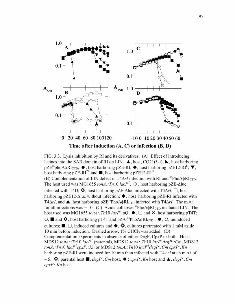
97
FIG. 3.3. Lysis inhibition by RI and its derivatives. (A) Effect of introducing lecines into the SAR domain of RI on LIN. ▲, host, CQ21(λ-t); , host harboring pZEssphoAφRICTD; , host harboring pZE-RI; , host harboring pZE12-RIL; , host harboring pZE-RI2L and , host harboring pZE12-RI3L. (B) Complementation of LIN defect in Τ4∆rI infection with RI and ssPhoAφRICTD. The host used was MG1655 tonA::Tn10 lacIq1. , host harboring pZE-∆luc infected with T4D; ◊, host harboring pZE-∆luc infected with T4∆rI; , host harboring pZE12-∆luc without infection; , host harboring pZE-RI infected with T4∆rI; and , host harboring pZEssPhoAφRICTD infected with T4∆rI. The m.o.i for all infections was ~ 10. (C) Azide collapses ssPhoAφRICTD mediated LIN. The host used was MG1655 tonA::Tn10 lacIq1 pQ. , and , host harboring pT4T;
, and ◊, host harboring pT4T and pZA-ssPhoAφRICTD. , , uninduced cultures; , , induced cultures and , ◊, cultures pretreated with 1 mM azide 10 min before induction. Dashed arrow, 1% CHCl3 was added. (D) Complementation experiments in absence of either DegP, CpxP or both. Hosts MDS12 tonA::Tn10 lacIq1 (parental), MDS12 tonA::Tn10 lacIq1degP::Cm, MDS12 tonA::Tn10 lacIq1cpxP::Kn or MDS12 tonA::Tn10 lacIq1degP::Cm cpxP::Kn harboring pZE-RI were induced for 10 min then infected with T4∆rI at an m.o.i of ~ 5. ◊, parental host; , degP::Cm host, ; cpxP::Kn host and ▲, degP::Cm cpxP::Kn host.
0 20 40 60 80 100120Time after induction (A) or infection (B)
A
B
0.1
0.1
1.0
1.0
A550
Time after induction (A, C) or infection (B, D)
C
0.1
1.0
-10 0 10 20 30 40 50 60
D
0.1
1.0A550
0 20 40 60 80 100120Time after induction (A) or infection (B)
A
B
0.1
0.1
1.0
1.0
A550
Time after induction (A, C) or infection (B, D)
C
0.1
1.0
-10 0 10 20 30 40 50 60
D
0.1
1.0A550
98
was below the limits of detection with our antibody, even when 5 ml of T4-infected
culture at A550 ~ 0.5 was used for SDS-PAGE and Western blot. Instead, we added
chloramphenicol to cultures undergoing LIN during the course of an infection with wild
type T4. This treatment resulted in a gradual decrease in culture A550 beginning
immediately after the addition of chloramphenicol (Fig. 3.5A). After 1 hour of infection,
nearly 70% of the total phage present in the treated culture had been released to the
medium compared to 10% of the phage in a parallel, untreated culture (Fig.3.5B). The
ability of chloramphenicol to partially abrogate LIN is consistent with the rapid turnover
of RI during phage infection.
The SAR domain is a major determinant of RI’s instability
Previously, we had observed that when the periplasmic C-terminal domain of RI
(RICTD) was fused to the cleavable signal sequence of alkaline phosphatase (PhoA), the
resulting chimera (Fig. 2.1), ssPhoAΦRICTD, was more effective at eliciting LIN than RI
itself (Fig 2.4A). Induction of the ssphoAΦrICTD gene resulted in the accumulation of
two anti-RI reactive species (Fig. 3.2E, lane 4 and 5). The larger product with a mass
approximately equal to that of RI was located while the smaller product with a mass
approximately the same as that of a polypeptide fragment comprising residues 25-97 of
RI was periplasmic (Fig. 3.2E lane 7). The latter product was not present in cultures
treated with azide (Fig. 3.2E lane 10 to 12), an inhibitor of the sec translocation pathway,
and represents the soluble, periplasmic RICTD released by cleavage of the ssPhoAΦRICTD

99
precursor by signal peptidase. Since azide treatment also eliminated LIN (Fig 3.3), it
was the periplasmic RICTD and not the precursor that inhibited the T holin. When
cultures expressing the ssphoAΦrICTD gene were treated with chloramphenicol, the
periplasmic RICTD polypeptide was found to be relatively stable (Fig. 3.4B and E).
Moreover, the membrane-restricted RI3L protein also had a half-life considerably longer
than RI (Fig. 3.4D and E). These data indicate that the degradation of RI occurred after
FIG. 3.4. Stability of RI. Panels, A- D show Western blots of samples taken from cultures expressing the indicated rI alleles. Samples were TCA precipitated, as described in Materials and Methods. (A) Wild type RI. Lane 1, purified RICTD
his, lanes 8-13: 0, 2, 3.5, 5.5, 7.5 and 9 min after addition of Cam. Lane 2-7: same time interval but no Cam added. Lane 14, molecular weight marker. (B) ssPhoAΦRICTD. Lane 1, molecular weight marker and lane 14, purified RICTD
his. Lanes 8-13:0, 2.3, 3.9, 5.5, 7 and 10 min after Cam addition. Lanes 2-7: same time interval but no Cam added. (C) RI in a DegP- strain. Lanes 9-15: 0, 2.4, 3.8, 5.3, 6.8, 8.3 and 9.2 min after addition of Cam. Lanes 1-7: same time intervals but no Cam added. Lane 8, molecular weight marker. (D) RI3L. Lanes 9-15: 0, 2.3, 3.7, 5.3, 6.7, 8.2 and 10 after Cam addition. Lane 1-7: same time interval but without Cam addition. Lane 8, molecular weight marker. (E) Kinetics of RI degradation.
, RI, ▲, ssPhoAΦRICTD, , RI3L and , RI in DegP-.
A1 2 3 4 5 6 7 8 9 10 11 12 13 14
1610
6
1 2 3 4 5 6 7 8 9 10 11 12 13 14
1610
6
B
C
1 2 3 4 5 6 7 8 9 10 11 12 13 14 1516
6
E
63
1 2 3 4 5 6 7 8 9 10 11 12 13 14 15
16
D
0 2 4 6 8 10
% i
nten
sity
Time after Cam addition (min)
100
10.0
A1 2 3 4 5 6 7 8 9 10 11 12 13 14
1610
6
1 2 3 4 5 6 7 8 9 10 11 12 13 141610
6
1 2 3 4 5 6 7 8 9 10 11 12 13 14
1610
6
1 2 3 4 5 6 7 8 9 10 11 12 13 14
1610
6
B
C
1 2 3 4 5 6 7 8 9 10 11 12 13 14 1516
6
E
63
1 2 3 4 5 6 7 8 9 10 11 12 13 14 15
16
63
1 2 3 4 5 6 7 8 9 10 11 12 13 14 15
16
D
0 2 4 6 8 10
% i
nten
sity
Time after Cam addition (min)
100
10.0

100
its release from the membrane and was promoted by the exposure of its SAR domain to
the periplasm.
Additional evidence for the destabilization of RI by its SAR domain was
obtained by complementing the LIN defective phage, T4∆rI, with plasmids carrying
either the rI or the ssphoAΦrICTD genes. In these experiments, the plasmids were induced
ten minutes before infection with T4∆rI. Since T4 infection results in the rapid
destruction of host DNA, synthesis of RI or ssPhoAΦRICTD should cease shortly
thereafter. Attempts to complement the LIN defect of T4∆rI with rI resulted in a 5
0
20
40
60
80
100
120
20
40
60
80
100
120
Perc
ent o
f pha
ge r
elea
se
― Ethanol Chloramphenicol
B
0 20 40 60 80 100
A
0.1
1.0
A550
0
20
40
60
80
100
120
20
40
60
80
100
120
Perc
ent o
f pha
ge r
elea
se
― Ethanol Chloramphenicol
B
0 20 40 60 80 100
A
0.1
1.0
A550
FIG. 3.5. Chloramphenicol collapses LIN. (A) Lysis profile. Host MDS12 tonA::Tn10 lacIq1 was infected with an m.o.i of ~ 10 using either T4D (solid symbols) or T4∆rI (open symbols). ◩, no infection, and , infected cultures without chemical treatment, and ◊, infected cultures treated with 95% ethanol at the indicated time, ▲ and , infected cultures treated with 300µg/mL of chloramphenicol at the indicated time at the times indicated by arrow (solid head: T4∆rI, open head: T4D). Dashed arrow, 1% chloroform was added to the indicated cultures. (B) Percent of phage release. Chemicals added to the cultures are indicated in the figure. Black bar, total phage and gray bar, free phage.

101
minute delay in lysis compared to the 30-40 minute delay seen with ssphoAΦrICTD (Fig.
3.3B). This result is consistent with the increased stability of the RICTD compared to RI
with its N-terminal SAR domain.
The DegP protease is required for the rapid turnover of RI
We reasoned that the rapid degradation of periplasmic RI might due to the
recognition of its exposed SAR domain by proteolytic machinery responsible for
removing misfolded proteins from this compartment. Since DegP is known to be
involved in the degradation of misfolded periplasmic proteins, we determined the effect
of mutations that eliminate DegP activity on the half-life and function of RI. The RI
protein was found to have a prolonged half-life in a degP mutant (Fig. 3.4C and E).
Somewhat unexpectedly, no significant lysis delay was observed when T4∆rI was
complemented with rI expressed from a plasmid in this mutant (Fig. 3.3D).
Recently, Isaac et al. (57) showed that degradation of a subset of misfolded
periplasmic proteins by DegP required the adaptor protein CpxP. We hypothesized that
both DegP and CpxP were necessary for RI degradation. According to this idea, when
DegP is absent, RI is stable, but no significant LIN could be observed because CpxP
binds to RI and prevents the interaction of RI with T. To test this hypothesis, we
performed the complementation experiments in the absence of CpxP, DegP or both.
Lysis delay in the double mutant was not different from the degP mutant alone,
indicating CpxP did not interfere with RI antiholin function (Fig.3.3D).
102
Lysis inhibition of ssPhoAΦRICTDY42am in T4 infection
Since ssPhoAΦRICTD was a better inhibitor than RI of T-mediated lysis in the
plasmid context, we wanted to study its function in phage T4. Multiple trials to generate
T4 ssphoAΦrICTD, in which the rI gene is replaced with ssphoAΦrICTD, failed. This
suggested that T4 ssphoAΦrICTD was not capable of forming a plaque. Therefore, we
made a different phage, T4 ssphoAΦrICTD Y42am. In this phage, the rI gene was replaced
with ssphoAΦrICTD Y42am, in which Tyr 42 of RI is replaced by an amber codon. We
expected this phage to make r plaques on a non-suppressor lawn and no plaques on a
suppressor lawn. However, T4ssphoAΦrICTDy42am made r plaques on both suppressor and
non suppressor lawns (Fig 3.6A and B). In addition, the lysis profile of T4
ssphoAΦrICTDY42am in liquid culture had an intermediate phenotype (Fig. 3.6 C). A likely
explanation for this phenotype was that the efficiency of suppression was not 100%. In
fact, supF is the best suppressor available, but the efficiency of suppression is ~ 50% in
uninfected cells (99); T4 infection perturbs all aspects of protein synthesis, so it would
not be surprising for suppression to be significantly less.
Discussion
Lysis inhibition (LIN) in T-even phage infection is a historically significant
phenomenon that was used to elucidate many basic concepts of molecular biology.
However, its molecular basis has been obscure. Recently, it was shown that holin T and
antiholin RI play a major role in LIN. With T4 infected cells, LIN only occurs when

103
there is superinfection with intact T4 particles but not with T4 ghosts, indicating that the
intact phages deliver some kind of signal to activate LIN. LIN can be reconstituted by
expressing T and RI from plasmids. In this system, RI is expressed at much higher
levels than in T4 infection suggesting that overexpression of RI compensates for the lack
of the signal of LIN. In Chapter II, we showed that T and RI interact via their C-
terminal domains, and the C-terminal domain of T could titrate the inhibition by RI,
implicating the C-terminal domain of T as the receptor for the LIN signal. How does the
LIN signal work? Here, we have established that RI possesses a N-terminal SAR
domain, which allows it to be released into the periplasm. Anchoring RI in the
A B
Non-suppressor Suppressor
T4D
T4∆rI
T4ssrICTDy42am
0 20 40 60 80100120
A550
Time after infection (min)
1.0
0.1
A B
Non-suppressor Suppressor
T4D
T4∆rI
T4ssrICTDy42am
Non-suppressor Suppressor
T4D
T4∆rI
T4ssrICTDy42am
0 20 40 60 80100120
A550
Time after infection (min)
1.0
0.1
FIG. 3.6. Plating phenotypes. The hosts used were either non-suppressor MG1655 tonA::Tn10 lacIq1 or suppressor LE392 tonA::Tn10 supE supF. (A) Plaque morphology of indicated phages on indicated host. (B) LIN phenotypes. Non-suppressor host ( , ▲, and ) or suppressor host ( , , and ◊) were infected with either T4D (▲ and ), T4∆rI ( and ) or T4ssphoAΦrICTD ( and ◊) or not infected ( and ). Dashed arrow indicates when CHCl3 was added.

104
membrane by leucine substitutions in the SAR domain significantly reduced its antiholin
function suggesting that it is the soluble periplasmic form of RI that normally
participates in LIN. In addition, the SAR domain of RI facilitates its DegP-dependent
degradation. Thus, while the release of RI into the periplasm is necessary for LIN, it is
also responsible for the rapid degradation of RI. Based on these findings, we propose
the model shown in Fig 3.7. When there is no superinfection, RI is made, is released
into the periplasm, is inactivated and is degraded by DegP. Changing from the active to
the inactive form, in the absence of superinfection, is rapid since the stability of RI in
DegP mutants is not accompanied by a commensurate enhancement of LIN. Thus, there
is no active RI available, there is no inhibition of T-mediated lysis; thus, lysis occurs at
the time programmed into the holin. When there is superinfection, one or both of the
following events may occur. (1) Superinfection signal enhances the release of RI into
FIG 3.7. Model of how superinfection leads to LIN.

105
the periplasm, or (2) superinfection signal slows down the inactivation of RI. Either or
both of these processes increase the concentration of active RI in the periplasm; thus RI
is now available to interact with T and inhibit lysis. Since both T4 genomic DNA and
the internal proteins are ectopically-localized in the periplasm during superinfection,
either or both of these elements can serve as the LIN signal.
Energetically, it would be difficult to release a TMD into the periplasm. In fact,
we do not know how proteins with N-terminal SAR domain are released into the
periplasm. It is possible that a protein with SAR domain is pulled out of the membrane
by an unknown factor. If this is the case, the LIN signal enhances the activity of this
factor, thus increasing the amount of RI being released into the periplasm. To slow
down the inactivation of RI, the LIN signal can either interact directly with RI and
prevents it to change into the inactive form or to inhibit an inhibitor of active RI. An
alternative theory positing that the LIN signal inhibits the activity of active RI inhibitor
is unlikely. If active RI inhibitor is a host protein, mutation in this protein would result
in a host mutant that supports T4 infection and replication, but do not allow lysis to
occur. This type of host mutant has never been isolated. If active RI inhibitor is a T4
protein, mutation in this protein would result in a T4 mutant that infects and replicate but
fail to lyse the host. This type of T4 mutant has never been isolated either. Therefore,
the most likely action that a superinfection signal allows LIN to occur is somehow to
prevent the inactivation of active RI. To remove the LIN signal, internal proteins would
be degraded by DegP protease, and the ectopically localized DNA would be degraded by
the periplasmic endonuclease I. Once the LIN signal is removed, inactivation of RI is

106
rapid, no active RI is available, and lysis occurs, unless another superinfection occurs.
This model would explain why superinfection has to occur continuously in order for LIN
to persist.
Why should RI possess a SAR domain instead of a signal sequence or a
transmembrane domain? We showed that ssPhoAΦRICTD, a version of RI with a
cleavable signal sequence that allowed the secretion of the inhibiting domain into the
periplasm, is a strong, stable inhibitor of T for the following reason. In the absence of
the SAR domain, the process of changing into the inactive state is much slower, which
explains the longer LIN time in the complementation where ssPhoAΦRICTD was used. In
addition, we could not obtain T4ssPhoAφrICTD as a plaque former, consistent with the
idea that LIN imposed by the secreted RICTD is too stable. Therefore, a metastable
inhibitor is the best fit for a system that senses the availability of new hosts. A SAR
domain serves two purposes. It allows the release of RI into the periplasm which can
either inhibit lysis or be inactivated and degraded, depending on a signal that reflects the
availability of new hosts.
107
CHAPTER IV
CHARACTERIZATION OF HOLIN T
Introduction
Holins are highly diverse: more than 50 unrelated families are known from phage
genomics. All but one of these holin families are in two topological classes: class I, with
three TMDs, N-out and C-in and class II, with two TMDs, N-in and C-in. The T holin
of T4 is the prototype of class III holins. It is unique because of its large size, 218 a a,
and large periplasmic domain. A topological study of T established that the C-terminal
domain (CTD) of T is in the periplasm (108). When this domain is deleted, the
truncated T holin is still partially functional. However, the truncated holin T is
insensitive to lysis inhibition mediated by the antiholin RI (108). In Chapter II, we
showed that the TCTD interacts with the antiholin RI of T4. In addition, the expression of
TCTD also delayed holin T-mediated lysis, indicating that it may play a role in T-T
oligomerization.
The topological study of T was carried out using PhoA-LacZ sandwich fusions
(108). In such studies, the results are only meaningful when the fusion proteins are
either PhoA+/LacZ- or PhoA-/LacZ+. This result was found with three fusions, all in the
C-terminal domain of T. The PhoA+/LacZ- nature of these fusions unambiguously
demonstrated the periplasmic localization of the TCTD. This left unsolved whether the N-
terminus was cytoplasmic or periplasmic. If T has two TMDs and an N-out and C-out
topology, a PhoA-LacZ fusion at the N-terminus would likely be non-permissive for

108
translocation of the first TMD into the membrane. Thus, fusion analysis is not a good
method for studying topology of the N-terminus lacking a signal sequence. Therefore,
one objective of this chapter is to determine whether T has one TMD with N-in and C-
out or two TMDs with N-out and C-out topology (Fig 4.1B).
In addition, we do not know the role of the N-terminal domain (NTD) of T in T-
mediated lysis or the distribution of T molecules in E. coli cells at the time of lysis. It
has been proposed that holin forms large aggregates in the membrane (48). These
aggregates have been called “death rafts”, and it was speculated that these death rafts act
as the locations of hole formation. The second objective of this chapter is to analyze the
function of the TNTD in T-mediated lysis and to determine how T is localized during the
lytic pathway.
Materials and methods
Bacterial strains, bacteriophages, plasmids, and culture growth
The bacterial strains, bacteriophages, and plasmids used in this work are
described in Tables 2.1, 2.2 and 2.3. T4 phage stocks were prepared as previously
described (109). Bacterial cultures were grown in standard LB medium supplemented
with ampicillin (100 µg/ml), kanamycin (40 µg/ml), chloramphenicol (10 µg/ml) when
appropriate. Growth and lysis of cultures were monitored by following the optical
density of cultures at A550 as previously described (109). When indicated, isopropyl β-

109
+ -
-+ +-
+ + + -
-+
MAAPRISFSPSDILFGVLDRLFKDNATGKVLASRVAVVILLFIMAIVWYRGDSFFEYYKQ………
-+
MDTYRYIAAPRISFSPSDILFGVLDRLFKDNATGKVLASRVAVVILLFIMAIVWYRGDSFFEYYKQ………
-+ ---
MEQKLISEEDLAAPRISFSPSDILFGVLDRLFKDNATGKVLASRVAVVILLFIMAIVWYRGDSFFEYYKQ………
10 20 30 40 50
60
T
AU
1 TC
myc
T
A B
N
N
CC
FIG
4.1
. A
min
o ac
id se
quen
ce o
f T, i
ts d
eriv
ativ
es a
nd p
ossi
ble
topo
logi
es o
f T.
(A) A
min
o ac
id se
quen
ce o
f T a
nd
mut
ant p
rote
ins.
Bol
d, T
MD
as p
redi
cted
by
TMH
MM
. G
ray
shad
ed,s
econ
d po
ssib
le T
MD
. So
lid a
rrow
, ins
ertio
n of
one
arg
inin
e re
sidu
e, o
pen
arro
w, i
nser
tion
of tw
o ar
gini
ne re
sidu
es.
(B) T
wo
poss
ible
topo
logi
es o
f T.
+ -
-+ +-
+ + + -
-+
MAAPRISFSPSDILFGVLDRLFKDNATGKVLASRVAVVILLFIMAIVWYRGDSFFEYYKQ………
-+
MDTYRYIAAPRISFSPSDILFGVLDRLFKDNATGKVLASRVAVVILLFIMAIVWYRGDSFFEYYKQ………
-+ ---
MEQKLISEEDLAAPRISFSPSDILFGVLDRLFKDNATGKVLASRVAVVILLFIMAIVWYRGDSFFEYYKQ………
10 20 30 40 50
60
T
AU
1 TC
myc
T
A B
N
N
CC
+ -
-+ +-
+ + + -
-+
MAAPRISFSPSDILFGVLDRLFKDNATGKVLASRVAVVILLFIMAIVWYRGDSFFEYYKQ………
-+
MDTYRYIAAPRISFSPSDILFGVLDRLFKDNATGKVLASRVAVVILLFIMAIVWYRGDSFFEYYKQ………
-+ ---
MEQKLISEEDLAAPRISFSPSDILFGVLDRLFKDNATGKVLASRVAVVILLFIMAIVWYRGDSFFEYYKQ………
10 20 30 40 50
60
T
AU
1 TC
myc
T
A B
+ -
-+ +-
+ + + -
-+
MAAPRISFSPSDILFGVLDRLFKDNATGKVLASRVAVVILLFIMAIVWYRGDSFFEYYKQ………
-+
MDTYRYIAAPRISFSPSDILFGVLDRLFKDNATGKVLASRVAVVILLFIMAIVWYRGDSFFEYYKQ………
-+ ---
MEQKLISEEDLAAPRISFSPSDILFGVLDRLFKDNATGKVLASRVAVVILLFIMAIVWYRGDSFFEYYKQ………
10 20 30 40 50
60
T
AU
1 TC
myc
T
+ -
-+ +-
+ + + -
-+
MAAPRISFSPSDILFGVLDRLFKDNATGKVLASRVAVVILLFIMAIVWYRGDSFFEYYKQ………
-+
MDTYRYIAAPRISFSPSDILFGVLDRLFKDNATGKVLASRVAVVILLFIMAIVWYRGDSFFEYYKQ………
-+ ---
MEQKLISEEDLAAPRISFSPSDILFGVLDRLFKDNATGKVLASRVAVVILLFIMAIVWYRGDSFFEYYKQ………
10 20 30 40 50
60
T
AU
1 TC
myc
T
A B
N
N
CC
FIG
4.1
. A
min
o ac
id se
quen
ce o
f T, i
ts d
eriv
ativ
es a
nd p
ossi
ble
topo
logi
es o
f T.
(A) A
min
o ac
id se
quen
ce o
f T a
nd
mut
ant p
rote
ins.
Bol
d, T
MD
as p
redi
cted
by
TMH
MM
. G
ray
shad
ed,s
econ
d po
ssib
le T
MD
. So
lid a
rrow
, ins
ertio
n of
one
arg
inin
e re
sidu
e, o
pen
arro
w, i
nser
tion
of tw
o ar
gini
ne re
sidu
es.
(B) T
wo
poss
ible
topo
logi
es o
f T.

110
D-thiogalactoside (IPTG), KCN, dinitrophenol (DNP) or CHCl3 was added to give final
concentrations of 1 mM, 10 mM, 10 mM or 1%, respectively.
Standard DNA manipulations, PCR, and DNA sequencing
Isolation of plasmid DNA, DNA amplification by PCR, DNA transformation,
and DNA sequencing were performed as previously described (145). Oligonucleotides,
shown in Table 2.4, were obtained from Integrated DNA Technologies, Coralville, IA,
and were used without further purification. Single base changes, small and large
insertions were introduced using commercially synthesized primers in conjunction with
the QuikChange kit from Stratagene. The Rapid DNA ligation kit from Roche
Molecular Biochemicals was used for ligation reactions. All other enzymes were
purchased from New England Biolabs except for Pfu polymerase, which was from
Stratagene. Automated fluorescent sequencing was performed at the Laboratory for
Plant Genome Technology at the Texas Agricultural Experiment Station.
Construction of plasmids
Positively charged residues and peptide tags were introduced into the N-terminal
domain of T by site-directed mutagenesis of T using QuikChange. The following primer
pairs, T5-R-6for/T5-R-6rev, T1-RR-2for/T1-RR-2rev, and pT4T (133) as the template
were used to generate pT4T5R6 and pT4T1RR2. To generate plasmids harboring t alleles
encoding T with a histidine tag at different positions, QuikChange reaction using pT4T

111
as the template and following primer pairs 28-G1H6G2-29-for/rev, 51-G1H6G2-29-
for/rev, 132-H6G2for/rev and 148-G1H6G2-149-for/rev were performed to obtain
pT4T28his, pT4T51his and pT4T148his, respectively. The nucleotide sequence coding for a
cmyc tag and an AU1 tag was introduced between codon 1 and 2 of t gene in plasmid
pT4This (133)using the primer pairs cymc-Tfor/cmyc-Trev and au1-Tfor/au1-Trev
respectively to generate pT4cmycThis and pT4au1This.
The rI gene was amplified from pZA-RI (133) using primer pair cmycNde1-
RIfor/Kpn1-RIrev. The PCR product was digested with NdeI and KpnI, and then ligated
into pET-duet1 digested with same enzymes to make pET-duet-RI. The DNA sequence
encoding This was amplified from pT4This using the primers ThisinpETfor and
ThisinpETrev. The PCR product was digested with NdeI1 and BamHI and ligated into
pET11a digested with the same enzymes to make pET11a- This. The This coding
sequence was cut out of pET11a- This using XbaI and BamHI and then ligated into pET-
duet-RI digested with the same enzymes to make pET-duet-RI-This.
Residue 2 to 28 of T was deleted by a PCR reaction that excluded the coding
region for these residues, using the primers T∆2-28down and T∆2-28up and pT4T as the
template. The PCR product was phosphorylated and ligated to make pT4T∆2-28.
To fuse GFP to the N-terminus of T, the coding region of GFP was amplified
using the primers Kpn1-gfp-for/CgfpNt-rev and pDS439 (120) as the template. Next,
the coding region of T was amplified using the primers CgfpNt-for/AvrIItrev and pT4T
as the template. The two PCR products were used as templates for an SOE PCR (54)
using the primers Kpn1-gfp-for and AvrIItrev. The resulting PCR product was digested
112
with KpnI1 and AvrII then ligated into pZA32-luc digested with the same enzymes to
make pZA-GFPΦT. An AGSAG linker was added between the coding region of GFP
and T using primer pair AG-T-for/Gfp-AGS-rev and pZA-GFPΦT as the template. The
PCR product was phosphorylated and ligated to make pZA-GFPΦAGSAGΦT plasmid.
Plasmids harboring rV alleles of t were created using QuikChange PCR with
pT4T as the template and the following primer pairs For/RevtR5K, For/RevtI39V and
For/RevT75I to obtain pT4TR5K, pT4TI39V and pT4TT75I respectively.
Purification and N-terminal sequencing of This
The plasmid pETduet-RI-This was transformed into C43(DE3)tonA::Tn10
slyD::Kn. The transformants were grown in 3L of LB-Amp at 37oC until A550 ~ 0.8.
The culture was induced with IPTG for 1hr and the cells were harvested by
centrifugation at 7,000 x g for 15 min. The cell pellet was resuspended in 30 ml of
French Press buffer (100 mM Tris, pH 8, 1 mM DTT, 1 mM PMSF and 30µl of protease
inhibitor cocktail(Sigma)), and the cells were lysed by passing through a French Press
cell at 16,000 psi twice. The unlysed cells were removed in a clearing spin at 2,000 x g
for 10 min. The membrane fraction was collected by centrifugation of the supernatant of
the clearing spin at 100,000 x g for 1hr. The membrane fraction was extracted using the
buffer 1% EBB, 0.5 M NaCl and 20 mM HEPES pH 8 overnight at 4oC. Insoluble
material was removed by centrifugation at 100,000 x g for 1hr. The soluble fraction was
filtered then used as the starting material for IMAC purification using His-trap HP
113
column (GE/Amersham Pharmacia). The protein was eluted using low pH buffer (1%
Ebb, 50 mM NaCl, 20 mM sodium acetate pH 2.5). The pH of the fractions was
adjusted to 7 using 1 M NaOH. The purity and the presence of T in each fraction were
assessed by SDS-PAGE and Western blot. Fractions containing This were pooled,
concentrated to 500 µl, analyzed using SDS-PAGE, Coomassie stain and western blot.
The band recognized by T antiserum was cut out and used for N-terminal sequencing.
The N-terminal sequencing was performed by the Protein Chemistry Laboratory of
Texas A&M University, Department of Biochemistry and Biophysics.
Results
His tag mutagenesis
To obtain purified T for in vitro studies, an allele of t that encoded for a T protein
with an oligohistidine-tag at its extreme C-terminus was constructed by Ramanculov
(108,109). However, lysis mediated by this his-tagged T protein is not saltatory like
wild type T-mediated lysis, indicating the holin function of this his-tagged T protein is
compromised. Therefore, this allele is not suitable for the overexpression and
purification of T for use in further in vitro studies. We attempted to generate a more
suitable allele encoding a his-tagged T protein by moving the his-tag to sites internal to
T. Four different positions were chosen for the insertion of the Gly2His6Gly2 sequence,
between residues 28 and 29, 51 and 52, 132 and 133 and 148 and 149, to make T28his,
T51his, T132his and T148his respectively. The lysis profiles of these alleles are shown in Fig
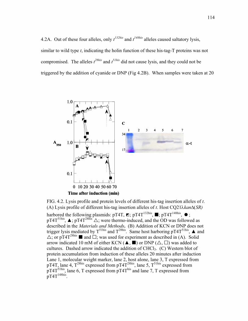
114
4.2A. Out of these four alleles, only t132his and t148his alleles caused saltatory lysis,
similar to wild type t, indicating the holin function of these his-tag-T proteins was not
compromised. The alleles t28his and t51his did not cause lysis, and they could not be
triggered by the addition of cyanide or DNP (Fig 4.2B). When samples were taken at 20
FIG. 4.2. Lysis profile and protein levels of different his-tag insertion alleles of t. (A) Lysis profile of different his-tag insertion alleles of t. Host CQ21λkan∆(SR) harbored the following plasmids: pT4T, ◩; pT4T132his, ; pT4T148his, ; pT4T51his, ▲; pT4T28his ; were thermo-induced, and the OD was followed as described in the Materials and Methods. (B) Addition of KCN or DNP does not trigger lysis mediated by T51his and T28his. Same host harboring pT4T51his, ▲ and
; or pT4T28his and ; was used for experiment as described in (A). Solid arrow indicated 10 mM of either KCN (▲, ) or DNP ( , ) was added to cultures. Dashed arrow indicated the addition of CHCl3. (C) Western blot of protein accumulation from induction of these alleles 20 minutes after induction Lane 1, molecular weight marker, lane 2, host alone, lane 3, T expressed from pT4T, lane 4, T28his expressed from pT4T28his, lane 5, T51his expressed from pT4T51his, lane 6, T expressed from pT4This and lane 7, T expressed from pT4T148his.
C1 2 3 54 6 7
α-t
34
17
A
0.1
1.0
0 10 20 30 40 50 60 70Time after induction (min)
B
0.1
1.0A550
C1 2 3 54 6 7
α-t
34
17
A
0.1
1.0
0 10 20 30 40 50 60 70Time after induction (min)
B
0.1
1.0A550
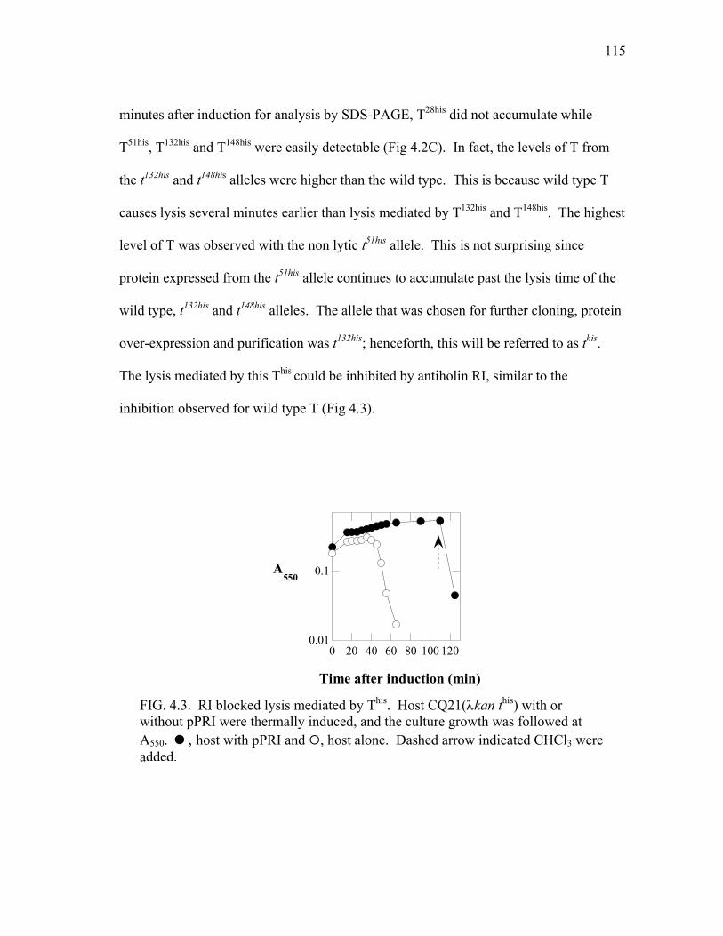
115
minutes after induction for analysis by SDS-PAGE, T28his did not accumulate while
T51his, T132his and T148his were easily detectable (Fig 4.2C). In fact, the levels of T from
the t132his and t148his alleles were higher than the wild type. This is because wild type T
causes lysis several minutes earlier than lysis mediated by T132his and T148his. The highest
level of T was observed with the non lytic t51his allele. This is not surprising since
protein expressed from the t51his allele continues to accumulate past the lysis time of the
wild type, t132his and t148his alleles. The allele that was chosen for further cloning, protein
over-expression and purification was t132his; henceforth, this will be referred to as this.
The lysis mediated by this This could be inhibited by antiholin RI, similar to the
inhibition observed for wild type T (Fig 4.3).
0.01
0.1
0 20 40 60 80 100 120
A550
Time after induction (min)
FIG. 4.3. RI blocked lysis mediated by This. Host CQ21(λkan this) with or without pPRI were thermally induced, and the culture growth was followed at A550. , host with pPRI and , host alone. Dashed arrow indicated CHCl3 were added.

116
Purification of T
Over-expression of this from a T7 promoter caused cell death soon after induction
allowing only a small amount of T to accumulate. For this reason, this was cloned into a
pETduet vector in tandem with rI. The pETduet vector possesses two T7 promoters
which allow the simultaneous over-expression of two proteins. The growth curve after
induction is shown in Fig 4.4A. Even with this system, This could not be seen on the
SDS gel stained with Coomassie, but was observed when the induced cells were
subjected to SDS-PAGE and Western blotting as shown in Fig 4.4B. This protein was
purified as described in the Materials and Methods with ~ 0.6 mg isolated from 6L of
culture.
0 20 40 60 800.5
1.0
2.0
Time after induction (min)
A
22
391 2 3 4
B
α-t
0 20 40 60 800.5
1.0
2.0
Time after induction (min)
A
22
391 2 3 4
B
α-t
FIG 4.4. Growth curve and Western blot analysis of cells harboring pETduet-RI-This. (A) Growth curves. induction of pETduet-1. , induction of pETduet-RI-This. (B) Western blot analysis of T using T antibodies. Lanes 1 and 2, pETduet1. Lanes 3 and 4, pETduet-RI-This. Lanes 1 and 3, uninduced. Lanes 2 and 4, induced.

117
Because the amount of purified full length This was not enough to raise a T
antiserum, the DNA fragment encoding the CTD of This was cloned into a pET vector,
and used for over-expression and purification as described in the Materials and Methods
of Chapter II. This protein was also used as the standard to determine the amount of T
present in the cell after 60 minutes of T4 wild type infection. According to the gel
shown in Fig 4.5, there is approximately 1.5 pmole or ~ 7.7 x 10¹⁰ T molecules in 1.5 x
108 cells at 60 min after infection with T4D. Thus, the number of T molecules per cell is
approximately 500.
Analysis of N-terminal function
Ramanculov’s fusion analysis (108) suggested that T has at least one TMD, with
the C-terminal domain from residue 70 in the periplasm and the N-terminal domain
(NTD) in the cytoplasm. The 69 residue NTD is long enough to possess either one or
two TMDs, as shown in Fig 4.1 B. If T has one TMD, its topology would be N-in and
C-out. As a result, the first methionine of T should be deformylated, because its N-
terminus is located in the cytoplasm, where the host enzyme deformylase has access to
FIG 4.5. Quantification of T in T4D infected cells. Lane 1, T4D infected cells, lane 2, molecular weight marker, lanes 3-5, purified TCTD
his. Lane 3, 64 pmol, lane 4, 6.4 pmol, lanes 5, 0.64 pmol. Lane 6, uninfected cells. * full length T, ** TCTD
his.
1 2 3 4 5 6
*** 17
34
α-t
1 2 3 4 5 6
*** 17
34
α-t

118
remove the formyl group of the first methionine. However, if T has a second TMD in
the NTD, its topology would be N-out and C-out. There are two possible ways to
achieve this. First, both TMDs could be translocated across the membrane by the
SecYEG translocon. Assuming T was translocated cotranslationally, the first
methionine could not be deformylated. Second, the TMD2 was translocated across the
membrane by the SecYEG translocon, and the first TMD could traverse the membrane
by a post-translational “flip up” pathway. The post-translational flipping of a TMD into
the membrane had been shown for the first TMD of leader peptidase and of S105, the
holin of bacteriophage λ (R. White et al., manuscript in preparation). In both cases, N-
flip up occurs so fast that the formyl group of the first methionine of these proteins was
not removed by the enzyme deformylase. In the case of phage λ holin, the purified S105
protein was not N-terminal sequenceable. The purified S105 could be sequenced only
when it was treated with deformylase prior to N-terminal sequencing (R. White et al.,
manuscript in preparation). The flipping of TMD1 of S105 across the membrane was
found be inhibited by the addition of positively charged residues at its N-terminus. The
addition of positive charges at the N-terminus also changed holin S105 to an antiholin
(44,45), which inhibited S105. The N-terminus of the purified λ antiholin could be
sequence without treatment by deformylase (R. White et al., manuscript in preparation).
To determine whether the formyl group of the first methionine of T is removed, purified
This was subjected to N-terminal sequencing. The N-terminus of purified This could be
sequenced by Edman degradation, indicating that the protein was deformylated. This
suggested that the N-terminus of T was in the cytoplasm.
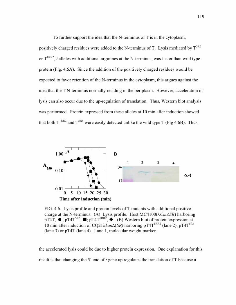
119
To further support the idea that the N-terminus of T is in the cytoplasm,
positively charged residues were added to the N-terminus of T. Lysis mediated by T5R6
or T1RR2, t alleles with additional arginines at the N-terminus, was faster than wild type
protein (Fig. 4.6A). Since the addition of the positively charged residues would be
expected to favor retention of the N-terminus in the cytoplasm, this argues against the
idea that the T N-terminus normally residing in the periplasm. However, acceleration of
lysis can also occur due to the up-regulation of translation. Thus, Western blot analysis
was performed. Protein expressed from these alleles at 10 min after induction showed
that both T1RR2 and T5R6 were easily detected unlike the wild type T (Fig 4.6B). Thus,
the accelerated lysis could be due to higher protein expression. One explanation for this
result is that changing the 5’ end of t gene up regulates the translation of T because a
0 5 10 15 20 25 30
A550
Time after induction (min)
A1.00
0.10
0.01
B
1 2 3 4
α-t34
17
0 5 10 15 20 25 30
A550
Time after induction (min)
A1.00
0.10
0.01
B
1 2 3 4
α-t34
17
FIG. 4.6. Lysis profile and protein levels of T mutants with additional positive charge at the N-terminus. (A) Lysis profile. Host MC4100(λCm∆SR) harboring pT4T, ; pT4T5R6, ; pT4T1RR2, . (B) Western blot of protein expression at 10 min after induction of CQ21λkan∆(SR) harboring pT4T1RR2 (lane 2), pT4T5R6 (lane 3) or pT4T (lane 4). Lane 1, molecular weight marker.

120
rare codon was used. When a rare codon is used in an mRNA, the rate of translation
shortly after induction is higher than for mRNA with common codons. This is because
the pool of tRNA recognizing the rare codon is fully charged. Since only a certain
number of holin molecules required for lysis, a higher rate of holin synthesis resulted in
faster lysis. Together these data strongly support the idea that T has one TMD with N-in
and C-out topology.
We also placed the negatively charged C-myc tag (EQKLISEEDL) and neutral
AU1 tag (DTYRYI) between residue 1 and 2 of T132his. Surprisingly, these alleles
caused lysis faster than the t132his allele, and they appeared to be insensitive to LIN
mediated by RI (Fig 4.7A). SDS-PAGE and Western blot (Fig 4.7B) showed that the
protein level of AU1T132his was higher than that of T132his, but the protein level of
CmycT132his was about the same as that of T132his. The higher protein level of AUIT132his
might explain the faster triggering of lysis and, perhaps, the insensitivity to RI.
However, the faster lysis phenotype of cmycThis suggested that the N-terminus plays a
functional role in lysis timing.
Analysis of rV alleles
The T4rV mutants isolated by Krylov in 1965 (74,76) were later sequenced by
Dressman and Drake in 1999 (29). These mutants contained mutations in the t gene that
caused T to become insensitive to LIN. We wanted to check the lysis profile and to test
the sensitivity of these mutants toward LIN mediated by RI in our plasmid system. All
three known rV mutants, tR5K, tI39V and tT75I, lysed faster than wild type t (Fig 4.8A). In
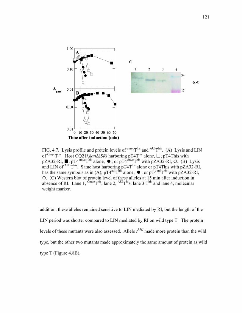
121
addition, these alleles remained sensitive to LIN mediated by RI, but the length of the
LIN period was shorter compared to LIN mediated by RI on wild type T. The protein
levels of these mutants were also assessed. Allele tR5K made more protein than the wild
type, but the other two mutants made approximately the same amount of protein as wild
type T (Figure 4.8B).
FIG. 4.7. Lysis profile and protein levels of cmycThis and AUIThis. (A) Lysis and LIN of CmycThis. Host CQ21λkan∆(SR) harboring pT4This alone, ; pT4This with pZA32-RI, ; pT4cmycThis alone, ; or pT4cmycThis with pZA32-RI, . (B) Lysis and LIN of AU1This. Same host harboring pT4This alone or pT4This with pZA32-RI, has the same symbols as in (A); pT4auIThis alone, ; or pT4auIThis with pZA32-RI,
. (C) Western blot of protein level of these alleles at 15 min after induction in absence of RI. Lane 1, CmycThis, lane 2, AUIThis, lane 3 This and lane 4, molecular weight marker.
C
1 2 3 4
α-t34
17
0 10 20 30 40 50 60 70
B
0.10
0.01
Time after induction (min)
A
0.01
0.10
1.00
A550
C
1 2 3 4
α-t34
17
0 10 20 30 40 50 60 70
B
0.10
0.01
Time after induction (min)
A
0.01
0.10
1.00
A550
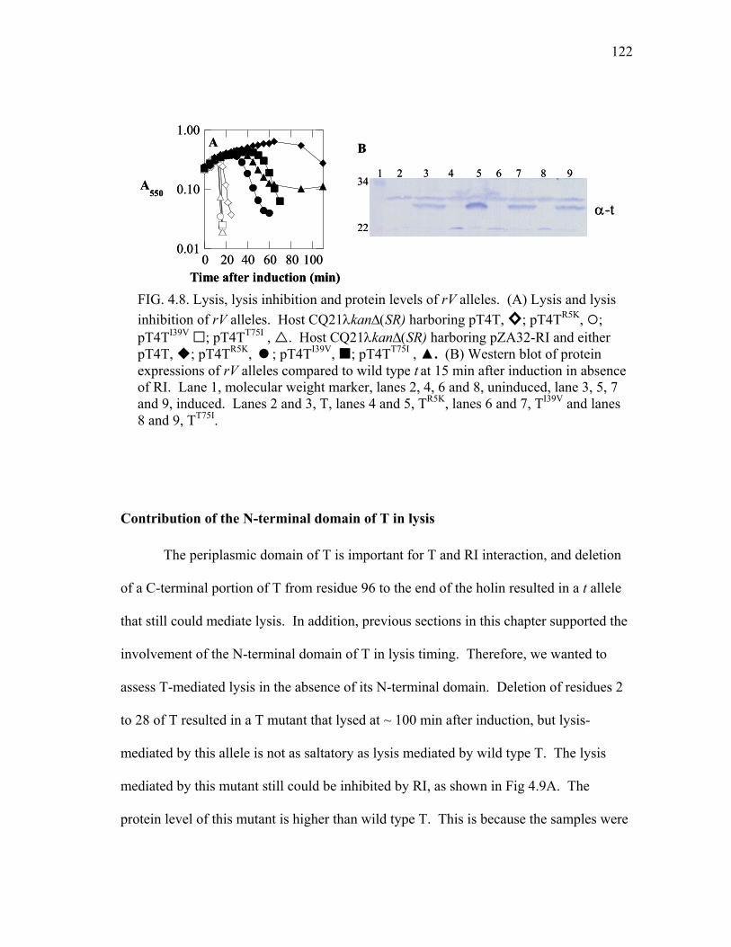
122
Contribution of the N-terminal domain of T in lysis
The periplasmic domain of T is important for T and RI interaction, and deletion
of a C-terminal portion of T from residue 96 to the end of the holin resulted in a t allele
that still could mediate lysis. In addition, previous sections in this chapter supported the
involvement of the N-terminal domain of T in lysis timing. Therefore, we wanted to
assess T-mediated lysis in the absence of its N-terminal domain. Deletion of residues 2
to 28 of T resulted in a T mutant that lysed at ~ 100 min after induction, but lysis-
mediated by this allele is not as saltatory as lysis mediated by wild type T. The lysis
mediated by this mutant still could be inhibited by RI, as shown in Fig 4.9A. The
protein level of this mutant is higher than wild type T. This is because the samples were
FIG. 4.8. Lysis, lysis inhibition and protein levels of rV alleles. (A) Lysis and lysis inhibition of rV alleles. Host CQ21λkan∆(SR) harboring pT4T, ◊; pT4TR5K, ; pT4TI39V ; pT4TT75I , . Host CQ21λkan∆(SR) harboring pZA32-RI and either pT4T, ; pT4TR5K, ; pT4TI39V, ; pT4TT75I , ▲. (B) Western blot of protein expressions of rV alleles compared to wild type t at 15 min after induction in absence of RI. Lane 1, molecular weight marker, lanes 2, 4, 6 and 8, uninduced, lane 3, 5, 7 and 9, induced. Lanes 2 and 3, T, lanes 4 and 5, TR5K, lanes 6 and 7, TI39V and lanes 8 and 9, TT75I.
0 20 40 60 80 100
A550
Time after induction (min)
A1.00
0.10
0.01
B
1 2 3 54 6 734
8 9
22α-t
0 20 40 60 80 100
A550
Time after induction (min)
A1.00
0.10
0.01
B
1 2 3 54 6 734
8 9
22α-t
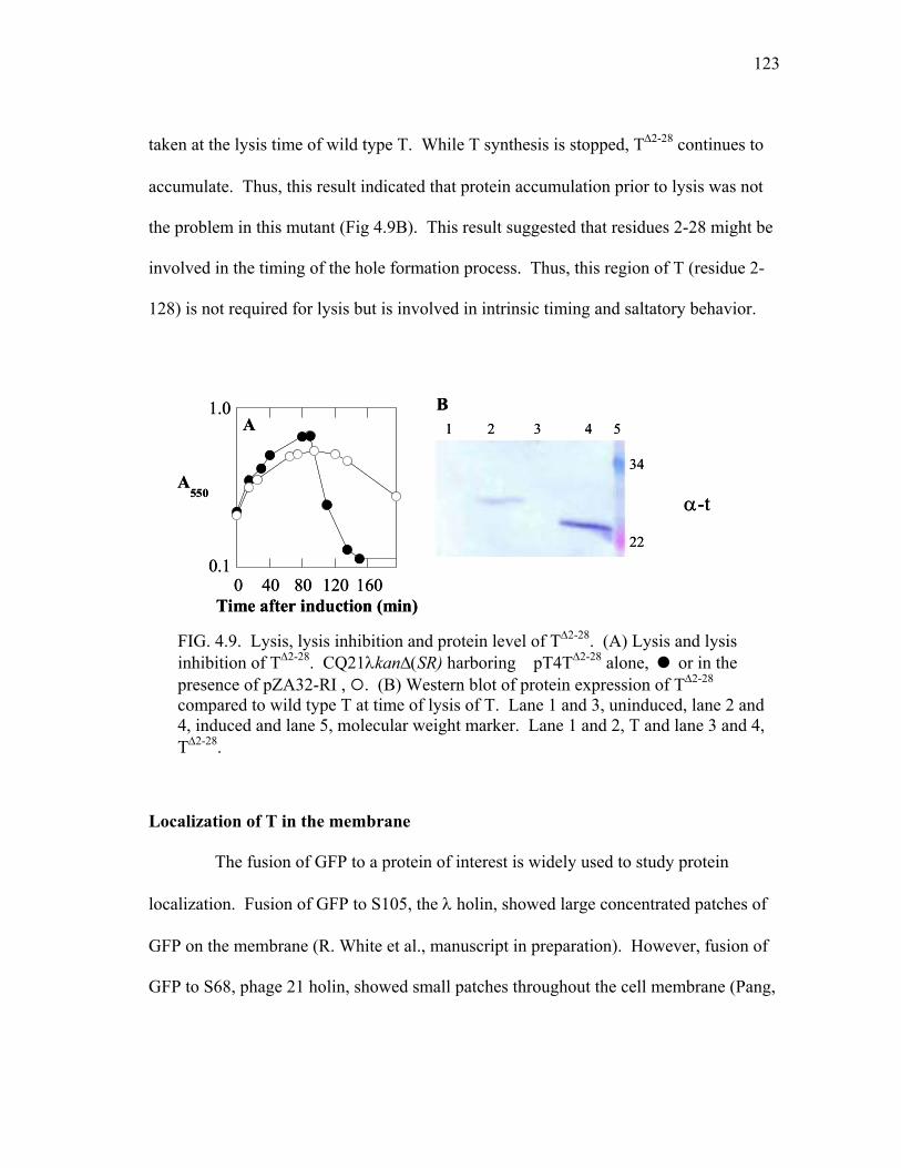
123
taken at the lysis time of wild type T. While T synthesis is stopped, T∆2-28 continues to
accumulate. Thus, this result indicated that protein accumulation prior to lysis was not
the problem in this mutant (Fig 4.9B). This result suggested that residues 2-28 might be
involved in the timing of the hole formation process. Thus, this region of T (residue 2-
128) is not required for lysis but is involved in intrinsic timing and saltatory behavior.
Localization of T in the membrane
The fusion of GFP to a protein of interest is widely used to study protein
localization. Fusion of GFP to S105, the λ holin, showed large concentrated patches of
GFP on the membrane (R. White et al., manuscript in preparation). However, fusion of
GFP to S68, phage 21 holin, showed small patches throughout the cell membrane (Pang,
FIG. 4.9. Lysis, lysis inhibition and protein level of T∆2-28. (A) Lysis and lysis inhibition of T∆2-28. CQ21λkan∆(SR) harboring pT4T∆2-28 alone, or in the presence of pZA32-RI , . (B) Western blot of protein expression of T∆2-28 compared to wild type T at time of lysis of T. Lane 1 and 3, uninduced, lane 2 and 4, induced and lane 5, molecular weight marker. Lane 1 and 2, T and lane 3 and 4, T∆2-28.
0 40 80 120 160
A550
Time after induction (min)
A1.0
0.1
B1 2 3 4 5
34
22
α-t
0 40 80 120 160
A550
Time after induction (min)
A1.0
0.1
B1 2 3 4 5
34
22
α-t

124
T., personal communication). It was proposed that before forming a hole, holin S105
concentrated into rafts in the membrane known as death rafts (48). Thus, the fluorescent
patches observed in S105ΦGFP and S68ΦGFP may represent the death rafts proposed
A
B
1 2 3 4 5 6 7
C
55
45α-GFP
D
1 2 3 4 5 6 755
17
65
35 α-t
*
* *
•
•••
A
B
1 2 3 4 5 6 7
C
55
45α-GFP
D
1 2 3 4 5 6 755
17
65
35 α-t
*
* *
•
•••
D
1 2 3 4 5 6 755
17
65
35 α-t
*
* *
•
•••
FIG. 4.10. Fluorescence and protein levels of GFPΦT. (A) Fluorescence image of cells expressing GFPΦT. (B) Fluorescence image of cells expressing GFPΦAGSAGΦT. (C) Western blot analysis of fusion protein using GFP antibodies. Lane 1, molecular weight marker, lane 2, 4 and 6, uninduced, lane 3, 5 and 7, induced. Lane 2 and 3, cells expressing T from pZA32-T, lanes 4 and 5, cells expressing GFPΦT from pZA32-GFPΦT and lanes 6 and 7, cells expressing GFPΦAGSAGΦT from pZA32-GFPΦAGSAGΦT. (D) Western blot analysis of fusion protein using T antibodies. Lanes 1, 3 and 5, uninduced, lane 2, 4 and 6, induced. Lane 1 and 2, cells expressing T from pZA32-T, lanes 3 and 4, cells expressing GFPΦT from pZA-GFPΦT, lanes 5 and 6, cells expressing GFPΦAGSAGΦT from pZA32-GFPΦAGSAGΦT and lane 7, molecular weight marker. *, intact fusion protein, **, T and •, cleavage products.

125
by Grundling et al. (48). Holin T is similar to S105 in that it forms a big hole in the
membrane; thus, we wanted to see if T forms rafts like S105. GFP was fused at the
extreme N-terminus of T either with or without a linker (AGSAG). Expression of these
fusion proteins caused cell death faster than expression of wild type T. Western blot
analysis using GFP antibodies showed that GFPΦT fusion proteins with the correct
molecular weight accumulated. Detection using T antibodies indicated that the majority
of fusion protein was intact; however, a small amount was cleaved into four smaller
species, none of which had the same molecular weight as T alone (Fig 4.10 C and D).
Since T antibodies were raised against the purified periplasmic domain of T, it is not
likely that the degradation was from the C-terminus of T but rather was from the N-
terminus of GFP. In addition, there was more GFPΦT compared to wild type T alone.
In E. coli cells expressing GFPΦT, many fluorescent patches were observed on the
membrane (Fig 4.8A, B). This suggested that death rafts similar to those seen with
S105ΦGFP also form with holin GFPΦT.
Discussion
A number of modifications of the N-terminal domain of T resulted in t alleles
with faster lysis times. In most cases, these modifications also increased the level of
holin accumulation. The shorter lysis time of t alleles with higher levels of protein
expression can be explained using the model proposed by Wang and Young in 2003.
According to this model, the pathway to form lesions in the membrane is a transition
126
from monomers to dimers to oligomers to protein rafts. At some critical holin
concentration, a small hole is formed in the middle of the raft which causes a local
depolarization. This local depolarization causes a tertiary or quaternary structural
change of the holins in the raft and leads to hole formation (139). Therefore, reaching a
critical concentration of holin in the membrane seems to be one of several important
factors contributing to the timing of lysis. It is reasonable to argue that t alleles that up
regulate the protein expression levels should have shorter lysis times because they reach
the critical protein concentration necessary for lysis faster. However, the same argument
cannot be used to explain faster lysis by alleles that have about the same protein
expression level as wild type T in vivo. These cases may be explained by considering a
cytoplasmic element that interacts with the N-terminus of T, thus interfering with the
oligomerization process. If modification of the N-terminus of T disrupts the interaction
between T and this cytoplasmic element, there is nothing to interfere with T
oligomerization, allowing faster hole formation and faster lysis.
Why are these fast lysers insensitive to RI-mediated LIN? In 2001, Ramanculov
proposed a model to explain how RI-mediated LIN takes place. In this model, T-
mediated lysis can only be inhibited by RI when T is in a form that is called sensitive T
or Ts, and these are Ts molecules may be in the monomer form. Once a T molecule
participates in the higher oligomer forms, it is no longer sensitive to RI-mediated LIN.
If the fast lysing mutants form oligomers faster than RI can form heterodimer with them,
these mutants become insensitive toward RI-mediated LIN.

127
We observed fluorescent patches on the E. coli cell membrane when GFPΦT
was expressed, indicating death raft formation. Since the GFPΦT fusion protein
accumulated at a higher level than wild type T, we do not know how much the
fluorescent patches reflect death rafts formed by T in T4 infection. We also observed
cleavage products from the fusion protein only when T antibodies were used for protein
detection, indicating the protein fusion proteins were degraded from the GFP end. This
gives rise to another problem; that is we do no know the contribution of these cleavage
products to fluorescent patch formation. To eliminate the possibility patch formation is
an over-expression artifact, we need to confirm this result using cells infected with
T4∆rIgfpΦt e-, T4 phage with defective endolysin, deletion of rI and GFPΦT. Also it
would be interesting to see the distribution of GFPΦT in the cell membrane in the
presence of RI. S68ΦGFP, the holin of phage 21, gave patches of fluorescence, but in the
presence of antiholin, the patches disappeared and a uniform fluorescence was observed
on the cell membrane (Pang, T., personal communication). If a similar result is observed
in T4 infection, it would support the lysis and lysis inhibition model proposed above.
That is, in the absence of antiholin, big rafts of holin form, and in the presence of
antiholin, only small rafts form and lysis is inhibited.

128
CHAPTER V
CONCLUSIONS AND FUTURE DIRECTIONS
The lysis-inhibition phenomenon is one of the "founding" problems of modern
molecular genetics, first characterized in the work of Delbrück's Phage Church in the
1940's. Despite its historical prominence, very thorough physiological and genetic
investigations, and apparent simplicity in terms of the number of genes involved, six
decades have seen very little progress made in terms of understanding this phenomenon
at the molecular level. This began to change with the work of Ramanculov et al. (107-
110), which established that the T protein of T4 functioned as a holin and that the RI
protein could act as an antiholin specific for the T4 holin T. The work in this
dissertation has followed on from that work and made a number of advances. First, the
work in this dissertation has provided evidence for the following conclusions: (1) RI is a
type II integral membrane protein, with an N-terminal TMD acting as a signal anchor
and the bulk of the protein in the periplasm; (2) RI is a SAR protein that is released from
the cytoplasmic membrane into the periplasm; (3) T and RI interact via their respective
periplasmic C-terminal domains; (4) the function of the periplasmic C-terminal domain
of RI is sufficient for LIN, and, in fact, is more efficient at this inhibition than wt RI with
its SAR domain; (5) the release of the C-terminal domain of RI into the periplasm is
required for efficient RI-mediated LIN to occur; (6) the SAR domain also makes RI
proteolytically unstable, and the proteolysis is largely mediated by the host periplasmic
protease DegP; (7) that T has one transmembrane domain, with its N-terminus in the
129
cytoplasm; (8) the N-terminal cytoplasmic domain of T is integrally involved in lysis
timing of T, and (9) holin T accumulates as oligomeric patches in the membrane in the
lytic pathway. Some of these conclusions warrant further discussion.
T-mediated lysis and interaction with RI
T and its relatives stand out from other known holins in terms of size. With 218
residues, T is twice as big as S105, the holin of λ, which has three TMDs, and three
times as big as S68, the holin of phage 21, which has 2 TMDs, yet T only has one TMD.
Since the large periplasmic C-terminal domain of T interacts with RI, and RI and T are
the only r proteins that are required for LIN in all host backgrounds, it seems likely that
the CTD of T can be regarded as the receptor of the LIN signal. Interestingly, co-
expression of secreted periplasmic domain and wild-type holin T delayed T-mediated
lysis by ~ 5 min. This indicates that this domain is involved in holin-holin interactions
leading to hole-formation. It may be possible to achieve the purification of substantial
quantities of the CTDs of both proteins, which have, up to now, been over-produced as
inclusion bodies in the cytoplasm of E. coli. If secreted CTDs of both RI and T could be
purified, the LIN interaction could be open to structural analysis.
Mutations at the N-terminus of T were found to change lysis timing. Insertion of
amino acids at the TNTD resulted in t alleles that lysed earlier. However, a deletion of the
complete cytoplasmic N-terminus resulted in an allele with a later onset of lysis. Even
though the rate of T synthesis is up-regulated in the N-terminal insertion alleles, this fact
alone cannot be used to account for the acceleration or deceleration of lysis time.

130
Acceleration of lysis did not occur in just one insertion but in all the insertion mutants,
indicating this is not a random event. Perhaps, addition of these amino acids at the N-
terminus of T disrupts an inhibitory interaction that normally delays the process of hole
formation. This could be a homotypic interaction of the N-terminus of T with itself, or a
heterotypic interaction with a cytoplasmic domain of a host protein. When the
interference is removed, lesion formation occurs faster, resulting in shorter lysis time.
Deletion of the 27 residues at the N-terminus of T caused a significant lysis delay,
indicating that the N-terminus may also be involved in facilitating hole-formation,
perhaps in the process of oligomerization. These ideas can be tested by following the
kinetics of oligomerization of each of these mutants, using pulse-labeling and cross-
linking studies.
Properties of RI
The periplasmic C-terminal domain of RI is sufficient for imposing LIN. LIN in
T4 infection is a metastable state of lysis inhibition, caused by superinfection events, and
collapses if superinfection ceases. Thus, finding the inhibition domain is not sufficient
to explain LIN. The simplest way to bring about LIN instability would be to make the
inhibitor, the antiholin RI, unstable. Indeed, when the life-time of RI was investigated
after the suppression of protein synthesis, it was determined that RI had a very short
half-life. Moreover, the proteolytic instability of RI was shown to be dependent on the
host periplasmic protease, DegP. Also, the presence of the N-terminal SAR domain,
which allows RI to be released in the periplasm, confers sensitivity of RI to DegP.

131
Mutational analysis could provide clues as to what it is about the SAR domain that
attracts the periplasmic protease.
Localization of RI in the periplasm is also required for efficient antiholin
function of RI. However, the mechanism by which superinfection activates RI-mediated
lysis inhibition is not known. Recall that RI is an early gene product, and T is a late
gene product. This means antiholin RI is made before holin T, and if RI is a stable and
active protein, then lysis would be permanently inhibited. This problem can be solved
by making the life time of active RI very short. When there is no superinfection which
equates to the availability of new hosts, T-mediated lysis can proceed. When there is
superinfection which equates to the absence of uninfected hosts, a LIN signal is sent into
the infected cell. This signal allows RI to stay active longer. When there is no further
superinfection, the LIN signal vanishes, and the RI inactivation process is speeded up.
Now, without active RI available to mediate LIN, LIN would collapse.
Even though ssPhoAΦRICTD and RI3L are more stable than wild-type, we could
not transfer these mutant rI alleles to the T4 chromosome. There are several possible
reasons why they could not be obtained. (1) The PhoA chimera may be such a potent
antiholin that it blocks plaque-formation, so our method, which involves screening for
plaques, would not work. (2) Even though we predict that T4rI3L should make a large, r
plaque due to partial lost of antiholin function, this may not be the case. In fact, we do
not know the plaque morphology of this mutant. Future progress here requires PCR-
based control experiments to demonstrate that the recombinant molecules are being
formed in the recombination lysates.
132
The signal for LIN
There is still a lot to be done to elucidate the mechanism of LIN. Most
importantly, what is the signal of LIN at the molecular level? There are two possible
general kinds of signals from the superinfection event: first, the ectopically-localized
genomic T4 DNA, or, second, the internal proteins of the capsid, which total over 300
molecules. In one model, the LIN signal would serve to stabilize active RI, at least
temporarily. Thus our ability to cause LIN by over-expressing RI results simply from
being able to swamp the proteolytic capacity of the cell, so that RI accumulates in the
membrane and periplasm. Assessing RI stability in an in vitro reconstitution of
superinfection will distinguish which of the two is the signal. This might be feasible
using spheroplasts. Inducing t in the presence of just a basal level of RI after formation
of spheroplasts should result in lysis. If this system works, the next step might be to add
the T4 head contents, DNA and proteins, to the spheroplasts, to see if the basal level of
RI could be activated to delay lysis.
Another question is what is the level of RI during the infectious cycle either in
the presence or absence of superinfection? Currently, it is a major handicap that we
cannot detect RI in T4 infection. To answer this question, we need more sensitive RI
antibodies. In addition, we also need T4 antibodies that neutralize T4 and prevent it
from infecting the cells. With these tools in hand, one can neutralize the infection at any
particular time and follow the protein level of RI.
We established the contribution of the N and the C-terminal domain of T in lysis
and lysis inhibition, but we do not know the boundary of the one TMD that T possesses.

133
Because we do not know where the TMD is, we do not know how it participates in the
hole formation process. In addition, how does T mediate lysis? Can we visualize the
hole after lysis occurs? These questions and more will arise as we make progress on this
most ancient phenomenon of classical molecular biology.

134
REFERENCES
1. Abedon, S. T. 1994. Lysis and the interaction between free phages and infected cells, p. 397-405. In J. D. Karam (ed.), Molecular biology of Bacteriophage T4. ASM Press, Washington, DC.
2. Alexeyev, M. F., and H. H. Winkler. 1999. Membrane topology of the Rickettsia prowazekii ATP/ADP translocase revealed by novel dual pho-lac reporters. J. Mol. Biol. 285:1503-1513.
3. Anderson, C. W., and J. Eigner. 1971. Breakdown and exclusion of superinfecting T-even bacteriophage in Escherichia coli. J. Virol. 8:869-886.
4. Anderson, C. W., J. R. Williamson, and J. Eigner. 1971. Localization of parental deoxyribonucleic acid from superinfecting T4 bacteriophage in Escherichia coli. J. Virol. 8:887-893.
5. Benzer, S. 1955. Fine structure of a genetic region in bacteriophage. Proc. Natl. Acad. Sci. USA 41:344-354.
6. Benzer, S. 1957. The elementary unit of heredity, p. 70-93. In W. D. McElroy and B. Glass (ed.), The chemical basic of heredity. The Johns Hopkins Press, Baltimore, MD.
7. Bernhardt, T. G., W. D. Roof, and R. Young. 2000. Genetic evidence that the bacteriophage φX174 lysis protein inhibits cell wall synthesis. Proc. Natl. Acad. Sci. USA 97:4297-4302.
8. Bernhardt, T. G., D. K. Struck, and R. Young. 2001. The lysis protein E of phi X174 is a specific inhibitor of the MraY-catalyzed step in peptidoglycan synthesis. J. Biol. Chem. 276:6093-6097.
9. Bernhardt, T. G., I. N. Wang, D. K. Struck, and R. Young. 2001. A protein antibiotic in the phage Qbeta virion: diversity in lysis targets. Science 292:2326-2329.
10. Bernhardt, T. G., I. N. Wang, D. K. Struck, and R. Young. 2002. Breaking free: "protein antibiotics" and phage lysis. Res. Microbiol. 153:493-501.
11. Bienkowska-Szewczyk, K., B. Lipinska, and A. Taylor. 1981. The R gene product of bacteriophage λ is the murein transglycosylase. Mol. Genet. Genomics 184:111-114.
135
12. Black, L. W., and C. Ahmad-Zadeh. 1971. Internal proteins of bacteriophage T4D: their characterization and relation to head structure and assembly. J. Mol. Biol. 57:71-92.
13. Blasi, U., C-Y. Chang, M. T. Zagotta, K. Nam, and R. Young. 1990. The lethal λS gene encodes its own inhibitor. EMBO J. 9:981-989.
14. Bode, W. 1967. Lysis Inhibition in Escherichia coli infected with bacteriophage T4. J. Virol. 1:948-955.
15. Brock, T. D. 1990. The emergence of bacterial genetic. Cold Spring Harbor Laboratory Press, Cold Spring Harbor, NY.
16. Broom-Smith, J. K., and B. G. Spatt. 1986. A vector for the construction of translational fusions to TEM beta-lactamase and the analysis of protein export signals and membrane protein topology. Gene 49:341-349.
17. Cairns, J., G. S. Stent, and J. D. Watson 1966. Phage and the origins of molecular biology. Cold Spring Harbor Laboratory of Quantative Biology, Cold Spring Harbor, NY.
18. Campbell, A. M. 1969. Episomes. Harper and Row, New York, NY.
19. Champe, S. P. 1963. Bacteriophage reproduction. Annu. Rev. Microbiol. 17:87-114.
20. Cheng, X., X. Zhang, J. W. Pflugrath, and F. W. Studier. 1994. The structure of bacteriophage T7 lysozyme, a zinc amidase and an inhibitor of T7 RNA polymerase. Proc. Natl. Acad. Sci. USA 91:4034-4038.
21. Childs, J. D. 1973. Superinfection exclusion by incomplete genome of bacteriophage T4. J. Virol. 11:1-8.
22. Coombs, D. H., and F. Arisaka. 1994. T4 tail structure and function, p. 259-281. In J. D. Karam (ed.), Molecular biology of bacteriophage T4. ASM Press, Washington, DC.
23. Cordonnier, C., and G. Bernardi. 1965. Localization of E. coli endonuclease I. Biochem. Biophys. Res. Commun. 20:555-559.
24. Cornett, J. B., and M. Vallee. 1973. The map position of the immunity (imm) gene of bacteriophage T4. Virology 51:506-508.

136
25. Daegelen, P., and E. Brody. 1990. The rIIA gene of bacteriophage T4. I. Its DNA sequence and discovery of a new open reading frame between genes 60 and rIIA. Genetics 125:237-248.
26. Delbrück, M. 1942. Interference between bacterial viruses: I. Interference between two bacterial viruses acting on the same host, and the mechanism of virus growth. Arch. Biochem. Biophys. 264:1238-1244.
27. Doermann, A. H. 1948. Lysis and lysis inhibition of Escherichia coli bacteriophage. J. Bacteriol. 55:257-276.
28. Doermann, A. H. 1952. The intracellular growth of bacteriophages. I. Liberation of intracellular bacteriophage T4 by premature lysis with another phage or with cyanide. J. Gen. Physiol. 35:645-656.
29. Dressman, H. K., and J. W. Drake. 1999. Lysis and lysis inhibition in bacteriophage T4: rV mutations reside in the holin t gene. J. Bacteriol. 181:4391-4396.
30. Driukas, A., and R. Nivinskas. 2003. Studies on the expression of gene rIII and 30.9 of bacteriophage T4 in vivo. Biologijia 3:13-15.
31. Duckworth, D. H. 1971. Inhibition of T4 bacteriophage multiplication by superinfecting ghost and the development of tolerance after bacteriophage infection. J. Virol. 7:8-14.
32. Duckworth, D. H. 1974. Who discovered bacteriophage? Bacteriological Reviews 40:793-802.
33. Eiserling, F. A. 1983. Structure of T4 Virion, p. 11-24. In C. K. Mathews, E. Kutter, G. Mosig, and P. B. Berget (ed.), Bacteriophage T4. ASM Press, Washington, DC.
34. Eiserling, F. A., and L. W. Black. 1994. Pathways in T4 Morphogenesis, p. 209-212. In J. D. Karam (ed.), Molecular biology of bacteriophage T4. ASM Press, Washington, DC.
35. Emrich, J. 1968. Lysis of T4-infected bacteria in the absence of lysozyme. Virology 35:158-165.
36. Emrich, J., and G. Streisinger. 1968. The role of phage lysozyme in the life cycle of phage T4. Virology 36:387-391.
37. Ennis, H. L., and K. D. Kievitt. 1973. Association of the RIIA protein with the bacterial membrane. Proc. Natl. Acad. Sci. USA 70:1468-1472.

137
38. Evrard, C., J. Fastrez, and J. P. Declercq. 1998. Crystal structure of the lysozyme from bacteriophage lambda and its relationship with V and C-type lysozymes. J. Mol. Biol. 76:151-164.
39. Fastrez, J. 1996. Phage lysozymes, p. 35-64. In P. Jollès (ed.), Lysozymes: Model enzymes in biochemistry and biology. Birkhäuser Verlag, Basel.
40. Fürste, J. P., W. Pansegrau, R. Frank, H. Blöcker, P. Scholz, M. Bagdasarian, and E. Lanka. 1989. Molecular cloning of the plasmid RP4 primase region in a multi-host-range tacP expression vector. Gene 48:119-131.
41. Furukawa, H., and S. Mizushima. 1982. Roles of cell surface components of Escherichia coli K-12 in bacteriophage T4 infection: interaction of tail core with phospholipids. J. Bacteriol. 150:916-924.
42. Goldberg, E., L. Grinius, and L. Letellier. 1994. Recognition, attachment and injection, p. 347-356. In J. D. Karam (ed.), Molecular biology of bacteriophage T4. ASM Press, Washington, DC.
43. Goldberg, E. B. 1980. Bacteriophage nucleic acid penetration, p. 115-141. In L. L. P. L. Randall (ed.), Virus receptors. Receptor and recognition. Chapman & Hall, Ltd., London.
44. Graschopf, A., and U. Blasi. 1999. Functional assembly of the lambda S holin requires periplasmic localization of its N-terminus. Arch. Microbiol. 172:31-39.
45. Graschopf, A., and U. Blasi. 1999. Molecular function of the dual-start motif in the lambda S holin. Mol. Microbiol. 33:569-582.
46. Grundling, A., U. Blasi, and R. Young. 2000. Biochemical and genetic evidence for three transmembrane domains in the class I holin, lambda S. J. Biol. Chem. 275:769-776.
47. Grundling, A., U. Blasi, and R. Young. 2000. Genetic and biochemical analysis of dimer and oligomer interactions of the lambda S holin. J. Bacteriol. 182:6082-6090.
48. Grundling, A., M. D. Manson, and R. Young. 2001. Holins kill without warning. Proc. Natl. Acad. Sci. USA 98:9348-9352.
49. Hausman, R. 1988. The T7 Group, p. 259-289. In R. Calendar (ed.), The bacteriophages. Plenum Press, New York, NY.
50. Herriott, R. M., and J. L. Barlow. 1957. Protein coats or "ghosts" of coli phage T2. II. The biological functions. J. Gen. Physiol. 41:307-331.

138
51. Hershey, A. D. 1946. Mutation in bacteriophage with respect to the type of plaque. Genetics 31:620-640.
52. Hershey, A. D. 1946. Spontaneous mutation in bacterial virus. Cold Spring Harb. Symp. Quant. Biol. 11:67-77.
53. Ho, K. 2001. Bacteriophage therapy for bacterial infections. Rekindling a memory from the pre-antibiotics era. Perspectives in Biology and Medicine 44:1-16.
54. Ho, S. N., H. D. Hunt, R. M. Horton, J. K. Pullen, and L. R. Pease. 1989. Site-directed mutagenesis by overlap extension using the polymerase chain reaction. Gene 77:51-59.
55. Höltje, J-V. 1998. Growth of the stress-bearing and shape-maintaining murein sacculus of Escherichia coli. Microbiology and Molecular Biology Reviews 62:181-203.
56. Inouye, M., N. Arnheim, and R. Sternglanz. 1973. Bacteriophage T7 lysozyme is an N-acetylmuramyl-L-alanine amidase. J. Biol. Chem. 248:7247-7252.
57. Isaac, D. D., J. S. Pinkner, S. J. Hultgren, and T. J. Silhavy. 2005. The extracytoplasmic adaptor protein CpxP is degraded with substrate by DegP. Proc. Natl. Acad. Sci. USA 102:17775-17779.
58. Israeli, M., and M. Artman. 1970. Leakage of b-galactosidase from phage-infected Escherichia coli: A re-evaluation. J. Gen. Virol. 7:137-142.
59. Jacob, F., and C. R. Fuerst. 1958. The mechanism of lysis by phage studied with defective lysogenic bacteria. J. Gen. Physiol 18:518-526.
60. Jacobson, R. H., X. J. Zhang, R. F. DuBose, and B. W. Matthews. 1994. Three-dimensional structure of beta-galactosidase from E. coli. Nature 369:761-766.
61. Josslin, R. 1970. The lysis mechanism of phage T4: mutants affecting lysis. Virology 40:719-726.
62. Josslin, R. 1971. Physiological studies on the t gene defect in T4-infected Escherichia coli. Virology 44:101-107.
63. Kai, T., H. Ueno, Y. Otsuka, W. Morimoto, and T. Yonesaki. 1999. Gene 61.3 of bacteriophage T4 is the spackle gene. Virology 260:254-259.
139
64. Kanamaru, S., P. G. Leiman, V. A. Kostyuchenko, P. R. Chipman, V. V. Mesyanzhinov, F. Arisaka, and M. G. Rossman. 2002. Structure of the cell puncturing device of bacteriophage T4. Nature 415:553-557.
65. Kao, S-H., and W. H. McClain. 1980. Baseplate protein of bacteriophage T4 with both structural and lytic function. J. Virol. 34:95-103.
66. Kao, S-H., and W. H. McClain. 1980. Role of bacteriophage T4 gene 5 and gene s products in cell lysis. J. Virol. 34:104-107.
67. Kim, D. Y., and K. K. Kim. 2005. Structure and function of HtrA family proteins, the key players in protein quality control. J. Biochem. Mol. Biol. 38:266-274.
68. Koch, G., and W. J. Dreyer. 1957. Characterization of an enzyme of phage T2 as a lysozyme. Virology 6:291-293.
69. Koch, G., and E. M. Jordan. 1957. Killing of E. coli B by phage free T2 lysate. Biochim. Biophys. Acta 25:437.
70. Kolesky, S., M. Ouhammouch, E. N. Brody, and E. P. Geiduschek. 1999. Sigma competition: the contest between bacteriophage T4 middle and late transcription. J. Mol. Biol. 291:267-281.
71. Kolisnychenko, V., G. Plunkett III, C. D. Herring, T. Fehér, J. Pósfai, F. R. Blattner, and G. Pósfai. 2002. Engineering a reduced Escherichia coli genome. Genome Research 12:640-647.
72. Kolmar, H., P. R. Waller, and R. T. Sauer. 1996. The DegP and DegQ periplasmic endoproteases of Escherichia coli: specificity for cleavage sites and substrate conformation. J. Bacteriol. 178:5925-5929.
73. Krojer, T., M. Garrido-Franco, R. Huber, M. Ehrmann, and T. Clausen. 2002. Crystal structure of DegP (HtrA) reveals a new protease-chaperone machine. Nature 416:455-459.
74. Krylov, V. N. 1966. A new r gene of bacteriophage T4B? Microb. Genet. Bull. 24:4-5.
75. Krylov, V. N., and N. K. Yanknovsky. 1975. Mutation of the new gene stIII of bacteriophage T4B suppressing the lysis defect of gene stII and a gene e mutant. J. Virol. 15:22-26.
76. Krylov, V. N., and A. Zapadnaya. 1965. Bacteriophage T4B r mutations sensitive to temperture (rts). Genetic 1:7-11.
140
77. Kutter, E., B. Guttman, and K. Carlson. 1994. The transition form host to phage metabolism after T4 infection, p. 343-356. In J. D. Karam (ed.), Molecular biology of bacteriophage T4. ASM Press, Washington, DC.
78. Kutter, E., T. Stidham, B. Guttman, E. Kutter, D. Batts, S. Peterson, T. D. Javakhishvili, F. Arisaka, V. Mesyanzhinov, W. Rüger, and G. Mosig. 1994. Genomic map of bacteriophage T4, p. 491-518. In J. D. Karam (ed.), Molecular biology of bacteriophage T4. ASM, Washington, DC.
79. Lederberg, J. 1952. Lysogenicity in E. coli K-12. Genetics 36:560-573.
80. Lipinska, B., M. Zylicz, and C. Georgopoulos. 1990. The HtrA (DegP) protein, essential for Escherichia coli survival at high temperatures, is an endopeptidase. J. Bacteriol. 172:1791-1797.
81. Loessner, M. J., G. Wendlinger, and S. Scherer. 1995. Heterogeneous endolysins in Listeria monocytogenes bacteriophages: a new class of enzymes and evidence for conserved holin genes within the siphoviral lysis cassettes. Mol. Microbiol. 16:1231-1241.
82. Los, M., G. Wegrzyn, and P. Neubauer. 2003. A role for bacteriophage T4 rI gene function in the control of phage development during pseudolysogeny and in slowly growing host cells. Res. Microbiol. 154:547-552.
83. Lu, M. J., and U. Henning. 1989. The immunity (imm) gene of Escherichia coli bacteriophage T4. J. Virol. 63:3472-3478.
84. Lu, M. J., and U. Henning. 1992. Lysis protein T of bacteriophage T4. Mol. Gen. Genet. 235:253-258.
85. Lu, M. J., Y. D. Stierhof, and U. Henning. 1993. Location and unusual membrane topology of the immunity protein of the Escherichia coli phage T4. J. Virol. 67:4905-4913.
86. Lutz, R., and H. Bujard. 1997. Independent and tight regulation of transcriptional units in Escherichia coli via the LacR/O, the TetR/O and AraC/I1-I2 regulatory elements. Nucleic Acids Res. 25:1205-1210.
87. Lwoff, A. 1953. Lysogeny. Bacteriological Reviews 17:269-337.
88. Mathew, B. W. 1996. Structural and genetic analysis of the folding and function of T4 lysozyme. FASEB J. 10:35-41.

141
89. Mathews, C. K. 1994. An overview of the T4 developmental program, p. 1-8. In J. D. Karam (ed.), Molecular biology of bacteriophage T4. ASM Press, Washington, DC.
90. McPheeters, D. S., A. Christensen, E. T. Young, G. Stormo, and L. Gold. 1986. Translational regulation of expression of the bacteriophage T4 lysozyme gene. Nucleic Acids Res. 14:5813-5826.
91. Meerman, H. J., and G. Georgious. 1994. Construction and characterization of a set of E. coli strains deficient in all known loci affecting the proteolytic stability of secreted recombinant proteins. Bio/Technology 12:1107-1110.
92. Miller, E. S., E. Kutter, G. Mosig, F. Arisaka, T. Kunisawa, and W. Ruger. 2003. Bacteriophage T4 genome. Microbiology and Molecular Biology Reviews 67:86-156.
93. Model, P., and M. Russel. 1988. Filamentous bacteriophage, p. 375-456. In R. Calendar (ed.), The bacteriophages. Plenum Press, New York, NY.
94. Moody, M. F. 1973. Sheath of bacteriophage T4. 3. Contraction mechanism deduced from partially contracted sheaths. J. Mol. Biol. 80:613-635.
95. Mosig, G., and F. Eiserling. 2006. T4 and related phages: Structure and development, p. 225-267. In R. Calendar (ed.), The bacteriophages, Oxford University Press, Inc., New York, NY.
96. Mukai, F., G. Streisinger, and B. Miller. 1967. The mechanism of lysis in phage T4-infected cells. Virology 33:398-404.
97. Murray, N. E., W. J. Brammar, and K. Murray. 1977. Lambdoid phages that simplify the recovery of in vitro recombinants. Mol. Gen. Genet. 150:53-61.
98. Nakagawa, H., F. Arisaka, and S-I. Ishii. 1985. Isolation and characterization of the bacteriophage T4 tail-associated lysozyme. J. Virol. 54:460-466.
99. Nene, V., and R. E. Glass. 1981. Direct measurement of absolute suppressor efficiency. Bioscience Reports 1:571-574.
100. Paddison, P., S. T. Abedon, H. K. Dressman, K. Gailbreath, J. Tracy, E. Mosser, J. Neitzel, B. Guttman, and E. Kutter. 1998. The roles of the bacteriophage T4 r genes in lysis inhibition and fine-structure genetics: A new perspective. Genetics 148:1539-1550.

142
101. Park, J. T. 2001. Identification of a dedicated recycling pathway for anhydro-N-acetylmuramic acid and N-acetylglucosamine derived from Escherichia coli cell wall murein. J. Bacteriol. 183:3842-3847.
102. Park, T., D. K. Struck, J. F. Deaton, and R. Young. 2006. Topological dynamics of holins in programmed bacterial lysis. Proc. Natl. Acad. Sci. USA 103:19713-19718.
103. Peterson, R. F, K. D. Kievitt, and H. L. Ennis. 1972. Membrane protein synthesis after infection of Escherichia coli B with phage T4: the RIIB protein. Virology 50:520-527.
104. Pribnow, D., D. C. Sigurdson, L. Gold, B. S. Singer, C. Napoli, J. Brosius, T. J. Dull, and H. F. Noller. 1981. rII cistron of bacteriophage T4. DNA sequence around the intercistronic divide and position of genetic landmark. J. Mol. Biol. 149:337-376.
105. Raab, R., G. Neal, J. Garrett, R. Grimaila, R. Fusselman, and R. Young. 1986. Mutational analysis of bacteriophage lambda lysis gene S. J. Bacteriol. 167:1035-1042.
106. Raab, R., G. Neal, C. Sohaskey, J. Smith, and R. Young. 1988. Dominance in lambda S mutations and evidence for translational control. J. Mol. Biol. 199:95-105.
107. Ramanculov, E. 2001. Bacteriophage T4 Lysis and Lysis Inhibition: A molecular perspective. Ph.D. dissertation Texas A&M University, College Station.
108. Ramanculov, E., and R. Young. 2001. Genetic analysis of the T4 holin: timing and topology. Gene 265:25-36.
109. Ramanculov, E., and R. Young. 2001. Functional analysis of the phage T4 holin in a lambda context. Mol. Genet. Genomics 265:345-353.
110. Ramanculov, E., and R. Young. 2001. An ancient player unmasked: T4 rI encodes a t-specific antiholin. Mol. Microbiol. 41:575-583.
111. Raudonikiene, A., and R. Nivinskas. 1992. Gene rIII is the nearest downstream neighbour of bacteriophage T4 gene 31. Gene 114:85-90.
112. Raudonikiene, A., and R. Nivinskas. 1993. The sequences of gene rIII of bacteriophage T4 and its mutants. Gene 134:135-136.

143
113. Riede, I. 1987. Lysis gene t of T-even bacteriophage: Evidence that colicins and bacteriophage genes have common ancestors. J. Bacteriol. 169:2956-2961.
114. Riede, I. 1987. Receptor specificity of the short tail fibers (gp12) of T-even type Escherichia coli phages. Mol. Gen. Genet. 206:110-115.
115. Rutberg, B., and L. Rutberg. 1965. Role of superinfecting phage in lysis inhibition with phage T4 in Escherichia coli. J. Bacteriol. 90:891-894.
116. Rutberg, L., and B. Rutberg. 1964. On the expression of the rII mutation of T-even bacteriophages in Escherichia coli strain B. Virology 22:280-281.
117. Salton, M. R. 1957. The properties of lysozyme and its action on microorganisms. Bacteriological Reviews 21:82-99.
118. Shapira, A., E. Giberman, and A. Kohn. 1974. Recoverable potassium fluxes variations following adsorption of T4 phage and their ghosts on Escherichia coli B. J. Gen. Virol. 23:159-171.
119. Shinedling, S., D. Parma, and L. Gold. 1987. Wild-type Bacteriophage T4 is restricted by the lambda rex genes. J. Virol. 61:3790-3794.
120. Siegele, D. A., and J. C. Hu. 1997. Gene expression from plasmids containing the araBAD promoter at subsaturating inducer concentrations represents mixed populations. Proc. Natl. Acad. Sci. USA 94:8168-8172.
121. Silhavy, T. J., and M. L. Berman. 1984. Bacterial strains, p. xi-xiii. In T. J. Silhavy and M. L. Berman (ed.), Experiments with gene fusions. Cold Spring Harbor Laboratory Press, Cold Spring Harbor, NY.
122. Silver, S., E. Levine, and P. M. Spielman. 1968. Cation fluxes and permeability changes accompanying bacteriophage infection of Escherichia coli. J. Virol. 2:763-771.
123. Simon, L. D., and T. F. Anderson. 1967. The infection of Escherichia coli by T2 and T4 bacteriophage as seen in the electron microscope. I. Attachment and penetration. Virology 32:279-297.
124. Singer, B. S., S. T. Shinedling, and L. Gold. 1983. The rII genes: a history and a prospectus, p. 327-333. In C. K. Mathews, E. M. Kutter, G. Mosig, and P. B. Berget (ed.), Bacteriophage T4. ASM, Washington, DC.
125. Slavcev, R. A., and S. Hayes. 2004. Overexpression of rexA nullifies T4rII exclusion in Escherichia coli K(λ) lysogen. Can. J. Microbiol. 50:133-136.
144
126. Smith, D. L., and R. Young. 1998. Oligohistidine tag mutagenesis of the λ holin gene. J. Bacteriol. 180:4199-4211.
127. Snyder, L., and G. Kaufmann. 1994. T4 phage exclusion mechanism, p. 391-396. In J. D. Karam (ed.), Molecular biology of bacteriophage T4. ASM Press, Washington, DC.
128. Snyder, L., and K. McWilliams. 1989. The rex genes of bacteriophage lambda can inhibit cell function without phage superinfection. Gene 81:17-24.
129. Stent, G. S. 1963. The Twort-D'Herelle phenomenon, p. 1-21. In R. Emerson, D. Kenedy, R. B. Park, G. Beadle, and D. M. Whiaker (ed.), Molecular biology of bacterial virus. W. H. Freeman and Company, San Francisco and London.
130. Strauch, K. L., and J. Beckwith. 1988. An Escherichia coli mutation preventing degradation of abnormal periplasmic proteins. Proc. Natl. Acad. Sci. USA 85:1576-1580.
131. Strauch, K. L., K. Johnson, and J. Beckwith. 1989. Characterization of degP, a gene required for proteolysis in the cell envelope and essential for growth of Escherichia coli at high temperature. J. Bacteriol. 171:2689-2696.
132. Streisinger, G., F. Mukai, W. J. Dreyer, B. Miller, and S. Horiuchi. 1961. Mutations affecting the lysozyme of phage T4. Cold Spring Harb. Symp. Quant. Biol. 26:25-30.
133. Tran, T. A., D. K. Struck, and R. Young. 2005. Periplasmic domains define holin-antiholin interactions in T4 lysis inhibition. J. Bacteriol. 187:6631-6640.
134. Tsugita, A., and M. Inouye. 1968. Complete primary structure of phage lysozyme from Escherichia coli T4. J. Mol. Biol. 37:201-212.
135. Vallee, M., and J. B. Cornett. 1972. A new gene of bacteriophage T4 determining immunity against superinfecting ghost and phage T4-infected Escherichia coli. Virology 48:777-784.
136. Vallee, M., J. B. Cornett, and H. Bernstein. 1972. The action of bacteriophage T4 ghost on Escherichia coli and the immunity to this action developed in cells preinfected with T4. Virology 48:766-776.
137. van Wezenbeek, P. M., T. J. Hulsebos, and J. G. Schoenmaker 1980. Nucleotide sequence of the filamentous bacteriophage M13 DNA genome: Comparison with phage fd. Gene 11:129-148.

145
138. Visconti, N. 1953. Resistance to lysis from without in bacteria infected with T2 bacteriophage. J. Bacteriol. 66:247-253.
139. Wang, I. N., J. Deaton, and R. Young. 2003. Sizing the holin lesion with an endolysin-β-galactosidase fusion. J. Bacteriol. 185:779-787.
140. Wang, I. N., A. E. Dykhuizen, and L. B. Slobodkin. 1996. The evolution of phage lysis timing. Evolution Ecology 10:545-558.
141. Wang, I. N., D. L. Smith, and R. Young. 2000. Holins: the protein clocks of bacteriophage infections. Annu. Rev. Microbiol. 54:799-825.
142. Weaver, L. H., and B. W. Matthews. 1987. Structure of bacteriophage T4 lysozyme refined at 1.7 Å resolution. J. Mol. Biol. 193:189-199.
143. Weintraub, S. B, and F. R. Frankle. 1972. Identification of the T4rIIB gene product as a membrane protein. J. Mol. Biol. 70:589-615.
144. Xu, M., A. Arulandu, D. K. Struck, S. Swanson, J. C. Sacchettini, and R. Young. 2005. Disulfide isomerization after membrane release of its SAR domain activates P1 lysozyme. Science 307:113-117.
145. Xu, M., D. K. Struck, J. Deaton, I. N. Wang, and R. Young. 2004. A signal-arrest-release sequence mediates export and control of the phage P1 endolysin. Proc. Natl. Acad. Sci. USA 101:6415-6420.
146. Young, R. 1992. Bacteriophage lysis: mechanism and regulation. Microbiol. Rev. 56:430-481.
147. Young, R. 2000. Phages will out: strategies of host cell lysis. Trends. Microbiol. 8:120-128.
148. Young, R. 2002. Bacteriophage holins: deadly diversity. J. Mol. Microbiol. Biotechnol. 4:21-36.
149. Zagotta, M. T., and D. B. Wilson. 1990. Oligomerization of the bacteriophage lambda S protein in the inner membrane of Escherichia coli. J. Bacteriol. 172:912-921.

146
VITA
Tram Anh Thi Tran received her Bachelor of Science degree in biology and
chemistry from Stephen F. Austin State University in the fall of 1998. In spring of 1999,
she entered the Biotechnology program at the same university and received the Master of
Science degree in 2001. In the fall of 2000, she started her doctorate study in
biochemistry at Texas A&M University and received her Ph.D. in the spring of 2007.
Her research focuses on the mechanism of bacteriophage T4 lysis and lysis inhibition.
Ms. Tran may be reached at Texas A&M University, TAMU 2128, Bio/Bio 315,
College Station, TX 77843. Her e-mail address is [email protected]

![BACTERIOPHAGE-RESISTANT AND BACTERIOPHAGE-SENSITIVE ...halsmith/phagemutantsubmitted_2.pdf · BACTERIOPHAGE-RESISTANT AND BACTERIOPHAGE-SENSITIVE BACTERIA IN A CHEMOSTAT ... [22],](https://img.dokumen.tips/doc/110x75/5b3839687f8b9a5a518d2ce1/bacteriophage-resistant-and-bacteriophage-sensitive-halsmithphagemutantsubmitted2pdf.jpg)


![Bacteriophage kX174: Gene A overlaps gene B · protein synthesis andcell lysis (13)], washedoncewithTPG, suspended in pre-warmed TPG medium (4 X 10⁸cells per ml), andaeratedfor 10min.This](https://img.dokumen.tips/doc/110x75/5f562d5546ea2838cd442770/bacteriophage-kx174-gene-a-overlaps-gene-b-protein-synthesis-andcell-lysis-13.jpg)


![Equilibrium distributions of simple biochemical reaction ...hespanha/published/20140054-full.pdfthe bacteriophage l lysis–lysogeny pathway [1]), or other methods (e.g. balanced truncation,](https://img.dokumen.tips/doc/110x75/5f49035e1111604a19761499/equilibrium-distributions-of-simple-biochemical-reaction-hespanhapublished20140054-fullpdf.jpg)