Embed Size (px)
Citation preview

SPEAKER: DR LUIZ ALBERTO BOMJARDIM PÔRTO
DERMATOLOGIST
BRAZIL
BACTERIAL SKIN INFECTIONS

MRSA INFECTIONS
• Concept: Methicillin-
resistant Staphylococcus aureus
• Epidemiology: Gradual increase of
resistance.
• Nosocomial MRSA risk factors:
Hospitalization, ICU, invasive procedures,
previous antibiotic therapy, health
professionals, diabetes mellitus, EV drugs,
immunosuppression and chronic diseases.

MRSA INFECTIONS
• Community MARSA risk factors: Children,
EV drugs, indigenous, homosexual men,
military, prisoners and athletes.
• Microorganisms more virulent by genetic
characteristics.

MRSA INFECTIONS
• Clinic caracteristics:
-Abscess, cellulitis, folliculitis, impetigo,
infected wounds, external otitis, paronychia
and colonization of the skin in cases of
atopic dermatitis.
- Increased morbidity.
• Propedeutics: Culture blood, tissue or
secretion.

MRSA INFECTIONS
• Treatment:
- Pathology-specific treatment.
- Prefer non-beta-lactam antibiotics, such
as: clindamycin, sulfamethoxazole-
trimethoprim and tetracyclines.
- On suspicion of MARSA infection, start
empirical antibiotics and stagger specific
antibiotics by culture with antibiograma.

MRSA INFECTIONS
• Treatment:
- Decolonization: systemic antibiotic
therapy, topical 2% mupirocin, personal
hygiene with antiseptic or antimicrobial
solutions (iodine-povidine, chlorhexidine
or triclosan).

MRSA INFECTIONS
• Prevention:
- Avoid skin-to-skin contact and share
personal belongings / clothing.
- Hand washing.
- Use of alcohol gels.
- Cover wounds.
- Isolation contact of MARSA carriers.
- Early treatment.

ERISIPELA AND CELULLITE
• Concept:
- Erysipelas is a bacterial infection of the
dermis with lymphatic involvement that is
frequently found in the lower limbs and face.
- Cellulite: It is the extension of the process
above to the subcutaneous tissue.
• Etiology: Streptococcus and S. aureus.

ERISIPELA AND CELULLITE
• Epidemiology:
- Incidence: 2: 1.000 / year.
- Location: lower limbs (85%) and face (10%).
- Gender: Female more affected.
- Risk factor: Obesity, erysipelas / cellulitis
recurrence, Peripheral venous insufficiency
and diabetes.

ERISIPELA AND CELULLITE
• Epidemiology:
- Entrance door: interdigital interdigital,
dermatophytosis and lower limb ulcers.

ERISIPELA AND CELULLITE
• Erisipela-> Clinic caracteristics:
- Incubation period: 2-5 days.
- Sudden onset and systemic signs /
symptoms (fever, chills, nausea and
malaise).
- Cutaneous changes with sharp edges,
such as: phlogistic signs, progressive
enlargement, blisters, vesicles and
hemorrhagic areas.


ERISIPELA AND CELULLITE
• Celullite-> Clinic caracteristics:
- Systemic signs/symptoms (fever, chills,
nausea and malaise).
- Skin damage with imprecise borders, such
as: phlogistic signs, blisters, vesicles,
pustules and necrotic tissues.

ERISIPELA AND CELULLITE
• Erisipela\Celullite-> Clinic caracteristics:
- After antibiotic onset, progressive
improvement after 2 days and complete
resolution in 2 weeks.
- Delayed treatment increases
complications.
- Complication: Abscess, necrosis and DVT.
- Relapses: inadequate antibiotic therapy
and persistence of risk factors.

ERISIPELA AND CELULLITE
• Diagnosis:
- Clinic caracteristics of the patient.
- Propedeutic (severe cases): Hemogram,
blood cultures, wound culture (blister,
needle punction or biopsy), swab interdigital
spaces and dosage of anti-streptolysin O
(group A beta-hemolytic streptococcus
label).

ERISIPELA AND CELULLITE
• Treatment:
- Rest and lift lower limb.
- Prophylactic antibiotic therapy in relapses:
Penicillin G benzathine 2,4 million IU 3/3
weeks or erythromycin 250mg twice daily.
- Treating risk factors.
- Emollients on the lesions.

ERISIPELA AND CELULLITE
• Treatment of erysipelas:
- Penicillin G 0.6-1.2 million Units IM twice
daily and Cefalexin 500mg 6 / 6hs 10-14
days.
- Hospitalization in children and
immunocompromised cases.

ERISIPELA AND CELULLITE
• Cellulite treatment:
- Minimum duration of 10 days.
- Cephalexin 500mg VO 6 / 6hs and oxacillin
1G EV 4 / 4hs.
- Severe cases: Vancomycin or linezolid.
- Immunocompromised hospitalization and
chronic disease
- Children younger than 3 years: Ceftriaxone.

NECROTIZING FASCIÍTE
• Concept: Severe infectious characterized
by rapidly progressive necrosis of the
subcutaneous and muscular fascia.
• Epidemiology: High mortality.
• Risk factors: obesity, immunosuppression,
drug addiction, diabetes mellitus, recent
surgeries and traumatic wounds.
• Location: lower limbs, perineum and
abdominal wall.

NECROTIZING FASCIÍTE
• Etiology:
- Type I: Polymicrobial (Aerobic and
anaerobic). Associated with diabetes
mellitus, surgical procedure or infected
wound.
- Type II: Community infection, mainly group
A streptococcus. Most common form of
necrotizing fasciitis. It affects young and
healthy.

NECROTIZING FASCIÍTE
• Clinic caracteristics :
- Beginning of the picture: Erythema, edema,
heat and subcutaneous hardened even
beyond the area of erythema. Pain
disproportionate to physical examination
findings.
- Second stage: Toxemia and sensory
reduction. Blue-gray skin and blisters.
NECROTIZING FASCIÍTE
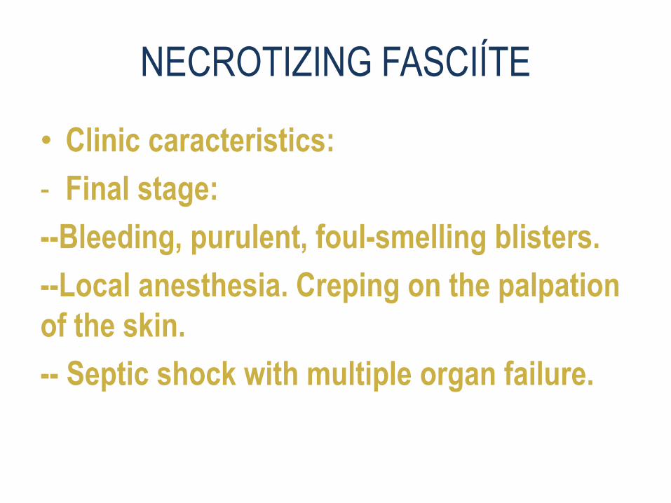
• Clinic caracteristics:
- Final stage:
--Bleeding, purulent, foul-smelling blisters.
--Local anesthesia. Creping on the palpation
of the skin.
-- Septic shock with multiple organ failure.

NECROTIZING FASCIÍTE
• Diagnosis:
- It is clinical and surgical.
- Radiological examinations can help identify
which muscle is involved.
- Propedeutics: surgical culture material and
blood, antibiogram, gram of secretion, CPK,
lactate, coagulogram, ions, blood gases and
hemogram.

NECROTIZING FASCIÍTE
• Treatment:
- Clinical support.
- Early surgical debridement.
- Broad spectrum empirical antibiotic
therapy followed by gram and cultured
secretion antibiogram study.

NECROTIZING FASCIÍTE
• Treatment:
- Antibiotic therapy: Carbapenems or
betalactamases inhibitors (pipe-tazo and
ampi-sulbactam) associated with
clindamycin or Antibiotic for MARSA
(vancomycin and linezolid).
- Duration: depends on the clinical picture.

ERYSIPELOID
• Concept: Acute cutaneous infection
caused by the gram-positive bacillus
Erysipelothrix rhusiopathiae.
• Epidemiology:
• Bacteria found in soil and animals.
• Human contamination when having direct
contact with contaminated food or objects.
Risk group: Butcher, fisherman, housewife
and veterinarian

ERYSIPELOID
• Clinic caracteristics:
- Localized skin infection (erysipeloid):
-- More common shape.
-- Incubation period from 1 to 7 days.
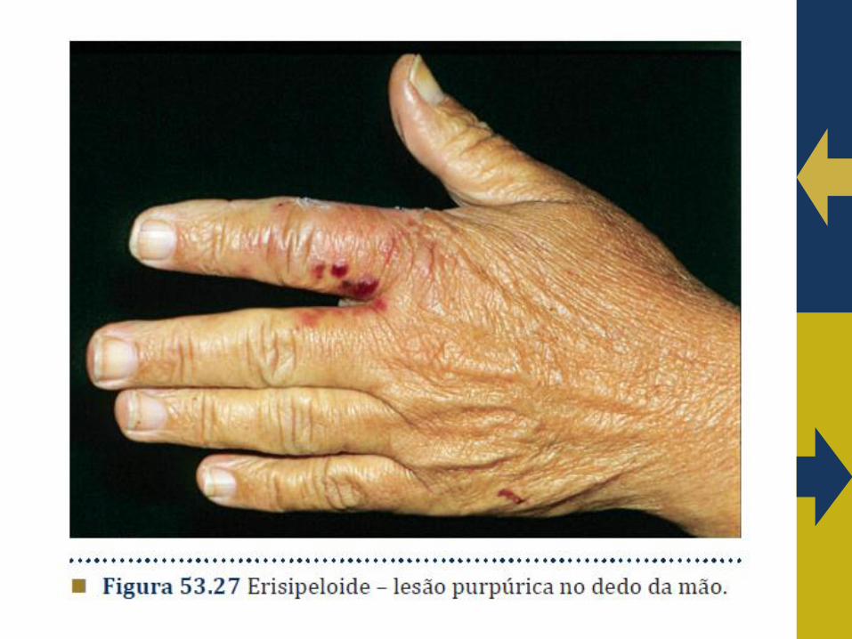
--Lesions with purpuric or violet coloration,
painful and warm with well-marked and
raised margins. Location in hands. Self-
limiting table in a period of 2 weeks.


ERYSIPELOID
• Clinic caracteristics:
- Diffuse cutaneous form:
- --Different parts of the body.
- - Spontaneous resolution and frequent
recurrences.
- --Negative cultures.

ERYSIPELOID
• Clinic caracteristics:
- Generalized infecction:
--Septic artritys, bone necrosys, brain
stroke, pleural efflusion e bacterial
endocarditys. Fever e wheigt loss.
--Polimorfic skin lesions with central necrotc
areas and elevated borders.
--Positive hemocultures.

ERYSIPELOID
• Diagnosis:
- Clínic and epidemiologic.
- Skin lesions or aspirate Gram study.
- Biopsy for culture e for histopathology.
- Histopathology: Vasodilatation of dermic
papiles and a inflamatory infiltration with
neutrophils and eosinophils with dermic
perivascular distribution.

ERYSIPELOID
• Treatment:
- Penicillin, erythromycin and ciprofloxacin.

ERYTHRASMA
• Concept: Localized chronic superficial
skin infection.
• Etiology: Gram-positive bacillus
Corinebacterium minutissimum.
• Epidemiology: It is frequent in tropical
climate and in adult men. Most common
bacterial infection of the feet.

ERYTHRASMA
• Risk factors: Obesity, diabetes, lack of
hygiene, hyperhidrosis and
immunodepression.
• Clinic caracteristics:
- It affects areas with maceration and
Humidity : underarms, inframammary,
intergluteal, groin and interdigital.

ERYTHRASMA
• Clinic caracteristics:
- Erythrasma interdigital: Chronic
asymptomatic maceration with
desquamation cracks interdigital (3rd and
4th Interdigital Spine).
- It is usually associated with candidiasis or
dermatophyte in 30% of cases.

ERYTHRASMA
• Clinic caracteristics:
- Initial caracteristics: Well-defined lesions
of irregular shape, reddish coloration and
asymptomatic.
- Progression: Brownish lesions, fine
scaling and slightly raised bordes.
- It may cause pruritus in the perianal
region.


ERYTHRASMA
• Diagnosis:
- Direct examination of the scales of the
lesion with KOH and stained by the gram:
Small coccoid forms and long filaments.
- Wood lamp: Red-coral fluorescence.

ERYTHRASMA
• Topic treatment:
- Ceratolytics: Salicylic acid 2 or 4%
- Imidazolic topical.
- Clindamycin 2%.
- Erythromycin 2%.
- Fusidic acid.

ERYTHRASMA
• Sistemic treatment:
- Single dose clarithromycin.
- Erythromycin 250mg VO 6 / 6hs 14 days.
• Prophylaxis:
- - Antiseptic soaps and avoid risk factors.

PSEUDOMONES INFECTION
• Etiology: Pseudomonas aeruginosa- gram
negative bacillus of low virulence.
• Primary cutaneous infection: healthy
individuals with loss of skin barrier and
good prognosis.
• Skin manifestations secondary to
septicemia: Immunocompromised are
affected and have a worse prognosis.

SYNDROME OF THE GREENEDNAILS
• Etiology: Pseudomonas aeruginosa.
• Risk factor: Humidity and nail trauma.
• Risk group: Hairdresser and housewife.
• Clinic caracteristics: Painful paronychia
with greenish nail.
• Diagnosis: Clinic + gram + culture.
• Treatment: Polimixin and bacitracin 6/6hs
in solution for 4 months. Avoid humidity.


SCARLATINA
• Concept: Acute disease caused by Group
A beta-hemolytic Streptococcus exotoxins.
• Epidemiology: Endemic in large centers.
Common in children aged 1-10 years.
• Clinic caracteristics:
• - Upper airways are the gateway through
contaminated droplets.

ESCARLATINA
• Clinic caracteristics:
- Incubation period 2-5 days.
- Membranous tonsillitis associated with
painful cervical lymphadenopathy.
- Sixth day: beginning exfoliative rash with
sanding appearance with capillary
fragility. Oral paleness.
- Tenth day: Discoloration disappears with
lamellar palmoplantar desquamation.
ESCARLATINA
• Clinic caracteristics:
- Fever, nausea, vomiting, abdominal pain
and tongue in raspberry.
- Diagnosis:
- Clinic caracteristics + elevation ASLO +
pharyngeal culture + leukocytosis with
deviation to left
- Treatment: Penicillin G benzathine,
amoxicillin and erythromycin.

LINFANGITE
• Concept: Inflammation of lymphatic
vessels, usually of bacterial origin.
• Etiology: Beta-hemolytic Streptococcus or
S. aureus both coagulase-positive, which
enter the lymphatic through trauma.

LINFANGITE
• Clinic caracteristics: Systemic reaction.
Erythema from local inoculation to
regional lymph nodes, which are swollen
and painful.
• Diagnosis: Clinic caracteristics + blood
count + culture.
• Treatment: systemic antibiotic therapy,
resting and elevated limbs.

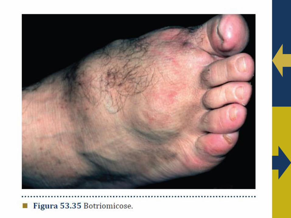
BOTHRIOMYCOSIS
• Etymology: Botrio (grape clusters) and
mycosis (fungi infection).
• Synonymy: Staphylococcal
actinophytosis, actinobacillosis and
granular bacteriosis.
• Etiology: S. aureus and other bacteria.
• Risk factor: Immunodepression and
patients with chronic diseases.

BOTHRIOMYCOSIS
• Immunology: Bacteria in symbiosis with
the host. Defects of immunity.
• Clinic caracteristics:
- Chronic, granulomatous and suppurative
infection that affects skin, soft tissues and
bones by contiguity. It can affect viscera.
- Skin: nodules, ulcers, verrucous plaques
and fistulas that drain purulent secretion
em mãos e pés.

BOTHRIOMYCOSIS
• Diagnosis:
- Histopathology:
- Basophilic grains surrounded by hyaline
material stained by gram.
- Phenomenon of Splendore-Hoeppli.
-Culture secretion for bacteria and fungi.
-Radiology examinations.

BOTHRIOMYCOSIS
• Treatment:
Sulfamethoxazole-trimethoprim,
minocycline, erythromycin, dapsone and
cefazolin for long periods.

CAT SCRATCH DISEASE
• Etiology: Bartonella henselae- Strongly
argirophic gram negative sticks.
• Epidemiogia: Rare. There are mild and
authoritarian cases.
• Reservoirs: Cats and other felines that are
contaminated by cat fleas.
• Transmission to humans: Licking or biting
the cat.

CAT SCRATCH DISEASE
• Clinic caracteristics:
- The most common bartonellosis.
- Cutaneous lesions begin 3-5 days after
trauma such as erythematous papules and
vesicles that develop into ulcers and
regress with residual macules.
- Painful unilateral lymph node enlargement
with possibility of suppuration.
- Mild constitutional symptoms and fever.

CAT SCRATCH DISEASE
• Diagnosis:
- Histopathology of the skin and lymph
nodes: Granulomatous infiltrate of
lymphocytes, histiocytes and neutrophils
with central necrotic area and pleomorphic
bacilli strongly stained by silver in
Warthin-Starry coloration.
- Serological test - Elisa.
- Culture and PCR - hardly accessible.

CAT SCRATCH DISEASE
• Treatmet:
- Doxycycline VO 100mg twice a day
associated with rifampicin 60mg once a
day or ciprofloxacin 500mg twice a day or
azithromycin 500mg once a day.


1. PORTO, L.A.B. Bacterial skin infections. www.drluizporto.com.br/dermatologia (accessed september 08, 2016).
2. BELDA JUNIOR, W. DI CHIACCHIO, N.; CRIADO, P. R. Tratado de Dermatologia 2ª edição. São Paulo Atheneu; 2014.
3. Baddour LM. Cellulitis and erysipelas. In: UpToDate. (Acessado em 22 de outubro de 2015).
4. Stevens DL, Baddour LM. Necrotizing soft tissue infections. UpToDate. (Acessado em 22 de outubro de 2015).
BIBLIOGRAPHY

THANK YOU!
DOUBTS?
Luiz Alberto Bomjardim Pôrto
http://www.drluizporto.com.br/