Embed Size (px)
Citation preview
Minireview
Bacterial cell biology outside the streetlight
Silvia Bulgheresi*
Department of Ecogenetics & Systems Biology,
University of Vienna, Althanstrasse 14, Vienna 1090,
Austria.
Summary
As much as vertical transmission of microbial sym-
bionts requires their deep integration into the host
reproductive and developmental biology, symbiotic
lifestyle might profoundly affect bacterial growth and
proliferation. This review describes the reproductive
oddities displayed by bacteria associated – more or
less intimately – with multicellular eukaryotes.
“We become what we behold. We shapeour tools and then our tools shape us”
Culkin, J.M. (1967). A schoolman’s guideto Marshall McLuhan. Saturday Review,pp. 51-53, 71-72.
Introduction
According to rough estimates, less than 0.1% of the up to
109 bacterial species thriving on planet Earth may be culti-
vated. Yet the reproduction of only a handful of lab-reared
bacteria is being studied (Goley, 2013). The current quest
to unravel the mechanisms underlying bacterial reproduc-
tion is therefore comparable to a drunkard’s search. Its
inherent observational bias – the so-called streetlight effect
– is treacherous as it may easily generate wrong assump-
tions or even beliefs. Here, I wish to review cell biological
studies on bacterial symbionts, some of which already con-
futed well-rooted tenets of bacterial cell growth and division
(Table 1). I will use the term symbiont sensu lato, that is to
say every organism living together with a differently named
organism (de Bary, 1879). Traditionally, symbionts are clas-
sified in ecto- and endosymbionts depending on whether
they are outside or inside the host body respectively. In this
review, although formally situated inside the host, I will
regard the gut epithelium as superficial and consider its
inhabitants – the gut microbiota – together with other ecto-
symbionts. Owing to space limitations, I will not review
systems in which the molecular mechanisms underlying
anomalous symbiont growth have not been investigated
yet. These are the vast majority and include – for example
– the ectosymbionts of unicellular (Desai et al., 2010; Stras-
sert et al., 2010; Brune, 2014; Brune and Dietrich, 2015)
and colonial eukaryotes (Bright et al., 2014), the endosym-
bionts of unicellular eukaryotes (Schulz and Horn, 2015)
and of abyssal bathymodiolin mussels Candidatus Endonu-
cleobacter bathymodioli (Zielinski et al., 2009). Finally, also
due to space limitations, I will be able neither to review our
knowledge about mitochondrial and plastid reproduction
and its control by the eukaryotic cell, nor to consider bacte-
ria whose cell biology and physiology is dramatically
affected by developmental programs [e.g., elementary
body–reticulate body transition in Protchlamidiae associ-
ated to free-living amoebae (Horn, 2008; Schrallhammer
and Schweikert, 2009)]. And now, before I embark on
describing abnormal reproductive modes, I must briefly
summarize how conventional bacteria grow and divide.
Bacterial growth and division for dummies
A wealth of exhaustive and well-written reviews is available
on the fundamental process of bacterial reproduction
[among the most recent ones: Bramkamp and Van Baarle
(2009), Young (2010); den Blaauwen (2013); Chang and
Huang (2014); Rowlett and Margolin (2015)]. The peptido-
glycan (PG or murein) wall or sacculus confers physical
strength and defines the shape of most bacteria (Egan and
Vollmer, 2013; de Pedro and Cava, 2015; Randich and
Brun, 2015). It overlies their cell membrane and consists of
a mesh-like macromolecule of glycan chains that are
cross-linked together via peptide chains. In model rods,
two protein assemblies (the elongasome and the divisome)
direct the modification and synthesis of the cell wall: during
growth, the elongasome inserts freshly synthetized PG
along the length of the rod, and during septation a cytoki-
nesis complex called the divisome (Nanninga, 1991)
completes the steps of constriction and new PG synthesis
at midcell (Typas et al., 2012). Both divisome and elonga-
some consist of scaffolding cytoskeletal-like proteins
(cytoplasmic), inner membrane spanning proteins and aReceived 28 September 2015; accepted 02 June 2016:
*For correspondence. [email protected].
VC 2016 The Authors. Environmental Microbiology Reports published by Society for Applied Microbiology and JohnWiley & Sons LtdThis is an open access article under the terms of the Creative Commons Attribution License, which permits use, distribution andreproduction in any medium, provided the original work is properly cited.
Environmental Microbiology (2016) 18(8), 2305–2318 doi:10.1111/1462-2920.13406

Tab
le1.
Exam
ple
sof
bacte
rial
sym
bio
nts
dis
pla
yin
gnon-c
anonic
aldiv
isio
nm
odes
Host
Sym
bio
nt
Environm
ent
surr
oundin
gth
esym
bio
nt
Repro
ductive
anom
aly
Pre
sence
of
Fts
Z
Host
facto
r(s)
affecting
sym
bio
nt
div
isio
n
Extr
acellu
lar
sym
bio
nts
Laxus
oneis
tus
nem
ato
de
Candid
atu
sT
hio
sym
bio
n
oneis
ti
(Gam
mapro
teobacte
ria)
Marine
sedim
ent
Wid
enin
gand
sym
metr
iclo
ngi-
tudin
alfissio
n
YE
SN
N
Eubostr
ichus
fert
ilis
nem
ato
de
Eubostr
ichus
fert
ilis
nem
a-
tode
ecto
sym
bio
nt
(Gam
mapro
teobacte
ria)
Marine
sedim
ent
Cell
elo
ngation
(up
to45mm
)
and
sym
metr
ictr
ansve
rse
fissio
n
YE
SN
N
Eubostr
ichus
dia
neae
nem
ato
de
Eubostr
ichus
dia
neae
nem
a-
tode
ecto
sym
bio
nt
(Gam
mapro
teobacte
ria)
Marine
sedim
ent
Cell
elo
ngation
(up
to120mm
)
and
sym
metr
ictr
ansve
rse
fissio
n
YE
SN
N
Epulo
pis
ciu
mspp.
Surg
eonfish
(Fir
mic
ute
s)
Gastr
oin
testinaltr
act
Multip
lein
tracellu
lar
offspring
Meta
bacte
rium
poly
spora
Guin
ea
pig
(Fir
mic
ute
s)
Gastr
oin
testinaltr
act
Multip
leendospore
form
ation
or
bin
ary
fissio
n
YE
SN
N
Mus
musculu
sS
egm
ente
dF
ilam
ento
us
Bacte
rium
(SF
B;
Fir
mic
ute
s)
Gastr
oin
testinaltr
act
Cell
elo
ngation,
segm
enta
tion
and
intr
acellu
lar
offsprin
gor
sporu
lation
YE
SN
N
Intr
acellu
lar
sym
bio
nts
Legum
eR
hiz
obia
(Alp
hapro
teobacte
ria
)
Modifie
dpla
nt
cell
(nodule
)
innodule
Cell
elo
ngation
(blo
ckof
cyto
kin
esis
)
YE
SN
odule
-specific
cyste
ine-r
ich
peptides
(NC
Rs)
Weevil
Sodalis
pie
ranto
niu
sM
odifie
din
sect
cell
(bacte
r-
iocyte
)in
bacte
riom
e
Cell
elo
ngation
(blo
ckof
cyto
kin
esis
)
YE
SC
ole
opte
ricin
A(C
olA
)
Aphid
Buchnera
aphid
icola
(Gam
mapro
teobacte
ria)
Modifie
din
sect
cell
(bacte
r-
iocyte
)in
bacte
riom
e
Gig
antism
(blo
ckof
cyto
kin
esis
)
YE
SS
ecre
ted
cyste
ine-r
ich
pro
tein
s?
Sulc
iam
uelle
riM
odifie
din
sect
cell
(bacte
r-
iocyte
)in
bacte
riom
e
Gig
antism
(blo
ckof
cyto
kin
esis
)
YE
SN
N
2306 S. Bulgheresi
VC 2016 The Authors. Environmental Microbiology Reports published by Society for Applied Microbiology and JohnWiley & Sons Ltd,Environmental Microbiology, 18, 2305–2318
profusion of periplasmic enzymes including PG synthases
and hydrolases. In nearly all bacteria, the tubulin-like
GTPase FtsZ regulates the localization and activity of divi-
some components. In canonical rod-shaped bacteria,
which divide by transverse binary fission, spatiotemporal
regulation of the divisome is fairly understood. During divi-
sion, the GTP-dependent polymerization of FtsZ creates a
ring-shaped structure called the Z-ring at the centre of the
Bacterial cell biology outside the streetlight 2307
VC 2016 The Authors. Environmental Microbiology Reports published by Society for Applied Microbiology and JohnWiley & Sons Ltd,Environmental Microbiology, 18, 2305–2318
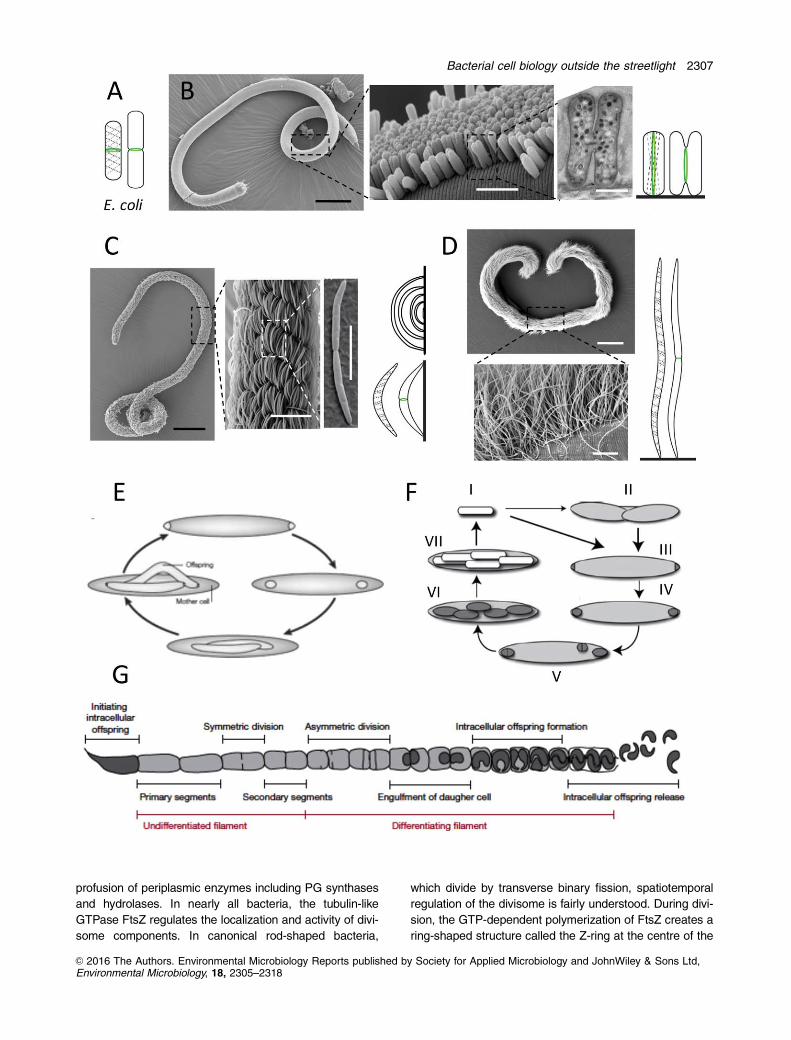
cell (Fig. 1A) and perpendicular to the axis of chromosome
segregation (Bi and Lutkenhaus, 1991; L€owe and Amos,
1998; Mukherjee and Lutkenhaus, 1998; Erickson et al.,
2010). The Z-ring is attached to the cytoplasmic face of
the membrane via membrane-associated proteins and
recruits ten essential proteins important for septum assem-
bly and Z-ring constriction (Hale et al., 1997; Pichoff and
Lutkenhaus, 2005). In Escherichia coli, the mechanism for
targeting the Z-ring to midcell involves the Min system, a
machinery that inhibits the formation of the contractile ring
at the cell poles (Raskin and de Boer, 1999; Hale et al.,
2001; Monahan et al., 2014), and nucleoid occlusion (NO),
which prevents nucleoid guillotining by the Z-ring (Wol-
dringh et al., 1990; 1991; Bernhardt and de Boer, 2005).
Much less is known about the spatiotemporal regulation of
the elongasome, for which the actin-like protein MreB
appears to be the major scaffold for coordinating PG pre-
cursor synthesis and polymerization (Fig. 1; Esue et al.,
2005, 2005; Salje et al., 2011; Ozyamak et al., 2013).
Although the bacteria treated in the following sections
are coccoid, rod-shaped or filamentous and therefore, mor-
phologically, are not significantly different from the
conventional ones, they display at least one of the following
peculiarities: (i) polymorphism, often within the same popu-
lation, (ii) reproduction modes other than transverse binary
fission and/or (iii) alternation between two different repro-
duction modes.
Dressed to cooperate: growth anomalies of
ectosymbionts
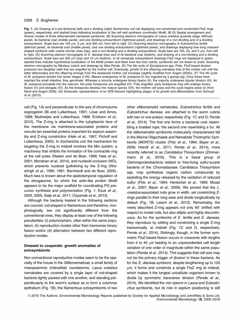
Non-conventional reproductive modes seem to be the spe-
cialty of the house in the Stilbonematinae, a small family of
mesopsammic (interstitial) roundworms: Laxus oneistus
nematodes are covered by a single layer of rod-shaped
bacteria tightly packed with one another, and standing per-
pendicularly to the worm’s surface as to form a columnar
epithelium (Fig. 1B); the filamentous ectosymbionts of two
other stilbonematid nematodes, Eubostrichus fertilis and
Eubostrichus dianeae, are attached to the worm cuticle
with two or one pole(s) respectively (Fig. 1C and D; Pende
et al., 2014). The first one forms a bacterial coat resem-
bling a braided rope, the second one resembling a fur. All
the stilbonematid symbionts molecularly characterized fall
in the Marine Oligochaete and Nematode Thiotrophic Sym-
bionts (MONTS) cluster (Polz et al., 1994; Bayer et al.,
2009; Heindl et al., 2011; Pende et al., 2014), more
recently referred to as Candidatus Thiosymbion (Zimmer-
mann et al., 2016). This is a basal group of
Gammaproteobacteria related to free-living sulfur-purple
bacteria of the Chromatiaceae. Candidatus Thiosymbion
spp. may synthesize organic carbon compounds by
exploiting the energy released by the oxidation of reduced
sulfur (Polz et al., 1994; Hentschel et al., 1999; Musat
et al., 2007; Bayer et al., 2009). We proved that the L.
oneistus-associated rods grow in width, set constricting Z-
rings parallel to their long axes and divide longitudinally by
default (Fig. 1B; Leisch et al., 2012). Remarkably, the
newly described Z-ring appears not only 908 shifted with
respect to model rods, but also elliptic and highly discontin-
uous. As for the symbionts of E. fertilis and E. dianeae,
they reproduce by setting and constricting a single Z-ring
transversally, at midcell (Fig. 1C and D, respectively;
Pende et al., 2014). Strikingly, though, in the former sym-
metric FtsZ-based fission occurs in crescents with lengths
from 4 to 45 mm leading to an unprecedented cell length
variation of one order of magnitude within the same popu-
lation (Pende et al., 2014). This suggests that cell size may
not be the primary trigger of division in these bacteria. As
for the E. dianeae symbiont, despite lengthening up to 120
mm, it forms and constricts a single FtsZ ring at midcell,
which makes it the longest unicellular organism known to
divide by symmetric transverse division (Pende et al.,
2014). We identified the min operon in Laxus and Eubostri-
chus symbionts, but its role in septum positioning is still
Fig. 1. (A) Drawing of a pre-divisional (left) and a dividing (right) Escherichia coli cell displaying non-constricted and constricted FtsZ rings(green), respectively, and dashed lines indicating localization of the cell wall synthesis coordinator MreB. (B–D) Spatial arrangement anddivision modes of three stilbonematid nematode symbionts; (B) Scanning electron micrographs of Laxus oneistus (juvenile stage, leftmostpanel), its bacterial coat (middle panel), and one dividing ectosymbiont (rightmost panel), and drawings of a non-dividing and of a dividingectosymbiont. Scale bars are 100 mm, 3 mm, and 500 nm, from left to right. (C) Scanning electron micrographs of Eubostrichus fertilis(leftmost panel), its bacterial coat (middle panel), and one dividing ectosymbiont (rightmost panel), and drawings displaying how long crescent-shaped symbiont cells overlie shorter ones (top), and a non-dividing and a dividing ectosymbiont. Scale bars are 100, 20, and 5 mm, from leftto right. (D) Scanning electron micrographs of E. dianeae (top) and of its bacterial coat (bottom), and drawing of a non-dividing and a dividingectosymbiont. Scale bars are 200 mm (top) and 10 mm (bottom). In all nematode ectosymbiont drawings FtsZ rings are displayed in green,dashed lines indicate hypothetical localization of the MreB protein and black lines the host cuticle; symbionts are not drawn to scale. Scanningelectron micrographs by Nikolaus Leisch and drawings by Nika Pende. (E) The life cycle of Epulopiscium spp. Polar, FtsZ-based divisionproduces two daughter cells that are engulfed by the mother cell. Eventually, growth of the offspring overtakes that of the mother cell until thelatter deteriorates and the offspring emerge from the weakened mother cell envelope (slightly modified from Angert (2005)). (F) The life cycleof M. polyspora divided into seven stages (I-VII). Mature endospores of M. polyspora (I) are ingested by a guinea pig. Once these havereached the small intestine, they germinate. Whereas a minority undergoes binary fission (II), the majority undergoes bipolar division (III). AsM. polyspora proceeds into the caecum, the polar forespores are engulfed (IV). Fully engulfed, early forespores may still undergo binaryfission (V) and elongate (VI). As the forespores develop into mature spores (VII), the mother cell lyses and the cycle begins anew (I) (fromWard and Angert 2008). (G) Schematic representation of an SFB filament highlighting stages of its growth and differentiation from Schnupfet al. (2015).
2308 S. Bulgheresi
VC 2016 The Authors. Environmental Microbiology Reports published by Society for Applied Microbiology and JohnWiley & Sons Ltd,Environmental Microbiology, 18, 2305–2318
unknown. Moreover, although we identified the mre operon
in all the aforementioned stilbonematid symbionts we do
not know how MreB coordinates cell wall growth in the
nematode ectosymbionts. Finally, we must determine the
exact number of genomes that get symmetrically localized
and their orientation and segregation mechanisms. Do the
different symbiont spatial dispositions, i.e., the different
symbiont reproductive strategies, represent adaptations to
different nematode hosts or to different nutritional regimes
(Vadia and Levin, 2015)? Did L. oneistus symbiont longitu-
dinal fission or did E. fertilis symbiont bipolar attachment
evolve to favour symbiont vertical transmission? Are these
two symbionts metabolically more dependent on their host
than the E. dianeae symbiont, which can afford to let one
daughter cell detach from the host surface?
We hope that omics-based comparisons among stilbo-
nematids occupying different habitats or carrying different
types of bacterial coats will clarify whether the latter serve
specific, host-symbiont metabolic networks or physiologi-
cal interdependencies, that – in turn – evolved as
adaptations to specific habitats.
Outside but inside: fish gut residents
Although extraordinarily long, the E. dianeae symbiont is
not the longest known. The size record holders are indeed
the surgeonfish intestinal symbionts Epulopiscium spp.
Their heterotrophic cigar-shaped cells are up to 600 3 80
mm and reproduce by forming at least two intracellular
daughter cells (Fig. 1E; Angert et al., 1993). Cells coloniz-
ing the gut of a given host fish synchronize internal
offspring initiation and development so that offspring
always grows during the day, concomitantly with host feed-
ing. Reproduction begins as two Z-rings form at the
mother cell’s poles. As the rings fully constrict, two polar
cells form, are engulfed by the mother cell and grow in a
membrane-bound cytoplasmic compartment until they
completely fill the mother cell. Finally, the mother cell
undergoes a form of programmed cell death that likely con-
serves the biochemical resources accumulated during
growth (Ward et al., 2009) and offspring emerge through a
split in the mother cell envelope. Notably, despite develop-
mental synchrony, inhabitants of a single population vary
up to five times in volume (Mendell et al., 2008). How can
Epulopiscium spp. accommodate growth of internal off-
spring? Although their genome size is unexceptional (ca. 4
Mb), each cell contains up to 105 copies (Mendell et al.,
2008). However, in contrast to most bacteria, in which
chromosomes are distributed throughout the cytoplasm,
Epulopiscium nucleoids are located at the cell periphery,
which may also allow Epulopiscium to respond promptly to
environmental stimuli.
Outside but inside: mammalian gut residents
Another curious gastrointestinal dweller is Metabacterium
polyspora. Unlike most endospore formers, this guinea pig
gut resident produces up to nine endospores per mother
cell (Chatton and P�erard, 1913; Robinow, 1957). Although
no Metabacterium-like symbiont has been maintained in
culture, morphologically similar symbionts have been
found in various rodent species (Kunstyr et al., 1988). The
natural life cycle of M. polyspora (Fig. 1F) requires the bac-
terium to cycle through the gastrointestinal tract and
therefore relies on the coprophagous nature of the guinea
pig for survival (Angert and Losick, 1998). Only mature
endospores survive passage through the mouth and stom-
ach of the host, and may germinate in the small intestine.
Here, some cells undergo binary fission, but most cells
begin to sporulate (Angert and Losick, 1998). From the
small intestine, M. polyspora cells are deposited in the
guinea pig caecum where they complete sporulation. Cells
with engulfed forespores or mature endospores do not
seem to undergo binary fission and, after traversing the
lower intestine, they are finally eliminated from the host
with its feces. If a guinea pig ingests the defecated spores,
the life cycle starts again. The process of endospore for-
mation in M. polyspora differs from that of prototypical
endospore-forming bacteria such as Bacillus subtilis
(Angert and Losick, 1998). The asymmetric cell division of
M. polyspora normally takes place at both cell poles (rather
than at one only) and DNA is partitioned into both polar
compartments and it is also retained in the mother cell.
Therefore, unlike sporulating B. subtilis, which is diploid,
M. polyspora must contain three or more genomes, which
makes coordinating DNA replication and segregation with
offspring formation even more challenging (Angert and
Losick, 1998). After engulfment, the forespores can
undergo division to produce multiple forespores that grow
and mature into endospores. Most endospore-forming Fir-
micutes produce a single, dormant, invulnerable spore
only to survive adverse environmental conditions or to
increase their dispersal. It is therefore stunning that the
reiteration of such a developmental program is part of the
normal M. polyspora life cycle. The coordination of multiple
endospore formation with transit through the gut, com-
bined with a coprophagous natural host, might favour this
reproduction mode over binary fission. Additionally, the
occasional experience of harsh conditions outside the host
would still trigger M. polyspora to initiate sporulation for
survival and dispersal.
There is another group of low-GC Gram-positive bacte-
ria that forms endospores for reproduction and dispersal in
a very unorthodox way: the segmented filamentous bacte-
rium (SFB) or Candidatus Arthromitus (Fig. 1G). SFB
reside in the intestinal tracts of many vertebrate species
such as mice and ourselves (Klaasen et al., 1992; Yin
Bacterial cell biology outside the streetlight 2309
VC 2016 The Authors. Environmental Microbiology Reports published by Society for Applied Microbiology and JohnWiley & Sons Ltd,Environmental Microbiology, 18, 2305–2318

et al., 2013; Schnupf et al., 2015) and have received much
interest because of their ability to educate the gut immune
system and to induce a healthy level of physiological
inflammation (Schnupf et al., 2013; Ivanov et al., 2009).
Already four decades ago, ultrastructural studies of murine
gut SFB supported the following life cycle: attachment to
epithelial cells via the holdfast tip of the so-called “initiating
intracellular offspring” leads to SFB embedding among the
microvilli followed by filamentous growth. A complex devel-
opmental program thereupon starts at the distal tip and
ultimately leads to intracellular offspring formation and
release (Davis and Savage, 1974; Chase and Erlandsen,
1976; Ferguson and Birch-Andersen, 1979). According to
this model, when filaments grow longer than 50 mm in
length (most filaments are �100 lm and can be up to
1 mm long), the large primary filament segments start to
undergo a symmetrical division to form smaller secondary
segments. These differentiate by dividing asymmetrically
to form a mother and a daughter cell. The latter becomes
engulfed and subsequently divides to form two intracellular
offspring within the surrounding mother cell segment. Intra-
cellular offspring are then released from the filament by
breakdown of the filament septa and reattach to the host.
Remarkably, the intracellular offspring have two possible
fates, either holdfast-producing differentiation, or matura-
tion to form a spore. In the first case, the active offspring
are released into the lumen of the intestine and they attach
to the intestinal epithelium to establish new filaments within
the host. In the second case, maturation results in two
intracellular offspring cells that are encased in a common
spore coat, which forms an endospore. As the cells of the
intestinal epithelium are constantly shed and renewed,
they are not a stable substrate. Therefore, the production
of intracellular offspring released from the dying parental
filament probably evolved to allow SFB to reposition itself
inside the same host. Additionally, the endospore provides
an effective alternative for the SFB to disperse. These
alternative forms of offspring (either active or dormant)
allow the SFB to maintain stable populations within a given
host and to colonize new hosts after surviving harsh envi-
ronments such as aerobic ones or the highly acidic upper
gut. The proposed SFB life cycle was recently and com-
pletely recapitulated in vitro (Schnupf et al., 2015). This is
exciting, as it will permit the investigation of the complex
developmental stages of SFB and the detailed dissection
of the unique SFB–host interaction at the cellular and
molecular levels.
What controls the growth of gut residents?
The host-secreted molecules that determine SFB cell fate,
as well as those putatively controlling that of other gut resi-
dents are not known. Where should we look for those? It is a
challenge for the gut to stably host 100 trillion bacteria
belonging to more than 100 different species without this
inducing inflammation or without the host falling victim of
pathogenic infections (estimates by Qin et al., 2010). Pertur-
bations in intestinal homeostasis are the basis of various
diseases such as obesity, diabetes and inflammatory bowel
disease. Given that immune homeostasis relies on several,
partly overlapping immunological mechanisms we still have
not fully grasped how it is achieved (Hooper and Macpher-
son, 2010). However, several effectors of the innate immune
system controlling the number of specific gut residents have
already been identified and these are antimicrobial peptides
(AMPs). This should not come as a surprise as the growing
consensus is that both pathogenic and mutualistic bacteria
share similar microbe associated molecular patterns
(MAMPs) and trigger an immune response. Nevertheless, its
outcome differs between pathogenic and mutualistic associa-
tions: in the former, the immune response triggers the
elimination of deleterious microbes, in the latter a molecular
dialogue is initiated which results in homeostasis and immu-
notolerance (Nussbaum and Locksley, 2012). In a mutualistic
context, antimicrobial peptides produced by the epithelia con-
tribute to maintaining the population structure of the
microbiota and prevent these from penetrating the underlying
tissues. The epithelia are covered with a thin mucus layer
that is kept nearly sterile, while microbes abound in the
above-lying lumen (Hooper and Macpherson, 2010). In the
small intestine, this is accomplished by the Paneth cells
which secrete into the gut AMPs and other antimicrobial pro-
teins thereby limiting contact between the microbiota and
epithelial tissues (Kobayashi et al., 2005; Vaishnava et al.,
2008). In addition, AMPs such as a-defensins also regulate
the composition of the microbiota in the lumen (Salzman
et al., 2003; 2010; Wilson et al., 1999). In particular, bacteria
known as Firmicutes decreased with higher defensin produc-
tion by Paneth cells. Similarly, Vaishnava et al. (2011)
showed that the antimicrobial lectin RegIII-g prevents any
contact between the mouse commensal microbiota and the
epithelial surface of the small intestine. Disruption of this
physical separation, by knocking out the RegIII-g gene,
resulted in bacterial proliferation at the intestinal epithelial sur-
face, which subsequently triggered an adaptive immune
response against the microbiota. Secretion of the RegIII-g
protein was shown to inhibit bacterial growth within a band
extending 50 mm from the epithelium, creating a “no-
microbe’s-land” where no commensal can grow or stimulate
the host immune system. Given that RegIII-g is not
expressed in the large intestine, this is one of the first cases
describing the involvement of the innate immune system in
the regulation of mutualism via a local immune response.
Also in the fruit flies gut a well-adjusted level of AMP expres-
sion under healthy, as well as pathogenic conditions is
essential for maintaining homeostasis (Ryu et al., 2008).
In conclusion, a respectable body of data indicates that
vertebrate-secreted AMPs target and regulate the number
2310 S. Bulgheresi
VC 2016 The Authors. Environmental Microbiology Reports published by Society for Applied Microbiology and JohnWiley & Sons Ltd,Environmental Microbiology, 18, 2305–2318

of gut micro-residents. Although their capacity to affect the
morphology or reproduction mode of commensals has not
been shown yet, this is likely, as research reviewed in the
following section indicates.
“Staying put”: reproductive anomalies of
endosymbionts with non-reduced genomes
If cell gigantism and associated polyploidy have been
observed in numerous microbial symbionts, only in those
of legumes and weevils the molecular triggers, i.e., the
host-secreted molecules that block bacterial cytokinesis
have been nailed (Van der Velde et al., 2010; Login et al.,
2011). In most cases, the benefit of plant and insect sym-
bionts is privileged acquisition of nutrients and a growth
niche. Whether the symbionts reside extracellularly, in
luminal spaces between cells and tissues, or live an intra-
cellular existence they are closely associated with the host
cells. When legumes interact with nitrogen-fixing rhizobia,
the symbiosis leads to formation of new organs, the root
nodules (Fig. 2A). These organs house millions of endo-
symbiotic rhizobia. Within symbiotic nodule cells, these
Alphaproteobacteria become capable of reducing atmos-
pheric nitrogen to ammonium, which is transferred to the
plant and used for its growth. These irreversibly differenti-
ated rhizobia (also referred to as bacteroids; Fig. 2B) have
altered physiology and metabolism. In some legumes, as
in the model plant Medicago truncatula, bacteroids have
increased membrane permeability, highly amplified
genome content and – whether elongated or branched –
are much larger than soil-dwelling rhizobia. These bacte-
roids are incapable of cell division and reproduction
(Mergaert et al., 2006; Maroti et al., 2011). Although this
terminal differentiation of bacteroids is not observed in all
legumes and is therefore not essential per se for symbiotic
nitrogen fixation, it improves the symbiotic efficiency of the
bacteroids (Oono and Denison, 2010). In M. truncatula,
symbiotic nodule cells produce nodule-specific AMPs of a
particular family called NCR for nodule-specific cysteine-
rich peptides (NCRs; Mergaert et al., 2003; Alunni et al.,
2007). Remarkably, the M. truncatula NCR gene family
consists of several hundred genes, which are all specifi-
cally expressed in Rhizobium-infected nodule cells. The
NCRs are responsible for the terminal differentiated state
of the endosymbiont Sinorhizobium meliloti in M. truncatula
nodules (Fig. 2C; Van der Velde et al., 2010). Similarly to
other plant and animal AMPs, they effectively kill both
Gram-positive and Gram-negative bacteria in vitro at proto-
typical concentrations. NCRs are transported via
exocytosis to the bacteroids and some of the NCRs enter
the bacterial cytosol and likely have intracellular bacterial
targets. The in vivo and in vitro effects of NCRs on S. meli-
loti are dramatically different. The peptides quickly kill the
rhizobia in vitro, whereas bacteroids do not grow but main-
tain active metabolism. The difference between the in vivo
and in vitro effect of NCRs could be explained if we
assume that, in vivo, several tens or hundreds of native
peptides, each likely present at very low concentrations,
have a different effect that a single, highly concentrated
peptide applied in vitro. Moreover, particular conditions
prevalent in nodules, such as the low free oxygen concen-
tration, needed for activity of the oxygen-sensitive
nitrogenase, could modulate the bacterial responses to the
NCRs in such a way that the bacteroids survive without
growing. Some NCRs inhibit bacterial division in vivo and
in vitro, leading to cell elongation. Such NCRs were local-
ized at the division site of S. meliloti cells, and may
therefore interfere with the bacterial cytokinetic machinery.
However, the high sequence variability of NCRs suggests
diversity in their functions, mode of actions and targets
involved in different aspects of bacteroid metabolism, but
could also be an adaptation to the high diversity of soil rhi-
zobia. Analysis of the sequence of NCRs showed that they
are subject to diversifying evolution, which is compatible
with such a hypothesis (Alunni et al., 2007). Moreover, it is
remarkable and yet unexplainable that most plants contain
hundreds of genes encoding for cysteine-rich proteins sim-
ilar to innate immunity-dedicated AMP genes (Silverstein
et al., 2007), even if they do not employ them to form sym-
bioses or to defend themselves. The NCRs are indeed
expressed only in nodules and are not induced during
treatment of M. truncatula with pathogens.
In conclusion, legumes may adopt effectors of the innate
immune system to dominate their endosymbionts in order
to maximize their own profits. In a striking case of conver-
gent evolution, this is also the case for Sitophilus weevils
(Fig. 2D–G; Login et al., 2011, Login and Heddi, 2013).
Cereal weevils house Sodalis pierantonius (Fig. 2F; Oake-
son et al., 2014; formerly known as Sitophilus Primary
Endosymbiont, or SPE) permanently, in the female germ
cells from which they are transmitted to the progeny. Early
during embryogenesis, S. pierantonius induces the differ-
entiation of bacteriocyte cells that form the bacteriome
(Heddi et al., 1999), a specific organ that secludes endo-
symbionts (Fig. 2E; Anselme et al., 2008). These host
cells, in response, express an adapted response in order
to maintain the symbionts within the bacteriome (Anselme
et al., 2008; Login et al., 2011). Sequestering symbiotic
bacteria in the bacteriome – where only a few immune
effectors are expressed – protects them from exposure to-
and elimination by a standard, non-tailored systemic
immune response. This humoral response does indeed
attack S. pierantonius, when this bacterium is injected in
the insect hemolymph (Nakabachi et al., 2005; Ratzka
et al., 2011). In the weevil bacteriome, instead, Coleopteri-
cin A (ColA) is the only antimicrobial expressed
constitutively and colA transcript levels correlate with S.
pierantonius density (Login and Heddi, 2012; Anselme
Bacterial cell biology outside the streetlight 2311
VC 2016 The Authors. Environmental Microbiology Reports published by Society for Applied Microbiology and JohnWiley & Sons Ltd,Environmental Microbiology, 18, 2305–2318

et al., 2008; Login et al., 2011; Masson et al., 2015). While
ColA exhibits bactericide activity against Gram-negative
bacteria and kills them at high concentrations, it exhibits a
bacteriostatic activity against S. pierantonius at the low
concentrations likely existing in the bacteriocytes. Micro-
scopic observations showed that inhibition of cytokinesis
by ColA led to filamentation, namely to up to 50 mm-long E.
coli cells (Fig. 2G) and up to 200 mm-long Nardonella cells,
the primary endosymbiont of the weevil Rhynchophorus
ferrugineus. Additionally, it was also showed that S.
2312 S. Bulgheresi
VC 2016 The Authors. Environmental Microbiology Reports published by Society for Applied Microbiology and JohnWiley & Sons Ltd,Environmental Microbiology, 18, 2305–2318

pierantonius reduced significantly in size in vivo following
functional knockout of the colA gene (Login et al., 2011).
However, whether this bacterial size reduction is due to a
resumption of bacterial cytokinesis or a multiplication of
small bacteria remains unclear. In vivo silencing of the
colA gene also resulted in an extensive dispersion of the
symbiont outside of the bacteriome, which strongly sup-
ports the idea that, besides its morphology, ColA controls
symbiotic bacteria through a specific and local inhibition of
bacterial cytokinesis. How does ColA block bacterial fis-
sion? Without affecting the eukaryotic cell, ColA might
interact with bacterial outer membrane protein C (OmpC)
and OmpA and, in the bacterial cytoplasm, with the chap-
eronin GroEL. groEl gene deletion led to filamentation due
to the misfolding of FtsE (Susin et al., 2006; Fujiwara and
Taguchi, 2007). Considering that both ColA peptide activity
and groEL gene deletion induce filamentation, it was pro-
posed that ColA hampers cell division through inhibition of
the GroEL chaperonin activity. The presence of accessible
hydrophobic patches is the major feature guiding the inter-
action of GroEL with its substrate. The hydropathy plots
showed that ColA N-terminus is positively charged and it
might therefore form stable complexes with GroEL. Inter-
estingly, the interaction of ColA with GroEL is extremely
specific since no interaction was detected with other
eukaryotic chaperones, which may explain why ColA does
not harm weevil bacteriocytes (Login et al., 2011).
The reproductive minimalists: reproduction of
intracellular endosymbionts with highly reduced
cytokinetic and/or cytoskeletal machineries
Could AMPs-induced cytokinesis also keep at bay sym-
bionts with extremely reduced genomes? This question is
not easy to answer given that many of these not only lack
divisome genes but also a canonical PG. Like weevils,
aphids carry an obligatory mutualistic endosymbiont,
Buchnera aphidicola, which harbours from 20 to several
hundreds genome copies, varying from cell to cell (Komaki
and Ishikawa, 1999; 2000). Notably, the genomic copy
number of this gammaproteobacterium is low in aphid
embryos, increases during postembryonic development to
adulthood, and decreases during insect ageing. Moreover,
aphids have two different morphs. While the aphid colony
usually consists mostly of apterae, the wingless morphs,
some environmental cues increase the population of ala-
tae, the winged morphs. Alatae-associated Buchnera have
twice as many genomic copies per cell as apterae-
associated ones. The amplification of the genomic DNA by
Buchnera might be a genetic counterbalance against accu-
mulating mutations drift, which leads to long-term massive
genome reduction (Moran, 1996; Baumann et al., 1996;
Andersson and Kurland, 1998; Fares et al., 2002). This
low genome copy number of Buchnera in aphid embryos
might result from the elimination of mutated copies during
symbiont transmission from mother to progeny. During
host postembryonic development, DNA replication not fol-
lowed by cytokinesis restores the high number of genomic
copies of Buchnera characteristic of adult host insect.
Apart from the copy number, also the physical conforma-
tion of the Buchnera genome varies in response to the
physiological state of the host insect (Komaki and Ishi-
kawa, 2000).
We do not know the molecular mechanisms that link
host physiology with the number of Buchnera genome cop-
ies per cell. Curiously, a novel class of genes that encodes
small, secreted, often cysteine-rich, proteins appears to be
transcribed in bacteriocytes (Shigenobu and Stern, 2013).
These genes are first expressed in developing aphids,
exactly when the prospective bacteriocytes engulf the sym-
bionts, and bacteriocyte-specific expression is maintained
throughout the aphid’s life. The expression pattern sug-
gests that recently evolved secretion proteins act within
bacteriocytes, perhaps to mediate the symbiosis with ben-
eficial bacterial partners, which is reminiscent of the
aforementioned leguminous plant NCRs (Shigenobu and
Stern, 2013).
Polyploidy has also been suspected (Wu et al., 2006)
and subsequently proven (Woyke et al., 2010) for the Bac-
teroidetes Sulcia muelleri, another sap-feeding insect
endosymbiont (Fig. 2H–J). This was estimated to contain
180–880 genome copies per cell. As in the case of Buch-
nera, it is not known what controls the number of Sulcia
genomes or cells.
How do obligate endosymbionts reproduce without a cell
wall? Since the original report of Klieneberger in 1935
Fig. 2. (A) Medicago truncatula nodules (http://www.isv.cnrs-gif.fr). (B) A M. truncatula symbiotic cell entirely filled with terminally differentiated,strongly elongated bacteroids; scale bar is 10 mm; from Kereszt et al. (2011). (C) Size, shape, and DNA content of S. meliloti bacteroids isolatedfrom nitrogen-fixing M. truncatula nodules (left) and free-living, cultured Synorhizobium meliloti bacteria (right). Nomarski (top) and fluorescence(bottom) microscopy of DAPI stained bacteria and bacteroids. Scale bars are 10 mm; from Mergaert et al. (2006). (D) A Sitophilus weevil; http://www.pestid.msu.edu/insects-and-arthropods/grain-weevils/. (E) Sodalis pierantonius endosymbionts within the weevil bacteriocytes (left) and thebacteriome of adult mesenteric caeca (right); scale bars are 10 mm (left) and 200 mm (right); from Login and Heddi (2013). (F) Stainedchromosomes of a S. pierantonius cell; scale bar is 20 mm; from Login et al. (2011). (G) Effect of low concentrations of ColA and ColB on E. colimorphology. Bacteria were incubated in LB broth (left, control), in LB with 8 mM ColA (middle, cell gigantism), or in LB with 8 mM ColB (right, nocell gigantism observed); scale bar is 20 mm; from Login et al. (2011). (H) The cicada Diceroprocta semicincta feeding on plant xylem sap; credit:Adam Fleishman. (I) Reproduction of drawing of symbionts dissected from the bacteriome of the cicada Philaenus spumarius; scale bar is 10 mm;from Moran et al. (2005). (J) Fluorescence in situ hybridization targeting the Bacteroidetes symbiont Candidatus Sulcia muelleri (Sm) dissectedfrom the bacteriomes of the cicada Clastoptera arizonana; scale bar is 10 mm; from Moran et al. (2005).
Bacterial cell biology outside the streetlight 2313
VC 2016 The Authors. Environmental Microbiology Reports published by Society for Applied Microbiology and JohnWiley & Sons Ltd,Environmental Microbiology, 18, 2305–2318

(Klieneberger, 1935) cell wall-deficient bacteria (CWDB or
L-forms) have been described many times in the literature.
Molecular genetic analysis of the L-form variant of B. subti-
lis showed that conversion into a form that can replicate
reasonably efficiently in the absence of a cell wall requires
only two genetic changes (Leaver et al., 2009). Remark-
ably, despite the limited mutational changes required, L-
form cells completely abandon the normally essential cell
division machinery used by virtually all extant bacterial
cells, and proliferate instead by a mechanism of membrane
tubulation or blebbing referred to as extrusion-resolution.
This process is, at least for B. subtilis, completely inde-
pendent of the cell wall precursor synthetic pathway and
the major cytoskeletal proteins, MreB and FtsZ. A recent
report on Listeria L-forms described vesiculation, a pro-
cess involving similarly complex membrane dynamics
(Dell’Era et al., 2009). A wide range of bacteria is thought
to be able to enter the L-form state, including both Gram-
positive and -negative lineages (Domingue and Woody,
1997). Modern extant cells may have retained L-form pro-
duction as a back-up process in case of cell wall defective
synthesis or damage. These eventualities are likely
ancient, given the widespread production of PG active anti-
biotics, such as b-lactams, glycopeptides and lipopeptides,
by various primitive groups of bacteria (Goodfellow and
Fiedler, 2010; Gupta, 2011). Consistently, a considerable
body of experimental and clinical evidence supports the
pathogenicity of CWDB (Domingue and Woody, 1997).
Probably all known bacterial species can be converted to
L-forms by a variety of inducing agents. Among the best
known of such agents are cell wall-inhibiting antibiotics,
high concentrations of amino acids, and peptidases and
PG lytic enzymes. It is therefore not surprising that (i) obli-
gate PG-deficient endosymbionts can reproduce and that
(ii) L-form bacteria have also been found to form non-
pathogenic symbioses with a wide range of plants, where
they confer resistance against subsequent challenge by
bacterial pathogens (Paton, 1987). Examples include the
systemic protection by L-forms of the pathogen Pseudo-
monas syringae of bean plants against halo-blight (Amijee
et al., 1992) and the protection of cabbage against Xantho-
monas campestris (Waterhouse et al., 1996). More
recently, L-forms of the endophyte Bacillus amyloliquefa-
ciens have been observed in vanilla crops where they
likely protect them from diseases (White et al., 2014).
But between bacterial symbionts bearing complete cyto-
kinetic machinery and L-forms other exotic cases have
been reported. Chlamydiae are important pathogens and
symbionts lacking the cell-division protein FtsZ but it was
recently shown that some environmental ones have cell
wall sacculi, albeit consisting of a novel PG type (Pilhofer
et al., 2014, Jacquier et al., 2015). The discovery of chla-
mydial PG challenges the current hypothesis that it is the
absence of a cell wall, to make FtsZ non-essential.
Concluding remarks and open questions
The recent extension of cell biological studies to the micro-
bial symbioses field already confuted a number of tenets
well-rooted in this discipline, underscoring the dangers of
the so-called streetlight effect. Even considering the few
systems discussed in this non-systematic – and therefore
not exhaustive – review only, it is obvious that many more
long-standing cell biological dogmas will be broken. More
generally, the study of cell wall growth, divisome assembly
and positioning, and chromosome segregation in organ-
isms displaying atypical growth modes will help us to
elucidate what lies at the core of the bacterial cytokinetic
machinery and how it evolved. This information is most
precious as it can be easily exploited to design new antibi-
otics, thereby filling the present “discovery void” (Silver,
2011; 2014; Li and Ma, 2015). Of note, Gammaproteobac-
teria are not only the most common microorganisms
associated with animals (Sachs et al., 2011), but they also
include several common, still challenging pathogens.
Besides informing cell biology and biomedicine, by study
how symbionts divide we can learn whether (and possibly
how) – in evolutionary time – the associative lifestyle
shaped bacterial reproduction. As symbiont morphologies
and spatial dispositions defy easy explanations, the host
role or that of abiotic factors in shaping them is an open
question. For example, the L. oneistus symbiont (Leisch
et al., 2012) and, possibly, that of Kentrophoros (Fenchel
and Finlay, 1989) divide longitudinally allowing both daugh-
ter cells to keep contact with the host surface.
Nevertheless, longitudinal division was also suggested for
endosymbionts of the gutless oligochaete Olavius (Giere
and Krieger, 2001; reviewed in Bright and Giere, 2005)
and of the deep-sea mussel B. puteoserpentis (Zielinski
et al., 2009) although these, as endosymbionts, unlikely
evolved this fission mode to better transmit the associative
lifestyle to both their offspring.
Unfortunately, many symbionts, including the vast major-
ity of the ones mentioned here are not cultivable yet.
Moreover, we ignore how their respective free-living coun-
terparts, if existing, divide. It is possible that they either
retained or lost their capacity to switch between canonical
and non-canonical reproduction depending on their free-
living or symbiotic condition. More efforts are necessary to
cultivate these symbionts or – at least – to characterize the
morphology and reproduction mode of their environmental
counterparts, if existing.
Finally, not to incur into yet another observational bias
by merely moving from one streetlight to the next, it is nec-
essary to extend cell biological investigations to other
environmental organisms from both the Archaea and
Eukarya domains (Bernander et al., 2011), as well as other
ecological niches such as extreme environments. Only by
bringing cell biology outside the streetlight we can identify
2314 S. Bulgheresi
VC 2016 The Authors. Environmental Microbiology Reports published by Society for Applied Microbiology and JohnWiley & Sons Ltd,Environmental Microbiology, 18, 2305–2318

conserved mechanisms of cell growth and reproduction
and find new ways to block it for biomedical purposes.
Acknowledgments
I do not have any conflict of interest to declare. I was sup-
ported by the Austrian Science Fund (FWF) project P22470.
Many thanks to Abdelaziz Heddi, Andreas Brune, Esther
Angert, Joerg A. Ott, Nikolaus Leisch, and Nika Pende for their
helpful comments on the manuscript’s first draft. I am deeply
grateful to two anonymous reviewers for their precious and
constructive comments.
References
Alunni, B., Kevei, Z., Redondo-Nieto, M., Kondorosi, A.,
Mergaert, P., and Kondorosi, E. (2007) Genomic organiza-
tion and evolutionary insights on GRP and NCR genes, two
large nodule-specific gene families in medicago truncatula.
Mol Plant Microbe Interact 20: 1138–1148.Amijee, F., Allan, E.J., Waterhouse, R.N., Glover, L.A., and
Paton, A.M. (1992) Nonpathogenic association of L-form
bacteria (Pseudomonas-syringae pv phaseolicola) with
Bean-plants (Phaseolus-vulgaris L) and its potential for bio-
control of halo blight disease. Biocontrol Sci Technnol 2:
203–214.Andersson, S.G., and Kurland, C.G. (1998) Reductive evolu-
tion of resident genomes. Trends Microbiol 6: 263–268.
Angert, E.R., Clements, K.D., and Pace, N.R. (1993) The larg-
est bacterium. Nature 362: 239–241.Angert, E.R. (2005) Alternatives to binary fission in bacteria.
Nat Rev Microbiol 3: 214–224.Angert, E.R., and Losick, R.M. (1998) Propagation by sporula-
tion in the guinea pig symbiont metabacterium polyspora.
Proc Natl Acad Sci U S A 95: 10218–10223.Angert, E.R., Clements, K.D., and Pace, N.R. (1993) The larg-
est bacterium. Nature 362: 239–241.Anselme, C., Perez-Brocal, V., Vallier, A., Vincent-Monegat,
C., Charif, D., Latorre, A., et al. (2008) Identification of the
weevil immune genes and their expression in the bacter-
iome tissue. BMC Biol 6: 43.
Baumann, P., Baumann, L., and Clark, M.A. (1996) Levels of
Buchnera aphidicola Chaperonin GroEL during growth of the
aphid Schizaphis graminum. Curr Microbiol 32: 279–285.Bayer, C., Heindl, N.R., Rinke, C., Lucker, S., Ott, J.A., and
Bulgheresi, S. (2009) Molecular characterization of the
symbionts associated with marine nematodes of the genus
Robbea. Environ Microbiol Rep 1: 136–144.Bernander, R., Lind, A.E., and Ettema, T.J. (2011) An arch-
aeal origin for the actin cytoskeleton: implications for eukar-
yogenesis. Commun Integr Biol 4: 664–667.Bernhardt, T.G., and de Boer, P.A. (2005) SlmA, a nucleoid-
associated, FtsZ binding protein required for blocking septal
ring assembly over chromosomes in E. coli. Mol Cell 18:
555–564.Bi, E.F., and Lutkenhaus, J. (1991) FtsZ ring structure associ-
ated with division in Escherichia coli. Nature 354: 161–164.Bramkamp, M., and van Baarle, S. (2009) Division site selection
in rod-shaped bacteria. Curr Opin Microbiol 12: 683–688.Bright, M., and Giere, O. (2005) Microbial symbiosis in Anne-
lida. Symbiosis 38: 1–45.
Bright, M., Espada-Hinojosa, S., Lagkouvardos, I., and
Volland, J.M. (2014) The giant ciliate Zoothamnium niveum
and its thiotrophic epibiont Candidatus thiobios zoothamni-
coli: a model system to study interspecies cooperation.
Front Microbiol 5: 145.Brune, A. (2014) Symbiotic digestion of lignocellulose in ter-
mite guts. Nat Rev Microbiol 12: 168–180.Brune, A., and Dietrich, C. (2015) The gut microbiota of ter-
mites: digesting the diversity in the light of ecology and Evo-
lution. Annu Rev Microbiol 69: 145–166.Chang, F., and Huang, K.C. (2014) How and why cells grow
as rods. BMC Biol 12: 54.
Chase, D.G., and Erlandsen, S.L. (1976) Evidence for a com-
plex life cycle and endospore formation in the attached, fila-
mentous, segmented bacterium from murine ileum.
J Bacteriol 127: 572–583.Chatton, E., and Perard, C. (1913) Schizophytes of the cae-
cum of the Guinea-pig - III metabacterium polyspora N G,
NSP. C R Soc Seances Biol Fil 74: 1232–1234.Davis, C.P., and Savage, D.C. (1974) Habitat, succession,
attachment, and morphology of segmented, filamentous
microbes indigenous to the murine gastrointestinal tract.
Infect Immun 10: 948–956.
de Bary, A. (1879) Die Erscheinung der symbiose. vortrag
gehalten auf der versammlung deutscher naturforscher und
aerzte zu Cassel. Strassburg K. J. Tr€ubner.de Pedro, M.A., and Cava, F. (2015) Structural constraints
and dynamics of bacterial cell wall architecture. Front Micro-
biol 6: 449.Dell’Era, S., Buchrieser, C., Couve, E., Schnell, B., Briers, Y.,
Schuppler, M., and Loessner, M.J. (2009) Listeria monocy-
togenes L-forms respond to cell wall deficiency by modify-
ing gene expression and the mode of division. Mol
Microbiol 73: 306–322.den Blaauwen, T. (2013) Prokaryotic cell division: flexible and
diverse. Curr Opin Microbiol 16: 738–744.
Desai, M.S., Strassert, J.F., Meuser, K., Hertel, H., Ikeda-
Ohtsubo, W., Radek, R., and Brune, A. (2010) Strict cospe-
ciation of devescovinid flagellates and bacteroidales ecto-
symbionts in the gut of dry-wood termites (kalotermitidae).
Environ Microbiol 12: 2120–2132.Domingue, G.J. Sr., and Woody, H.B. (1997) Bacterial persist-
ence and expression of disease. Clin Microbiol Rev 10:
320–344.Egan, A.J., and Vollmer, W. (2013) The physiology of bacterial
cell division. Ann N Y Acad Sci 1277: 8–28.Erickson, H.P., Anderson, D.E., and Osawa, M. (2010) FtsZ in
bacterial cytokinesis: cytoskeleton and force generator all in
one. Microbiol Mol Biol Rev 74: 504–528.
Esue, O., Cordero, M., Wirtz, D., and Tseng, Y. (2005) The
assembly of MreB, a prokaryotic homolog of actin. J Biol
Chem 280: 2628–2635.Fares, M.A., Ruiz-Gonzalez, M.X., Moya, A., Elena, S.F., and
Barrio, E. (2002) Endosymbiotic bacteria: groEL buffers
against deleterious mutations. Nature 417: 398.Fenchel, T., and Finlay, B.J. (1989) Kentrophoros – a mouth-
less ciliate with a symbiotic kitchen Garden. Ophelia 30:
75–93.Ferguson, D.J., and Birch-Andersen, A. (1979) Electron
microscopy of a filamentous, segmented bacterium
attached to the small intestine of mice from a laboratory
Bacterial cell biology outside the streetlight 2315
VC 2016 The Authors. Environmental Microbiology Reports published by Society for Applied Microbiology and JohnWiley & Sons Ltd,Environmental Microbiology, 18, 2305–2318

animal colony in Denmark. Acta Pathol Microbiol Scand B87: 247–252.
Fujiwara, K., and Taguchi, H. (2007) Filamentous morphologyin GroE-depleted Escherichia coli induced by impaired fold-ing of FtsE. J Bacteriol 189: 5860–5866.
Giere, O., and Krieger, J. (2001) A triple bacterial endosymbio-sis in a gutless oligochaete (annelida): ultrastructural andimmunocytochemical evidence. Invertebr Biol 120: 41–49.
Goley, E.D. (2013) Tiny cells meet big questions: a closer look
at bacterial cell biology. Mol Biol Cell 24: 1099–1102.Goodfellow, M., and Fiedler, H.P. (2010) A guide to successful
bioprospecting: informed by actinobacterial systematics.Antonie Van Leeuwenhoek 98: 119–142.
Gupta, R.S. (2011) Origin of diderm (Gram-negative) bacteria:
antibiotic selection pressure rather than endosymbiosislikely led to the evolution of bacterial cells with two mem-branes. Antonie Van Leeuwenhoek 100: 171–182.
Hale, C.A., and de Boer, P.A. (1997) Direct binding of FtsZ toZipA, an essential component of the septal ring structure
that mediates cell division in E. coli. Cell 88: 175–185.Hale, C.A., Meinhardt, H., and de Boer, P.A. (2001) Dynamic
localization cycle of the cell division regulator MinE in Esch-erichia coli. Embo J 20: 1563–1572.
Heddi, A., Grenier, A.M., Khatchadourian, C., Charles, H., and
Nardon, P. (1999) Four intracellular genomes direct weevilbiology: nuclear, mitochondrial, principal endosymbiont, andWolbachia. Proc Natl Acad Sci U S A 96: 6814–6819.
Heindl, N.R., Gruber-Vodicka, H.R., Bayer, C., Lucker, S., Ott,
J.A., and Bulgheresi, S. (2011) First detection of thiotrophicsymbiont phylotypes in the pelagic marine environment.FEMS Microbiol Ecol 77: 223–227.
Hentschel, U., Berger, E.C., Bright, M., Felbeck, H., and Ott,J. (1999) Metabolism of nitrogen and sulfur in ectosymbiotic
bacteria of marine nematodes (nematoda, stilbonematinae).Mar Ecol Prog Ser 183: 149–158.
Hooper, L.V., and Macpherson, A.J. (2010) Immune adapta-tions that maintain homeostasis with the intestinal micro-biota. Nat Rev Immunol 10: 159–169.
Horn, M. (2008) Chlamydiae as symbionts in eukaryotes.Annu Rev Microbiol 62: 113–131.
Ivanov, I.I., Atarashi, K., Manel, N., Brodie, E.L., Shima, T.,Karaoz, U., et al. (2009) Induction of intestinal Th17 cells bysegmented filamentous bacteria. Cell 139: 485–498.
Jacquier, N., Viollier, P.H., and Greub, G. (2015) The role ofpeptidoglycan in chlamydial cell division: towards resolvingthe chlamydial anomaly. FEMS Microbiol Rev 39: 262–275.
Kereszt, A., Mergaert, P., Mar�oti, G., and Kondorosi, E. (2011)
Innate immunity effectors and virulence factors in symbio-sis. Curr Opin Microbiol 14: 76–81.
Klaasen, H.L., Koopman, J.P., Poelma, F.G., and Beynen,A.C. (1992) Intestinal, segmented, filamentous bacteria.FEMS Microbiol Rev 8: 165–180.
Klieneberger, E. (1935) The natural occurrence ofpleuropneumonia-like organisms in apparent symbiosis withStreptobacillus moniliformis and other bacteria. J PatholBacteriol 40: 93–105.
Kobayashi, K.S., Chamaillard, M., Ogura, Y., Henegariu, O.,
Inohara, N., Nunez, G., and Flavell, R.A. (2005) Nod2-dependent regulation of innate and adaptive immunity inthe intestinal tract. Science 307: 731–734.
Komaki, K., and Ishikawa, H. (1999) Intracellular bacterialsymbionts of aphids possess many genomic copies perbacterium. J Mol Evol 48: 717–722.
Komaki, K., and Ishikawa, H. (2000) Genomic copy number ofintracellular bacterial symbionts of aphids varies in
response to developmental stage and morph of their host.Insect Biochem Mol Biol 30: 253–258.
Kunstyr, I., Schiel, R., Kaup, F.J., Uhr, G., and Kirchhoff, H.(1988) Giant gram-negative noncultivable endospore-
forming bacteria in rodent intestines. Naturwissenschaften75: 525–527.
Leaver, M., Dominguez-Cuevas, P., Coxhead, J.M., Daniel,R.A., and Errington, J. (2009) Life without a wall or divisionmachine in bacillus subtilis. Nature 457: 849–853.
Leisch, N., Verheul, J., Heindl, N.R., Gruber-Vodicka, H.R.,Pende, N., den Blaauwen, T., and Bulgheresi, S. (2012)Growth in width and FtsZ ring longitudinal positioning in agammaproteobacterial symbiont. Curr Biol 22: R831–R832.
Li, X., and Ma, S. (2015) Advances in the discovery of novel
antimicrobials targeting the assembly of bacterial cell divi-sion protein FtsZ. Eur J Med Chem 95: 1–15.
Login, F.H., and Heddi, A. (2013) Insect immune system main-tains long-term resident bacteria through a local response.J Insect Physiol 59: 232–239.
Login, F.H., Balmand, S., Vallier, A., Vincent-Monegat, C.,Vigneron, A., Weiss-Gayet, M., et al. (2011) Antimicrobialpeptides keep insect endosymbionts under control. Science334: 362–365.
L€owe, J., and Amos, L.A. (1998) Crystal structure of the bac-terial cell-division protein FtsZ. Nature 391: 203–206.
Maroti, G., Kereszt, A., Kondorosi, E., and Mergaert, P. (2011)Natural roles of antimicrobial peptides in microbes, plantsand animals. Res Microbiol 162: 363–374.
Masson, F., Vallier, A., Vigneron, A., Balmand, S., Vincent-Monegat, C., Zaidman-Remy, A., and Heddi, A. (2015) Sys-temic infection generates a Local-like immune response of thebacteriome organ in insect Symbiosis. J Inn Immun 7: 290–301.
Mendell, J.E., Clements, K.D., Choat, J.H., and Angert, E.R.
(2008) Extreme polyploidy in a large bacterium. Proc NatlAcad Sci U S A 105: 6730–6734.
Mergaert, P., Nikovics, K., Kelemen, Z., Maunoury, N.,Vaubert, D., Kondorosi, A., and Kondorosi, E. (2003) Anovel family in Medicago truncatula consisting of more than
300 nodule-specific genes coding for small, secreted poly-peptides with conserved cysteine motifs. Plant Physiol 132:161–173.
Mergaert, P., Uchiumi, T., Alunni, B., Evanno, G., Cheron, A.,
Catrice, O., et al. (2006) Eukaryotic control on bacterial cellcycle and differentiation in the Rhizobium-legume symbio-sis. Proc Natl Acad Sci U S A 103: 5230–5235.
Monahan, L.G., Liew, A.T., Bottomley, A.L., and Harry, E.J.(2014) Division site positioning in bacteria: one size does
not fit all. Front Microbiol 5: 19.Moran, N.A. (1996) Accelerated evolution and muller’s rachet
in endosymbiotic bacteria. Proc Natl Acad Sci U S A 93:2873–2878.
Moran, N.A., Tran, P., and Gerardo, N.M. (2005) Symbiosis
and insect diversification: an ancient symbiont of sap-feeding insects from the bacterial phylum Bacteroidetes.Appl Environ Microbiol 71: 8802–8810.
2316 S. Bulgheresi
VC 2016 The Authors. Environmental Microbiology Reports published by Society for Applied Microbiology and JohnWiley & Sons Ltd,Environmental Microbiology, 18, 2305–2318

Mukherjee, A., and Lutkenhaus, J. (1998) Purification, assem-
bly, and localization of FtsZ. Methods Enzymol 298: 296–
305.
Musat, N., Giere, O., Gieseke, A., Thiermann, F., Amann, R.,
and Dubilier, N. (2007) Molecular and morphological char-
acterization of the association between bacterial endosym-
bionts and the marine nematode Astomonema sp. from the
Bahamas. Environ Microbiol 9: 1345–1353.Nanninga, N. (1991) Cell division and peptidoglycan assembly
in Escherichia coli. Mol Microbiol 5: 791–795.
Nakabachi, A., Shigenobu, S., Sakazume, N., Shiraki, T.,
Hayashizaki, Y., Carninci, P., et al. (2005) Transcriptome
analysis of the aphid bacteriocyte, the symbiotic host cell
that harbors an endocellular mutualistic bacterium, Buch-
nera. Proc Natl Acad Sci U S A 102: 5477–5482.Nussbaum, J.C., and Locksley, R.M. (2012) Infectious (non)-
tolerance-frustrated commensalism gone awry? Cold
Spring Harb Perspect Biol 4:Oakeson, K.F., Gil, R., Clayton, A.L., Dunn, D.M., von
Niederhausern, A.C., Hamil, C., et al. (2014) Genome
degeneration and adaptation in a nascent stage of symbio-
sis. Genome Biol Evol 6: 76–93.Oono, R., and Denison, R.F. (2010) Comparing symbiotic effi-
ciency between swollen versus nonswollen rhizobial Bacte-
roids. Plant Physiol 154: 1541–1548.Ozyamak, E., Kollman, J.M., and Komeili, A. (2013) Bacterial
actins and their diversity. Biochemistry 52: 6928–6939.Paton, A.M. (1987) L-forms: evolution or revolution? J Appl
Bacteriol 63: 365–371.
Pende, N., Leisch, N., Gruber-Vodicka, H.R., Heindl, N.R.,
Ott, J., den Blaauwen, T., and Bulgheresi, S. (2014) Size-
independent symmetric division in extraordinarily long cells.
Nat Commun 5: 4803.Pichoff, S., and Lutkenhaus, J. (2005) Tethering the Z ring to
the membrane through a conserved membrane targeting
sequence in FtsA. Mol Microbiol 55: 1722–1734.Pilhofer, M., Aistleitner, K., Ladinsky, M.S., Konig, L., Horn,
M., and Jensen, G.J. (2014) Architecture and host interface
of environmental chlamydiae revealed by electron cryoto-
mography. Environ Microbiol 16: 417–429.Polz, M.F., Distel, D.L., Zarda, B., Amann, R., Felbeck, H., Ott,
J.A., and Cavanaugh, C.M. (1994) Phylogenetic analysis of
a highly specific association between ectosymbiotic, sulfur-
oxidizing bacteria and a marine nematode. Appl Environ
Microbiol 60: 4461–4467.Qin, J., Li, R., Raes, J., Arumugam, M., Burgdorf, K.S.,
Manichanh, C., et al. (2010) A human gut microbial gene
catalogue established by metagenomic sequencing. Nature
464: 59–65.Randich, A.M., and Brun, Y.V. (2015) Molecular mechanisms
for the evolution of bacterial morphologies and growth
modes. Front Microbiol 6: 580.Raskin, D.M., and de Boer, P.A. (1999) Rapid pole-to-pole
oscillation of a protein required for directing division to the
middle of Escherichia coli. Proc Natl Acad Sci U S A 96:
4971–4976.Ratzka, C., Liang, C., Dandekar, T., Gross, R., and
Feldhaar, H. (2011) Immune response of the ant Campo-
notus floridanus against pathogens and its obligate
mutualistic endosymbiont. Insect Biochem Mol Biol 41:
529–536.
Robinow, C.F. (1957) Short note on metabacterium polyspora.
Z Tropenmed Parasitol 8: 225–227.Rowlett, V.W., and Margolin, W. (2015) The min system and
other nucleoid-independent regulators of Z ring positioning.
Front Microbiol 6: 478.Ryu, J.H., Kim, S.H., Lee, H.Y., Bai, J.Y., Nam, Y.D., Bae,
J.W., et al. (2008) Innate immune homeostasis by the
homeobox gene caudal and commensal-gut mutualism in
Drosophila. Science 319: 777–782.Sachs, J.L., Skophammer, R.G., and Regus, J.U. (2011) Evo-
lutionary transitions in bacterial symbiosis. Proc Natl Acad
Sci U S A 108(Suppl 2): 10800–10807.Salje, J., van den Ent, F., de Boer, P., and L€owe, J. (2011)
Direct membrane binding by bacterial actin MreB. Mol Cell
43: 478–487.Salzman, N.H., Ghosh, D., Huttner, K.M., Paterson, Y., and
Bevins, C.L. (2003) Protection against enteric salmonellosis
in transgenic mice expressing a human intestinal defensin.
Nature 422: 522–526.Salzman, N.H., Hung, K., Haribhai, D., Chu, H., Karlsson-
Sjoberg, J., Amir, E., et al. (2010) Enteric defensins are
essential regulators of intestinal microbial ecology. Nat
Immunol 11: 76–83.Schnupf, P., Gaboriau-Routhiau, V., and Cerf-Bensussan, N.
(2013) Host interactions with segmented filamentous bacte-
ria: an unusual trade-off that drives the post-natal matura-
tion of the gut immune system. Semin Immunol 25: 342–
351.
Schnupf, P., Gaboriau-Routhiau, V., Gros, M., Friedman, R.,
Moya-Nilges, M., Nigro, G., et al. (2015) Growth and host
interaction of mouse segmented filamentous bacteria in
vitro. Nature 520: 99–103.
Schrallhammer, M., and Schweikert, M. (2009) The killer
effect of paramecium and its causative agents. In Endosym-
bionts in Paramecium, Vol. 12, Fujishima, M. (ed). Berlin
Heidelberg, Germany: Springer, pp. 227–246.
Schulz, F., and Horn, M. (2015) Intranuclear bacteria: inside
the cellular control center of eukaryotes. Trends Cell Biol
25: 339–346.Shigenobu, S., and Stern, D.L. (2013) Aphids evolved novel
secreted proteins for symbiosis with bacterial endosym-
biont. Proc Biol Sci 280: 20121952.Silver, L.L. (2011) Challenges of antibacterial discovery. Clin
Microbiol Rev 24: 71–109.Silver, L.L. (2014) Antibacterials for any target. Nat Biotechnol
32: 1102–1104.Silverstein, K.A.T., Moskal, W.A., Wu, H.C., Underwood, B.A.,
Graham, M.A., Town, C.D., and VandenBosch, K.A. (2007)
Small cysteine-rich peptides resembling antimicrobial pep-
tides have been under-predicted in plants. Plant J 51: 262–
280.Strassert, J.F., Desai, M.S., Radek, R., and Brune, A. (2010)
Identification and localization of the multiple bacterial sym-
bionts of the termite gut flagellate Joenia annectens. Micro-
biology 156: 2068–2079.Susin, M.F., Baldini, R.L., Gueiros-Filho, F., and Gomes, S.L.
(2006) GroES/GroEL and DnaK/DnaJ have distinct roles in
stress responses and during cell cycle progression in Cau-
lobacter crescentus. J Bacteriol 188: 8044–8053.
Typas, A., Banzhaf, M., Gross, C.A., and Vollmer, W.
(2012) From the regulation of peptidoglycan synthesis to
Bacterial cell biology outside the streetlight 2317
VC 2016 The Authors. Environmental Microbiology Reports published by Society for Applied Microbiology and JohnWiley & Sons Ltd,Environmental Microbiology, 18, 2305–2318
bacterial growth and morphology. Nat Rev Microbiol 10:123–136.
Vadia, S., and Levin, P.A. (2015) Growth rate and cell size: are-examination of the growth law. Curr Opin Microbiol 24:96–103.
Vaishnava, S., Behrendt, C.L., Ismail, A.S., Eckmann, L., andHooper, L.V. (2008) Paneth cells directly sense gut com-mensals and maintain homeostasis at the intestinal host-microbial interface. Proc Natl Acad Sci U S A 105: 20858–
20863.Vaishnava, S., Yamamoto, M., Severson, K.M., Ruhn, K.A.,
Yu, X., Koren, O., et al. (2011) The antibacterial lectinRegIIIgamma promotes the spatial segregation of micro-biota and host in the intestine. Science 334: 255–258.
Van der Velde, W., Zehirov, G., Szatmari, A., Debreczeny, M.,Ishihara, H., Kevei, Z., et al. (2010) Plant peptides governterminal differentiation of bacteria in symbiosis. Science327: 1122–1126.
Ward, R.J., Clements, K.D., Choat, J.H., and Angert, E.R.
(2009) Cytology of terminally differentiated epulopisciummother cells. DNA Cell Biol 28: 57–64.
Ward, R.J., and Angert, E.R. (2008) DNA replication duringendospore development in metabacterium polyspora. MolMicrobial 67: 1360–1370.
Waterhouse, R.N., Buhariwalla, H., Bourn, D., Rattray, E.J.,and Glover, L.A. (1996) CCD detection of lux-marked pseu-domonas syringae pv phaseolicola L-forms associated withChinese cabbage and the resulting disease protection
against Xanthomonas campestris. Lett Appl Microbiol 22:262–266.
White, J.F., Jr., Torres, M.S., Sullivan, R.F., Jabbour, R.E.,Chen, Q., Tadych, M., et al. (2014) Occurrence of bacillusamyloliquefaciens as a systemic endophyte of vanilla
orchids. Microsc Res Technol 77: 874–885.
Wilson, C.L., Ouellette, A.J., Satchell, D.P., Ayabe, T., Lopez-Boado, Y.S., Stratman, J.L., et al. (1999) Regulation ofintestinal alpha-defensin activation by the metalloproteinasematrilysin in innate host defense. Science 286: 113–117.
Woldringh, C.L., Mulder, E., Huls, P.G., and Vischer, N. (1991)
Toporegulation of bacterial division according to the nucle-oid occlusion model. Res Microbiol 142: 309–320.
Woldringh, C.L., Mulder, E., Valkenburg, J.A., Wientjes, F.B.,Zaritsky, A., and Nanninga, N. (1990) Role of the nucleoid
in the toporegulation of division. Res Microbiol 141: 39–49.Woyke, T., Tighe, D., Mavromatis, K., Clum, A., Copeland, A.,
Schackwitz, W., et al. (2010) One bacterial cell, one com-plete genome. PLoS One 5: e10314.
Wu, D., Daugherty, S.C., Van Aken, S.E., Pai, G.H., Watkins,
K.L., Khouri, H., et al. (2006) Metabolic complementarityand genomics of the dual bacterial symbiosis of sharp-shooters. PLoS Biol 4: e188.
Yin, Y., Wang, Y., Zhu, L., Liu, W., Liao, N., Jiang, M., et al.(2013) Comparative analysis of the distribution of seg-
mented filamentous bacteria in humans, mice and chickens.ISME J 7: 615–621.
Young, K.D. (2010) Bacterial shape: two-dimensional ques-tions and possibilities. Annu Rev Microbiol 64: 223–240.
Zielinski, F.U., Pernthaler, A., Duperron, S., Raggi, L., Giere,
O., Borowski, C., and Dubilier, N. (2009) Widespread occur-rence of an intranuclear bacterial parasite in vent and seepbathymodiolin mussels. Environ Microbiol 11: 1150–1167.
Zimmermann, J., Wentrup, C., Sadowski, M., Blazejak, A.,
Gruber-Vodicka, H., Kleiner, M., et al. (2016) Closelycoupled evolutionary history of ecto- and endosymbiontsfrom two distantly-related animal phyla. Mol Ecol doi:10.1111/mec.13554
2318 S. Bulgheresi
VC 2016 The Authors. Environmental Microbiology Reports published by Society for Applied Microbiology and JohnWiley & Sons Ltd,Environmental Microbiology, 18, 2305–2318