Embed Size (px)
Citation preview

Veterinarni Medicina, 53, 2008 (11): 573–584 Review Article
573
1. Introduction
Over recent years, there has been a growing inter-est in bacteria from the genus Anaplasma, especially in the species A. marginale, A. ovis and A. phago-cytophilum. It is connected with the pathogenic activity of these bacteria in farm animals, and also, though to a lesser degree, in people. Anaplasmosis,
a disease caused by various species of anaplasma, is an especially important issue for animal breeders. Apart from the costs of the additional veterinary care, this disease reduces the animal’s body weight, causes abortions, reduces milk production and fre-quently leads to death (Splitter et al., 1955; Sainz et al., 1999; Melendez, 2000; Stuen et al., 2002, 2003). When it comes to effects on people, only the spe-
Bacteria of the genus Anaplasma – characteristics of Anaplasma and their vectors: a review
A. Rymaszewska1, S. Grenda2
1University of Szczecin, Szczecin, Poland2Voivodship Inspectorate for Environmental Protection, Gorzow WLKP, Poland
ABSTRACT: Over recent years, there has been a growing interest in bacteria from the genus Anaplasma, especially the species A. marginale, A. ovis and A. phagocytophilum. It is connected with the pathogenic activity of these bacteria in farm animals, and also, though to a lesser degree, in humans. Anaplasmosis, a disease caused by various species of anaplasma, is an especially important issue for animal breeders. The main vectors of the Anaplasma bacteria are ticks, common arachnida occurring everywhere in the world, especially the genera Ixodes, Dermacen-tor, Rhipicephalus and Amblyomma. The genus Anaplasma includes obligate intracellular bacteria, parasitizing in the vacuoles of cells in eukaryotic hosts. A. marginale, A. centrale, A. ovis and A. bovis are obligate intracellular bacteria parasitizing in erythrocytes and monocytes of higher vertebrates, mostly ruminants. A. platys is mainly a pathogen of canines (displaying tropism to thrombocytes) and the species A. phagocytophilum (displaying tro-pism to granulocytes) is pathogenic to people and domestic animals. In this paper we present characteristics and differentiation of six species of the genus Anaplasma and their vectors in the world.
Keywords: ticks; Anaplasma vectors; anaplasmosis; bacteria from the genus Anaplasma
Contents
1. Introduction2. Taxonomical position of bacteria from the genus
Anaplasma 3. History of Anaplasma discovery4. Characterization of bacterium from the genus Ana-
plasma, pathogenic to farm animals and people4.1. Anaplasma marginale4.2. Anaplasma centrale4.3. Anaplasma bovis
4.4. Anaplasma ovis 4.5. Anaplasma platys 4.6. Anaplasma phagocytophilum
5. Vectors of the bacteria from the genus Ana-plasma
5.1. Morphology and behaviour of questing ticks5.2. Vectors of the bacteria from the genus Ana-
plasma in Europe5.3. Vectors of the bacteria from the genus Ana-
plasma outside Europe

Review Article Veterinarni Medicina, 53, 2008 (11): 573–584
574
cies A. phagocytophilum is pathogenic; it causes Human Granulocytic Anaplasmosis (HGA). The disease usually has mild symptoms, but in some cases, especially in individuals with a weakened im-mune system, it may be fatal (Fishbein et al., 1994; Dumler and Bakken, 1998; Dumler and Broqui, 2004).
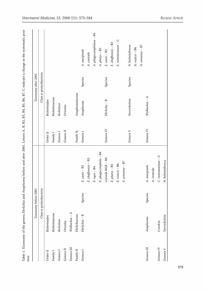
Bacteria from the genus Anaplasma belong to the Procaryota kingdom, the family Anaplasmataceae. The present classification has been valid since 2001 (Table 1), when many independent research-ers reorganized the families Rickettsiaceae and Ehrlichiaceae (Dumler et al., 2001).
Anaplasma belong to obligate intracellular mi-croorganisms, Gram-negative bacteria, living in the blood cells of mammals. Vertebrates can be their reservoir, i.e. an environment where the pathogen can live and proliferate for many years. However, in many cases bacteria from the genus Anaplasma cause diseases in domestic animals and people.
An essential part in the life cycle of the bacteria is played by vectors, organisms which contribute to their circulation in the environment. The main vec-tors of the Anaplasma bacteria are ticks, common arachnida occurring everywhere in the world, espe-cially the genera Ixodes, Dermacentor, Rhipicephalus and Amblyomma. Numerous research results have confirmed that a vector for anaplasma in Europe is the universally occurring tick I. ricinus, which can also be found in the African part of the Mediterranean Sea Basin (Ogden et al., 1998; Liz et al., 2000; Walker et al., 2001; Skotarczak et al., 2003). In the south of Europe, another vector of anaplasma is R. sanguineus (Sainz et al., 1999), and on the eastern borders of the continent and in Asia I. persulcatus. R. sanguineus, or the Brown dog tick, appears universally outside the European continent as well, i.e. in regions of Northern Africa, India, and in the territory of the United States (Stafford III, 2007).
Outside Europe, the most frequently mentioned vectors of the Anaplasma bacteria are the ticks I. scapularis (Black-legged tick) and I. pacyficus (Western black-legged tick). These species occur mostly in the United States of America and Canada (Parola and Raoult, 2001; Stafford III, 2007). In North and Central America, bacteria are also trans-mitted by ticks from the species A. americanum – known as the Lone star tick (Parola and Raoult, 2001; Stafford III, 2007).
Other vectors of anaplasma are the species from the genus Haemaphysalis, such as H. leporispalus-tris, H. lagrangei and H. longicornis, which are com-
mon in Japan, India, Russia, China, Korea, Thailand and in North America (Goethert and Telford III, 2003; Parola et al., 2003; Kawahara et al., 2006; Kim et al., 2006).
2. Taxonomical position of bacteria from the genus Anaplasma
In 2001, significant reorganizational changes were carried out within the order Rickettsiales. As a re-sult of this reorganization, the family Ehrlichiaceae was replaced by the family Anaplasmataceae, and some changes were made in the classification of the bacteria within the genera (Table 1) (Dumler et al., 2001).
The genera Anaplasma, Ehrlichia, Neorickettsia and Wolbachia include obligate intracellular bac-teria, parasitizing in the vacuoles of cells in eu-karytic hosts. Their systematic classification was performed on the basis of morphological, eco-logical, epidemiological and clinical characteri-zation. However, the development of molecular methods over recent years has enabled the partial recognition of the genome of these bacteria and consequently the partial change in their system-atic position. A comparison was performed for all sequences of conservative genes 16S rDNA (the gene coding 16S rRNA for the small subunit of the ribosome) and groESL (the gene coding the thermal shock protein), for those deposited in the GenBank up to the year 2000 (Dumler et al., 2001). A phylogenetic analysis was then carried out. The independent comparative analysis of the sequence of both genes gave similar results. Dendrograms of the sequences of the genes 16S rRNA and groESL have a similar arrangement, and bacteria are posi-tioned in clades similarly on both independent phy-logenetic trees, a further proof of the correctness of the research performed. Their accuracy is also shown by the high bootstrap obtained in both cases. Moreover, the new systematical division, emerging from the relationship analyses, is confirmed by the biological and antigenic characterization of each species (Dumler et al., 2001). Details concerning the changes in taxonomy in these groups of the bacteria are presented in Table 1.
Animal pathogens were attributed to the genus Anaplasma, such as A. centrale, A. marginale, A. platys, A. ovis and A. bovis, and also the aetio-logical factor of human anaplasmosis, A. phagocy-tophilum (Table 1).
Veterinarni Medicina, 53, 2008 (11): 573–584 Review Article
575
Tabl
e 1.
Tax
onom
y of
the
gene
ra E
hrlic
hia
and
Ana
plas
ma
befo
re a
nd a
fter
200
1. L
ette
rs A
, B, B
2, B
3, B
4, B
5, B
6, B
7, C
indi
cate
a c
hang
e in
the
syst
emat
ic p
osi-
tion
Taxo
nom
y be
fore
200
1Ta
xono
my
afte
r 200
1
Cla
ss α
-pro
teob
acte
ria
Cla
ss α
-pro
teob
acte
ria
Ord
er II
Rick
etts
iale
sO
rder
IIRi
cket
tsia
les
Fam
ily I
Rick
etts
iace
aeFa
mily
IRi
cket
tsia
ceae
Gen
era
IRi
cket
tsia
Gen
era
IRi
cket
tsia
Gen
era
IIO
rien
tiaG
ener
a II
Ori
entia
Gen
era
III
Wol
bach
ia –
A
Fam
ily II
Ehrl
ichi
acea
eFa
mily
II.
Ana
plas
mat
acea
e
Gen
era
IEh
rlic
hia
– B
Spec
ies
E. c
anis
– B
2G
ener
a I
Ana
plas
ma
Spec
ies
A. m
argi
nale
E. ch
affee
nsis
– B
3A
. cen
tral
e
E. e
qui –
B4
A. p
hago
cyto
philu
m –
B4
E. p
hago
cyto
phila
– B
4A
. pla
tys –
B5
czyn
nik
HG
E –
B4G
ener
a IV
Ehrl
icha
– B
Spec
ies
E. c
anis
– B
2
E. p
laty
s – B
5E.
cha
ffeen
sis –
B3
E. ri
stic
ii –
B6E.
rum
inan
tium
– C
E. se
nnet
su –
B7
Gen
era
VN
eori
cket
tsia
Spec
ies
N. h
elm
inth
oeca
N. r
istic
ii –
B6
N. s
enne
tsu
– B7
Gen
era
III
Ana
plas
ma
Spec
ies
A. m
argi
nale
Gen
era
VI
Wol
bach
ia –
A
A. c
entr
ale
Gen
era
IVC
owdr
iaC
. rum
inan
tium
– C
Gen
era
VN
eori
cket
tsia
N. h
elm
inth
oeca

Review Article Veterinarni Medicina, 53, 2008 (11): 573–584
576
3. History of Anaplasma discovery
Cases of tick fever occurring in goats, sheep and calves have been reported in Europe since 1780. The aetiological factor of this disease was not known but the description of symptoms corresponds well to the modern characterizations of anaplasmosis. Later reports frequently described the occurrence of tick fever in farm animals, mostly in small rumi-nants, and in dogs as well. Sometimes the disease led to the death of the animals, however its aetiol-ogy was not known yet. 150 years later, the first report was released that the disease in question was caused by a small bacterium E. phagocytophila (presently known as A. phagocytophilum) display-ing tropism to granulocytes (Jenkins et al., 2001).
The first reports on A. marginale appeared as early as in 1894, when Salmon and Smith detected the presence of inclusion in the cells of calf eryth-rocytes. The first full description came from Sir Arnold Theiler, who observed bacteria in eryth-rocytes of South African cattle in 1910 (Kocan et al., 2003). He discriminated two subspecies: the one more frequently creating “marginal points” in erythrocytes of calves (A. marginale) and the oth-er more frequently creating concentrations in the central part of blood cells, hence the name A. cen-trale. The latter of the subspecies was found to be less pathogenic to domestic animals (Kocan et al., 2003). The following years saw the discoveries of other species of anaplasmosis, pathogenic to ani-mals (Table 2), such as A. ovis, A. platys (formerly
known as E. platys) and A. bovis (Harvey et al., 1978; Kuttler, 1984).
After some research, it was shown that anaplas-mosis may affect humans; the first case of human HGA was observed in 1994 in the United States. It was caused by A. phagocytophilum (an agent of human granulocytic anaplasmosis, at first de-scribed as an agent of granulocytic ehrlichiosis) (Chen et al., 1994; Dumler et al., 2001). In 1996 this disease was also diagnosed in Europe, in Slovenia (Petrovec et al., 1997) and reported in Poland in 2001 (Tylewska-Wierzbanowska et al., 2001).
4. Characterization of bacterium from the genus Anaplasma, pathogenic to farm animals and people
4.1. Anaplasma marginale
A. marginale are obligate intracellular bacteria parasitizing in erythrocytes of higher vertebrates, mostly ruminants (Table 2). It causes so called bovine anaplasmosis which has been described in domestic and wild animals, i.e. in calves, water buf-falo, bison, African antelopes and the mule deer (Kocan et al., 2003). In infected animals, red blood cells were observed to have from 4 to 8 bacterial inclusions (Barbet et al., 1999; Kocan et al., 2004). The time of incubation of the disease depends on the degree of infection and can last from 7 to 60 days, on average approximately 28 days. During
Table 2. The characteristic of pathogens of genus Anaplasma
Aetiological agentDisease Vector Infected organism
or host Infected cellbefore 2001 after 2001
Ehrlichia bovis Anaplasma bovis bovine anaplasmosis
Haemaphysalis sp. Rhipicephalus sp. Amblyomma sp.
ruminants farming, small mammals monocytes
Anaplasma ovis Anaplasma ovis bovine anaplasmosis Dermacentor sp. small ruminants
(sheep, goats) erythrocytes
Anaplasma marginale Anaplasma marginale bovine anaplasmosis
Ixodes sp. Dermacentor sp. ruminants farming erythrocytes
Anaplasma centrale Anaplasma centrale bovine anaplasmosis
Ixodes sp. Haemaphysalis sp. ruminants farming erythrocytes
E. equi E. phagocytophilaCzynnik HGE
Anaplasma phagocy-tophilum (HGA agent)
human and animal granulocytic anaplasmosis
Ixodes sp. Dermacentor sp.
small ruminants forming and wild,
horses, dogs, humans
granulocyte
E. platys Anaplasma platys canine cyclic thrombocytopenia
Rhipicephalus sanguineus dogs platelets
Veterinarni Medicina, 53, 2008 (11): 573–584 Review Article
577
that time, the number of infected blood cells in-creases geometrically and in the acute phase of the disease 70% or more erythrocytes can be infected. During infection, cattle erythrocytes are phago-cyted by reticuloendothelial cells, which leads to anaemia and icterus without haemoglobinaemia and haemoglobinuria. Moreover, a clinical pic-ture of the disease also includes fever, weight loss, abortions and lethargy. Animals older than two years may even die due to the infection. Calves that experience the acute phase of the disease are permanently infected with the cyclic low-level rickettsemia or become carriers of A. marginale, and consequently in both cases they are reservoirs for the bacteria (Barbet et al., 1999; Kocan et al., 2003).
A. marginale is a tick-borne pathogen whose presence is mainly connected with tropical or subtropical regions, although these bacteria are detected more and more frequently in other re-gions of the world, e.g. Europe (Sicily, Hungary and Spain), Australia, South Africa (De Waal, 2000; de la Fuente et al., 2004, 2005; Kocan et al., 2004; Naranjo et al., 2006; Hornok et al., 2007).
From an economic point of view, A. marginale plays an important role in the world economy. De Waal (2000) revealed that more than 99% of the total cattle population in South Africa was at risk. Rajput et al. (2005) informed us that in Pakistan, Anaplasma parasites in buffalo and cat-tle Bubalus bubalis were 30% and 52%, respective-ly. It is estimated that in the United States alone, losses incurred by A. marginale may reach ap-prox. 300 million dollars a year (McCallon, 1973). Losses in Latin America reach over 800 million dol-lars a year (Lonibardo, 1976). Such great losses are also recorded in Africa, where cattle are often the only means of income. This has bred the idea of ge-netically modifying the cattle to achieve an inborn resistance to A. marginale (Melendez, 2000).
4.2. Anaplasma centrale
A. centrale is a parasite in the erythrocytes of ruminants, mainly cattle (Table 2). As opposed to A. marginale, it creates concentrations in the central part of the cell. This bacterium is widely-spread throughout the world. In spite of morpho-logical differences, occurrence and virulence, A. centrale is closely related to A. marginale. A. cen-trale is the cause of mild anaemia in most cases
of cattle infection (Kuttler, 1984; Kocan et al., 2003; Rajput et al., 2005). According to Theiler, A. cent-rale is less pathogenic to cattle than A. marginale but, most importantly, occasionally gives resistance against the latter. Hence it is used for the preparation of live vaccine strains, assuring immunological pro-tection against bovine anaplasmosis. Such vaccines are produced in Africa, Australia, Latin America and Israel (Kocan et al., 2003).
4.3. Anaplasma bovis
A. bovis is a bacterium detected mainly in cattle, but also observed in small mammals which are prob-ably a reservoir of this bacterium (Table 2) (Goethert and Telford III, 2003). It occurs in monocytes, and the disease it causes is called monocytic anaplasmo-sis. The symptoms of the disease are most visible in calves, but also in adult animals; symptoms include weakening of the body, marked reduction in weight, elevated temperature, enlargement of prescapular lymph nodes, paling of the mucous membranes, and in many cases an elevated amount of secreted mucus (Uilenberg, 1997; Santos and Carvalho, 2006).
A. bovis has been detected in Brazil, North America, Africa and Japan (Goethert and Telford III, 2003; Kawahara et al., 2006; Santos and Carvalho, 2006).
4.4. Anaplasma ovis
A. ovis mainly parasitizes small ruminants, i.e. sheep and goats, and its presence has been confirmed in most regions of the world both in farm and wild animals (Kuttler, 1984). Similarly to A. marginale and A. centrale, these bacteria live in erythrocytes, hence the anaemia of the infected animals (Splitter et al., 1956). In the case of A. ovis, bacterial inclu-sions are found 35–40% of the time in the central or submarginal part of the erythrocyte of the host, and the remaining 60–65% of the time in the marginal part (Shompole et al., 1989).
Experimental research shows that animals expe-riencing an acute phase of the disease display de-pression, debility, marked decline in body weight, fever and progressive anaemia whose consequence is among other things a reduction in milk production. The infection is also potentially lethal. The symp-toms of the disease depend on age, on the general condition of the animals and their breed (Splitter et al., 1955; Shompole et al., 1989).
Review Article Veterinarni Medicina, 53, 2008 (11): 573–584
578
Clinical symptoms occurring in animals result-ing from the presence of A. ovis in blood cause huge losses in farming stock. This has considerable significance for poorer countries in tropical and subtropical regions where animal stocks consist mainly of sheep and goats. They are the basic and frequently only producers of milk and meat, and also provide farms with natural manure used in agriculture (Jensen, 1955). Hence the losses A. ovis causes in the economy, especially in those regions, are significant and create a need for further re-search concerning the spreading and pathogenicity of A. ovis and possibilities for prophylaxis (Kuttler, 1984).
Contemporary research conducted by various scientific centres shows that A. ovis is a problem not only in poorer countries. These bacteria have been detected in the U.S.A., Italy and Hungary (de la Fuente et al., 2002, 2005; Hornok et al., 2007). The occurrence of A. ovis is not widespread there, but one ought to allow for the fact that a return to tradi-tional methods of farming (pasturage on meadows) can facilitate the expansion of this pathogen. The spread of this bacterium may also be enhanced by modern climatic changes.
4.5. Anaplasma platys
For the first time (1978), A. platys was detected in the U.S.A. (Florida; Harvey et al., 1978). It was also detected in Central America (Venezuela), Asia (Korea, Japan, China, Taiwan and Thailand), Australia and Africa. In Europe, the presence of A. platys was confirmed around the Mediterranean Basin, in France, Greece, Spain and Italy (Sainz et al., 1999; Suksawat et al., 2001; Sparagano et al., 2003; de la Fuente et al., 2006; Kawahara et al., 2006; Kim et al., 2006).
A. platys is mainly a pathogen of canines, usu-ally dogs. It lives in blood cells, causing Canine Cyclic Thrombocytopenia (CCT) (Table 2). Clinical symptoms of this disease vary depending on the species of the dog, but fundamentally CCT causes weakening of the animal, lethargy, anorexia, res-piratory distress, fever, increased mucous secre-tion, purulent ocular discharge, splenomegaly and muzzle hyperkeratosis (Sainz et al., 1999). These bacteria do not cause death, and treatment with suitable antibiotics is usually successful after four weeks. Doxycycline is one of such effective antibi-otic (Sainz et al., 1999).
4.6. Anaplasma phagocytophilum
A. phagocytophilum is a frequently described bacterium from the genus Anaplasma. It has been detected in domestic animals, mostly small rumi-nants, but also in people (Table 2). These bacteria are an aetiological factor of granulocytic anaplas-mosis which in the case of human infections is de-fined as Human Granulocytic Anaplasmosis – HGA (Human Anaplasmosis – HA).
Human anaplasmosis is a multisystemic disease, difficult to diagnose due to unspecific symptoms which are similar to the common flu. It may have mild or heavy symptoms, depending on the pa-tient’s condition. One estimate states that about 40% of those infected require hospitalization. Early detected HGA is curable. In Europe no deadly cases have been reported, and in the U.S.A. the mortal-ity rate was 7–10% (Fishbein et al., 1994; Dumler and Bakken, 1998; Blanco and Oteo, 2002; Dumler and Broqui, 2004). Although anaplasmosis alone is not perhaps a life-threatening disease, frequent complications such as renal failure, intravascular coagulation, cardiomegaly, seizures or coma, may all be fatal. These involved mostly the elderly, who became ill after receiving immunosuppressive med-icines, and children (Fishbein et al., 1994; Dumler and Bakken, 1998).
In the case of farm animals anaplasmosis caused by A. phagocytophilum is a disease with subclinical or heavy symptoms. It has unspecific symptoms, such as fever, drowsiness, anorexia, abortions, drop in body weight and reduction in milk production. Left unattended, especially in weaker individuals, it can lead to death (Stuen et al., 2002; Garcia-Perez et al., 2003).
5. Vectors of bacteria from the genus Anaplasma
5.1. Morphology and behaviour of questing ticks
In epidemiology, a vector is an organism which contributes to the spreading of a pathogen in a pop-ulation of hosts. Among saprophytes, it is ticks that play the greatest part in the epidemiology of disease transmission. Anaplasmosis is a typical tick-borne zoonosis, in which ticks are the vectors, i.e. they transfer bacteria to the blood of the higher verte-brates on which they feed.

Veterinarni Medicina, 53, 2008 (11): 573–584 Review Article
579
The world fauna of ticks, representative of arach-nida (phylum Arachnida), includes about 860 spe-cies divided into four families. All ticks are obligate, blood-sucking arthropods (Arthropoda), belonging to the subdivision Acari (mites) (Siuda, 1991). All of them parasitize vertebrate animals. Their con-tact with hosts is usually temporary, restricted in most species to only receiving nourishment, but their medical, veterinary and economic impact is immense. Two families of proper ticks especially – Ixodidae and Amblyommidae – have an impact on the economy and medicine. Parasitizing ticks cause immediate results, such as damage to the host’s skin or local inflammations; and more importantly being reservoirs of viruses, bacteria or protozoans, ticks may transfer pathogens to people and animals (Siuda, 1991).
Ticks do not have a segmentation characteristic of most other arachnids. Their bodies are oval, nar-rowing towards the front, with the gnathosoma em-bedded on their idiosoma. The gnathosoma made from mouth organs is also the main adhesive organ. The idiosoma, from the dorsal side, is coated with so-called scutum, which in males covers almost all the dorsum, and in females only the forepart. The remaining part of the dorsal surface of the idiosoma is covered by an extensible epidermis, so called alloscutum, which enables the extraction of large amounts of blood by females, nymphs and larvae. In males, the epidermis covers only the side surfaces of the idiosoma. The size of the scutum is a means of differentiation between male and female ticks. On the abdominal side, there are four pairs of limbs. On the feet of the first pair, on the dorsal side, the Haller’s organ can be noticed. It is the most impor-tant sensory organ of ticks, enabling the search for a host (Siuda, 1991; Buczek and Magdon, 1999). Some species, e.g. from the genera Amblyomma and Rhipicephalus, have eyes, while other ticks have photosensitive cells which react to light intensity changes (Buczek and Magdon, 1999).
Some species of ticks actively seek hosts (e.g. I. scapularis, I. pacyficus or A. americanum), others assume the wait-and-see attitude (e.g. I. ricinus). They cling to plants, and when a potential host ap-proaches, they raise up their first pair of legs and hook onto the host (hair, clothes, etc.) with small claws (Buczek and Magdon, 1999).
The tick’s reaction to a host depends on several factors. Research has shown that a major factor influencing the behaviour of questing individuals is an increased concentration of CO2 in the air, i.e.
exhalations from a host. Changes in the concen-tration of carbon dioxide in the air, registered by ticks, make them adopt an expectant position (in passive species) or move towards the source of the gas (in active species). The scent of a host and its body temperature can also significantly influence the tick’s choice. Other impulses that activate ticks are pheromones, urine, sounds given off by animals or an environmental factor – light intensity (Buczek and Magdon, 1999). Stimulating factors vary and characterize different species of ticks.
The behaviour of ticks may also be influenced by seasonal and diurnal rhythms characteristic of local populations. Seasonal activity is conditioned by ambient temperature, and diurnal activity by potential host activity, ambient temperature and humidity.
5.2. Vectors of bacteria from the genus Anaplasma in Europe
The most widespread European tick is the com-mon tick I. ricinus, a vector for bacteria from the ge-nus Anaplasma. Sometimes I. ricinus is also called the European sheep tick (Parola and Raoult, 2001). This common tick is a blood-sucking arachnid, belonging to the order Ixodida, the genus Ixodes (Siuda, 1991). Its parasitizing activity on verte-brates, including humans, causes many pathologi-cal alterations, sometimes even life-threatening for its hosts (swelling, inflammatory disorders of skin, allergy, paralysis). I. ricinus belongs to extranida-mental ectoparasites (which feed on transmigrant animals in the open environment), with a three-host life cycle (each active developmental phase of the tick has an independent host) (Siuda 1991).
The life cycle of the tick I. ricinus includes the following phases: larva, nymph and adult (imago). This species is characterized by seasonality, or in other words dependence of the activity and course of each phase of the life cycle on the seasons, char-acteristic of a temperate climate. The life cycle of the common tick in temperate climates lasts several years, most frequently approximately three years, but sometimes under favourable environmental conditions even longer (Siuda, 1991).
In the annual cycle, ticks show seasonal activity which depends on their geographical location. In temperate climates it starts from mid-April (some-times earlier, even in March) and lasts to the be-ginning of November with two peaks – the first

Review Article Veterinarni Medicina, 53, 2008 (11): 573–584
580
from May to mid-June, the second in September. After hot summers, it is possible that the autum-nal peak will not occur. It is accepted that mature forms can be active already at temperatures of 16°C, with nymphs active at temperatures of 25°C. The embryonic period lasts, depending on the ambi-ent temperature, from 24 to 50 days (Siuda, 1991; Skotarczak et al., 1999).
Besides seasonal activity, ticks are also observed by diurnal activity. It is connected, among other things, with local environmental conditions, such as temperature and humidity, and with the host activity. Species of ticks parasitizing on animals are characterized by diurnal activity, and will feed most intensely in these hours. I. ricinus is the most active in morning hours (about 8) and in the after-noon and at night (15–24), but can attack at other times of the day as well (Siuda, 1991; Buczek and Magdon, 1999).
Among European ticks from the genus Ixodes, the taiga tick – I. persulcatus, is also known as a vector for anaplasma (Figure 1). The geographical range of this species covers north-eastern Europe, areas of the taiga and forested steppes. Similarly to I. ricinus, the taiga tick is an extranidamental parasite, i.e. it has three hosts in its lifetime. Also like the common tick, it is characterized by a lack of host specificity (Siuda, 1991). The life cycle of I. persulcatus can last from two to five years.
R. sanguineus, the dog tick (Brown dog tick), be-longs to cosmopolitan ticks (the detailed character-
istics of R. sanguineus in chapter 5.3). Its presence has been established in all regions of Africa, India, in the United States and around the Mediterranean Basin (Parola and Raoult, 2001; Stafford III, 2007). In Europe, the occurrence of another tick from the genus Rhipicephalus was observed in Spain, namely R. bursa, which is also probably a vector for patho-gens from the genus Anaplasma (de la Fuente et al., 2004).
5.3. Vectors of bacteria from the genus Anaplasma outside Europe
The most frequently mentioned pathogen vector outside Europe is the tick I. scapularis (Black-legged tick), which is also called a deer tick in some regions. I. scapularis can be found in the eastern and south-eastern United States and Canada (Figure 1). The biotope of this species is forests with a rich forest floor. I. scapularis hosts include mainly deer, dogs, small birds and rodents, but also people. Males of this tick reach a length of 2–2.7 mm, females are larger, approximately 3–3.7 mm. The imago has a red-brown colour, with younger ones being brighter. I. scapularis prefers a deciduous forest and a habitat containing litter (Stafford III, 2007).
I. scapularis is a species with the two-host devel-opment cycle. Juvenile phases, larvae and nymphs feed on one host and adult forms on the other. A female feeds for about 3–7 days, then falls off and
Figure 1. The world distribution of vectors of bacteria from the genus Anaplasma

Veterinarni Medicina, 53, 2008 (11): 573–584 Review Article
581
oviposits in the litter, laying 2 000 or more eggs. Larvae hatch from eggs on the ground, and then at-tach to the first host. After feeding, the larva moults into a nymph which feeds on the same host. Only after filling itself with blood does the nymph fall from the host and transform into an adult on the ground. The imago actively seeks a second host, where it feeds (Stafford III, 2007). Another species of ticks which is physically similar to I. scapularis is I. pacificus, occurring mostly in the west of the United States of America (Pacific US and British Columbia). I. pacificus, also called the Western black-legged tick, is found mostly on the edges of forests, coppices or in open spaces, such as mead-ows and pastures. Its hosts are people, deer, dogs, coyotes, small rodents, wild hogs, horses and liz-ards (Stafford III, 2007).
A. americanum (Lone Star Tick) is also considered a vector for bacteria from the genus Anaplasma. It appears in regions of the southeastern United States, and also in some regions of Central America. Its biotope is wooded areas with brush, especially hickory-oak forests, along creeks and rivers (Parola and Raoult, 2001). The colour of the A. americanum imago varies from brown to tan, and in females the dorsum has a characteristic silvery white spot, so-called “lone star”. Males of the A. americanum have some small spots or streaks around the perimeter of the body. The average size of an unfed imago ranges from 3 to 4 mm. All developmental phases can feed on humans, and both small and large mam-mals as well. Juvenile phases have been observed mainly on small birds and rodents. A female of A. americanum lays up to 3 000 eggs (Parola and Raoult, 2001; Stafford III, 2007).
An essential role in the transmission of Anaplasma is played by ticks from the genus Dermacentor sp., most of all D. variabilis (American dog tick). This tick, also called the wood tick, occurs in grassy meadows, wooded areas, near barns, kennels, and anywhere the animals are kept. The imago of D. variabilis is dark brown. Adult and juvenile forms feed mostly on small rodents, opossums, raccoons, horses, deer and cattle. The female of this tick attains 13 mm in length after feeding. It feeds for about 10–12 days, and after falling off it can lay from 3 000 to 7 000 eggs. The life cycle lasts about two years. Among ticks from the genus Dermacentor, D. andersoni (Rocky Mountain wood tick) and D. aurutus are included. D. andersoni is also a vector of anaplasma, living outdoors in low vegetation. This tick is brown, and the scutum is
grey. D. andersoni hosts are rodents, sheep, cattle, deer and humans. The ticks D. andersoni and D. au-rutus occur in the United States and Asia (Parola and Raoult, 2001; Parola et al., 2003; Stafford III, 2007).
Both within and outside Europe, one of the com-mon ticks as a vector for Anaplasma is the dog tick R. sanguineus (Brown dog tick, or interchange-ably Red dog tick). These ticks are characterized by their elongated body without ornamentation, with a reddish-brown colour. These ticks occur most frequently in houses, kennels and dog runs (Parola and Raoult, 2001). R. sanguineus has a three-host developmental cycle, but contrary to the aforementioned species from the genus Ixodes, each stage of R. sanguineus has a high specificity to one host – the dog, while humans are only an accidental host. The life cycle of R. sanguineus may last 4–5 months. The female lays a minimum of 4 000 eggs. Humans are parasitized by R. sangineus extremely seldom, and only when there is no other host in its environment. This tick occurs most of all in Northern Africa, India, United States and Europe, mainly around the Mediterranean Basin (Figure 1) (Stafford III, 2007).
Over recent years, some reports have shown that ticks from the genus Haemaphysalis also play an important part in the transmission of anaplasma. Most frequently mentioned are the species H. lep-orispalustris (A. bovis) and H. longicornis (A. bo-vis, A. centrale, A. phagocytophilum). These ticks have two-host life cycles, and the length of the life cycle does not exceed one year. Ticks from the ge-nus Haemaphysalis occur mostly in Japan, Russia (Far East), China, Koreas, Northern America and southeastern Australia (Parola and Raoult, 2001; Goethert and Telford III, 2003; Kawahara et al., 2006; Kim et al., 2003, 2006).
The tick I. ricinus, a vector for Anaplasma, has been reported in Northern Africa, in around Morocco and Tunisia (Parola and Raoult, 2001; Sarih et al., 2005).
The recent increasing popularity of recreation in forested areas or in the vicinity of forests raises the risk of contact with ectoparasites, for example with various species of ticks. Such grounds are typical biotopes of hard ticks. The end of the 20th century also brought about changes in the manner of ani-mal breeding. In many industrialized countries the number of large farms is decreasing, where animals used to be raised in a closed system. Traditional methods of breeding animals are more popular and

Review Article Veterinarni Medicina, 53, 2008 (11): 573–584
582
natural pastures are more and more popular for both large ruminants (cattle) and small ones (sheep, goats). Apart from changes in people’s lifestyles and farming techniques, we can also observe en-vironmental changes. Environmental factors such as climate changes, deforestation, an increase in the roe deer population, or the introduction of new animal species which are potential pathogen reservoirs and hosts of ticks, favour an increase in the number of various tick populations (Wielinga et al., 2006).
It is also important to remember that in the present world both the humans and the animals bred by them are subject not only to indigenous do-mestic pathogens but also to imported ones. Hence the diagnosis becomes more and more difficult and laborious. It seems that the development of mo-lecular techniques can facilitate the identification of these diseases, and information on the occur-rence of ticks and pathogens transmitted by them will help veterinary services diagnose diseased farm animals correctly.
REFERENCES
Barbet A.F., Blentlinger R.Y.J., Lundgren A.M., Blouin E.F., Kocan K.M. (1999): Comparison of surface pro-tein of Anaplasma marginale grown in tick cell cul-ture, tick salivary glands, and cattle. Infection and Immunity, 67, 102–107.
Blanco J.R., Oteo J.A. (2002): Human granulocytic ehr-lichiosis in Europe. Clinical Microbiology and Infec-tion, 8, 763–772.
Buczek A., Magdon T. (1999): Host location by ticks (Acari: Ixodida).Wiadomosci Parazytologiczne, 45, 3–12.
Chen S.M., Dumler J.S., Bakken J.S., Walker D.H. (1994): Identification of a granulocytotropic Ehrlichia species as the etiologic agent of human disease. Journal of Clinical Microbiology, 32, 589–595.
de la Fuente J., Garcia-Garcia J.C., Blouin E., Saliki J.T., Kocan K.M. (2002): Infection of tick cells and bovine erythrocytes with one genotype of the intracellular ehrlichia Anaplasma marginale excludes infection with other genotypes. Clinical and Diagnostic Labora-tory Immunology, 9, 658–668.
de la Fuente V.J., Hofle U., Ruiz-Fons F., Fernandez de Mera I.G., Van Den Bussche R.A., Kocan K.M., Gor-tazar C. (2004): Anaplasma infection in free-ranging Iberia red deer in the region of Castilla-La Mancha, Spain. Veterinary Microbiology, 100, 163–173.
de la Fuente J., Naranjo V., Ruiz-Fons F., Hofle U., Fern-andez de Mera I.G., Villanua D., Almazan C., Torina A., Caracappa S., Kocan K.M.., Gortazar C. (2005): Potential vertebrate hosts and invertebrate vectors of Anaplasma marginale and Anaplasma phagocy-tophilum in central Spain. Vector Borne and Zoonotic Diseases, 5, 390–401.
de la Fuente J., Torina A., Naranjo V., Nicosia S., Alongi A., Mantia F., Kocan K.M. (2006): Molecular charac-terization Anaplasma platys strains from dogs in Sici- ly, Italy. BMC Veterinary Research, 2, 24.Dumler J.S., Bakken J. (1998): Human ehrlichiosis: Newly recog-nized infections transmitted by ticks. Annual Review of Medicine, 49, 201–213.
De Waal D.T. (2000): Anaplasmosis control and diagnsis in South Africa. Annals of the New York Academy of Science, 916, 474–483.
Dumler J.S., Broqui P. (2004): Molecular diagnosis of human granulocytic anaplasmosis. Expert Review of Molecular Diagnostics, 4, 89–98.
Dumler J.S., Barbet A.F., Bekker C.P., Dasch G.A., Palmer G.H., Ray S.C., Rikihisia Y., Rurangirwa F.R. (2001): Reorganization of genera in the families Rickettsiaceae and Anaplasmataceae in the order Rickettsiales: uni-fication of some species of Ehrlichia with Anaplasma, Cowdria with Ehrlichia and Ehrlichia with Neorick-ettsia, descriptions of six new species combinations and designation of Ehrlichia equi and ’HGE agent’ as subjective synonyms of Ehrlichia phagocytophila. In-ternational Journal of Systematic and Evolutionary Microbiology, 51, 2145–2165.
Fishbein D.B., Dawson J.E., Robinson L.E. (1994): Human ehrlichiosis in the United States, 1985 to 1990. Annals of Internal Medicine, 120, 736–743.
Garcia-Perez A.L., Baradika J., Oporto B., Povedano I., Juste R.A. (2003): Anaplasma phagocytophila as an abortifacient agent in sheep farms from Northern Spain. Annals of the New York Academy of Science, 990, 429–432.
Goethert H.K., Telford III S.R. (2003): Enzootic transmis-sion of Anaplasma bovis in Nuntucket Cottontial Rab-bits. Journal of Clinical Microbiology, 41, 744–3747.
Harvey J.W., Simpson C.F., Gaskin J.M. (1978): Cyclic trombocytophenia induced by a rickettsiae-like agent in dogs. Journal of Infectious Diseases, 137, 182–188.
Hornok S., Elek V., de la Fuente J., Naranjo V., Farkas R., Majoros G., Foldvari G. (2007): First serological and molecular evidence on the endemicity of Anaplasma ovis and A. marginale in Hungary. Veterinary Micro-biology, 122, 316–322.
Jenkins A., Kristiansen B.E., Allum A.G., Aakre R.K., Strand L., Kleveland E.J., van de Pol I., Schouls L.

Veterinarni Medicina, 53, 2008 (11): 573–584 Review Article
583
(2001): Borrelia burgdorferi sensu lato and Ehrlichia spp. in ixodes ticks from southern Norway. Journal of Clinical Microbiology, 39, 3666–3671.
Jensen R. (1955): Suspected anaplasmosis in sheep. North American Veterinarian, 36, 117–118.
Kawahara M., Rikihisa Y., Lin Q., Isogo E., Tahara K., Itagaki A., Hiramitsu Y., Tajima T. (2006): Novel ge-netic variants of Anaplasma phagocytophilum, Ana-plasma bovis, Anaplasma central, and novel Ehrlichia sp. in wild deer and ticks on two Major Islands in Ja-pan. Applied and Environmental Microbiology, 72, 1102–1109.
Kim C.M., Kim M.S., Park M.S., Park J.H., Chae J.S. (2003): Identification of Ehrlichia chaffeensis, Ana-plasma phagocytophilum, and A. bovis in Heamaphys-alis longicornis and Ixodes persulcatus ticks from Korea. Vector Borne and Zoonotic Diseases, 3, 17–26.
Kim C.M., Yi Y.H., Yu D.H., Lee M.J., Cho M.R., Desai A.R., Shringi S., Klein T.A., Kim H.C., Song J.W., Baek L.J., Chong S.T., O’Guinn M.L., Lee J.S., Lee I.Y., Park J.H., Foley J., Chae J.S. (2006): Tick-borne Rickettsial patho-gens in ticks and small mammals in Korea. Applied and Environmental Microbiology, 72, 5766–5776.
Kocan K.M., de la Fuente J., Guglielmone A.A., Melen-dez R.D. (2003): Antigens and alternatives for control of Anaplasma marginale infection in cattle. Clinical Microbiology Reviews, 16, 698–712.
Kocan K.M., de la Fuente J., Blouin E.F., Garcia-Garcia J.C. (2004): Anaplasma marginale (Rickettsiales: An-aplasmataceae): recent advances in defining host-pathogen adaptations of a tick-borne Rickettsia. Parasitology, 129, 285–300.
Kuttler K.L. (1984): Anaplasma infection in wild and domestic ruminants: a review. Journal of Wildlife Dis-eases, 20, 12–20.
Liz J.S., Anderes L., Sumner J.W., Massung R.F., Gern L., Rutti B., Brossard M. (2000): PCR detection of granulocytic ehrlichiae in Ixodes ricinus ticks and wild small mammals in western Switzerland. Journal of Clinical Microbiology, 38, 1002–1007.
Lonibardo R.A. (1976): Socioeconomic importance of the tick problem in the Americas. PAHO Science Pub-lication, 316, 79.
McCallon B.R. (1973): Prevalence and economic aspects of anaplasmosis. In: Jones E.W. (ed): Proceedings of the Sixth National Anaplasmosis: Conference, March 19–20, 1973, Las Vegas, Nevada, 1–3.
Melendez R.D. (2000): Future perspective on veterinary hemoparasite research in the tropic at the start of this century. Annals of the New York Academy of Science, 916, 253–258.
Naranjo V., Ruiz-Fons F., Hofle U., Fernandez de Mera I.G., Villanua D., Almazan C., Torina A., Caracappa S., Kocan K.M., Gortazar C., de la Fuente J. (2006): Molecular epidemiology of human and bovine ana-plasmosis in southern Europe. Annals of the New York Academy of Science, 1078, 95–99.
Ogden N.H., Bown K., Horrocks B.K., Woldenhiwet Z., Bennett M. (1998): Granulocytic Ehrlichia infection in Ixodid ticks and mammals in woodlands and up-lands of the U.K. Medical and Veterinary Entomology, 12, 423–429.
Parola P., Raoult D. (2001): Ticks and tickborne bacterial diseases in Humans: an emerging infectious threat. Clinical Infectious Diseases, 32, 897–928.
Parola P., Cornet J.P., Sanogo Y.O., Miller R.S., Thien H.V., Gonzalez J.P., Raoult D., Telford III S.R., Wong-srichanalai C. (2003): Detection of Ehrlichia spp., Anaplasma spp., Rickettsia spp., and other Eubacteria in ticks from the Thai-Myanmar and Vietnam. Journal of Clinical Microbiology, 41, 1600–1608.
Petrovec M., Lotric-Furlan S., Zupanc T.A., Strle F., Brouqui P., Roux V., Dumler J.S.(1997): Human disease in Europe caused by a granulocytic Ehrlichia species. Journal of Clinical Microbiology, 35, 1556–1559.
Rajput Z.I., Hu S.H., Arijo A.G., Habib M., Khalid M. (2005): Comparative study of Anaplasma parasites in tick carrying buffaloes and cattle. Journal of Zhejiang University Science, 6B, 1057–1062.
Sainz A., Amusategui I., Tesouro M.A. (1999): Ehrlichia platys infection and disease in dogs in Spain. Journal of Veterinary Diagnostic Investigation, 11, 382–384.
Santos C.F., Carvalho C.B. (2006): First report of Ana-plasma bovis (Donatien and Lestoquard, 1936) Dum-ler et al. (2001) at micro region of Campos dos Goytacazes, State of Rio de Janeiro, Brazil. Revista Brasileira de Parasitologia Veterinaria, 15, 126–127.
Sarih M., M’Ghirbi Y., Bouattour A., Gern L., Baranton G., Postic D. (2005): Detection and identification of Ehrlichia spp. in ticks collected in Tunisia and Morocco. Journal of Clinical Microbiology, 43, 1127–1132.
Shompole S., Waghela S.D., Rurangirwa F.R., McGuire T. (1989): Cloned DNA probes identify Anaplasma ovis in goats and reveal a high prevalence of infection. Journal of Clinical Microbiology, 27, 2730–2735.
Siuda K. (1991): Ticks (Acari: Ixodida) from Poland. Part I. PWN, Warszawa, Wroclaw. 8–274.
Skotarczak B., Soroka M., Wodecka B. (1999): The oc-currence of I. ricinus in the select recreative areas in the province of Szczecin. Part 1. Wiadomosci Parazy-tologiczne, 45, 507–517.
Skotarczak B., Rymaszewska A., Wodecka B., Sawczuk M. (2003): Molecular evidence of coinfection of Bor-

Review Article Veterinarni Medicina, 53, 2008 (11): 573–584
584
relia burgdorferi sensu lato, human granulocytic ehr-lichiosis agent, and Babesia microti in ticks from North-Western Poland. Journal of Parasitology, 89, 194–196.
Sparagano O.A.E., de Vos A., Paoletti B., Camma C., de Santis P., Otranto D., Giangaspero A. (2003): Molecu-lar detection of Anaplasma platys in dogs using polymerase chain reaction and reverse line blot hy-bridization. Journal of Veterinary Diagnostic Investi-gation, 15, 527–534.
Splitter E.J., Twiehaus M.J., Castro E.R. (1955): Anaplas-mosis in sheep in the United State. Journal of the Amer-ican Veterinary Medical Association, 127, 244–245.
Splitter E.J., Anthony H.D., Twiehaus M.J. (1956): Ana-plasma ovis in the United States: experimental studies with sheep and goats. American Journal of Veterinary Research, 17, 487–491.
Stafford III K.C. (2007): Tick management handbook. In: The Connecticut Agricultural Experiment Station, Bulletin No.1010. EPS Printing II, LLC, South Wind-sor, Connecticut. 9–18.
Stuen S., Bergstrom K., Palmer E. (2002): Reduced weight gain due to subclinical Anaplasma phagocytophilum (formerly Ehrlichia phagocytophila) infection. Ex-perimental and Applied Acarology, 28, 209–215.
Stuen S., Nevland S., Moum T. (2003): Fatal cases of tick-borne fever (TBF) in sheep caused by several 16S rRNA gene variants of Anaplasma phagocytophilum. Annals of the New York Academy of Science, 990, 433–434.
Suksawat J., Pitulle C., Arraga-Alvarado C., Madrigal K., Hancock S.I., Breitschwerdt E.B. (2001): Coinfection with tree Ehrlichia species in dogs from Thailand and Venezuela with emphasis on consideration of 16S ri-bosomal DNA secondary structure. Journal of Clinical Microbiology, 39, 90–93.
Tylewska-Wierzbanowska S., Chmielewski T., Kondrusik M., Hermanowska-Szpakowicz T., Sawicki W., Sulek K. (2001): First cases of acute human granulocytic ehrlichiosis in Poland. European Journal of Clinical Microbiology & Infectious Diseases, 20, 196–198.
Uilenberg G. (1997): General review of tick-borne dis-eases of sheep and goats word-wide. Parasitology, 39, 161–165.
Walker A.R., Alberdi M.P., Urquhart K.A. (2001): Risk factors in habitats of the ticks Ixodes ricinus in fluenc-ing human exposure to Ehrlichia phagocytophila bac-teria. Medical and Veterinary Entomology, 15, 40–49.
Wielinga P.R., Gaasenbeek C., Fonville M., de Boer A., de Vries A., Dimmers W., Jagers G.A.O., Schouls L.M., Borgsteede F., van der Giessen J.W.B. (2006): Longi-tudinal analysis of tick densities and Borrelia, Ana-plasma, and Ehrlichia infections of Ixodes ricinus ticks in different habitat areas in the Netherlands. Applied and Environmental Microbiology, 72, 7594–7601.
Received: 2008–07–24Accepted after corrections: 2008–11–12
Corresponding Author:
Anna Rymaszewska, University of Szczecin, Department of Genetics, al. Piastow 40B, 71-065 Szczecin, PolandTel. +48 91 444 2782, e-mail: [email protected]