Embed Size (px)
Citation preview

Back To Chiropractic CE Seminars
MRI ~ 6 Hours
Welcome to Back To Chiropractic Online CE exams:
This course counts toward your California Board of Chiropractic Examiners CE. (also accepted in other states, check our website or with your Chiropractic State Board)
The California Board requires that you complete all of your CE hours BEFORE
the end of your Birthday month. We recommend that you send your chiropractic
license renewal form and fee in early to avoid any issues.
COPYRIGHT WARNING
The copyright law of the United States (Title 17, United States Code) governs the making of photocopies or other
reproductions of copyrighted material. Under certain conditions specified in the law, libraries and archives are authorized
to furnish a photocopy or other reproduction. One of these specified conditions is that the photocopy or reproduction is not
to be "used for any purpose other than private study, scholarship, or research." If a user makes a request for, or later uses,
a photocopy or reproduction for purposes in excess of "fair use," that user may be liable for copyright infringement. This
site reserves the right to refuse to accept a copying order if, in its judgment, fulfillment of the order would involve violation
of the copyright law.

Exam Process: Read all instructions before starting!
1. You must register/pay first. If you haven't, please return to: backtochiropractic.net
2. Open a new window or a new internet tab & drag it so it's side-by-side next to this page.
3. On the new window or new tab you just opened, go to: backtochiropractic.net website.
4. Go directly to the Online section. DON'T register again.
5. Click on the Exam for the course you want to take. No passwords needed.
6. Follow the Exam instructions.
7. Upon passing exam (70%), you’ll be able to immediately download your certificate,
and it’ll also be emailed to you. If you don’t pass, you must repeat the exam.
Please retain the certificate for 5 years. DON’T send it to the state board.
If you get audited and lose your records, I’ll have a copy.
I’m always a phone call away... 707.972.0047 or email: [email protected]
Marcus Strutz, DC
Back To Chiropractic CE Seminars
33000 North Highway 1
Ft Bragg CA 95437

SPINAL MRI: WHERE DO I START?
JC Carter DC, DACBR4480-H S Cobb Dr #325Smyrna, GA 30080770-984-5395jcradiology.com

Thank You For Providing Many of the Images in This Presentation!
▪ Sunnyvale Imaging Center
568 South Mathilda Ave.
Sunnyvale, CA 94086
408.738.0232

References
▪ Musculoskeletal MRI, Helms et. Al., Saunders, 2001, Phil PA
▪ Magnetic Resonance Imaging in Ortopaedics and Sports Medicine, D Stoller, Lippincott, Williams and Wilkins, 2007, Baltimore, MD.
▪ Skeletal Imaging Atlas of the Spine and Extremities Taylor Hughes Resnick, Saunders Elsevier , 2010, Maryland Heights, MO.
▪ Diagnostic Imaaging Orthopedics, Stoller et. Al., Amirys, 2004, Salt Lake City, UT

How does MR work?
▪ Hydrogen ions (most prevalent in body) all spin creating a magnetic moment but do so randomly cancelling each other out
▪ When the body is near a strong magnetic field the H+ align with the magnetic field of the magnet
▪ The H+ are excited by adding energy in the form of a radio frequency (RF) using a surface coil
▪ The protons realign by “precessing” in unison
▪ Their chaining magnetic moment causes an electric current in the surface coil that is transferred back to the computer creating the image

Field Strength
▪ Magnetic strength measured in Tesla
▪ 0.3-0.5: low field strength
▪ 0.5-1.0: intermediate field strength
▪ 1.5-3.0: high field strength
▪ Above 4.0 eyes can’t see difference and creates too much heat in patient
▪ The higher the Tesla the prettier the image
▪ Small extremities should be obtained on high field magnets

MR Contra-indications
▪ Brain aneurysm clips
▪ Intra-ocular foreign bodies esp. metal
▪ Subcutaneous metal or shrapnel
▪ Pacemakers and some heart valves
▪ Neurotransmitters
▪ Cochlear implants
▪ Tatoos????
▪ Early pregnancy?

MR Terms
▪ High (bright) signal-white
▪ Intermediate signal-light gray
▪ Low signal-dark gray
▪ Signal void-black (air is a signal void on ALL imaging sequences)
▪ Hypo intense-darker than adjacent tissue
▪ Hyper intense-brighter than adjacent tissue
▪ Iso intense-same

MR Sequences
▪ T1
▪ T2
▪ PD
▪ Fat Suppressed
STIR
FS PD FSE

T1
▪ Fat- white
▪ Water-black
▪ Good anatomical detail

T2
▪ Fat- dark gray
▪ Water-white
▪ Gives physiologic information especially edema (bone or soft tissue)

T1 from T2
▪ Locate the CSF
▪ If black, it is T1
▪ If white, it is T2, STIR, or FS PD FSE
▪ If gray, it is likely PD

T1 T2

Proton Density (PD)
▪ Fat- light gray
▪ Water-medium gray
▪ Good for cartilage evaluation
▪ Not very common


Fat Suppressed Proton Density Fast Spin Echo (FS PD FSE)
▪ Fat- black
▪ Water-bright
▪ Good for bone marrow edema, synovial fluid, tendons, ligaments, and cartilage evaluation

▪ A: bone bruise
▪ B: MCL tear
▪ C: ACL tear

Short T1 Inversion Recovery (STIR)
▪ Fat- black
▪ Water-bright
▪ Takes longer to do than FS PD FSE
▪ Good for bone marrow edema, synovial fluid, tendons, ligaments, and cartilage evaluation

STIR
▪ Anything bright is fluid
▪ Great for evaluation of tumor (especially metastasis), fracture and bone bruising

The bright areas in the vertebrae represent tumor

Normal Lumbar Anatomy

T1 T2

Psoas
TP
Sacrum
Erector sp.

Find
▪ TP
▪ Erector spinae
▪ Psoas
▪ Sacrum

Pedicle
Nerve
Facet jnt

Find
▪ Pedicle
▪ Nerve
▪ Facet joint

Vena C
Disc
Nerve
Vessel
Facet
Liver
Gall Bl
R Kidney
Multifidii
Quadratus
Psoas
Aorta
Vena c


L4 N L4 lamina
Vena c

Find
▪ Spinal nerve
▪ Lamina
▪ IVC

Find
▪ Liver
▪ GB
▪ IVC
▪ Aorta
▪ Psoas
▪ Quadratus
▪ Multifidi
▪ Kidney

L5 N
Lig. flavum

Find
▪ L4 sup end plate
▪ L5 nerve
▪ L4 lamina

Super sp. lig
Inter sp lig
Basivertebral aa/vn

Find
▪ Basivertebral a/v
▪ S1 N
▪ SP
▪ Superspinous lig
▪ Interspinous lig

ALL
Conus Med
Normal disc

Find
▪ Conus
▪ Basivertebral a/v
▪ L5 N
▪ ALL
▪ Nucleus p

ES
Psoas
IVC
Aorta
Quad lumb
L3N

Find
▪ Psoas
▪ IVC
▪ Aorta
▪ Erector sp.
▪ Exiting spinal N

Inf L3 endplate
L3 Sp N
L4 N Rt

Annulus
Nucleus p
L3 Sp n
L4 N Rt
L3 Inf Facet

Find
▪ Annulus
▪ Nucleus p
▪ Exiting sp N
▪ Nerve root
▪ Superior facet
▪ Floating N roots

L3 Sp N
L4 N Rt
Lig FlavL3 Inf Facet
L4 Sup Facet

Find
▪ Exiting sp N
▪ N root
▪ Lig flavum

Lat recessL4 N
Iliac aa

Find
▪ Lateral recess
▪ Nerve
▪ Inferior facet
▪ Iliac aa
▪ Lig flavum

L5 disc
L5 body
L5 N

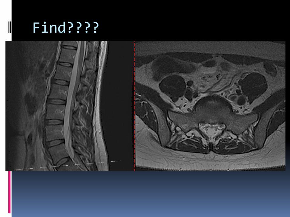
Find????

L5 discS1 N
Sacral facet
L5 inf fac

Find????

Glut Med
Lat recess
S1 N

Find????

SI
Find????

Glut max

Find????

Lumbar MRI Search Pattern
▪ What follows is my search pattern
▪ Examples of some of the things I’m looking for are provided as well

Lumbar Spine: Sagittal
▪ Alignment
Overall curve
ADI
Anterolisthesis
Retrolisthesis

L4 anterolisthesis
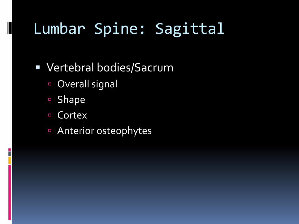
Lumbar Spine: Sagittal
▪ Vertebral bodies/Sacrum
Overall signal
Shape
Cortex
Anterior osteophytes

Hemangioma
▪ Typically decreased signal T1 and increased signal T2
▪ VERY common
▪ Rarely of any significance

Mets: Abnormal signal/shape and end-plate destruction

Anterior bulge/osteophyte

Lumbar Spine: Sagittal
▪ Posterior arch
Overall signal
Shape
IVF

Active pars defects: bright marrow on T2

Lumbar Spine: Sagittal
▪ Discs/End-plate
End-plate signal
Height
Signal
Contour
Posterior spurs
Schmorl’s nodes

Modic Changes
▪ Indicate progressive changes of degenerative disc disease as it affects the end-plate
▪ Not always seen but helpful when present

Endplate Changes
▪ Modic Type I
Decreased T1
Increased T2
Sign of acute degeneration
Invariably associated with PAINFUL DISCS
Spine 2003; 28: 715-20
Radiology 2001; 218: 420-7
Eur Spine J 1998; 7: 363-8
J Spine Discord 2000; 15: 438-43

Modic Type I

Endplate Changes
▪ Modic Type II
Increased T1
Isointense T2
Represents the endplate changing but is not yet visible on X-ray.
IT IS INVOLVED WITH CHANGE IN THE NUTRTION TO THE DISC.

Modic Type II

Endplate Changes
▪ Modic Type III
Decreased T1
Decreased T2
Sclerosis visible on X-ray
No active marrow
End-stage endplate change

Modic Type III

DJD Facts
▪ Scientific studies suggest that spondylosis deformans is the consequence of normal aging, whereas intervertebral osteochondrosis (AKA deteriorated disc), results from a clearly pathologic process with (or without) symptoms.
J Bone Jnt Surg 1962; 44: 243-68
Acta Ortop Scan 1985; 56: 496-99
Cin Orthop Rl Res 1987; 224: 97-104
Spine 2004; 4(6suppl): 167s-72s

Disc
▪ Intervertebral osteochondrosis
Loss of disc height
Vacuum phenomenon
Disc calcification
Posterior spur/osteocartilagenous ridge

PG/GAG: The Water Binders

Importance of Water in Cartilage▪ Although the tensile strength of the collagen
is that of steel wire, it cannot support compressive load since it would fold or crumble. It is the hydrostatic pressure of water bound to proteoglycans, retained and restrained by the collagen meshwork, that gives cartilage its resilience and load bearing properties.
Sem In Arth & Rheum 1984; 14(2): 110

GAGs and DDD
▪ In cadaveric spines, decreased T2 signal in degenerated discs was closely associated with glucoseaminoglycan (GAG) concentration rather than absolute water content.
New Orleans Ortho Rsrch Soc; 1994; 166-20

PGs and DDD
▪ The degenerated PG has a higher keratin sulfate to chondroitin sulfate ratio reducing the tensile strength and the disc becomes progressively more fibrous and disorganized.
Rehabil 1977; 16: 22-9
Orthop Clin N Am 1971; 2: 59-70
Arth Rheum 1981; 24: 12-21

Disc Bulge
▪ Physiologic
1-3mm
Due to compressive forces during course of a day
▪ Degenerative
Not a herniation
Can contribute to stenosis
Due to lack of water binding from decreased glucosaminoglycans (GAGs)

Disc Bulge and Dessication: loss of signal on T2

Annular Tears
▪ Typically on periphery of disc
▪ Contributes to DDD
▪ When in a disc herniation signifies it is recent

Annular Tear: localized bright signal in disc on T2

Significance of Annular Tears▪ Simply an incision of the annulus can produce
morphologic and functional changes in the adjacent nerves, such as increased capillaries and reduced nerve conduction velocities.
Spine 1996; 21: 2539-43

Lumbar Spine: Sagittal
▪ Spinal canal
End of cord
Contour
Signal

Tarlov Cysts
▪ Benign out-pouchings of dura through sacral foramina or against the sacrum
▪ Almost ALWAYS asymptomatic

Tarlov Cyst

Arachnoid (Tarlov) cysts/Sacral meningioceles (rarely symptomatic): follow water

Lumbar Spine: Sagittal
▪ Soft Tissues
Abdominal aorta
Pelvic cavity
Paravertebral muscles

Neurofibroma

Lumbar Spine: Transaxial
▪ Vertebral body
Signal
Shape

Pedicle/body destruction

Lumbar Spine: Transaxial
▪ Posterior arch
Signal
Shape
Articular facets

Synovial Cyst
▪ Due to increased hydrostatic pressure from facet OA
▪ Acts like a disc herniation
▪ Usually found incidentally on a patient sent in with a clinical suspicion of disc herniation

Synovial Cyst

Lumbar Spine: Transaxial
▪ Disc contour
Neural compression

Signs of Disc Herniation: Need 3 of 5 (consistent to same N level)
▪ Primarily leg pain
▪ Leg pain confined to dermatome
▪ Neural stretch tests recreate or exacerbate the leg pain
▪ At least 2 of 4 neurologic findings consistent with dermatome
Muscle weakness Decreased reflex
Abnormal pinwheel
Atrophy
▪ MR or CT correlating to dermatome

Disc Displacement
▪ PROTRUSION
Present if the width of the base is wider than the length of the posterior extension
Contained

Disc Displacement
▪ EXTRUSION
Present if the width of the base is narrower than the length of the posterior extension
Contained

Disc Displacement
▪ SEQUSTRATION
Present if the displaced disc material has lost completely any continuity with the parent disc. The sequestered disc may migrate.
Can result in a free fragment

Protrusion vs. Extrusion

Disc Protrusion

Disc Extrusion

Disc Sequestration

Foraminal protrusion: Often compress the N fasicles which are in the inf. IVF
N compression

Foraminal bulge: Can compress the N fasicles which are in the inf. IVF

Lumbar Spine: Transaxial
▪ Canal Size
Signal
Nerve compression
Ligamentum flavum
Lateral recess

Signs of Degenerative Spondylolisthesis in Lumbar Spine▪ Primarily scleratogenous leg pain (one or both legs)
▪ Comes and goes
▪ Often reduced by leaning forward or sitting down
▪ No neurologic findings
▪ Very common
▪ 4 F’s: fat, female (I didn’t come up with this ladies), forty, L4

Central Stenosis

Lateral Recess Stenosis
Resnick

Lateral Recess Stenosis

Lumbar Spine: Transaxial
▪ Soft Tissues
Aorta
Kidneys
Paravertebral muscles
Pelvic cavity

Large uterine fibroids

Cervical Spine

T1 T2

Ext Car a
Facet jnt
IVF

Find
▪ Ext carottid
▪ Facet joint
▪ IVF

Int Car a
Vert aN
Pillar
Ped
Lat mass
Occ Cereb

Find????

Clivus Cruciate lig
Lig flavum

Find???

For Mag
Dens
Ant tub
Post tub
Sup Sp lig
Trachea
Glottis
Tonsils

Find

SCM
Int Jug v
Int car a
Vert a
Unco jnt
Sup fac C5
Inf fac C4Ext Jug v

Find????

DiscCord
Uncinate

Find????

Vert a
TP
Paraspinal mm
Trap
Vocal ch

Find????

Cervical Spine
▪ Sagittal
▪ Transaxial

Cervical Spine: Sagittal
▪ Alignment
Overall curve
ADI
Anterolisthesis
Retrolisthesis
Basilar invagination

RA: Stair stepping

Cervical Spine: Sagittal
▪ Vertebral bodies
Overall signal
Shape
Cortex
Anterior osteophytes

Mets: Btight signal in bone on T2

Mets: Abnormal signal and shape

Cervical Spine: Sagittal
▪ Posterior arch
Overall signal
Shape

Mets: Abnormal signal, shape, size

Cervical Spine: Sagittal
▪ Discs
Height
Signal
Contour
Posterior spurs

Decreased ht, herniation, annular tear, cord compression

Cervical Spine: Sagittal
▪ Spinal canal
Cord thickness
Cord signal
Ligamentum flavum
Brain stem

Arnold-Chiari Malformation
▪ Type I: 1-4 mm. Usually asymptomatic
▪ Type II: 5 mm or more. Usually symptomatic at some point
▪ Both may cause syrinx formation (usually Type II)
▪ Both may be associated with upper cervical anomalies
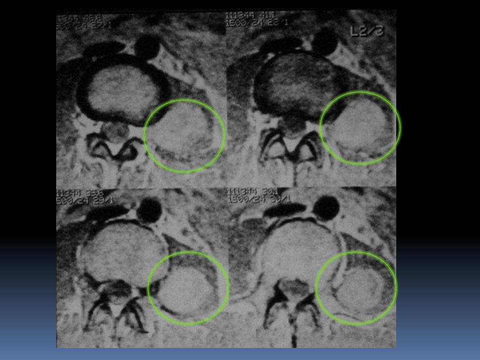
Syrynx/Syringomyelia
▪ CSF filled cavity within the parenchyma of the spinal cord
▪ Causes:
Arnold-Chiari
Cord tumor
Cord trauma
Idiopathic
Left sided thoracic scoliosis

Syrinx: follows water

Type 2 Chiari with syrinx

Cervical Spine: Sagittal
▪ Soft Tissues
Soft tissue space
Thyroid
Paravertebral muscles
Vertebral artery

Parathyroid adenoma
Cervical Spine: Transaxial
▪ Vertebral body
Signal
Shape
Cervical Spine: Transaxial
▪ Posterior arch
Signal
Shape
Articular facets
Uncovertebral joints
Neural foramen

Cervical Spine: Tansaxial
▪ Disc contour
Neural compression

Extrusion with neural compression

Protrusion without neural compression

Cervical Spine: Transaxial
▪ Canal
Cord
Size
Signal
Compression
Ligamentum flavum

Cord compression, post sours/bulge, buckling of lig flavum

Cervical Spine: Transaxial
▪ Soft Tissues
Soft tissue space
Thyroid
Paravertebral muscles
Vertebral artery

Thyroid carcinoma

SHOULDER AND KNEE MRI: WHERE DO I START?
JC Carter DC, DACBR4480-H S Cobb Dr #325Smyrna, GA 30080770-984-5395jcradiology.com

Thank You For Providing Many of the Images in This Presentation!
▪ Sunnyvale Imaging Center
568 South Mathilda Ave.
Sunnyvale, CA 94086
408.738.0232

References
▪ Diagnostic Imaging Orthopedics, Stoller et. Al., Amirys, 2004, Salt Lake City, UT
▪ Musculoskeletal MRI, Helms et. Al., Saunders, 2001, Phil PA
▪ Magnetic Resonance Imaging in Ortopaedics and Sports Medicine, D Stoller, Lippincott, Williams and Wilkins, 2007, Baltimore, MD.
▪ Skeletal Imaging Atlas of the Spine and Extremities Taylor Hughes Resnick, Saunders Elsevier , 2010, Maryland Heights, MO.

MR Terms
▪ High (bright) signal-white
▪ Intermediate signal-light gray
▪ Low signal-dark gray
▪ Signal void-black (air is a signal void on ALL imaging sequences)
▪ Hypo intense-darker than adjacent tissue
▪ Hyper intense-brighter than adjacent tissue
▪ Iso intense-same

MR Sequences
▪ T1
▪ T2
▪ PD
▪ Fat Suppressed
STIR
FS PD FSE

T1
▪ Fat- white
▪ Water-black
▪ Good anatomical detail

T2
▪ Fat- dark gray
▪ Water-white
▪ Gives physiologic information especially edema (bone or soft tissue)

T1 from T2
▪ Locate the synovial fluid
▪ If black, it is T1
▪ If white, it is T2, STIR, or FS PD FSE
▪ If gray, it is likely PD

Proton Density (PD)
▪ Fat- light gray
▪ Water-medium gray
▪ Good for cartilage evaluation
▪ Not very common


Fat Suppressed Proton Density Fast Spin Echo (FS PD FSE)
▪ Fat- black
▪ Water-bright
▪ Good for bone marrow edema, synovial fluid, tendons, ligaments, and cartilage evaluation

▪ A:bone bruise
▪ B: MCL tear
▪ C: ACL tear

Short T1 Inversion Recovery (STIR)
▪ Fat- black
▪ Water-bright
▪ Takes longer to do than FS PD FSE
▪ Good for bone marrow edema, synovial fluid, tendons, ligaments, and cartilage evaluation

STIR
▪ Anything bright is fluid
▪ Great for evaluation of tumor (especially metastasis), fracture and bone bruising

Normal Shoulder Anatomy
▪ Don’t get too frustrated, it takes a long time to learn

Coracoid
Clavicle
Coracoclav. lig
Short headbiceps

Find????

Subscap M/TBiceps m
and t

Find????

Biceps and coracobrach
Trap Supraspin
Glenoid
Ant labrum
Clav
Delt

Find????
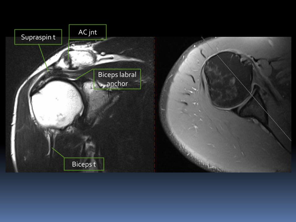
AC jntSupraspin t
Biceps t
Biceps labralanchor

Find????

Inf GH lig
Sup labrum
Suprascap a/n
Subscap and t

Find????

SubscapTeres mnr
Post labrum
IGL
Acromion

Find????

Infraspin
Teres mnr

Clav
Acromion

Suprasp and central t

Labrum
Subscap t
Infrasp
Coracoid p

Find???

Labrum
Biceps and trans lig
coracobrach

Find????

Thickened IGL

Delt

Ant deltSupra t
Biceps t

AC
Bicep t
SupraspInfrasp
Teres
Coracoacromlig

AC Coracoacromlig
Suprasp
Coracoid
Infrasp
Teres
Bicep t
Biceps

Triceps
Coracohum lig
Sup labrum
Ant GHL

Triceps
Labrum
Ant inf GHL

Coracoclav lig
Coracoid
Clavicle
Infrasp Suprasp
Post Delt
Tricep

Coracoclav lig

Shoulder Search Pattern

Coronal Oblique
▪ Overall signal
▪ Supraspinatus and infraspinatus
▪ AC joint
▪ Acromion
▪ GH joint
▪ Sup and inf labrum
▪ IGL

Sagittal Oblique
▪ Supraspin, infraspin, teres, subscap
▪ Rotator interval
▪ Biceps
▪ Glenohumeral ligs

Axial
▪ Long head biceps
▪ Subscap
▪ Ant and post labrum
▪ Glenohumeral ligs

Normal Knee Anatomy
▪ Don’t get too frustrated, it takes a long time to learn

Quad t
Patellar t

MCL
Med Meniscus
Iliotib band
ACL
Lat meniscus
PCL

ACL
Iliotib band
MCL
Lat meniscusMed
Meniscus
PCL

Bicep fem
Fib col lig
PCL

Fib col lig
Femur
Biceps
Vastus lat
Lat gast
Tib ant

Body of lat men

Pat t
Hoffa’s pad
Arcuatelig
Posthorn
Anthorn

Semimembr
Pop a
Gastroc
PCL
ACL
Hoffa’s pad
Popliteus
Quad t

PCL
ACL
Hoffa’s pad

Posthorn
Anthorn
Vast med
Gracilus
Semimembr

Med retin
Synovium
Lat retin
SartorSemimebr
Gasrtr
Pop a
Plantaris
Bicep fem

Medplica
MCL
Fib col lig
Pop v
Tib N

Med menisc
Lat menisc
Pop tendon
Iliotib band

Knee Checklist

Coronal Checklist
▪ Overall signal
▪ MCL
▪ LCL
▪ Meniscii
▪ Articular cartilage
▪ ACL
▪ PCL
▪ Iliotibial band

Sagittal Checklist
▪ Overall signal
▪ Meniscii
▪ Articular cartilage
▪ ACL
▪ PCL
▪ Patellar and quad tendons
▪ Suprapatellar pouch

Axial Checklist
▪ Overall signal
▪ Patellofemoral joint
▪ ACL/PCL
▪ Collateral ligs
▪ Meniscii
▪ Iliotibial band
▪ Joint effusion?

Shoulder Search Pattern

Coronal Oblique
▪ Overall signal
▪ Supraspinatus and infraspinatus
▪ AC joint
▪ Acromion
▪ GH joint
▪ Sup and inf labrum
▪ IGL

Rotator Cuff Tears
▪ Chronic repetitive stress
▪ Pain with or without motion
▪ Pain with movement or isometric contraction of the involved muscle group

Types of Rotator Cuff Tears

A Must

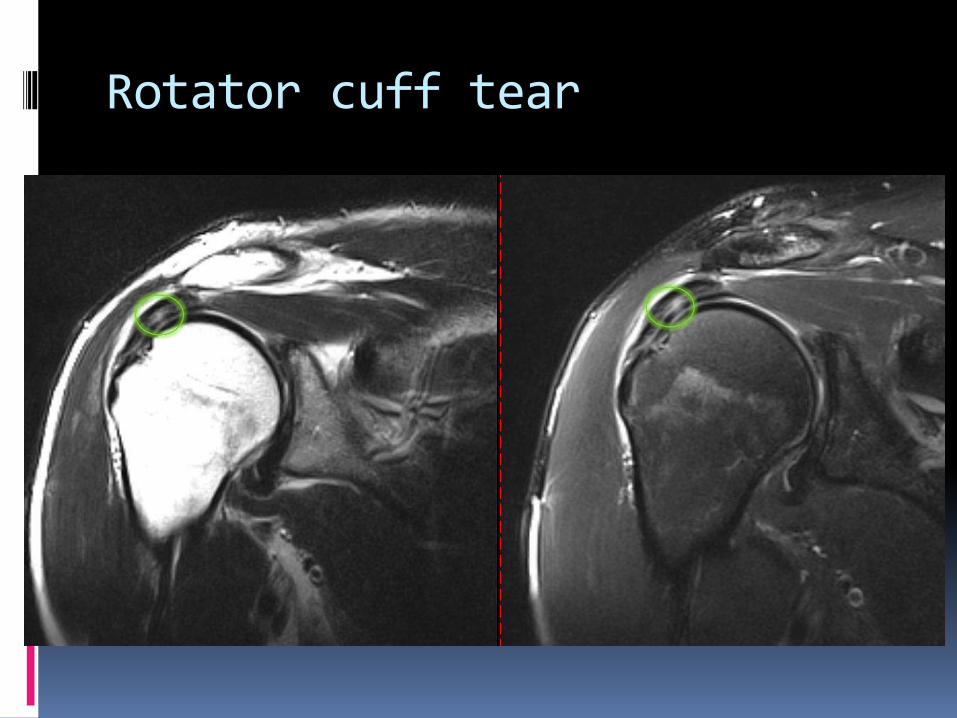
Rotator cuff tear

AC OA with compression of supraspinatus

SLAP Lesions
▪ Superior labral anterior to posterior lesion
▪ Popping, clicking, or catching in the shoulder.
▪ Pain when you move your arm over your head or throw a ball.
▪ A feeling of weakness or instability in the shoulder.
▪ Aching pain. People often have a hard time describing or pointing to exactly where the pain is
▪ Causes: Fall on your outstretched arm.
Fall on your shoulder.
Brace yourself with your outstretched arm in a car accident.
Lift heavy objects repeatedly or too suddenly.
Do a lot of overhead activities, such as throwing a baseball.

SLAP Tears
▪ Superior labral anterior to posterior tears
▪ Best on sagital obliques with increased signal in the superior labrum
▪ Very common and cause instability

In my opinion, the ABSOLUTE BEST
book on extremity MR

Sagittal Oblique
▪ Supraspin, infraspin, teres, subscap
▪ Rotator interval
▪ Biceps
▪ Glenohumeral ligs

Tear with retraction: suprspinatus stops before humeral head

Cuff and glenohumeral lig tear: harder to see on saggital but visible

Axial
▪ Long head biceps
▪ Subscap
▪ Ant and post labrum
▪ Glenohumeral ligs

Biceps dislocation ( not common) and transverse ligament tear: best seen on transaxial

Anterior Labral Tear
▪ Pain, usually with overhead activities
▪ Catching, locking, popping, or grinding
▪ Occasional night pain or pain with daily activities
▪ A sense of instability in the shoulder
▪ Decreased range of motion
▪ Loss of strength
▪ Causes: Falling on an outstretched arm
A direct blow to the shoulder
A sudden pull, such as when trying to lift a heavy object
A violent overhead reach, such as when trying to stop a fall or slide

Anterior labral tear: seen on transaxials, equator (half way down glenoid), and cause instability

Posterior Labral Tears
▪ Clicking or popping
▪ Sharp pain when torn labrum is pinched or displaced
▪ Sense of instability or apprehension with activity
▪ Causes:
Usually trauma

Posterior labral tear: not as common as anterior or SLAP tears, best seen transaxial, equator, cause instability

Knee Checklist

Coronal Checklist
▪ Overall signal
▪ MCL
▪ LCL
▪ Meniscii
▪ Articular cartilage
▪ ACL
▪ PCL
▪ Iliotibial band

Osteochondral defect: helmet shaped defect along the articular surface of femur

MCL Tears
▪ Pain at the sides of your knee. If there is an MCL injury, the pain is on the inside of the knee
▪ Swelling over the site of the injury.
▪ Instability — the feeling that your knee is giving way.
▪ Increased movement with valgus stress

MCL Tear: interruption in fibers with adjacent increased signal, best on coronal images
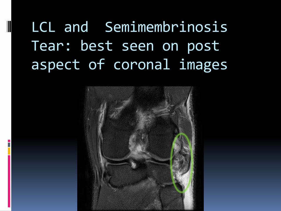
LCL and Semimembrinosis Tear: best seen on post aspect of coronal images

Meniscal Tears
▪ Pain
▪ Stiffness and swelling
▪ Catching or locking of your knee
▪ The sensation of your knee "giving way"
▪ You are not able to move your knee through its full range of motion
▪ Causes: Sudden meniscal tears often happen during sports. Players may
squat and twist the knee, causing a tear. Direct contact, like a tackle, is sometimes involved.
Older people are more likely to have degenerative meniscal tears. Cartilage weakens and wears thin over time. Aged, worn tissue is more prone to tears. Just an awkward twist when getting up from a chair may be enough to cause a tear, if the menisci have weakened with age.

Medial Meniscus Vertical Tear

Lateral Meniscus Tear

ACL Tears
▪ On coronal do not see adjacent to lateral femoral condyle
▪ Should see continuous fibers (even if small) on the sagittal images
▪ On sagittal look for bone bruising on anterior femur and posterior tibia

ACL Tears
▪ Pain with swelling
▪ Loss of full range of motion
▪ Tenderness along the joint line
▪ Discomfort while walking with a feeling your knee will “give out”
▪ Causes
▪ Changing direction rapidly Stopping suddenly
Slowing down while running
Landing from a jump incorrectly
Direct contact or collision, such as a football tackle (least common)

ACL Tear: no ACL adjacent to lateral condyle

ACL tear: interruption in fibers

Bone Bruise Pattern In ACL Tears

ACL Tear

Sagittal Checklist
▪ Overall signal
▪ Meniscii
▪ Articular cartilage
▪ ACL
▪ PCL
▪ Patellar and quad tendons
▪ Suprapatellar pouch

Meniscal Patterns/Grades

Grade I Meniscus
▪ Increased signal in central meniscus
▪ Indicates degeneration
▪ Not a true tear

Grade II Meniscus
▪ Linear increased signal which does not extend to meniscal surface
▪ That area of meniscus predisposed to tear

Grade III Meniscus
▪ Linear increased signal which does extend to meniscal surface
▪ A true tear

PCL Tears
▪ Pain with swelling that occurs steadily and quickly after the injury
▪ Swelling that makes the knee stiff and may cause a limp
▪ Difficulty walking
▪ The knee feels unstable, like it may "give out“
▪ Causes: A direct blow to the front of the knee (such as a bent knee hitting
a dashboard in a car crash, or a fall onto a bent knee in sports)
Pulling or stretching the ligament (such as in a twisting or hyperextension injury)
Simple misstep

PCL Tear

Patellar Tendon Tear (Jumper’s Knee)▪ Pain, tenderness, swelling, warmth, or redness over the patellar
tendon, most often at the lower pole of the patella (kneecap) or at the tibial tubercle (bump on the upper part of the lower leg)
▪ Pain and loss of strength (occasionally) with forcefully straightening the knee (especially when jumping or when rising from a seated or squatting position) or bending the knee completely (squatting or kneeling)
▪ Crepitation (a crackling sound) when the tendon is moved or touched
▪ Causes: Sports that require sudden, explosive quadriceps contraction (jumping, quick
starts, or kicking)
Running sports, especially running down hills
Poor physical conditioning (strength and flexibility, such as with weak quadriceps or tight hamstrings)
Flat feet

Patellar Tendon Tear

Axial Checklist
▪ Overall signal
▪ Patellofemoral joint
▪ ACL/PCL
▪ Collateral ligs
▪ Meniscii
▪ Iliotibial band
▪ Joint effusion?

Bone Bruises From Patellar Dislocation

MCL Tear


The End

Thanks for taking Online Courses with Back To Chiropractic CE Seminars.
I hope you enjoyed the course. Please feel free to provide feedback.
Check out: Back To Chiropractic Resource Pages
Chiropracticpedia Informational website for chiropractic patients
Free Materials Over 200 files: Posters, newsletters & more
Adjusting & Office Skills Free help from DCs that care
DCs Looking For DCs Looking to hire, or for a job?
Chiropractic Neurologists
Classifieds Looking to buy or sell a Practice
Memorials Tributes to great DCs who have passed
Marcus Strutz DC
Back To Chiropractic CE Seminars
33000 North Highway 1
Ft Bragg CA 95437
707.972.0047