Embed Size (px)
Citation preview

of May 19, 2018.This information is current as
Neutrophil NADPH Oxidase Activity Toxins Inhibit HumanBacillus anthracis
Bourdeau and Gary M. BokochMatthew A. Crawford, Caroline V. Aylott, Raymond W.
http://www.jimmunol.org/content/176/12/7557doi: 10.4049/jimmunol.176.12.7557
2006; 176:7557-7565; ;J Immunol
Referenceshttp://www.jimmunol.org/content/176/12/7557.full#ref-list-1
, 33 of which you can access for free at: cites 63 articlesThis article
average*
4 weeks from acceptance to publicationFast Publication! •
Every submission reviewed by practicing scientistsNo Triage! •
from submission to initial decisionRapid Reviews! 30 days* •
Submit online. ?The JIWhy
Subscriptionhttp://jimmunol.org/subscription
is online at: The Journal of ImmunologyInformation about subscribing to
Permissionshttp://www.aai.org/About/Publications/JI/copyright.htmlSubmit copyright permission requests at:
Email Alertshttp://jimmunol.org/alertsReceive free email-alerts when new articles cite this article. Sign up at:
Print ISSN: 0022-1767 Online ISSN: 1550-6606. Immunologists All rights reserved.Copyright © 2006 by The American Association of1451 Rockville Pike, Suite 650, Rockville, MD 20852The American Association of Immunologists, Inc.,
is published twice each month byThe Journal of Immunology
by guest on May 19, 2018
http://ww
w.jim
munol.org/
Dow
nloaded from
by guest on May 19, 2018
http://ww
w.jim
munol.org/
Dow
nloaded from

Bacillus anthracis Toxins Inhibit Human Neutrophil NADPHOxidase Activity1
Matthew A. Crawford,* Caroline V. Aylott,* Raymond W. Bourdeau,† and Gary M. Bokoch2*
Bacillus anthracis, the causative agent of anthrax, is a Gram-positive, spore-forming bacterium. B. anthracis virulence is ascribedmainly to a secreted tripartite AB-type toxin composed of three proteins designated protective Ag (PA), lethal factor, and edemafactor. PA assembles with the enzymatic portions of the toxin, the metalloprotease lethal factor, and/or the adenylate cyclaseedema factor, to generate lethal toxin (LTx) and edema toxin (ETx), respectively. These toxins enter cells through the interactionof PA with specific cell surface receptors. The anthrax toxins act to suppress innate immune responses and, given the importanceof human neutrophils in innate immunity, they are likely relevant targets of the anthrax toxin. We have investigated in detail theeffects of B. anthracis toxin on superoxide production by primary human neutrophils. Both LTx and ETx exhibit distinct inhibitoryeffects on fMLP (and C5a) receptor-mediated superoxide production, but have no effect on PMA nonreceptor-dependent super-oxide production. These inhibitory effects cannot be accounted for by induction of neutrophil death, or by changes in stimulatoryreceptor levels. Analysis of NADPH oxidase regulation using whole cell and cell-free systems suggests that the toxins do not exertdirect effects on NADPH oxidase components, but rather act via their respective effects, inhibition of MAPK signaling (LTx), andelevation of intracellular cAMP (ETx), to inhibit upstream signaling components mediating NADPH oxidase assembly and/oractivation. Our results demonstrate that anthrax toxins effectively suppress human neutrophil-mediated innate immunity byinhibiting their ability to generate superoxide for bacterial killing. The Journal of Immunology, 2006, 176: 7557–7565.
B acillus anthracis is a Gram-positive bacterium whose en-dospores enter the body through skin abrasions, inhala-tion, or ingestion to cause the often fatal disease anthrax.
Once infected, the B. anthracis spores germinate into vegetativebacilli and start secreting multiple virulence factors, including atoxin and an antiphagocytic poly-D-glutamic capsule (1). The ac-tion of the toxin is thought to play a critical role in the pathogen-esis of anthrax. Purified toxin preparations cause death in labora-tory animals (2), and B. anthracis strains missing genes thatencode for toxin are less virulent (3).
Anthrax toxin is a classic AB toxin, which is composed of threepolypeptides. The B component, protective Ag (PA),3 binds to twoalternative catalytically active A components, lethal factor (LF)and edema factor (EF), to produce lethal toxin (LTx) and edematoxin (ETx), respectively (reviewed in Ref. 1). For the toxins togain entry into cells, PA first binds to cell surface receptors, two ofwhich have been characterized, tumor endothelial marker 8 andcapillary morphogenesis protein 2 (4, 5). Receptor-bound PA isthen cleaved by cell surface proteases, leaving a 63-kDa fragment,
which assembles into a heptameric prepore complex (6, 7). Up tothree molecules of LF and/or EF bind to the prepore complex, andthe entire toxin complex undergoes receptor-mediated endocytosis(8, 9). As the complex traffics through the endocytic pathway, it isexposed to increasingly acidic pH, which initiates pore formationby the PA heptamer, ultimately enabling EF and LF to translocateinto the cytosol (10, 11).
LF is a zinc-dependent metalloprotease that cleaves and inacti-vates six of the seven MEKs (12, 13). These enzymes are directactivators of the MAPK pathways, which include ERK, p38, andJNK. MAPKs are key signaling mediators involved in prolifera-tion, differentiation, inflammation, stress response, and cell sur-vival (14). EF is a calcium- and calmodulin-dependent adenylylcyclase that increases intracellular levels of cAMP (15, 16), sub-sequently activating protein kinase A (PKA), cyclic nucleotide-gated ion channels, and the Epac (exchange proteins directly ac-tivated by cAMP) Rap1 guanine nucleotide exchange factors (17).Increased cAMP has been shown to cause edema, and impairphagocyte microbicidal activity and migration (18–20).
Clinical findings in individuals infected with B. anthracis sug-gest an early inhibition of the innate immune system (21, 22).Considerable research has centered on the effects of LTx uponmacrophages, which are clearly critical for the etiology of anthrax;however, there is little information on the effects of the B. anthra-cis toxin on other cells of the innate immune system, particularlyin humans. LTx causes death of sensitized murine macrophages(23–25), human peripheral mononuclear leukocytes (26), and en-dothelial cells (27). LTx has also been shown to modulate cytokinerelease from macrophages (28–31). In addition, both ETx and LTxhave been found to impair the function of dendritic cells (32, 33)and T cells (34, 35). Despite the fact that neutrophils constitute oneof the most powerful host defenses against bacteria and are amongthe first cells recruited to sites of anthrax infection (36, 37), thereis only a limited amount of research investigating the effects ofanthrax toxins on human neutrophil innate immune responses, andmuch of this work has reported contradictory results.
*Department of Immunology and Department of Cell Biology, The Scripps ResearchInstitute, La Jolla, CA 92037; and †Ben May Institute for Cancer Research, Universityof Chicago, Chicago, IL 60637
Received for publication December 30, 2005. Accepted for publication March28, 2006.
The costs of publication of this article were defrayed in part by the payment of pagecharges. This article must therefore be hereby marked advertisement in accordancewith 18 U.S.C. Section 1734 solely to indicate this fact.1 This work was supported by the Center for Disease Control and National Center forInfectious Disease, Grant CI000095.2 Address correspondence and reprint requests to Dr. Gary M. Bokoch, Department ofImmunology, The Scripps Research Institute, 10550 North Torrey Pines Road, LaJolla, CA 92037. E-mail address: [email protected] Abbreviations used in this paper: PA, protective Ag; LF, lethal factor; EF, edemafactor; LTx, lethal toxin; ETx, edema toxin; PKA, protein kinase A; Epac, exchangeproteins directly activated by cAMP; ROS, reactive oxygen species; IBMX, 3-isobu-tyl-1-methylxanthine; GSP, neutrophil membrane fraction; GSS, neutrophil cytosolfraction; PGB, phosphate glucose BSA.
The Journal of Immunology
Copyright © 2006 by The American Association of Immunologists, Inc. 0022-1767/06/$02.00
by guest on May 19, 2018
http://ww
w.jim
munol.org/
Dow
nloaded from
Neutrophils are recruited to inflammatory foci through the gen-eration of chemoattractants at these sites. Two early studies re-ported that both ETx and LTx increased neutrophil chemotaxistoward the peptide chemoattractant fMLP (38, 39). However, it hasbeen recently shown that LTx potently inhibits neutrophil chemo-taxis (40). Once recruited to the inflammatory site, neutrophilsphagocytically ingest bacteria and induce bacterial killing via thegeneration of superoxide anion and other reactive oxygen species(ROS), as well as by release of antimicrobial peptides and pro-teases into the phagocytic vesicle. Neutrophil ROS are generatedby the phagocyte NADPH oxidase, and include the primary spe-cies, superoxide anion (O2
��), a precursor of hydroxyl radical(OH�), H2O2, and hyperchlorous acid (reviewed in Ref. 41). Theimportance of ROS production in host defense is exemplified bythe immune disorder chronic granulomatous disease. Chronicgranulomatous disease patients are unable to produce ROS andsuffer from life-threatening bacterial and fungal infections dueto genetic defects in NADPH oxidase components (reviewed inRef. 42).
The human neutrophil NADPH oxidase is a multicomponentenzyme consisting of membrane-associated cytochrome b558,made up of 91- and 22-kDa subunits, and the cytosolic regulatorycomponents p47phox, p67phox, p40phox, and the Rac2 GTPase.These proteins are induced by appropriate neutrophil stimuli toassemble at the phagosomal or plasma membrane to form a func-tional NADPH oxidase. Much of the biochemistry of the NADPHoxidase has been elucidated using cell-free systems in which na-tive and/or recombinant regulatory components can be manipu-lated (reviewed in Ref. 43).
Early studies on the ability of B. anthracis LTx to induce celldeath in host macrophages suggested that LTx itself induced anincrease in leukocyte ROS production, leading to cell lysis (30).Data on the effects of the B. anthracis toxins on human neutrophil-mediated ROS generation remains limited. ETx, but not LTx, wasreported to inhibit opsonin-dependent ROS production; however,this was associated with a reduction in phagocytosis, precludingany conclusions as to whether there were direct NADPH oxidaseinhibitory effects (16). ETx was also reported to inhibit PMA-induced ROS formation by neutrophils (16). However, subsequentstudies have reported that neither LTx nor ETx have effects on thispathway (Refs. 39, 40, see also this study). Finally, both LTx andETx were shown to inhibit fMLP-mediated superoxide productionin LPS and muramyl dipeptide-primed neutrophils (39). Due to theimportance of neutrophil ROS production in human host resistanceand bacterial killing, we have investigated in detail the effects of B.anthracis toxins on ROS production by primary human neutro-phils. We report in this study that both LTx and ETx exhibit dis-tinct inhibitory effects on fMLP (and C5a) receptor-mediated ROSproduction, but have no effect on nonreceptor-dependent PMA-mediated ROS production. Our results indicate that anthrax toxinseffectively suppress neutrophil-mediated innate immunity by in-hibiting their ability to generate ROS for bacterial killing.
Materials and MethodsIsolation of human neutrophils
This study received ethical approval from The Scripps Research Institu-tional (TSRI) Review Board. Human blood was collected from the TSRINormal Blood Donor Service donor pool by venipuncture and anticoagu-lated with acid citrate dextrose. Neutrophils were isolated using dextransedimentation and Ficoll gradient centrifugation as described previously(44) with minor changes. Briefly, RBC were lysed in lysis buffer containing0.15 M ammonium chloride (NH4Cl), 12 mM sodium bicarbonate(NaHCO3), and 100 �M EDTA (Na2EDTA) for 5 min on ice. Cells werealso treated with 2.7 mM diisopropyl fluorophosphate (Sigma-Aldrich) for10 min on ice. Following isolation, neutrophils were resuspended at 2 �
107cells/ml in PBS containing 1 mM CaCl2, 20 mM glucose, and 0.25%BSA (phosphate glucose BSA, PGB buffer). Neutrophil preparations wereroutinely �95% pure and �99% viable. Experimental data was obtainedfrom separate donors.
Analysis of MAPK kinase and NADPH oxidase proteolysis byLTx
Neutrophils were either left untreated or treated with PA (1 �g/ml, final)and/or LF (0.3 �g/ml, final) at 37°C with continuous rotation for the in-dicated periods of time. Both toxins were provided by S. Leppla (NationalInstitutes of Health, Bethesda, MD). After incubation, treated samples werespun down and resuspended in 100 �l of PGB buffer. Cells were then lysedwith 100 �l of lysis buffer (50 mM Tris, 200 mM NaCl, 5 mM MgCl2, 1mM DTT, 1% Nonidet P-40, and 10% glycerol with 1 mM PMSF, 10�g/ml aprotinin, and 10 �g/ml leupeptin added fresh). Total protein con-centrations of the lysates were determined with a bicinchonic acid proteinassay (Pierce). Lysates were diluted 1/1 in 4� SDS sample buffer, boiledfor 5 min, then equal amounts of total protein were separated by 12%SDS-PAGE and electrotransferred onto polyvinylidene fluoride membrane(Millipore). MEK2 Western blots were washed in blocking buffer (10% 10mM HEPES (pH 7.4), 500 mM NaCl, and 3% BSA) overnight at 4°C.MEK2 was detected with an anti-N-terminal polyclonal Ab (anti-MEK2N20, 1/1000 dilution; Santa Cruz Biotechnology). MEK3 Western blotswere washed in TBS with 5% BSA overnight at 4°C. MEK3 was detectedwith an anti-C-terminal polyclonal Ab (anti-MEK3 C19, 1/1,000 dilution;Santa Cruz Biotechnology). p67phox was detected using an anti-p67phox Ab3958, 1/10,000 dilution, provided by B. Babior (TSRI). p47phox was de-tected using an anti-p47phox Ab 039, 1/5,000 dilution, in house; Rac2 wasdetected using an anti-Rac2 Ab R785, 1/2,000 dilution, in house; gp91phox
was detected using an anti-gp91phox Ab 54.1, 1/500 dilution, provided byM. Quinn (Montana State University, Bozeman, MT).; p22phox was de-tected using an anti-p22phox Ab 44.1, 1/1,000 dilution, provided by M.Quinn. Blots were developed using SuperSignal West Pico chemilumines-cent substrate according to the manufacturer’s instructions (Pierce).
Measurement of whole cell cAMP and superoxide production
cAMP levels in human neutrophils were determined using the cAMP Bio-trak Enzymeimmunoassay System (Amersham Biosciences). Human neu-trophils were suspended at 6.25 � 106 cells/ml in DMEM and were eitherleft untreated or were treated with the indicated amounts of toxins for 3 hat 37°C. Positive controls were treated with forskolin (10 �M, final) plus3-isobutyl-1-methylxanthine (IBMX) (100 �M, final) for 15 min at 37°C.Neutrophils (1 � 106) from each group were assayed in duplicate on 96-well tissue culture plates and lysed using the supplied lysis buffer. Super-natants were then collected and cAMP concentrations determined accord-ing to the manufacturer’s instructions.
During the review of this manuscript, we were made aware by a re-viewer that the EF used throughout these studies, obtained from List Bi-ological Laboratories, contains an amino acid mutation that reduces itsenzymatic activity. We had originally determined that we were using theList Biological Laboratories’ EF at concentrations that we observed toeffectively elevate neutrophil cAMP in our experiments. Native wild-typeEF (provided by Wei-Jen Tang, University of Chicago, IL) gave similarmaximum levels of neutrophil cAMP, but was active at �10-fold lowerconcentrations. We have subsequently verified our experimental resultsusing maximally effective concentrations of recombinant wild-type EF.
Superoxide production in human neutrophils was measured using thecytochrome c reduction assay (45). Neutrophils with or without preincu-bation at 37°C with continuous rotation with PA (1 �g/ml), LF (0.3 �g/ml), and/or EF (0.3 �g/ml; List Biological Laboratories) were aliquotedinto wells of a 96-well plate, 2 � 106 cells total. Cytochrome c (1 mg/ml,final; Sigma-Aldrich) was added, and the reaction was brought to a finalvolume of 200 �l with PGB buffer without BSA. The reactions were in-cubated at 37°C for 10 min followed by stimulation with fMLP (2 �M,final; Sigma-Aldrich), C5a (50 nM, final; Sigma-Aldrich), or PMA (2 �g/ml, final; Sigma-Aldrich); control wells contained superoxide dismutase(0.1 mg/ml, final; Sigma-Aldrich). Upon stimulation, the rate and extent ofcytochrome c reduction by superoxide was monitored for 8–15 min at 550nm using a Versamax microplate reader (Molecular Devices). All experi-ments were performed at least in duplicate, and initial rates were used tocalculate Vmax using Softmax Pro software (Molecular Devices).
Measurement of superoxide production in experiments with cAMP-el-evating agents, cAMP analogs, PKA/Epac1-specific activators, and MAPKinhibitors were preformed as described above with or without preincuba-tion with forskolin (10 �M, final; Sigma-Aldrich); IBMX (100 �M, final;Sigma-Aldrich); PGE1 (10 �M, final; Sigma-Aldrich); PGE2 (10 �M,final; Sigma-Aldrich); the cAMP analogs adenosine 3�,5�-cyclic
7558 ANTHRAX TOXINS INHIBIT NEUTROPHIL ROS PRODUCTION
by guest on May 19, 2018
http://ww
w.jim
munol.org/
Dow
nloaded from

monophosphate,N6,O2’-dibutyryl-sodium salt (db-cAMP) (2 mM, final;Calbiochem) and adenosine 3�,5�-cyclic monophosphate,8-bromo-sodiumsalt (br8-cAMP) (2 mM, final; Calbiochem); the Epac1-specific activator8-(4-chlorophenylthio)-2�-O-methyladenosine-3�,5�-cyclic monophos-phate (cPT-cAMP) (2 mM, final; Biolog); the PKA-specific activatorN6-benzoyladenosine-3�,5�-cyclic monophosphate (BNZ-cAMP) (2 mM,final; Biolog); the p38 inhibitor SB203580 (10 �M, final; Calbiochem); theMEK1 and MEK2 inhibitor U0126 (30 �M, final; Calbiochem); and theJNK inhibitor (30 �M, final; Calbiochem) with appropriate DMSO, etha-nol, and water controls.
Cell viability assays
Cell viability of anthrax toxin-treated neutrophils was routinely assayedusing trypan blue exclusion. A sample of the untreated, LF (0.3 �g/ml), EF(0.3 �g/ml), LTx (PA � 1 �g/ml; LF � 0.3 �g/ml), ETx (PA � 1 �g/ml;EF � 0.3 �g/ml), and LTx � ETx (PA � 1 �g/ml; LF and EF � 0.3�g/ml) treated cell suspensions were diluted 1/1.5 in PGB buffer and thenfurther diluted 1/1 in 0.4% trypan blue solution (Sigma-Aldrich). Afterstanding for 10 min, the total number of cells and the number of cells withdisrupted membranes were determined by light microscopy. Viability re-sults were verified using the MTT reduction assay (46), annexin V andpropidium iodine staining (Vybrant apoptosis assay kit no. 2; MolecularProbes), and by morphological examination.
Cell-free NADPH oxidase system
Human neutrophil membrane fractions (GSP) and neutrophil cytosolicfractions (GSS) were prepared according to the method of Curnutte et al.(47). To assess superoxide formation using neutrophil membrane and cy-tosol, the cell-free system of Curnutte et al. (47, 48) was used. Briefly,neutrophil membrane and cytosol were preincubated for indicated timeperiods with or without PA (1 �g/ml), LF (0.3 �g/ml), and/or EF (0.3�g/ml) at 37°C with continuous rotation. A total of 10 �g of purifiedneutrophil membrane and 100 �g of purified neutrophil cytosol (per reac-tion) were then aliquoted to wells of a 96-well plate. Cytochrome c (100�M, final), guanosine 5�-(3-O-thio)triphosphate (GTP�S) (10 �M, final;Sigma-Aldrich), flavin adenine dinucleotide (10 �M, final; Sigma-Aldrich), and SDS (100 �M, final; Sigma-Aldrich) were then added to eachreaction. Control wells also contained superoxide dismutase (0.1 mg/ml,final). After a 5-min equilibration at 25°C, the formation of superoxide wasinitiated by the addition of nicotinamide adenine dinucleotide phosphate,the reduced form of NADPH (400 �M, final; Sigma-Aldrich). The rate andextent of cytochrome c reduction was monitored for 8 min at 550 nm usinga Versamax microplate reader (Molecular Devices). All experiments weredone in duplicate, and initial rates were used to calculate Vmax using Soft-max Pro software (Molecular Devices).
Cell-free systems were also reconstituted using neutrophil membraneand cytosol fractions isolated from anthrax toxin-treated neutrophils (47,48) with minor modification. Briefly, human neutrophils were isolatedfrom human whole blood as described above. Neutrophils (5 � 108) werethen left untreated, whereas 5 � 108 neutrophils were treated with eitherLTx (PA � 1 �g/ml; LF � 0.3 �g/ml) or ETx (PA � 1 �g/ml; EF � 0.3�g/ml). At regular time points, NADPH oxidase activity in the treated anduntreated samples was checked using the cytochrome c reduction assay.Once effective NADPH oxidase inhibition was established, the cells weredisrupted by nitrogen cavitation, and the membrane and cytosol fractionswere isolated. Cell-free systems were reconstituted as described abovecombining toxin-treated and untreated membrane with GSS cytosol andcombining toxin-treated and untreated cytosol with GSP membrane.
Statistical analysis
Values are given in this study as mean � SEM of at least three duplicateexperiments. Statistically significant differences between sample groupswere determined using the one-way ANOVA test with a Dunnett and/orBonferroni posttest. A p value of 0.05 was considered significant.
ResultsAnthrax LTx and ETx are active in human neutrophils
Prior studies have established that LTx is a zinc metalloproteasecapable of cleaving the N terminus of MEK1–4, MEK6, andMEK7, whereas ETx is a calcium- and calmodulin-dependent ad-enylate cyclase capable of increasing intracellular cAMP levels(13, 16, 49, 50). To ensure that both LTx and ETx were able toenter and exert their catalytic activities in primary human neutro-phils, MEK cleavage and intracellular cAMP levels were assayed.
Lysates were prepared from LTx-treated human neutrophilsover a 7-h time course, and cleavage of MEK2 and MEK3 wasexamined by immunoblotting. Preliminary concentration-responsestudies indicated that maximum MEK cleavage took place at LF �0.3 �g/ml, with PA � 1 �g/ml (data not shown). As shown in Fig.1, MEK2 cleavage in the LTx-treated samples was detected by 1 h,and complete cleavage was achieved by 5 h. Similarly, MEK3cleavage in the LTx-treated samples was seen to begin as early as1 h, with complete cleavage observed by 5 h. When neutrophilswere treated with both LTx and ETx (EF � 0.3 �g/ml), the timecourse and extent of MEK2 and MEK3 cleavage was no differentfrom that observed with LTx alone (data not shown). No MEK2 orMEK3 cleavage was seen in untreated or LF only controls, andthere was no cleavage of the endogenous Rac2 GTPase used as aprotein control.
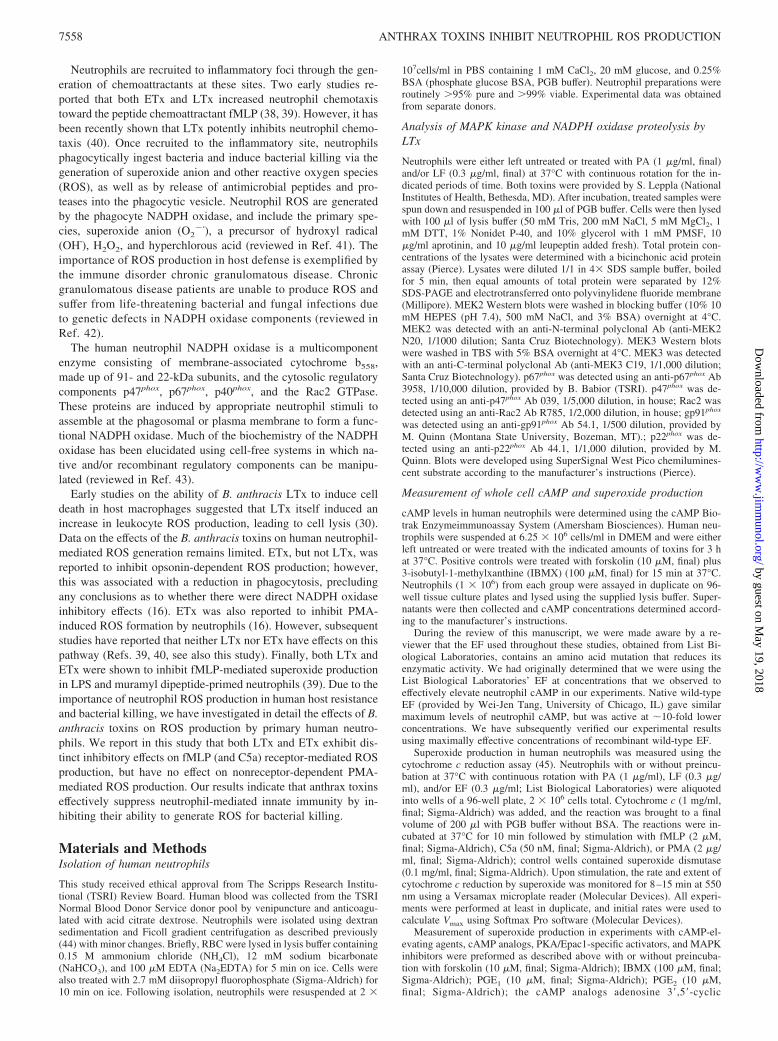
ETx was also effectively taken up and was active in primaryhuman neutrophils. As reported previously (15), neutrophilstreated with ETx (PA � 1 �g/ml; EF � 0.3–1 �g/ml) demon-strated a �2-fold increase in cAMP levels within 3 h of treatment,and this elevation was observed to persist at least out to 7 h oftreatment (Fig. 2). Maximum elevation of cAMP occurred at EFconcentrations of 0.3 �g/ml or above (data not shown). Althoughmodest, this level of cAMP elevation was similar to the maximumachieved by treatment with the potent cAMP-elevating agents, for-skolin plus IBMX. These results indicate that MEK cleavage andintracellular elevations in cAMP were induced by B. anthracisLTx and ETx, respectively, and that this required the action of thePA pore complex to enable entry into the cell.
Anthrax LTx and ETx inhibit receptor-stimulated, but notPMA-stimulated, superoxide production by human neutrophils
Inherent to the neutrophil’s ability to kill ingested microbes is theactivity of the NADPH oxidase system, which is responsible forthe generation of superoxide and subsequently formed ROS. Theimportance of MEKs in the upstream activation of the NADPHoxidase system, as well as existing literature indicating that in-creases in intracellular cAMP can inhibit superoxide production,suggested that LTx and ETx may potentially inhibit NADPH ox-idase-mediated ROS production, thereby promoting evasion of this
FIGURE 1. Cleavage of MEK2 and MEK3 in LTx-treated human neu-trophils. Human neutrophils (2 � 107/ml) were either left untreated ortreated with LF (0.3 �g/ml) or LTx (PA � 1 �g/ml; LF � 0.3 �g/ml) at37°C for indicated amounts of time. Total MEK2 levels (top panels),MEK3 levels (middle panels), and Rac2 levels (loading control, bottompanels) were determined by Western blot analysis. A representative exper-iment from three separate experiments is shown.
7559The Journal of Immunology
by guest on May 19, 2018
http://ww
w.jim
munol.org/
Dow
nloaded from
innate immune killing mechanism (51–53). To evaluate this pos-sibility, neutrophils were treated with maximally effective concen-trations of anthrax toxins (PA � 1 �g/ml; LF and/or EF � 0.3�g/ml) for various times, and the production of superoxide wasdetermined.
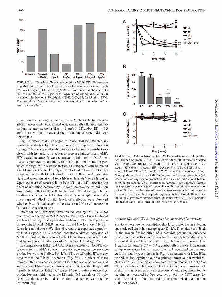
Fig. 3A shows that LTx began to inhibit fMLP-stimulated su-peroxide production by 3 h, with an increasing degree of inhibitionthrough 7 h as compared with untreated or LF only controls. Con-sistent with its rapidity of action to increase intracellular cAMP,ETx-treated neutrophils were significantly inhibited in fMLP-me-diated superoxide production within 1 h, and this inhibition per-sisted through the 7 h of incubation as compared with untreatedand EF only controls. This rapid onset of inhibition by ETx wasobserved both with EF (obtained from List Biological Laborato-ries) and recombinant wild-type EF (see Materials and Methods).Upon exposure of neutrophils to both LTx and ETx together, theonset of inhibition occurred by 1 h, and the severity of inhibitionwas similar to that of the cells treated with ETx alone. By 7 h, theinhibition seen in the LTx plus ETx-treated samples reached amaximum of �80%. Similar levels of inhibition were observedwhether Vmax (initial rates) or the extent (at 300 s) of superoxideformation was considered.
Inhibition of superoxide formation induced by fMLP was notdue to any reduction in fMLP receptor levels after toxin treatmentas determined by flow cytometry analysis of the binding of thefluorescein-labeled fMLP analog, formyl-Nle-Leu-Phe-Nle-Tyr-Lys (data not shown). We also observed that superoxide produc-tion in response to a second receptor-mediated activator ofNADPH oxidase, the chemoattractant C5a, was effectively inhib-ited by similar concentrations of LTx and/or ETx (Fig. 3B).
In contrast with fMLP and C5a receptor-mediated NADPH ox-idase activity, PMA-induced, receptor-independent superoxideproduction was not inhibited by LTx, ETx, or LTx plus ETx at anytime within the 7 h of incubation (Fig. 3C). No effect of thesetoxins on this nonreceptor-mediated stimulus was observed even atsubmaximal PMA concentrations near or below the IC50 (�40ng/ml). Neither the fMLP, C5a, nor PMA-stimulated superoxideproduction was inhibited in the LF only (0.3 �g/ml) or EF only(0.3 �g/ml) controls, indicating that the toxins were actingintracellularly.
Anthrax LTx and ETx do not affect human neutrophil viability
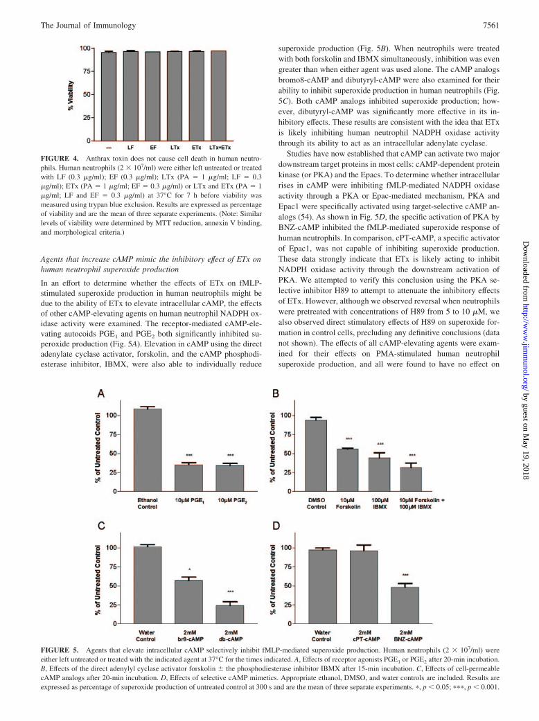
Previous literature has established that LTx is effective in inducingapoptotic cell death in macrophages (23–25). To exclude cell deathas the reason for inhibition of superoxide production observedupon treatment with B. anthracis toxins, neutrophil viability wasexamined. After 7 h of incubation with the anthrax toxins (PA �1 �g/ml; LF and/or EF � 0.3 �g/ml), cells from each treatmentgroup were stained with trypan blue and visualized microscopi-cally for viability. As shown in Fig. 4, treatment with LTx, ETx,or both toxins together had no significant effect on neutrophil vi-ability over a 7-h period as compared with untreated, LF only andEF only controls. The lack of effect of the anthrax toxins on cellviability was confirmed with annexin V and propidium iodidestaining as measured by flow cytometry, with the MTT assay forviability and proliferation, and by morphological examination(data not shown).
FIGURE 2. Elevation of human neutrophil cAMP by ETx. Human neu-trophils (1 � 106/well) that had either been left untreated or treated withPA only (1 �g/ml), EF only (1 �g/ml), or various concentrations of ETx(PA � 1 �g/ml; EF � 1 �g/ml or 0.5 �g/ml or 0.3 �g/ml) at 37°C for 3 hor treated with forskolin (10 �M) plus IBMX (100 �M) for 15 min at 37°C.Total cellular cAMP concentrations were determined as described in Ma-terials and Methods.
FIGURE 3. Anthrax toxin inhibits fMLP-mediated superoxide produc-tion. Human neutrophils (2 � 107/ml) were either left untreated or treatedwith LF (0.3 �g/ml); EF (0.3 �g/ml); LTx (PA � 1 �g/ml; LF � 0.3�g/ml); ETx (PA � 1 �g/ml; EF � 0.3 �g/ml) or LTx and ETx (PA � 1�g/ml; LF and EF � 0.3 �g/ml) at 37°C for indicated amounts of time.Neutrophils were tested for fMLP-stimulated superoxide production (A);C5a-stimulated superoxide production at 3 h (B); or PMA-stimulated su-peroxide production (C) as described in Materials and Methods. Resultsare expressed as percentage of superoxide production of the untreated con-trol at 300 s and are the mean of six separate experiments (A), two separateexperiments (B), and three separate experiments (C). Essentially identicalinhibition curves were obtained when the initial rates (Vmax) of superoxideproduction were plotted (data not shown). ���, p 0.001.
7560 ANTHRAX TOXINS INHIBIT NEUTROPHIL ROS PRODUCTION
by guest on May 19, 2018
http://ww
w.jim
munol.org/
Dow
nloaded from
Agents that increase cAMP mimic the inhibitory effect of ETx onhuman neutrophil superoxide production
In an effort to determine whether the effects of ETx on fMLP-stimulated superoxide production in human neutrophils might bedue to the ability of ETx to elevate intracellular cAMP, the effectsof other cAMP-elevating agents on human neutrophil NADPH ox-idase activity were examined. The receptor-mediated cAMP-ele-vating autocoids PGE1 and PGE2 both significantly inhibited su-peroxide production (Fig. 5A). Elevation in cAMP using the directadenylate cyclase activator, forskolin, and the cAMP phosphodi-esterase inhibitor, IBMX, were also able to individually reduce
superoxide production (Fig. 5B). When neutrophils were treatedwith both forskolin and IBMX simultaneously, inhibition was evengreater than when either agent was used alone. The cAMP analogsbromo8-cAMP and dibutyryl-cAMP were also examined for theirability to inhibit superoxide production in human neutrophils (Fig.5C). Both cAMP analogs inhibited superoxide production; how-ever, dibutyryl-cAMP was significantly more effective in its in-hibitory effects. These results are consistent with the idea that ETxis likely inhibiting human neutrophil NADPH oxidase activitythrough its ability to act as an intracellular adenylate cyclase.
Studies have now established that cAMP can activate two majordownstream target proteins in most cells: cAMP-dependent proteinkinase (or PKA) and the Epacs. To determine whether intracellularrises in cAMP were inhibiting fMLP-mediated NADPH oxidaseactivity through a PKA or Epac-mediated mechanism, PKA andEpac1 were specifically activated using target-selective cAMP an-alogs (54). As shown in Fig. 5D, the specific activation of PKA byBNZ-cAMP inhibited the fMLP-mediated superoxide response ofhuman neutrophils. In comparison, cPT-cAMP, a specific activatorof Epac1, was not capable of inhibiting superoxide production.These data strongly indicate that ETx is likely acting to inhibitNADPH oxidase activity through the downstream activation ofPKA. We attempted to verify this conclusion using the PKA se-lective inhibitor H89 to attempt to attenuate the inhibitory effectsof ETx. However, although we observed reversal when neutrophilswere pretreated with concentrations of H89 from 5 to 10 �M, wealso observed direct stimulatory effects of H89 on superoxide for-mation in control cells, precluding any definitive conclusions (datanot shown). The effects of all cAMP-elevating agents were exam-ined for their effects on PMA-stimulated human neutrophilsuperoxide production, and all were found to have no effect on
FIGURE 4. Anthrax toxin does not cause cell death in human neutro-phils. Human neutrophils (2 � 107/ml) were either left untreated or treatedwith LF (0.3 �g/ml); EF (0.3 �g/ml); LTx (PA � 1 �g/ml; LF � 0.3�g/ml); ETx (PA � 1 �g/ml; EF � 0.3 �g/ml) or LTx and ETx (PA � 1�g/ml; LF and EF � 0.3 �g/ml) at 37°C for 7 h before viability wasmeasured using trypan blue exclusion. Results are expressed as percentageof viability and are the mean of three separate experiments. (Note: Similarlevels of viability were determined by MTT reduction, annexin V binding,and morphological criteria.)
FIGURE 5. Agents that elevate intracellular cAMP selectively inhibit fMLP-mediated superoxide production. Human neutrophils (2 � 107/ml) wereeither left untreated or treated with the indicated agent at 37°C for the times indicated. A, Effects of receptor agonists PGE1 or PGE2 after 20-min incubation.B, Effects of the direct adenylyl cyclase activator forskolin � the phosphodiesterase inhibitor IBMX after 15-min incubation. C, Effects of cell-permeablecAMP analogs after 20-min incubation. D, Effects of selective cAMP mimetics. Appropriate ethanol, DMSO, and water controls are included. Results areexpressed as percentage of superoxide production of untreated control at 300 s and are the mean of three separate experiments. �, p 0.05; ���, p 0.001.
7561The Journal of Immunology
by guest on May 19, 2018
http://ww
w.jim
munol.org/
Dow
nloaded from
activation through this nonreceptor-mediated mechanism, as ob-served with ETx (data not shown).
The inhibition of p38 MAPK mimics the inhibitory effect of LTxon human neutrophil superoxide production
LTx proteolytically degrades MEK family members, resulting indefective activity of the downstream ERK, p38, and JNK pathways(55, 56). To gain insight into the possible mechanism of LTx in-hibition of fMLP-mediated NADPH oxidase activity, the effects ofa MEK1/2 inhibitor capable of blocking ERK activation, and se-lective p38 and JNK inhibitors on fMLP-induced superoxide pro-duction by human neutrophils were examined (57). Fig. 6 showsthat the p38 inhibitor, SB203580, significantly inhibited fMLP-stimulated superoxide production. Inhibition was concentrationdependent and occurred at concentrations previously shown to in-hibit p38 selectively (data not shown). The MEK1/2 inhibitor,U0126, also reduced fMLP-mediated superoxide production,whereas the JNK II inhibitor had no effect. Inhibition observedwhen all three drugs were added together was comparable to in-hibition produced by SB203580 alone (data not shown). All threeinhibitors had no effect on PMA-induced superoxide production atvarious concentrations in human neutrophils (data not shown).
Anthrax LTx and ETx do not inhibit superoxide production in ahuman neutrophil cell-free system
To gain additional insights into NADPH oxidase inhibition by an-thrax LTx and ETx, we used a cell-free NADPH oxidase system toinvestigate whether these toxins exerted any direct effects on theassembly or function of the NADPH oxidase. Human neutrophilmembrane and cytosol were either left untreated or treated withLTx (PA � 1 �g/ml; LF � 0.3 �g/ml) or ETx (PA � 1 �g/ml;EF � 0.3 �g/ml) for varying amounts of time before being used ina reconstituted cell-free system. The data from these experimentsare summarized in Table I. ETx did not inhibit cell-free superoxideproduction at any of the time points assayed. The inclusion of 2mM ATP, a required substrate for the production of cAMP, did notpromote inhibitory activity. Similarly, LTx was not capable of in-hibiting superoxide production in the cell-free system, even thoughwe established that MEK2 cleavage had taken place by the 7-htime point. Consistent with the lack of inhibitory effect of LTx inthis cell-free system, we also observed no LF-induced proteolysisof the NADPH oxidase components, gp91phox, p22phox, p47phox,
p67phox, or Rac2 (Fig. 7). No effects in the cell-free assay weredetected even at higher EF and LF concentrations, up to 1 �g/ml.
To determine whether the anthrax toxin-mediated NADPH ox-idase inhibition established in whole cell assays would be main-tained once the cells were broken, human neutrophils were treatedwith LTx or ETx before the isolation of cytosol and membranefractions. The untreated and toxin-treated membrane and cytosolfractions were then combined with membranes and cytosols (GSPand GSS, respectively) derived from normal neutrophils for mix-and-match experiments. As summarized in Table II, neither LTx
FIGURE 6. Effects of inhibitors of MAPK pathways on superoxide pro-duction. Human neutrophils (2 � 107/ml) were either left untreated ortreated with the p38 inhibitor SB203580, the MEK1 and MEK2 inhibitorU0126, or the JNK inhibitor II with appropriate DMSO and ethanol con-trols. After 15 min of incubation at 37°C, the cells were tested for fMLP-mediated superoxide production as described in Materials and Methods.Results are expressed as the percentage of superoxide production of un-treated control at 300 s and are the mean of three separate experiments. �,p 0.05.
Table I. LTx and ETx do not inhibit neutrophil superoxide productionin a cell-free systema
GroupInitial rate of O2
� production(nmol O2
�/min�mg Mb protein)
Untreated, 2 h 115.88 � 1.96b
Lethal toxin, 2 h 118.81 � 3.65Edema toxin, 2 h 130.26 � 3.02Untreated, 7 h 148.78 � 2.27Lethal toxin, 7 h 147.34 � 3.61Edema toxin, 7 h 164.59 � 5.96
a Human GSP and GSS were preincubated with or without PA (1 �g/ml), LF (0.3�g/ml), and/or EF (0.3 �g/ml) at 37°C with continuous rotation for indicated amountsof time. The GSS and GSP were then used in the reconstitution of the cell-free systemas described in Materials and Methods.
b Shown are the mean Vmax (nmol O2�/min�mg membrane protein) � SEM for
three independent experiments.
FIGURE 7. Anthrax LTx does not cleave cytosolic or membrane com-ponents of the NADPH oxidase complex. A, Human neutrophils (2 � 107/ml) were either left untreated or treated with LF alone (LF � 0.3 �g/ml)or LTx (PA � 1 �g/ml; LF � 0.3 �g/ml) at 37°C for 7 h. A, total p67phox
levels (top panel), p47phox levels (middle panel), and Rac2 levels (bottompanel) were determined by Western blot analysis. A representative exper-iment from three separate experiments is shown. B, Human neutrophils(2 � 107/ml) were either left untreated or treated with LTx (PA � 1 �g/ml;LF � 0.3 �g/ml) at 37°C for 7 h. Membrane fractions were then isolated,and total gp91phox levels (top panel) and p22phox levels (bottom panel) weredetermined by Western blot analysis. A representative experiment fromtwo separate experiments is shown.
7562 ANTHRAX TOXINS INHIBIT NEUTROPHIL ROS PRODUCTION
by guest on May 19, 2018
http://ww
w.jim
munol.org/
Dow
nloaded from
nor ETx-treated membranes were deficient in their ability to sup-port cell-free superoxide production in the presence of normal cy-tosol. Similarly, neither LTx nor ETx-treated cytosols were defi-cient in their ability to support cell-free superoxide production inthe presence of normal membranes. These results support the ideathat the anthrax toxins do not directly affect NADPH oxidase com-ponents, and that these toxins may exert their effects upstream ofNADPH oxidase assembly.
DiscussionAs major virulence factors of B. anthracis, LTx and ETx havebeen thought to play critical roles in the ability of this organism toevade innate immune responses of the host. We show in this studythat both toxins at relatively low concentrations are capable ofinhibiting one of the major microbicidal pathways of human neu-trophils, the NADPH oxidase. Interestingly, both toxins effectivelyinhibited NADPH oxidase activation by chemoattractant (fMLP orC5a) receptor-mediated stimulation, but not activation by the non-receptor activator, PMA. These inhibitory effects of the anthraxtoxins were concentration-dependent. Unlike the effects of anthraxLTx to induce an apoptotic response in macrophages, the NADPHoxidase inhibition we observed in human neutrophils was not as-sociated with significant cell death (Fig. 4), indicating thatNADPH oxidase inhibition is not a secondary consequence of de-creased cell viability. This is in agreement with the recent report byDuring et al. (40), which showed that anthrax LTx is also able toinhibit human neutrophil chemotactic responses to the chemoat-tractant fMLP. The combined inhibition of neutrophil chemotaxisto infectious sites, coupled with inhibition of the oxidative killingresponse, would effectively enable B. anthracis to avoid innateimmune clearing. This would allow a systemic infection to be es-tablished before an adaptive immune response could be achieved.
Our studies clearly demonstrate the ability of both LTx and ETxindividually to inhibit human neutrophil NADPH oxidase activityinitiated through receptor-mediated stimulation. A single priorstudy over 20 years ago using fMLP as a stimulus in human neu-trophils primed with bacterial LPS or muramyl dipeptide reportedinhibition by both LTx and ETx individually is essentially inagreement with our findings (39). We observed no effect of LTxand ETx on PMA-stimulated ROS formation at all PMA concen-trations examined. Our results differ from those reported byO’Brien et al. (16), in which chemiluminescence induced by thephorbol ester PMA was reported to be inhibited by ETx. We note
that this effect was only observed in the aforementioned study aftervery long times of stimulation with this agonist. The same studyreported that phagocytic uptake and chemiluminescence inducedby the avirulent Sterne strain of B. anthracis was inhibited by ETx.
NADPH oxidase inhibition by anthrax LTx is well correlatedwith the entry of the toxin into primary human neutrophils and theresulting proteolytic cleavage of the MEKs (Fig. 1). Indeed, theeffect of LTx is mimicked by inhibitors of the p38 MAPK path-way, suggesting that LTx is likely to induce NADPH oxidase in-hibition through this mechanism. Previous reports have shown thatinhibition of p38 MAPK can decrease NADPH oxidase output (58,59). It has been shown that p38 MAPK phosphorylates the cyto-solic regulatory component, p47phox, and that this phosphorylationis likely to be necessary for chemoattractant-initiated assembly andactivation of the NADPH oxidase (60, 61).
The inhibition of NADPH oxidase activity by anthrax ETx alsoappears to be a direct result of its activity as an adenylate cyclase(Fig. 2). The inhibitory effects of ETx were mimicked by agentsthat elevated cAMP in neutrophils through both receptor-mediatedand nonreceptor-mediated mechanisms (Fig. 5). Through the useof selective cAMP analogs, we were able to show that inhibitionwas dependent upon the ability to activate the downstream medi-ator cAMP-dependent protein kinase or PKA. Activation of Epacshad no inhibitory effect, indicating that the effects of ETx were notmediated through the activation of Rap1 exchange factors. Thesewere significant observations, because Rap1 has been shown toassociate with components of the phagocyte NADPH oxidase andhad been suggested as a possible mechanism for the inhibitoryaction of cAMP (62, 63).
The molecular basis for the effects of the anthrax toxins onNADPH oxidase activity was investigated in a cell-free assay sys-tem (Table I). We observed no direct inhibitory effects on cell-freeNADPH oxidase activity, nor proteolysis of NADPH oxidase reg-ulatory components by the LTx protease. This is consistent withthe inability of LTx to inhibit PMA-stimulated oxidant formation,contrary to what would be expected if regulatory components hadbeen degraded. Similarly, there was no change in the levels ofthese components after treatment with ETx. We attempted to lo-calize the effect of the LTx or ETx to either a membrane-associatedNADPH oxidase component or a cytosolic regulatory componentby performing mix-and-match experiments with materials isolatedfrom toxin-pretreated cells. However, we could detect no defect ineither fraction after toxin treatment (Table II). These results can beinterpreted in several ways: first, the inhibitory effects of the toxinscould be reversible, and not maintained after breakage of the neu-trophils and isolation of subcellular fractions; second, the intrinsicnature of the cell-free system, in which activation must beachieved through the addition of anionic amphiphiles (e.g., SDS),may obviate the inhibitory effect of the toxins. Indeed, it is clearthat the use of such in vitro-activating agents bypasses many of thenormal signaling events (e.g., phosphorylation) required for nor-mal NADPH oxidase activation. Thus, it may also be that the tox-ins act on upstream signaling pathways necessary for NADPH ox-idase assembly and activation.
In conclusion, we have shown that both anthrax LTx and ETxare individually able to effectively block NADPH oxidase-initiatedformation of microbicidal oxidants in human neutrophils. Theseeffects are potent and are likely to aid in the evasion of innateimmune clearing mechanisms by B. anthracis, thereby contribut-ing to the establishment and severity of the resulting anthraxinfection.
AcknowledgmentsWe acknowledge the technical advice and support of Ben P. Bohl.
Table II. Anthrax-mediated oxidase inhibition is not stable throughmembrane and cytosol isolationa
GroupInitial rate of O2
� production(nmol O2
�/min�mg Mb protein)
Untreated cytosol � GSP 94.86 � 6.42b
LTx-treated cytosol � GSP 93.53 � 6.95Untreated membrane � GSS 55.22 � 3.14LTx-treated membrane � GSS 60.05 � 8.14Untreated cytosol � GSP 106.62 � 23.24ETx-treated cytosol � GSP 93.39 � 19.76Untreated membrane � GSS 43.45 � 2.36ETx-treated membrane � GSS 46.34 � 1.53
a Cell-free systems were reconstituted using neutrophil membrane and cytosolfractions isolated from anthrax toxin-treated neutrophils. Cells were either left un-treated or treated with LTx (PA � �g/ml; LF � 0.3 �g/ml) or ETx (PA � 1 �g/ml;EF � 0.3 �g/ml) and once oxidase inhibition was established, the cells were used inmembrane and cytosol isolation. Cell-free systems were reconstituted by combiningtoxin-treated and untreated membrane with GSS cytosol and combining toxin-treatedand untreated cytosol with GSP membrane.
b Shown are the mean Vmax (nmol O2�/min�mg membrane protein) � SEM for
three independent experiments.
7563The Journal of Immunology
by guest on May 19, 2018
http://ww
w.jim
munol.org/
Dow
nloaded from

DisclosuresThe authors have no financial conflict of interest.
References1. Collier, R. J., and J. A. Young. 2003. Anthrax toxin. Annu. Rev. Cell Dev. Biol.
19: 45–70.2. Cui, X., M. Moayeri, Y. Li, X. Li, M. Haley, Y. Fitz, R. Correa-Araujo,
S. M. Banks, S. H. Leppla, and P. Q. Eichacker. 2004. Lethality during contin-uous anthrax lethal toxin infusion is associated with circulatory shock but notinflammatory cytokine or nitric oxide release in rats. Am. J. Physiol. 286:R699–R709.
3. Pezard, C., P. Berche, and M. Mock. 1991. Contribution of individual toxincomponents to virulence of Bacillus anthracis. Infect. Immun. 59: 3472–3477.
4. Bradley, K. A., J. Mogridge, M. Mourez, R. J. Collier, and J. A. Young. 2001.Identification of the cellular receptor for anthrax toxin. Nature 414: 225–229.
5. Scobie, H. M., G. J. Rainey, K. A. Bradley, and J. A. Young. 2003. Humancapillary morphogenesis protein 2 functions as an anthrax toxin receptor. Proc.Natl. Acad. Sci. USA 100: 5170–5174.
6. Milne, J. C., D. Furlong, P. C. Hanna, J. S. Wall, and R. J. Collier. 1994. Anthraxprotective antigen forms oligomers during intoxication of mammalian cells.J. Biol. Chem. 269: 20607–20612.
7. Petosa, C., R. J. Collier, K. R. Klimpel, S. H. Leppla, and R. C. Liddington. 1997.Crystal structure of the anthrax toxin protective antigen. Nature 385: 833–838.
8. Abrami, L., S. Liu, P. Cosson, S. H. Leppla, and F. G. van der Goot. 2003.Anthrax toxin triggers endocytosis of its receptor via a lipid raft-mediated clath-rin-dependent process. J. Cell Biol. 160: 321–328.
9. Abrami, L., M. Lindsay, R. G. Parton, S. H. Leppla, and F. G. van der Goot. 2004.Membrane insertion of anthrax protective antigen and cytoplasmic delivery oflethal factor occur at different stages of the endocytic pathway. J. Cell Biol. 166:645–651.
10. Rainey, G. J., D. J. Wigelsworth, P. L. Ryan, H. M. Scobie, and R. J. Collier.2005. Receptor-specific requirements for anthrax toxin delivery into cells. Proc.Natl. Acad. Sci. USA 102: 13278–13283.
11. Friedlander, A. M. 1986. Macrophages are sensitive to anthrax lethal toxinthrough an acid-dependent process. J. Biol. Chem. 261: 7123–7126.
12. Duesbery, N. S., C. P. Webb, S. H. Leppla, V. M. Gordon, K. R. Klimpel,T. D. Copeland, N. G. Ahn, M. K. Oskarsson, K. Fukasawa, K. D. Paull, andG. F. Vande Woude. 1998. Proteolytic inactivation of MAP-kinase-kinase byanthrax lethal factor. Science 280: 734–737.
13. Chopra, A. P., S. A. Boone, X. Liang, and N. S. Duesbery. 2003. Anthrax lethalfactor proteolysis and inactivation of MAPK kinase. J. Biol. Chem. 278:9402–9406.
14. Johnson, G. L., and R. Lapadat. 2002. Mitogen-activated protein kinase pathwaysmediated by ERK, JNK, and p38 protein kinases. Science 298: 1911–1912.
15. Leppla, S. H. 1982. Anthrax toxin edema factor: a bacterial adenylate cyclase thatincreases cyclic AMP concentrations of eukaryotic cells. Proc. Natl. Acad. Sci.USA 79: 3162–3166.
16. O’Brien, J., A. Friedlander, T. Dreier, J. Ezzell, and S. Leppla. 1985. Effects ofanthrax toxin components on human neutrophils. Infect. Immun. 47: 306–310.
17. Bos, J. L. 2003. Epac: a new cAMP target and new avenues in cAMP research.Nat. Rev. Mol. Cell Biol. 4: 733–738.
18. Warren, J. B., A. J. Wilson, R. K. Loi, and M. L. Coughlan. 1993. Opposing rolesof cyclic AMP in the vascular control of edema formation. FASEB J. 7:1394–1400.
19. Ydrenius, L., L. Molony, J. Ng-Sikorski, and T. Andersson. 1997. Dual action ofcAMP-dependent protein kinase on granulocyte movement. Biochem. Biophys.Res. Commun. 235: 445–450.
20. Zalavary, S., O. Stendahl, and T. Bengtsson. 1994. The role of cyclic AMP,calcium and filamentous actin in adenosine modulation of Fc receptor-mediatedphagocytosis in human neutrophils. Biochim. Biophys. Acta 1222: 249–256.
21. Guarner, J., J. A. Jernigan, W. J. Shieh, K. Tatti, L. M. Flannagan, D. S. Stephens,T. Popovic, D. A. Ashford, B. A. Perkins, and S. R. Zaki. 2003. Pathology andpathogenesis of bioterrorism-related inhalational anthrax. Am. J. Pathol. 163:701–709.
22. Jernigan, J. A., D. S. Stephens, D. A. Ashford, C. Omenaca, M. S. Topiel,M. Galbraith, M. Tapper, T. L. Fisk, S. Zaki, T. Popovic, et al. 2001. Bioterror-ism-related inhalational anthrax: the first 10 cases reported in the United States.Emerg. Infect. Dis. 7: 933–944.
23. Kim, S. O., Q. Jing, K. Hoebe, B. Beutler, N. S. Duesbery, and J. Han. 2003.Sensitizing anthrax lethal toxin-resistant macrophages to lethal toxin-inducedkilling by tumor necrosis factor-�. J. Biol. Chem. 278: 7413–7421.
24. Lin, C. G., Y. T. Kao, W. T. Liu, H. H. Huang, K. C. Chen, T. M. Wang, andH. C. Lin. 1996. Cytotoxic effects of anthrax lethal toxin on macrophage-like cellline J774A.1. Curr. Microbiol. 33: 224–227.
25. Friedlander, A. M., R. Bhatnagar, S. H. Leppla, L. Johnson, and Y. Singh. 1993.Characterization of macrophage sensitivity and resistance to anthrax lethal toxin.Infect. Immun. 61: 245–252.
26. Popov, S. G., R. Villasmil, J. Bernardi, E. Grene, J. Cardwell, A. Wu, D. Alibek,C. Bailey, and K. Alibek. 2002. Lethal toxin of Bacillus anthracis causes apo-ptosis of macrophages. Biochem. Biophys. Res. Commun. 293: 349–355.
27. Kirby, J. E. 2004. Anthrax lethal toxin induces human endothelial cell apoptosis.Infect. Immun. 72: 430–439.
28. Cordoba-Rodriguez, R., H. Fang, C. S. Lankford, and D. M. Frucht. 2004. An-thrax lethal toxin rapidly activates caspase-1/ICE and induces extracellular re-lease of interleukin (IL)-1� and IL-18. J. Biol. Chem. 279: 20563–20566.
29. Erwin, J. L., L. M. DaSilva, S. Bavari, S. F. Little, A. M. Friedlander, andT. C. Chanh. 2001. Macrophage-derived cell lines do not express proinflamma-tory cytokines after exposure to Bacillus anthracis lethal toxin. Infect. Immun. 69:1175–1177.
30. Hanna, P. C., B. A. Kruskal, R. A. Ezekowitz, B. R. Bloom, and R. J. Collier.1994. Role of macrophage oxidative burst in the action of anthrax lethal toxin.Mol. Med. 1: 7–18.
31. Pellizzari, R., C. Guidi-Rontani, G. Vitale, M. Mock, and C. Montecucco. 1999.Anthrax lethal factor cleaves MKK3 in macrophages and inhibits the LPS/IFN�-induced release of NO and TNF�. FEBS Lett. 462: 199–204.
32. Agrawal, A., J. Lingappa, S. H. Leppla, S. Agrawal, A. Jabbar, C. Quinn, andB. Pulendran. 2003. Impairment of dendritic cells and adaptive immunity byanthrax lethal toxin. Nature 424: 329–334.
33. Tournier, J. N., A. Quesnel-Hellmann, J. Mathieu, C. Montecucco, W. J. Tang,M. Mock, D. R. Vidal, and P. L. Goossens. 2005. Anthrax edema toxin cooper-ates with lethal toxin to impair cytokine secretion during infection of dendriticcells. J. Immunol. 174: 4934–4941.
34. Fang, H., R. Cordoba-Rodriguez, C. S. Lankford, and D. M. Frucht. 2005. An-thrax lethal toxin blocks MAPK kinase-dependent IL-2 production in CD4� Tcells. J. Immunol. 174: 4966–4971.
35. Paccani, S. R., F. Tonello, R. Ghittoni, M. Natale, L. Muraro, M. M. D’Elios,W. J. Tang, C. Montecucco, and C. T. Baldari. 2005. Anthrax toxins suppress Tlymphocyte activation by disrupting antigen receptor signaling. J. Exp. Med. 201:325–331.
36. Welkos, S. L., R. W. Trotter, D. M. Becker, and G. O. Nelson. 1989. Resistanceto the Sterne strain of B. anthracis: phagocytic cell responses of resistant andsusceptible mice. Microb. Pathog. 7: 15–35.
37. Lebowich, R. J., B. G. McKillip, and J. R. Conboy. 1943. Cutaneous anthrax: apathological study with clinical correlation. Am. J. Clin. Pathol. 13: 505–515.
38. Wade, B. H., G. G. Wright, E. L. Hewlett, S. H. Leppla, and G. L. Mandell. 1985.Anthrax toxin components stimulate chemotaxis of human polymorphonuclearneutrophils. Proc. Soc. Exp. Biol. Med. 179: 159–162.
39. Wright, G. G., and G. L. Mandell. 1986. Anthrax toxin blocks priming of neu-trophils by lipopolysaccharide and by muramyl dipeptide. J. Exp. Med. 164:1700–1709.
40. During, R. L., W. Li, B. Hao, J. M. Koenig, D. S. Stephens, C. P. Quinn, andF. S. Southwick. 2005. Anthrax lethal toxin paralyzes neutrophil actin-basedmotility. J. Infect. Dis. 192: 837–845.
41. Segal, A. W. 2005. How neutrophils kill microbes. Annu. Rev. Immunol. 23:197–223.
42. Babior, B. M. 2004. NADPH oxidase. Curr. Opin. Immunol. 16: 42–47.43. Groemping, Y., and K. Rittinger. 2005. Activation and assembly of the NADPH
oxidase: a structural perspective. Biochem. J. 386: 401–416.44. Badwey, J. A., J. T. Curnutte, C. B. Berde, and M. L. Karnovsky. 1982. Cy-
tochalasin E diminishes the lag phase in the release of superoxide by humanneutrophils. Biochem. Biophys. Res. Commun. 106: 170–174.
45. Bokoch, G. M., and V. Prossnitz. 1992. Isoprenoid metabolism is required forstimulation of the respiratory burst oxidase of HL-60 cells. J. Clin. Invest. 89:402–408.
46. Denizot, F., and R. Lang. 1986. Rapid colorimetric assay for cell growth andsurvival: modifications to the tetrazolium dye procedure giving improved sensi-tivity and reliability. J. Immunol. Methods 89: 271–277.
47. Curnutte, J. T., R. Kuver, and P. J. Scott. 1987. Activation of neutrophil NADPHoxidase in a cell-free system: partial purification of components and character-ization of the activation process. J. Biol. Chem. 262: 5563–5569.
48. Curnutte, J. T., R. Kuver, and B. M. Babior. 1987. Activation of the respiratoryburst oxidase in a fully soluble system from human neutrophils. J. Biol. Chem.262: 6450–6452.
49. Bardwell, A. J., M. Abdollahi, and L. Bardwell. 2004. Anthrax lethal factor-cleavage products of MAPK (mitogen-activated protein kinase) kinases exhibitreduced binding to their cognate MAPKs. Biochem. J. 378: 569–577.
50. Kumar, P., N. Ahuja, and R. Bhatnagar. 2002. Anthrax edema toxin requiresinflux of calcium for inducing cyclic AMP toxicity in target cells. Infect. Immun.70: 4997–5007.
51. Downey, G. P., J. R. Butler, H. Tapper, L. Fialkow, A. R. Saltiel, B. B. Rubin,and S. Grinstein. 1998. Importance of MEK in neutrophil microbicidal respon-siveness. J. Immunol. 160: 434–443.
52. Spisani, S., M. C. Pareschi, M. Buzzi, M. L. Colamussi, C. Biondi, S. Traniello,Z. G. Pagani, P. M. Paglialunga, I. Torrini, and M. E. Ferretti. 1996. Effect ofcyclic AMP level reduction on human neutrophil responses to formylated pep-tides. Cell. Signal. 8: 269–277.
53. Wright, C. D., P. J. Kuipers, D. Kobylarz-Singer, L. J. Devall, B. A. Klinkefus,and R. E. Weishaar. 1990. Differential inhibition of human neutrophil functions:role of cyclic AMP-specific, cyclic GMP-insensitive phosphodiesterase. Bio-chem. Pharmacol. 40: 699–707.
54. Christensen, A. E., F. Selheim, J. de Rooij, S. Dremier, F. Schwede, K. K. Dao,A. Martinez, C. Maenhaut, J. L. Bos, H. G. Genieser, and S. O. Doskeland. 2003.cAMP analog mapping of Epac1 and cAMP kinase: discriminating analogs dem-onstrate that Epac and cAMP kinase act synergistically to promote PC-12 cellneurite extension. J. Biol. Chem. 278: 35394–35402.
7564 ANTHRAX TOXINS INHIBIT NEUTROPHIL ROS PRODUCTION
by guest on May 19, 2018
http://ww
w.jim
munol.org/
Dow
nloaded from
55. Duesbery, N. S., and G. F. Vande Woude. 1999. Anthrax lethal factor causesproteolytic inactivation of mitogen-activated protein kinase kinase. J. Appl. Mi-crobiol. 87: 289–293.
56. Vitale, G., R. Pellizzari, C. Recchi, G. Napolitani, M. Mock, and C. Montecucco.1998. Anthrax lethal factor cleaves the N-terminus of MAPKKs and inducestyrosine/threonine phosphorylation of MAPKs in cultured macrophages. Bio-chem. Biophys. Res. Commun. 248: 706–711.
57. Park, J. M., F. R. Greten, Z. W. Li, and M. Karin. 2002. Macrophage apoptosisby anthrax lethal factor through p38 MAP kinase inhibition. Science 297:2048–2051.
58. Rane, M. J., S. L. Carrithers, J. M. Arthur, J. B. Klein, and K. R. McLeish.1997. Formyl peptide receptors are coupled to multiple mitogen-activatedprotein kinase cascades by distinct signal transduction pathways: role in ac-tivation of reduced nicotinamide adenine dinucleotide oxidase. J. Immunol.159: 5070 –5078.
59. Nick, J. A., N. J. Avdi, S. K. Young, C. Knall, P. Gerwins, G. L. Johnson, and
G. S. Worthen. 1997. Common and distinct intracellular signaling pathways inhuman neutrophils utilized by platelet activating factor and FMLP. J. Clin. Invest.99: 975–986.
60. Laroux, F. S., X. Romero, L. Wetzler, P. Engel, and C. Terhorst. 2005. Cuttingedge: MyD88 controls phagocyte NADPH oxidase function and killing of gram-negative bacteria. J. Immunol. 175: 5596–5600.
61. Brown, G. E., M. Q. Stewart, S. A. Bissonnette, A. E. Elia, E. Wilker, andM. B. Yaffe. 2004. Distinct ligand-dependent roles for p38 MAPK in primingand activation of the neutrophil NADPH oxidase. J. Biol. Chem. 279:27059 –27068.
62. Quilliam, L. A., H. Mueller, B. P. Bohl, V. Prossnitz, L. A. Sklar, C. J. Der, andG. M. Bokoch. 1991. Rap1A is a substrate for cyclic AMP-dependent proteinkinase in human neutrophils. J. Immunol. 147: 1628–1635.
63. Bokoch, G. M., L. A. Quilliam, B. P. Bohl, A. J. Jesaitis, and M. T. Quinn. 1991.Inhibition of Rap1A binding to cytochrome b558 of NADPH oxidase by phos-phorylation of Rap1A. Science 254: 1794–1796.
7565The Journal of Immunology
by guest on May 19, 2018
http://ww
w.jim
munol.org/
Dow
nloaded from


![Bacillus anthracis - As Biological WeaponsBacillus anthracis - as biological weapons :JOLN (Bacillus anthracis) ± MDNREUR ELRORJLF]QD miotr Daniszewski Department of Invertebrate](https://img.dokumen.tips/doc/110x75/613e1f0259df642846165479/bacillus-anthracis-as-biological-weapons-bacillus-anthracis-as-biological-weapons.jpg)