Embed Size (px)
Citation preview

B Lymphocytes Secrete Antigen-presentingVesicles By Graqa tLaposo,* Hans W. N i j m a n , ~ W i l l e m Stoorvogel ,* R i c h t j e Le i jendekker ,* CliffordV. Hard ingf l Corne l i s J .M. Melief , r
and Hans J. Geuze*
From the *Department of Cell Biology, Faculty of Medicine and Institute for Biomembranes, Utrecht University, 3584 CX Utrecht, The Netherlands; ~Department of lmmunohaematology and Blood Bank, University Hospital, 2300 RC Leiden, The Netherlands; and ~The Institute of Pathology, Case Western Reserve University, Cleveland, Ohio 44 I06
Summary Antigen-presenting cells contain a specialized late endocytic compartment, MIIC (major histo- compatibility complex [MHC] class II-enriched compartment), that harbors newly synthesized MHC class II molecules in transit to the plasma membrane. MIICs have a limiting membrane enclosing characteristic internal membrane vesicles. Both the limiting membrane and the inter- nal vesicles contain MHC class II. In this study on B lymphoblastoid cells, we demonstrate by immunoelectron miroscopy that the limiting membrane of MIICs can fuse directly with the plasma membrane, resulting in release from the cells of internal MHC class II-containing vesi- cles. These secreted vesicles, named exosomes, were isolated from the cell culture media by differential centrifugation followed by flotation on sucrose density gradients. The overall sur- face protein composition of exosomes differed significantly from that of the plasma membrane. Exosome-bound MHC class II was in a compact, peptide-bound conformation. Metabolically labeled MHC class II was released into the extracellular medium with relatively slow kinetics, 10 - 4% in 24 h, indicating that direct fusion of MIICs with the plasma membrane is not the major pathway by which MHC class II reaches the plasma membrane. Exosomes derived from both human and murine B lymphocytes induced antigen-specific MHC class II-restricted T cell responses. These data suggest a role for exosomes in antigen presentation in vivo.
H elper T lymphocytes recognize exogenous antigens bound to MHC class II molecules, displayed at the
surface of a variety of APC such as macrophages, B lym- phocytes, and dendritic cells including Langerhans cells of the epidermis. MHC class II molecules are heterodimers of two transmembrane proteins, an ot and 13 subunit with mo- lecular masses of 33-35 and 25-30 kD, respectively. After biosynthesis, ot and 13 subunits assemble in the endoplasmic reticulum with trimers of a nonpolymorphic type II trans- membrane polypeptide, the invariant (I) chain (1, 2). After transport to the Golgi complex, most of the class II mole- cules are targeted to endocytic compartments (3, 4), where the I chains are dissociated from the cx/[3 dimers, and de- graded, leaving MHC class II molecules ready to bind pep- tides derived from endocytosed proteins (5-7). Recent evi- dence has shown that I chain dissociation and peptide binding are facilitated by HLA-DM molecules (8).
We have recently demonstrated that in human B cells (9) and in dendritic cells, including Langerhans cells (10-12) and macrophages (13), most of the intracellular class II molecules reside in MHC class II-enriched compartments
(MIICs). 1 MIICs have lysosomal characteristics: they con- tain lysosomal marker molecules (LAMP1, CD63, [3-hex- osaminidase, and cathepsin D), are positioned late in the endocytic pathway, and are acidic (14). A further charac- terization of MIICs was accomplished with their purifica- tion from murine macrophages (15), human B cells (16), murine B cells (17, 18), and melanoma cells (19). Interest- ingly, it has recently been shown by immunoelectron mi- croscopy that the majority of HLA-DM molecules also re- sides in MIICs (20). Together, these studies indicate that MIICs are involved in antigen processing and peptide binding to class II molecules. However, functionally differ- ent subclasses of MIICs may exist (16, 18). In all APC stud- ied so far, MIICs and related compartments contain inter- nal membranes (9, 14, 16). These membranes probably originate from inward vesiculation of the limiting mem- brane of MIICs (21). Two types of MIICs could be distin- guished by morphological criteria: those displaying numer-
~Abbreviations used in this paper: BSAG, gold particles derivitized with BSA; MIIC, MHC class II-enriched compartment; TilL, transferrin receptor.
1161 j. Exp. Med.�9 The Rockefeller University Press �9 0022-1007/96/03/1161/12 $2.00 Volume 183 March 1996 1161-1172
Dow
nloaded from http://rupress.org/jem
/article-pdf/183/3/1161/1107814/1161.pdf by guest on 01 Decem
ber 2021

ous internal vesicles and those containing membrane sheets (10, 16, 22). Whether these two types of MIICs have dif- ferential functions in antigen processing and presentation remains to be established.
A major unresolved question in M H C class II trafficking concerns the pathway via which intracellular class II mole- cules are transferred to the cell surface. In macrophages, vesicles emanating from class II-enriched phagolysosomes have been suggested to provide for transport of M H C class II molecules to the plasma membrane (13). However, so far such vesicles have not been identified in other APC. MIICs in B cells and Langerhans cells are spherical structures with- out attached vesicles and tubules (9-12). Alternatively, M H C class II molecules may be transported to the plasma membrane by trans-Golgl network-derived vesicles (1, 23). So far no evidence has been presented for either of these pathways.
Using immunoelectron microscopy, biochemistry, and antigen presentation assays, we now show that in B cells MIICs are exocytic compartments. After fusion with the plasma membrane, small vesicles contained within the lu- men of the MIICs are released into the extracellular milieu. The externalized vesicles, termed exosomes analogous to vesicles released by reticulocytes (24-26), exhibited abun- dant M H C class II molecules at their surface, and specifi- cally presented antigenic peptides to T cells. Possible roles ofexosomes in vivo are discussed.
Materials and Methods
Cell Lines and Antibodies. The EBV-transformed human B cell lines RN (HLA-DR15 +) and JY (HLA-DR15-) were cultured in RPMI medium (Gibco Laboratories, Paisley, Scotland) supple- mented with 10% FCS (Hyclone Laboratories, Inc., Logan, UT) and 100 IU penicillin. The T cell clone 2F10 recognizing the peptide 418-427 from the HSP65 antigen of Mycobacterium leprae in the context of HLA-DR15 was generated as described (27, 28) and cultured in IMDM (Gibco Laboratories) supplemented with 10% heat-inactivated pooled human serum. The murine B cell line TA3 (H-2 k,d) (29) and the ribonuclease (90-105)-l-E~-spe- cific T cell hybridoma WA.23 (30) were maintained in DMEM with 10% FCS. The antibodies used in this study were rabbit anti-HLA DR (3, 9), mouse monoclonal anti-DR DA6.231 (31), mouse monoclonal anti-DR Tfi-36 (32); mouse monoclonal anti-HLA DR. B.8.11.2, and mouse monoclonal anti-HLA-DP B7.21 (gift of Dr. A. Mulder, Department of Immunohematol- ogy and Blood Bank, University Hospital, Leiden, The Nether- lands); mouse mAb directed against the lysosomal protein CD63 (33), rabbit anti-human LAMP1 (34), and rabbit anti-human transferrin receptor (TFR.) (gift of Dr. Alan Schwartz; Washing- ton University, St. Louis, MO).
Isolation and Puri~qcation qf Exosomes. RN cells were washed by centrifugation and recultured in fresh medium for 2 d. Cell culture media (35 nil) containing 2-5 X 10 v cells were centri- fuged for 10 mm at 300g to remove the cells. After a second cen- trifugation at 300 g, the medium was centrifuged for 10 rain at 1,200 g (2 • 30 nfin at 10,000 g, 60 rain at 70,000 g, and 60 min at 100,000 g sequentially, using a rotor (SW27; Beckman Instru- ments, Inc., Fullerton, CA). The pellets were solubilized in non- reducing SDS sample buffer at room temperature for analysis by
SDS-PAGE and Western blotting as described previously using 12SI-protein A (35). When indicated, aliquots of the samples were incubated at 100~ in the presence of [$-ME. For electron nil- croscopy, the 70,000-g pellet was resuspended in 100 pA of RPMI medium. For further purification of exosomes, the 70,000-g pellet was resuspended in 5 nil of 2.5 M sucrose, 20 mM Hepes/NaOH, pH 7.2. A linear sucrose gradient (2-0.25 M su- crose, 20 mM Hepes/NaOH, pH 7.2) was layered on top of the exosome suspension in a tube (SW27; Beckman Instruments, Inc.), and the sample was centrifugated at 100,000 g for 15 h. Gradient fractions (18 • 2 ml) were collected from the bottom of the tube, diluted with 3 ml PBS, and ultracentrifuged for 1 h at 200,000 g using a rotor (SW50; Beckman Instruments, Inc.). The pellets were solubilized in SDS sample buffer lacking [3-ME at room temperature and analyzed by SDS-PAGE and Western blot- ting. Protein concentrations were determined using the BCA as- say from Pierce Chemical Co. (Rockford, IL). Quantitation of MHC class II detected by Western blotting was realized using a Phospholmager (Molecular Dynamics, Sunnyvale, CA).
Biotinylation. 10 s RN cells were washed three times with PBS at 0~ by centrifugation and incubated for 30 min at 0~ in the presence of 1 mg/ml Sulfo-NHS-biotin (Pierce Chemical Co.). Nonreacted biotin was then quenched for 30 min with 15 mlVl NH4C1. The cells were washed with PBS at 0~ and lysed in 1 ml lysis buffer (50 mM Tris-HCl, pH 7.5, 0.5% NP-40, 5 mM MgCI~, 0.5 p,g/ml leupeptin, 0.7 p~g/ml pepstatin, 20 I~g/ml aprotinin, 0.1 mM PMSF). Nuclei were removed from the lysate by centrifugation. Exosomes secreted by 2 • l0 s cells were iso- lated by differential centrifugation followed by flotation on su- crose gradients as described above. Exosome pellets obtained from pooled sucrose gradient fractions were resuspended in PBS and biotinylated as described above for cells. Biotinylated exosomes were then pelleted through 0.3 M sucrose, 20 mM Hepes/NaOH, pH 7.2, and solubilized in lysis buffer. MHC class II was immu- noprecipitated from samples containing lysed biotinylated cells and exosomes using the anti-class II mAbs DA6.231 or Tii-36. The mAbs were adsorbed from hybridoma culture supernatants using protein A-Sepharose beads (Pharmacia Biotech Inc., Pisca- taway, NJ). Antibody-coated beads were washed with inununomix (50 mM Tris-HC1, pH 7.5, 0.5% NP-40, 5 mM EDTA, 150 mM NaC1, 0.1 mM PMSF) and added to the lysates that had been precleared with protein A-Sepharose. After a 16-h incubation at 4~ the beads were washed extensively with immunomix, lm- munoprecipitated MHC class II and samples of the total cell and exosome lysates were eluted from the beads in SDS sample buffer at reducing conditions at 95~ and analyzed by SDS-PAGE and Western blotting. After the transfer to Immobilon-P membranes (Millipore Corp., Milford, MA), the membranes were blocked in PBS containing 0.1% gelatin, 0.05 Triton X-100, and 0.01% anti- foam (Sigma Chemical Co., St. Louis, MO). Biotinylated proteins were labeled using USl-streptavidin (0.1 p~g/nil, 2 • l0 ~ cpm/p~g) and detected using a Phospholmager (Molecular Dynamics).
Afetabolic Labeling. Cells were washed three times with MEM lacking methionine and cysteine and preincubated for 45 rain at 37~ in a 5% CO, atmosphere. Cells were then pulsed for 45 rain with 50 Mbq/ml [3SS]methionine (Tran-S-label; ICN, Irvine, CA) and chased for different periods of time in RPMI supple- mented with 10% FCS, 1 mM methionine, and 1 mM cysteine (5 • 10 v cells/time point). Afi:er pulse-chase labeling, the cells were pelleted by centrifugation ~br 10 min at 300g. The superna- tants were collected and centrifuged for 5 rain at 10,000 g fol- lowed by 30 rain at 200,000 g in a rotor (model SW60; Beckman Instruments, Inc.). Cells and the 200,000-g pellets were lysed in
1162 B Lymphocytes Secrete Antigen-presenting Vesicles
Dow
nloaded from http://rupress.org/jem
/article-pdf/183/3/1161/1107814/1161.pdf by guest on 01 Decem
ber 2021

lysis buffer as described above. Nuclei were removed from the cell lysates by centrifugation for 5 min at 10,000 g. MHC class II was immunoprecipitated as described above. T i l l was immuno- precipitated as described previously (35). Precipitated MHC class II was eluted from the beads in SDS sample buffer at nonreducing conditions at room temperature. Precipitated Tfl~ was eluted from the beads in SDS-sample buffer at reducing conditions at 95~ Immunoprecipitates were analyzed by SDS-PAGE and fluorogra- phy. MHC class II was quantified using a Phospholmager.
hnmunoelectron Microscopy. The ILN B cell line and the T2-DR.3 cell line were pulse incubated with 5-nm gold particles deriv- itized with BSA (BSAG ODs20 = 5 in RPMI medium lacking se- rum) for 10 rain at 37~ The cells were washed extensively at 4~ with RPMI medium by centrifugation and chase incubated at 37~ for 10, 20, 50 or 80 rain in the absence of BSAG. The cells were fixed in a mixture of 2% paraformaldehyde-0.2% glu- taraldehyde in 0.1 M phosphate buffer, pH 7.4, and processed for ultracryomicrotomy and immunolabeling (36). To improve ultra- structure and visualization of membranes, the ultrathin cryosec- tions were collected as described by Liou and Slot (37). For elec- tron microscopy of the isolated exosomes, droplets of RPMI with suspended membranes from 70,000-g pellets (see above) were put on Formvar-carbon-coated electron microscopy grids, fixed as above, immunolabeled, and stained using the method described for ultrathin cryosecrions.
Antigen Presentation Assays. The EBV-B cell lines R N (HLA- DR 15 +) andJY (HLA-DR 15-) were incubated in the presence or absence of purified HSP 65 protein from M. leprae (50 p,g/ml) (38) for 4 h in 10 ml serum-free RPMI at 2 • 106 cells/ml. Then 30 ml of RPMI supplemented with 10% FCS was added, and the cells were incubated for 20 h at 37~ The cells were then washed to remove free antigen and incubated further for 24 h in RPMI/ 10% FCS medium at 37~ Exosomes were prepared by differen- tial centrifugation as described above. The efficiency of HSP 65 antigen presentation was measured by culturing 10,000 cells of the T cell clone 2F10 with irradiated (6,000 rad) EBV cells. As indi- cated in the legend to Fig. 6, increasing numbers of cells or dilu- tions of exosome preparations were used. The highest concentra- tion of exosomes corresponded to material that was secreted by 1.6 • 10 ~ cells in 24 h. B cells or exosomes resuspended in 100 ~1 IMDM/10% pooled human serum were added to the T cell clone (50 ~1 IMDM/10% pooled human serum per well) in 96- well flat-bottom microtiter plates (Costar Corp., Badhoevedorp, The Netherlands) for 4 d at 37~ 5% CO2 in humidified air. An- tibodies to HLA class II molecules were added at 10 pMwell in a final concentration of 10 gzg/ml. When indicated, 5 ~g/ml of HLA-DR15-restricted epitope of HSP65 (peptide 418-427) was added to the exosomes. 16 h before termination, 0.5 gtCi of [3H]thymidine was added to the wells. The cells were then har- vested on glass fiber filters using an automatic cell harvester, and the [~H]thymidine incorporation into cell DNA was determined by liquid scintillation counting. The results are expressed as the mean of triplicate measurements. Exosomes were prepared simi- larly from TA3 cells and incubated together with ribonuclease (90-105) peptide and WA.23 T hybridoma cells. After 24 h, su- pernatants were harvested, and the level of IL-2 secretion by WA.23 cells was determined by CTI_ line bioassay (39).
Results
MIICs Can Fuse with the Plasma Membrane. MIICs con- taining abundant M H C class II molecules are a general fea-
1163 R.aposo et al.
ture o f human and murine B lymphoblastoid cell lines (3, 9, 16, 22). In this study we analyzed immunogold- labeled ultrathin cryosections o f the human B cell line R N by im- munoelec t ron microscopy and found typical MIICs with internal membranes abundantly labeled for M H C class II (Fig. 1). Both multivesicular and mult i laminar MIICs were present.
The R N cells displayed many exocytic profiles suggest- ing fusion o f the l imiting membrane o f MIICs with the plasma membrane. These profiles contained vesicles remi- niscent o f those present in the multivesicular MIICs (Fig. 2). Membrane sheets were not found in the fusion profiles, indicating that p redominandy multivesicular MIICs fused with the plasma membrane. W e named the externahzed MIIC-de r ived vesicles exosomes analogous to vesicles that are released by reticulocytes as a consequence of fusion o f multivesicular bodies with the plasma membrane (25, 26). Both exosomes and the l imiting membranes o f the exocytic profiles were immunolabeled for M H C class II (Fig. 2) and for the lysosomal membrane proteins LAMP1 (Fig. 2 B) and CD63 (not shown).
To further demonstrate that the contents o f MIICs can be exocytosed, we tested whether previously endocytosed BSAG particles were reextemalized and could be found in exocytic profiles. R.N cells were allowed to internalize BSAG particles for 10 rain at 37~ washed extensively at 4~ to remove extracellular BSAG, and chased in the ab- sence of BSAG for varying periods o f t ime at 37~ BSAG was detected in MIICs after 20 rain o f chase (Fig. 1), con- sistent with previous observations on dendritic cells (10- 12), t h e J Y B cell line (9), and other B cells (16, 22). MIICs containing internal vesicles (early MIICs) were reached by the BSAG before those accommodat ing membrane sheets (late MIICs) (Fig. 1). BSAG began to appear in exocytic profiles after 20 rain o f chase (Fig. 2 A) and was abundant after a 50-rain chase (Fig. 2 B). The observation that BSAG was not associated with the plasma membrane after 10 min o f chase indicated that BSAG recovered in exocytic profiles after prolonged chase times indeed resulted from reexter- nalization.
Isolation of Exosomes. Exosomes were isolated from the culture media o f R N cells by differential centrifugation (25). Pelleted membranes were analyzed by Western blot- ting. The majority o f M H C class I I -conta in ing membranes sedimented at 70,000 g (Fig. 3 A, lane 6). To test whether secreted M H C class II molecules were membrane bound, membranes from the 70,000-g pellets were floated into lin- ear sucrose gradients. Western blot anlaysis o f nonboi led and nonreduced gradient fractions showed that compact, pep t ide-bound M H C class II molecules floated to an equi- l ibrium density o f I. 13 g /ml , confirming their association with membrane vesicles (Fig. 3 B). Next, membranes from the 70,000-g pellet were analyzed morphologically. As can be seen in Fig. 3 C, the 70,000-g pellet was composed o f a homogeneous populat ion o f vesicles o f 60--80 nm that la- beled for M H C class II. The vesicles were morphological ly similar to those found in sections o f MIICs (Fig. 1) and exocytic profiles (Fig. 2). W e conclude that the secreted
Dow
nloaded from http://rupress.org/jem
/article-pdf/183/3/1161/1107814/1161.pdf by guest on 01 Decem
ber 2021

Figure 1. KN B cells contain typical MIICs. Ultrathin cryosection of an RN cell immunolabeled for MHC class II with 10-nm gold particles as indicated in the figure. Cells had internalized 5 nm BSAG (BSAG ~) for 10 min and were chased in the absence of BSAG for 20 rain before fixation. MHC class II molecules are lo- calized in multivesicular and multilaminar MIICs. Two types of MIICs containing either vesicles or membrane sheets are shown. Note that after 30 rain of internal- ization, BSAG is present only in multive- sicnlar MIICs (arrows). N, nucleus; M, mi- tochondrion. Bar, 0.1 ~m.
M H C class II molecules were complexed with peptides and associated with membrane vesicles.
Exosome Membranes Are Distinct horn the Plasma Membrane. To investigate the possibility that the M H C class I I - con- taining vesicles that sedimented at 70,000 g represent shed plasma membrane fragments, the presence o f the T fK was monitored by Western blotting. Tfl~s are absent from MIICs (9) and from class II-enriched cell fractions in B cells (16). In most cell types, including B cells, Tfl~s are predominantly present at the plasma membrane and in early endosomes. Tfl~s have been localized in the limiting membrane of multivesicular bodies in most cell types, but not in their internal vesicles (40). Fig. 4 A shows that Tfl~s were only detectable in the cell lysate (lane 3) but not in the 70,000-g pellet (lane 4). The significant enrichment o f M H C class II over TftL in the 70,000-g pellet compared with the cell lysate (compare lanes 1 and 2 with lanes 3 and 4, respectively) suggests that little if any shed plasma mem- brane contaminated the exosome preparation. To further analyze differences in protein composition o f the plasma membrane and exosomes, plasma membrane proteins and exosomal membrane proteins were biotinylated. Probing Western blots with 125I-streptavidin revealed at least four proteins that are highly enriched in plasma membranes and four proteins that are highly enriched in exosomes (Fig. 4 B). M H C class II was identified among the enriched bio-
tinylated proteins in plasma membrane (lanes 3-5) and in exosomes (lanes 6-8) by immunoprecipitation. The differ- ential membrane protein composition of the plasma mem- brane and exosomes again strongly indicates that exosomes did not derive from shed plasma membrane.
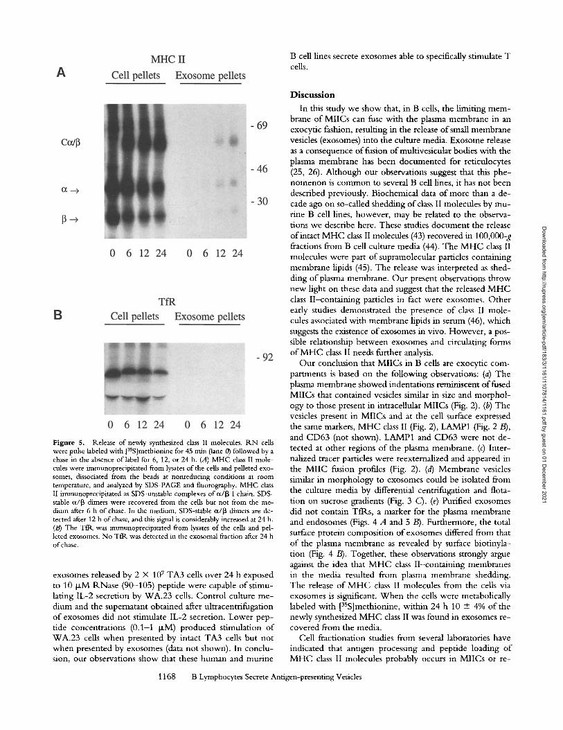
Release of Newly Synthesized Class II Molecules. To deter- mine the kinetics and the extent to which newly synthe- sized M H C class II molecules are released into the medium, R N cells were metabolically pulse labeled with [3SS]methio- nine for 45 min and chased for different periods o f time as indicated in Fig. 5. Cells and exosomes were collected by centrifugation, lysed, and M H C class II was immunoprecipi- tated. After pulse labeling, the majority of 3SS-MHC class II in the cells was immunoprecipitated as SDS-unstable c~/[3-I chain complexes (Fig. 5 A). These complexes were converted to SDS-stable, a/[3-peptide complexes N3 h after synthesis (not shown), consistent with the kinetics reported for other human B cell lines (3, 41). After 6 h o f chase, 3SS-MHC class II appeared in the compact configuration. Only very little 3SS-MHC class II was detected in the exosome frac- tion at this time. Increasing amounts of35S-compact M H C class II were recovered from the exosomes after 12 and 24 h o f chase. After 24 h o f chase, 10 + 4% (n = 5 from three independent experiments) o f the total newly synthesized M H C class II was recovered from exosomes. As a control, newly synthesized 3SS-Tt~ was immunoprecipitated and
1164 B Lymphocytes Secrete Antigen-presenting Vesicles
Dow
nloaded from http://rupress.org/jem
/article-pdf/183/3/1161/1107814/1161.pdf by guest on 01 Decem
ber 2021

Figure 2. MIICs can fuse with the plasma membrane. RN B cells were allowed to inter- nalize 5 nm BSAG particles for 10 min and chased for 20 min (A) or 50 rain (/3) in the absence of BSAG. (A) Ultrathirt cryosections immunogold labeled for MHC class II and LAMP1 as indicated in the figure. The fusion profile is defined by the presence of external- ized BSAG that had previously been endocy- tosed. In addition to BSAG, the exocytic pro- file contains MHC class II-labeled vesicles (exosomes). (B) Exocytic profde containing BSAG and exosomes labeled for MHC class 1I. Bars, 0.1 ~m.
analyzed by SDS-PAGE (Fig. 5 B). After pulse labeling, the precursor form o f the Tfl~ was detected. Complex glycosy- lated 3SS-Tfl~, migrat ing at a slightly lower mobility, was detected after chasing the cells. In contrast to 3SS-MHC class II, no 3SS-Tt~ was found in the exosome fraction, even after 24 h o f chase, again illustrating the selective in- corporat ion o f M H C class II in exosomes.
Exosomes Can Stimulate T Cells. The recovery o f SDS- stable M H C class II molecules from exosomes suggested their association wi th peptides (41, 42). Thus exosomes may be able to present antigens to T cells. W e tested this possibility using antigen presentation assays. H L A - D R 1 5 - positive ILN cells and exosomes isolated from culture me- dia o f R N cells were al lowed to bind pept ide 418-427 from the model antigen HSP 65 o f M. leprae. The cells and exosome preparations were then added to the T cell clone
1165 Raposo et al.
2F10, which recognizes this peptide in the context o f HLA- D R 1 5 (28, 29). In a parallel experiment , R N cells were allowed to endocytose the intact HSP 65 protein cont inu- ously for 24 h, washed, and incubated in the absence o f an- tigen for another 24 h. Cells and exosornes isolated from the chase med ium were then used to stimulate 2F10 cells as above. Both cells and exosomes incubated with antigenic pept ide (Fig. 6, A and C) and cells and exosomes derived from media o f cells that were preincubated with antigen (Fig. 6, B and D) were able to induce specific T cell responses. As a control, exosomes were prepared from culture media o f an equivalent number o f DP, A5-negat ive JY cells that had been incubated either in the presence or absence o f the antigen. Al though JY cells secreted an equivalent amount o f exosomes, these were ineffective in stimulating 2F10 T cell proliferation (not shown). For exosomes derived from
Dow
nloaded from http://rupress.org/jem
/article-pdf/183/3/1161/1107814/1161.pdf by guest on 01 Decem
ber 2021
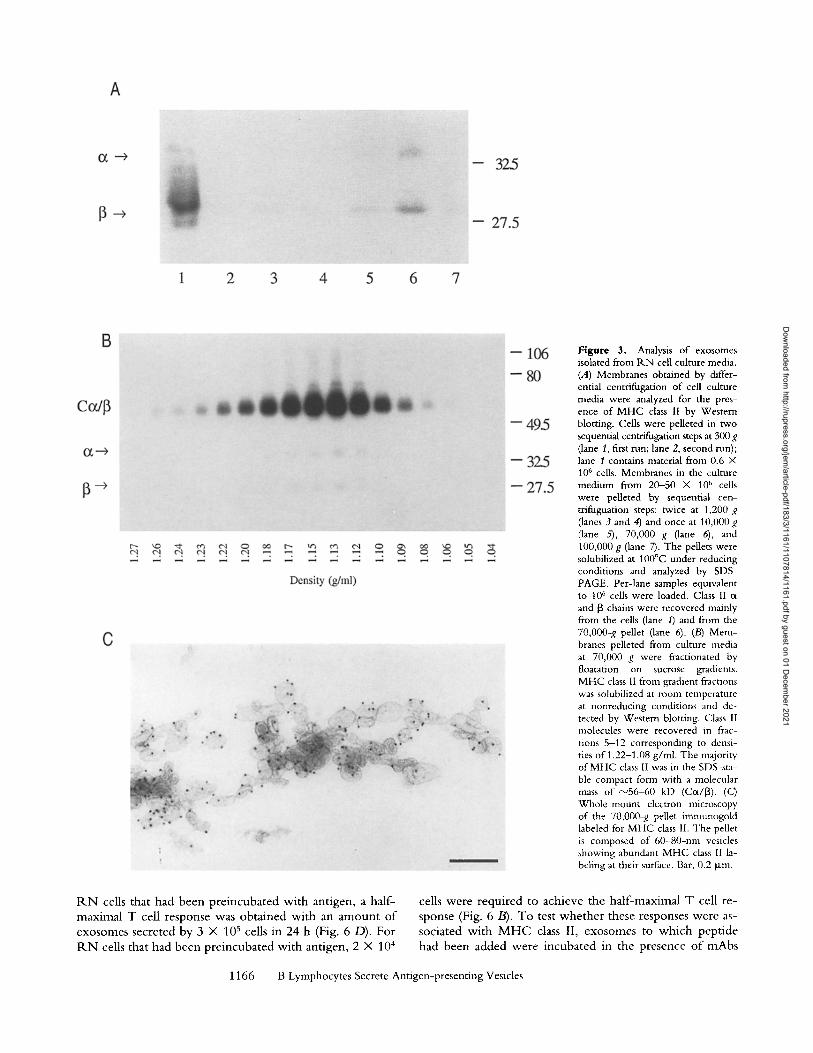
Figure 3. Analysis of exosomes isolated from P,.N cell culture media. (A) Membranes obtained by differ- ential centrifugation of cell culture media were analyzed for the pres- ence of MHC class 1I by Western blotting. Cells were pelleted in two sequential centrifugation steps at 300 g (lane 1, first run; lane 2, second nan); lane 1 contains material from 0.6 • 106 cells. Membranes in the culture medium from 20-50 • 10 ~' cells were pelleted by sequential cen- trifuguation steps: twice at 1,200 g (lanes 3 and 4) and once at 10,000g (lane 5), 70,000 g (lane 6), and 100,000 g (lane 7). The pellets were solubilized at 100~ under reducing conditions and analyzed by SDS- PAGE. Per-lane samples equivalent tO 106 cells were loaded. Class II c~ and 13 chains were recovered mainly from the cells (lane 1) and from the 70,O00-g pellet (lane 6). (/3) Mem- branes pelleted from culture media at 70,000 g were fractionated by floatation on sucrose gradients. MHC class II from gradient fractions was solubilized at room temperature at nonreducing conditions and de- tected by Western blotting. Class 1I molecules were recovered in frac- uons 5-12 corresponding to densi- ties of 1.22-1.08 g/ml. The majority of MHC class II was in the SDS-sta- ble compact form with a molecular mass of ~"56-60 kD (Cot/[~). (C) Whole-mount electron microscopy of the 70,000-g pellet imnmnogold labeled for MHC class 1I. The pellet is composed of 60-80-nm vesicles showing abundant MHC class I1 la- beling at their surface. Bar, 0.2 ~m.
R N cells tha t h a d b e e n p r e i n c u b a t e d w i t h a n t i g e n , a ha l f -
m a x i m a l T cell r e s p o n s e was o b t a i n e d w i t h an a m o u n t o f
e x o s o m e s s e c r e t e d b y 3 X 10 s cells in 24 h (Fig. 6 D) . F o r
P-,N cells tha t h a d b e e n p r e i n c u b a t e d w i t h a n t i g e n , 2 • 104
cells w e r e r e q u i r e d to a c h i e v e t h e h a l f - m a x i m a l T cell r e -
s p o n s e (Fig. 6 B). T o tes t w h e t h e r t h e s e r e s p o n s e s w e r e as-
s o c i a t e d w i t h M H C class II, e x o s o m e s to w h i c h p e p t i d e
h a d b e e n a d d e d w e r e i n c u b a t e d in t h e p r e s e n c e o f m A b s
1166 B Lymphocytes Secrete Antigen-present ing Vesicles
Dow
nloaded from http://rupress.org/jem
/article-pdf/183/3/1161/1107814/1161.pdf by guest on 01 Decem
ber 2021
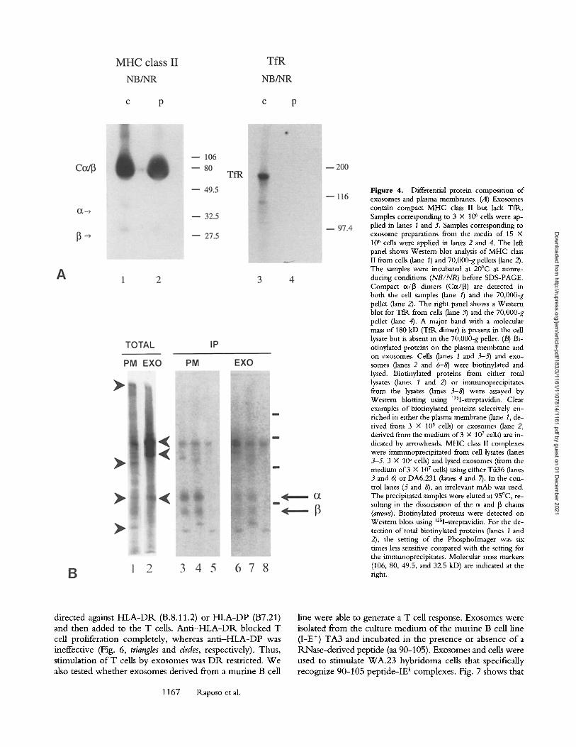
Figure 4. Differential protein composition of exosomes and plasma membranes. (A) Exosomes contain compact MHC class II but lack TfR.. Samples corresponding to 3 • 106 cells were ap- plied in lanes 1 and 3, Samples corresponding to exosome preparations from the media of 15 • 106 cells were apphed in lanes 2 and 4. The left panel shows Western blot analysis of MHC class II from cells (lane I) and 70,000-g pellets (lane 2). The samples were incubated at 20~ at nonre- ducing conditions (NB/NR) before SDS-PAGE. Compact c~/13 timers (Cot/13) are detected in both the cell samples (lane 1) and the 70,000-g pellet (lane 2). The fight panel shows a Western blot for TfR from cells (lane 3) and the 70,000-g pellet (lane 4). A major band with a molecular mass of 180 kD (Tftk dimer) is present in the cell lysate but is absent in the 70,000-g pellet. (B) Bi- otinylated proteins on the plasma membrane and on exosomes. Cells (lanes 1 and 3-5) and exo- somes (lanes 2 and 6-8) were biotinylated and lysed. Biotinylated proteins from either total lysates (lanes 1 and 2) or immunoprecipitates from the lysates (lanes 3-8) were assayed by Western blotting using 125I-streptavidin. Clear examples of biotinylated proteins selectively en- riched in either the plasma membrane (lane 1, de- rived from 3 • 105 cells) or exosomes (lane 2, derived from the medium of 3 • 107 cells) are in- dicated by arrowheads. MHC class II complexes were immunoprecipitated from cell lysates (lanes 3-5, 3 • 106 cells) and lysed exosomes (from the medium of 3 • 107 cells) using either T/i36 (lanes 3 and 6) or DA6.231 (lanes 4 and 7). In the con- trol lanes (5 and 8), an irrelevant mAb was used. The precipitated samples were eluted at 95~ re- suiting in the dissociation of the c~ and 13 chains (arrows). Biotinylated proteins were detected on Western blots using 125I-streptavidin. For the de- tection of total biotinylated proteins (lanes 1 and 2), the setting of the Phospholmager was six times less sensitive compared with the setting for the immunoprecipitates. Molecular mass markers (106, 80, 49.5, and 32.5 kD) are indicated at the fight.
d i r e c t e d aga ins t H L A - D R ( B . 8 . 1 1 . 2 ) o r H L A - D P (B7 .21 )
a n d t h e n a d d e d to t h e T cells. A n t i - H L A - D R b l o c k e d T cel l p r o h f e r a t i o n c o m p l e t e l y , w h e r e a s a n t i - H L A - D P was
i n e f f e c t i v e (Fig. 6, triangles a n d circles, r e sp ec t i v e ly ) . T h u s , s t i m u l a t i o n o f T cells b y e x o s o m e s was D R re s t r i c t ed . W e
also t e s t e d w h e t h e r e x o s o m e s d e r i v e d f r o m a r o u t i n e B cell
1167 P,.aposo et al.
l ine w e r e able to g e n e r a t e a T cell r e sponse . E x o s o m e s w e r e i so l a t ed f r o m t h e c u l t u r e m e d i u m o f t h e m u r i n e B cel l l i ne
( I -E +) T A 3 a n d i n c u b a t e d i n t h e p r e s e n c e o r a b s e n c e o f a R N a s e - d e r i v e d p e p t i d e (aa 9 0 - 1 0 5 ) . E x o s o m e s a n d cells w e r e
u s e d to s t i m u l a t e W A . 2 3 h y b r i d o m a cells t h a t spec i f ica l ly
9 0 - 1 0 5 p e p t i d e - I E c o m p l e x e s . Fig. 7 s h o w s t h a t r e c o g n i z e k
Dow
nloaded from http://rupress.org/jem
/article-pdf/183/3/1161/1107814/1161.pdf by guest on 01 Decem
ber 2021
B ceil hnes secrete exosomes able to specifically stimulate T cells.
Figure 5. Release of newly synthesized class II molecules, tLN cells were pulse labeled with [35S]methionine for 45 min (lane 0) followed by a chase in the absence of label for 6, 12, or 24 h. (A) MHC class I! mole- cules were immunoprecipitated from lysates of the cells and pelleted exo- somes, dissociated from the beads at nonreducing conditions at room temperature, and analyzed by SDS-PAGE and fluorography. MHC class II immunoprecipitated as SDS-unstable complexes of cl/13 I chain. SDS- stable ~x/13 dimers were recovered from the cells but not from the me- dium after 6 h of chase. In the medium, SDS-stable ot/13 dimers are de- tected after 12 h of chase, and this signal is considerably increased at 24 h. (/3) The Tfl~ was immunoprecipitated from lysates of the cells and pel- leted exosomes. No TfR was detected in the exosomal fraction after 24 h of chase.
exosomes released by 2 • 107 TA3 cells over 24 h exposed to 10 ~ M P, Nase (90-105) peptide were capable o f stimu- lating IL-2 secretion by WA.23 cells. Control culture me- dium and the supernatant obtained after ultracentrifugation o f exosomes did not stimulate IL-2 secretion. Lower pep- tide concentrations (0.1-1 p~M) produced stimulation o f WA.23 ceils when presented by intact TA3 cells but not when presented by exosomes (data not shown). In conclu- sion, our observations show that these human and rnurine
Discussion
In this study we show that, in B ceils, the bruiting m e m - brane o f MIICs can fuse with the plasma membrane in an exocytic fashion, resulting in the release o f small membrane vesicles (exosomes) into the culture media. Exosome release as a consequence o f fusion ofmultivesicular bodies with the plasma membrane has been documented for reticulocytes (25, 26). Although our observations suggest that this phe- nomenon is common to several B ceil lines, it has not been described previously. Biochemical data o f more than a de- cade ago on so-called shedding of class II molecules by mu- rine B cell lines, however, may be related to the observa- tions we describe here. These studies document the release o f intact M H C class II molecules (43) recovered in 100,000-g fractions from B ceil culture media (44). The M H C class II molecules were part o f supramolecular particles containing membrane hpids (45). The release was interpreted as shed- ding of plasma membrane. Our present observations throw new light on these data and suggest that the released M H C class II-containing particles in fact were exosomes. Other early studies demonstrated the presence o f class II mole- cules associated with membrane hpids in serum (46), which suggests the existence of exosomes in vivo. However, a pos- sible relationship between exosomes and circulating forms o f M H C class II needs further analysis.
Our conclusion that MiICs in B cells are exocytic com- partments is based on the following observations" (a) The plasma membrane showed indentations reminiscent o f fused MIICs that contained vesicles similar in size and morphol- ogy to those present in intracellular MIICs (Fig. 2). (b) The vesicles present in MIICs and at the ceil surface expressed the same markers, M H C class II (Fig. 2), LAMP1 (Fig. 2 B), and CD63 (not shown). LAMP1 and CD63 were not de- tected at other regions o f the plasma membrane. (c) Inter- nalized tracer particles were reexternalized and appeared in the M I I C fusion profiles (Fig. 2). (d) Membrane vesicles similar in morphology to exosomes could be isolated from the culture media by differential centrifugation and flota- tion on sucrose gradients (Fig. 3 C). (e) Purified exosomes did not contain TilLs, a marker for the plasma membrane and endosomes (Figs. 4 A and 5 B). Furthermore, the total surface protein composition o f exosomes differed from that o f the plasma membrane as revealed by surface biotinyla- tion (Fig. 4 /3). Together, these observations strongly argue against the idea that M H C class II-containing membranes in the media resulted from plasma membrane shedding. The release o f M H C class II molecules from the ceils via exosomes is significant. W h e n the cells were metabolically labeled with [35S]methionine, within 24 h 10 • 4% of the newly synthesized M H C class I! was found in exosomes re- covered from the media.
Cell fractionation studies from several laboratories have indicated that antigen processing and peptide loading of M H C class II molecules probably occurs in MIICs or re-
1168 B Lymphocytes Secrete Antigen-presenting Vesicles
Dow
nloaded from http://rupress.org/jem
/article-pdf/183/3/1161/1107814/1161.pdf by guest on 01 Decem
ber 2021

200o0
1000o
1000 10000 100000
e . x o s o m ~
A
30000"
Number of cells
B
. . . . . . , ~ - - - - . w - . , . u . . . . . . . ,
1 0 0 0 1 0 0 0 0 1 0 0 0 0 0
Number of cells
10000 C
8000
6000'
4000
2000,
0 . . . . . . --=, " . . . . . . . . --- --: . . . . .01 .1 1 .01 . 1 I
D
4 0 0 0 0 "
e X O S O I I ~ S
Figure 6. Presentation of HSP 65 antigen by HLA- DR15 + R.N B cells and exosomes to the CD4 + T cell clone 2F10. Proliferative responses to naive cells (A), to cells preincubated with antigen (B), to exosomes de- rived from naive cells (C), and to exosomes derived from cells preincubated with antigen (D). The solid symbols show proliferation measurements after addi- tion of HSP 65-derived peptide (418-427); the open symbols show where peptide was not added. HLA class II restriction was determined by adding 10 ~g/ml anti- DR antibody (triangles), anti-DP (circles), or no antibody (squares). The exosomes at the highest concentration were derived from media of 1.6 • 106 cells. All assays were performed in triplicate, and results are expressed in cpm [3H]thymidine incorporated into T cells. The SEM for triphcate cpm measurements was <10%. Re- sults shown form a representative example of experi- ments performed in duplicate.
lated endocytic compartments (15-19). The pathway(s) via which intracellular M H C class II molecules are transported from the MIICs to the cell surface are presently unknown. Since the l imit ing membrane o f MIICs contains M H C class II molecules and is incorporated in the plasma membrane during exocytic fusion, this pathway may contribute to M H C class II transport to the plasma membrane. Howeve r fusion o f MIICs with the plasma membrane is not likely to be the only, or even the major, pathway o f M H C class II delivery to the cell surface. First, our metabolic labeling data show that the release o f newly synthesized M H C class II into the med ium (Fig. 5 A) occurred with kinetics much slower than transport o f newly synthesized M H C class II mole- cules from M I I C to the plasma membrane (3, 41). A possi- ble caveat o f this interpretat ion is that sticking o f released exosomes to the plasma membrane may slow down their recovery from the medium. Second, we found that p redom- inantly multivesicular MIICs fused with the plasma m e m - brane, whereas electron microscopic and cell fractionation data (16) suggest that significant amounts o f M H C class II are transported to other types o f MIICs. W e have identified two types ofMIICs : multivescular ones that received endocy-
1169 Raposo et al.
tosed tracer first (early MIICs) and mult i laminar MIICs p o - sitioned later in the endocytic pathway (late MIICs). The exocytic profiles, and the 70,000-g pellets obtained from the culture media, contained vesicles (exosomes) rather than membrane sheets, suggesting that secretion is mainly re- stricted to the multivesicular MIICs. The internal M I I C vesicles are formed by inward budding o f the limiting mem- brane o f MIICs (see reference 21, Figs. 16 and 17) similar to those described for multivesicular bodies in other cell types (47). Therefore the exosomes were expected to expose the luminal domain o f M H C class II molecules at their surface. This orientation was confirmed by immunogold labehng on cryosections o f exosomes and by immunogo ld labeling o f isolated intact exosomes.
In multivesicular endosomes, which share many features with MIICs, internal vesicles contain receptors destined for degradation, whereas recycling receptors are sorted to the l imiting membrane (40, 48). MIICs in APC may represent a similar type o f sorting device to spatially segregate M H C class II molecules to the intemal vesicles, in order to dis- charge them from the cells through exocytosis. Thus, fu- sion o f MIICs with the plasma membrane may allow cells
Dow
nloaded from http://rupress.org/jem
/article-pdf/183/3/1161/1107814/1161.pdf by guest on 01 Decem
ber 2021

40000
=S a.
30000.
m t - o D. m 20000 ' G) n"
(9
'r162 10000,
�9 control T
M e d i u m S u p t , E x o s o m e s T A 3
Antigen Presenting Preparation
Figure 7. Peptide presentation by the exosomes isolated from the cul- ture medium of the murine B cell line TA3. WA.23 T hybridoma cells (10 s cells/well) were incubated with 10 p,M ribonuclease (90-105) peptide in the presence of intact TA3 cells (10 s cells/well), exosomes (70,000-g pe/let), supernatant from the 70,O00-g spin, or normal medium. The wells with exosornes contained material isolated from medium containing 2 • 107 cells. T cell response was determined by IL-2 secretion, measured us- ing a CTLL cell proliferation and [3H]methyl thymidine incorporation bioassay. Lower concentrations ofpeptide (0.1-1 p,M) produced stimula- tion with TA3 cells but not with exosornes (data not shown).
to discard M H C molecules via exosomes. However , we now document the presence o f presentable p e p t i d e - M H C class II complexes at the surface o f exosomes. Peptide gen- erated and bound to M H C class II intracellularly, as well as those bound to M H C class II on exosomes in vitro, p ro- duced a strong, peptide-specific and M H C class II-restricted stimulation o f T cells. Exosomes therefore fulfill the re-
quirements to induce T cell responses, including the ex- pression o f ubiquitous accessory molecules (49). Prel imi- nary observations have indeed indicated that several o f these molecules (]37, ICAM, LFA-3) are present in exosome prep- arations (our unpublished data). Our study does not provide conclusive information about the efficiency o f antigen pre- sentation by exosomes. From the data presented in Fig. 6, it can be estimated that exosomes are ~ 1 0 - 2 0 times less effi- cient in antigen presentation than cells. However , it should be considered that in antigen presentation assays, contact between B and T cells may be optimized selectively because o f sedimentation o f cells but not o f exosomes.
Secretion o f exosomes by B lymphocytes is reminiscent o f that o f the internal vesicles in cytolytic granules o f CTL (50, 51). Both MIICs and cytolytic granules are lysosome- like compartments. The internal vesicles o f cytolytic gran- ules are exocytosed by the CTL upon CTL- ta rge t cell in- teraction, and presumably mediate the killing o f target cells (50). Whe the r B cell exosomes also have an extracellular physiological role in vivo remains to be established. It has been suggested that follicular dendritic cells are able to pick up M H C class II molecules released by surrounding B cells by an unknown mechanism (52). The possibility that exo- somes provide for transfer units o f M H C class I I -pept ide complexes be tween different cells o f the immune system is wor th studying. Preliminary observations on physiological APC like dendritic cells, monocytes, and macrophages, which all contain multivesicular MIICs, have indicated the pres- ence o f exosomes at their surface. It can be speculated that in vivo, in the circulation, exosomes may function as trans- por t vehicles for M H C class I I -pept ide complexes respon- sible for maintenance o f long- term T cell memory or T cell tolerance. Further investigations are required to explore the usefulness o f exosomes, in particular as biological vehicles, in immunotherapy.
We thank Dr. J.W. Slot for discussions during the course of this work. We thank Dr. H. Ploegh and Dr. J. Neefjes for providing anti-HLA-DR polyclonal antibodies, Dr. A. Mulder for anti-HLA-DR and anti- HLA-DP antibodies, Dr. K. Guy for anti-HLA-DR. DA6.231, Dr. J. Sixma for anti-CD-63, Dr. M. Fukuda for anti-LAMP1, and Dr. A. Schwartz for anti-Tflk. T. van Rijn, tk. Scriwanek, and M. Niekerk are grate- fully acknowledged for their excellent photographical work.
This work was supported by Nederlandse Organisatie voor Wetenschappelijk Onderzoek program grant 900-523-094 to H. Geuze. G. Raposo is supported by the Commission of the European Communities (Hu- man Capital and Mobility), H.W. Nijman by Klinisch Wetenschappelijk Onderzoek program grant 900- 716-075, and W. Stoorvogel by the Koninklijke Nederlandse Akademie voor Wetenschappen. C.V. Hard- ing was supported by National Institutes of Health grant AI-35726.
Address correspondence to Hans J. Geuze, Utrecht University Faculty of Medicine, Dept. of Cell Biology, AZU HO2. 314, Heidelberglaan 100, 3584CX Utrecht, The Netherlands. G. Raposo's present address is Institut Curie, Section de R~cherche, 26 Rue d'Ulm, 75004, Paris, France.
Received for publication 28 April 1995 and in revised form 23 October 1995.
1170 B Lymphocytes Secrete Antigen-presenting Vesicles
Dow
nloaded from http://rupress.org/jem
/article-pdf/183/3/1161/1107814/1161.pdf by guest on 01 Decem
ber 2021

References 1. Cresswell, P. 1994. Assembly, transport, and function of
MHC class II molecules. Annu. Rev. Immunol. 12:259-293. 2. Wolf, P.R., and H.L. Ploegh. 1995. How MHC class II mol-
ecules acquire peptide cargo: biosynthesis and trafficking through the endocytic pathway. Annu. Rev. Cell Dev. Biol. 11:267-306.
3. Nee~es, J.J., V. Stollorz, P.J. Peters, H.J. Geuze, and H.L. Ploegh. 1990. The biosynthetic pathway of MHC class II but not class I molecules intersects the endocytic route. Cell. 61: 171-183.
4. Benaroch, P.J., M. Yilla, M.G. Raposo, K. lto, K. Miwa, H.J. Geuze, and H.L. Ploegh. 1995. How MHC class II mol- ecules reach the endocytic pathway. EMBO (Eur. Mol. Biol. Organ.)J. 14:37-49.
5. Roche, P.A., and P. Cresswell. 1991. Proteolysis of the class II-associated invariant chain generates a peptide binding site in intracellular HLA-DR molecules. Proc. Natl. Acad. Sci. USA. 88:3150-3154.
6. Harding, C.V., and H.J. Geuze. 1993. Antigen processing and intracellular traffic of antigens and MHC molecules. Curr. Opin. Cell Biol. 5:596-605.
7. Germain, R.N. 1994. MHC-dependent antigen processing and peptide presentation: providing ligands for T lymphocyte activation. Cell. 76:287-299.
8. Denzin, L.K., and P. Cresswell. 1995. HLA-DM induces CLIP dissociation from MHC class II ot[3 dimers and facilitates pep- tide loading. Cell. 82:155-165.
9. Peters, P.J., J.J. Nee0es, V. Oorschot, H.L. Ploegh, and H.J. Geuze. 1991. Segregation of MHC class II molecules from MHC class I molecules in the Golgi complex for transport to lysosomal compartments. Nature (Lond.). 349:669-676.
10. Kleijmeer, M., V. Oorschot, and H.J. Geuze. 1994. Human Langerhans cells display a tysosomal compartment enriched in MHC class II.J. Invest. Dermatol. 103:516-523.
11. Kleijmeer, M.J., M.A. Ossevoort, C.J.H. Van Veen, J.J. van Hellemond, J.J. Neeijes, W.M. Kast, C.J.M. Mehef, and H.J. Geuze. 1995. MHC class II compartments and the kinetics of antigen presentation in activated mouse spleen dendritic cells. J. lmmunol. 154:5715-5724.
12. Nijman, H.W., M.J. Kleijmeer, M.A. Ossevoort, V.M.J. Oor- schot, M.P.M. Vierboom, M. van de Keur, P. Kenemans, W.M. Kast, H.J. Geuze, and C.J.M. Melief. 1995. Antigen capture and MHC class II compartments in freshly isolated and cultured blood dendritic cells.J. Exp. Med. 182:163-174.
13. Harding, C.V., and H.J. Geuze. 1992. Class II molecules are present in macrophage lysosomes and phagolysosomes that function in the phagocytic processing of Listeria monocytogenes for presentation to T cells.J. Cell Biol. 119:531-542.
14. Geuze, H.J. 1994. A novel lysosomal compartment engaged in antigen presentation. Eur.J. Cell Biol. 64:3-6.
15. Harding, C.V., and H.J. Geuze. 1993. Immunogenic peptides bind to class II MHC molecules in an early lysosomal com- partment.J, lmmunol. 151:3988-3998.
16. West, M.A., J.M. Lucocq, and C. Watts. 1994. Antigen pro- cessing and class II MHC peptide-loading compartments in human B-lymphoblastoid cells. Nature (Lond.). 369:147-151.
17. Amigorena, S.,J.R. Drake, P. Webster, and L Mellman. 1994. Transient accumulation of new class II MHC molecules in a novel endocytic compartment in B lymphocytes. Nature (Lond.). 369:113--120.
18. Qiu, Y., X. Xu, A. Wandinger-Ness, D.P. Dalke, and S.K.
Pierce. 1994. Separation of subcellular compartments con- taining distinct functional forms of MHC class ll .J. Cell Biol. 125:595--605.
19. Tulp, A., D. Verwoerd, B. Dobberstein, H.L. Ploegh, andJ. Pieters. 1994. Isolation and characterization of the intracellu- lar MHC class II compartment. Nature (Lond.). 369:120-126.
20. Sanderson, F., M.J. Kleijmeer, A. Kelly, D. Verwoerd, A. Tulp, J.J. Neeijes, H.J. Geuze, andJ. Trowsdale. 1994. Accu- mulation of HLA-DM, a regulator of antigen presentation, in MHC class II compartments. Science (Wash. DC). 266:1566- 1569.
21. R_iberdy, J.M., R.R. Awa, H.J. Geuze, and P. Cresswell. 1994. Transport and intracellular distribution of MHC class II molecules and associated invariant chain in normal and anti- gen-processing mutant cell lines.J. Cell Biol. 125:1225-1237.
22. Peters, P., G. Raposo, J.J. Neefjes, V. Oorschot, R.L. Leijendekker, H.J. Geuze, and H.L. Ploegh. 1995. MHC class II compartments in human B lymphoblastoid cells are distinct from early endosomes.J. Exp. Med. 182:325-334.
23. Roche, P., C. Teletski, E. Stang, O. Bakke, and E. Long. 1993. Cell surface HLA-DR-invariant chain complexes are targeted to endosomes by rapid internalization. Proc. Natl. Acad. Sci. USA. 90:8581-8585.
24. Harding, C., J. Heuser, and P. Stahl. 1983. Receptor-medi- ated endocytosis of transferrin and recycling of the transferrin receptor in rat reticulocytes. J. Cell Biol. 97:329-339.
25. Harding, C., J. Heuser, and P. Stahl. 1984. Endocytosis and intracellular processing of transferrin and colloidal-gold trans- ferrin in rat reticulocytes: demonstration of a pathway for re- ceptor shedding. Eur. J. Cell Biol. 35:256-263.
26. Pan, B.T., K. Teng, C. Wu, M. Adam, and R.M. Johnstone. 1985. Electron microscopic evidence for externalization of the transferrin receptor in vesicular form in sheep reticulo- cytes.J. Cell Biol. 101:942-948.
27. Haanen, J.B.A.G., R. De Waal-Malefijt, P.C.M. Res, E.M. Kraakman, T.H.M. Ottenhof, R.R.P. de Vries, and H. Spits. 1991. Selection of a human Thl-like T cell subset by myco- bacteria.J. Exp. Med. 174:583-592.
28. Ottenhof, T.H.M., P.R. Klatser, B.C. Elferink, M.Y.L. de Wit, and R.R.P. de Vries. 1986. Mycobacterium leprae specific protein antigens defined by cloned helper T cells. Nature (Lond.). 319:66-68.
29. Glimcher, L.H., T. Hamano, R. Asofsky, D.H. Sachs, M. Pierres, L.E. Samuelson, S.O. Sharrow, and W.E. Paul. 1983. Ia mutant functional antigen-presenting cell lines. J. lmmunol. 130:2287-2294.
30. Chen, J.S., R.G. Lorenz, J. Goldberg, and P.M. Allen. 1991. Identification and characterization of a T cell-inducing epitope of bovine ribonuclease that can be restricted by multiple class II molecules.J. Immunol. 147:3672-3678.
31. Guy, K., V. van Heyningen, B.B. Cohen, D.L. Deane, and C.M. Steel. 1982. Differential expression and serologically distinct subpopulations of human la antigens detected with monoclonal antibodies to Ia alpha and beta chains. Eur. J. lm- munol. 12:942-948.
32. Shaw, S., A. Ziegler, and R. de Maas. 1985. Specificity of monoclonal antibodies directed against human and murine class II histocompatibility antigens as analyzed by binding to HLA-deletion mutant ceil lines. Hum. Immunol. 12:191-211.
33. Metzelaar, M.J., P.L.J. Wijngaard, P.J. Peters, J.J. Sixma, H.K. Nieuwenhuis, and H.C. Clevers. 1991. CD63 antigen: a novel
1171 Raposo et al.
Dow
nloaded from http://rupress.org/jem
/article-pdf/183/3/1161/1107814/1161.pdf by guest on 01 Decem
ber 2021

lysosomal membrane glycoprotein, cloned by a screening pro- cedure for intracellular antigens in eukaryotic cells. J. Biol. Chem. 266:3239-3245.
34. Carlsson, S.R., J. Roth, F. Piller, and M. Fukuda. 1988. Iso- lation and characterization of human lysosomal membrane glycoproteins, h-lamp-1 and h--lamp-2. Major asialosialogly- coproteins carrying polylactosaminoglycan. J. Biol. Chem. 263:18911-18917.
35. Stoorvogel, W., H.J. Geuze, J.M. Gri~th, A.L. Schwartz, and G.J. Strous. 1989. Relations between the intracellular path- ways of the receptors for transferrin, asialoglycoprotein, and mannose 6-phosphate in human hepatoma cells. J. Cell Biol. 108:2137-2148.
36. Slot, J.W., H.J. Geuze, S. Gigengack, G.E. Lienhard, and D. James. 1991. lmmuno-localization of the insulin regulatable glucose transporter in brown adipose tissue of the rat. J. Cell Biol. 113:123-135.
37. Liou, W., and J.W. Slot. 1994. Improved fine structure in mmmnolabeled cryosections after modifying the sectioning and pick-up conditions. Pro& Intl. Con. Electron Microsc. 13:253- 254.
38. Thole, J.E.R., P. Hinderson, J. de Bruyn, F. Cremers, J. van der Zee, H. de Cock, J. Tommassen, W. van Eden, and J.D.A. van Embden. 1988. Antigenic relatedness of a strongly immunogenic 65 kDA mycobacterial protein with a similarly sized ubiquitous bacterial common antigen. Microbial Patho- genesis. 4:71-83.
39. Harding, C.V. 1994. Techniques for studying phagocytic processing of bacteria for class I or II MHC-restricted antigen recognition by T lymphocytes. Methods Cell Biol. 34:307- 320.
40. Hopkins, C.R., A. Gibson, M. Shipman, and K. Miller. 1990. Movement of internalized hgand-receptor complexes along a continuous endosomat reticulum. Nature (Lond.). 346:335-339.
41. Neefjes, J.J., and H.L. Ploegh. 1992. inhibition ofendosomal proteolytic activity by leupeptin blocks surface expression of MHC class II molecules and their conversion to SDS resistant
alpha beta heterodimers in endosomes. EMBO (Eur..~lol. Biol. Organ.)J. 11:411-416.
42. Sadegh-Nasseri, S., and R.N. Germain. 1991. A role for pep- tide in determining MHC-class II structure. Nature (Lond.). 353:167-170.
43. Emerson, S.G., and R.E. Cone. 1979. Turnover and shed- ding of Ia antigens by murine spleen cells in culture. J. Immu- nol. 122:892-899.
44. Sachs, D.H., P. Kiszkiss, and K.J. Kim. 1980. Release of Ia antigens by a cultured B cell line.J. Immunol. 124:2130-2136.
45. Emerson, S.C., and R.E. Cone. 1981. l-Kk and H-2Kk anti- gens are shed as supramolecular particles in association with membrane lipids.J. Immunol. 127:482-486.
46. Callahan, G.N., S. Ferrone, M.D. Poulik, R.A. Reisfeld, and J. Klein. 1976. Characterization of la antigens in mouse se- rum.J. Immunol. 117:1351-1355.
47. Van Deurs, B., P.K. Holm, L. Kayser, K. Sandvig, and S.H. Hansen. 1993. Multivesicular bodies in HEp-2 cells are mat- urating endosomes. Eur. J. Cell Biol. 61:208-224.
48. Felder, S., K. Miller, G. Moehren, A. Ullrich, J. Schlessinger, and C.R. Hopkins. 1990. Kinase activity controls the sorting of the epidermal growth factor receptor within the multive- sicular body. Cell. 61:623-634.
49. Dustin, M.L., and T.A. Springer. 1991. Role of lymphocyte adhesion receptors in transient interactions and cell locomo- tion. Annu. Rev. Immunol. 9:27-66.
50. Peters, P., H.J. Geuze, H.A. van der Donk, andJ. Borst. 1990. A new model for lethal hit delivery by cytotoxic T lympho- cytes. Immunol. Today. 11:28-32.
51. Peters, P.J., J. Borst, V. Oorschot, M. Fukuda, O. Kr~henbiihl, J. Tschopp, J.W. Slot, and H.J. Geuze. 1991. Cytotoxic T lymphocyte granules are secretory lysosomes, containing both perforin and granzymes.J. Exp. Med. 173:1099-1109.
52. Gray, D., M. Kosco, and B. Stockinger. 1991. Novel path- ways of antigen presentation for the maintenance of memory. Int. lmmunol. 3:141-148.
1172 B Lymphocytes Secrete Antigen-presenting Vesicles
Dow
nloaded from http://rupress.org/jem
/article-pdf/183/3/1161/1107814/1161.pdf by guest on 01 Decem
ber 2021