Embed Size (px)
Citation preview

Volume 2 • Issue 3 • 1000122J Pulmon Resp MedISSN: 2161-105X JPRM, an open access journal
Aziz, J Pulmon Resp Med 2012, 2:3 DOI: 10.4172/2161-105X.1000122
Case Report Open Access
Efficacy of Percutaneous Pigtail Catheters for Thoracostomy at BedsideFahad Aziz*Department of Internal Medicine, Jersey City Medical Center, New Jersey, USA
AbstractGiven the potential morbidity of traditional chest tube insertion, use of pigtail is desirable. The purpose of this
case series is to determine the efficacy of bedside pigtail thoracostomy catheters in Adult population by using bedside ultrasound by the primary internist.
*Corresponding author: Department of Internal Medicine, Jersey City Medical Center, New Jersey, USA, E-mail: [email protected]
Received October 22, 2011; Accepted April 25, 2012; Published April 27, 2012
Citation: Aziz F (2012) Efficacy of Percutaneous Pigtail Catheters for Thoracostomy at Bedside. J Pulmon Resp Med 2:122. doi:10.4172/2161-105X.1000122
Copyright: © 2012 Aziz F . This is an open-access article distributed under the terms of the Creative Commons Attribution License, which permits unrestricted use, distribution, and reproduction in any medium, provided the original author and source are credited.
Keywords: Pigtail catheters; Bedside; Ultrasound
IntroductionThoracostomy tubes are a mainstay of treatment for removing fluid
or air from the pleural space. Placement of a chest tube is, however, an invasive procedure with potential morbidity. Complications include hemothorax, perforation of intrathoracic organs, diaphragmatic laceration, empyema, pulmonary edema, and Horner’s syndrome [1-3]. In an effort to reduce these complications, Fuhrman et al. [4] and subsequently Lawless et al. [5] described the use of percutaneous pigtail catheters in place of traditional large-bore tubes for thoracostomy and pleural drainage. The Seldinger needle-guide wire method of placement and smaller, more flexible catheters avoid the force required to place a large-bore chest tube by the dissection of trocar methods.
Materials & MethodsHere, we will describe 5 cases in which successful bed site pigtail
thoracostomy catheters were placed at bed site.
Technique
Percutaneous pigtail catheters were all single-lumen polyurethane coiled catheters, 7 to 8.5 F, used in conjunction with a wire and dilator, connected to a negative-pressure drainage system. The catheters were inserted using the modified Seldinger technique, with insertion of needle and syringe over a rib, with gentle aspiration of a syringe to locate either fluid or air in the pleura space. A J-tipped wire was then inserted and the needle removed. A dilator and scalpel were used to enlarge the insertion sire, and the catheter was then inserted over the wire. Finally, the wire was removed and the catheter was attached to a drain. The procedure was placed under ultrasound guidance at bedside
by the primary internist. No radiological or surgical help was requested (Figure 1).
Case description
Case 1: 44-year old African American male with a past medical history of HIV and hypertension presented to hospital with complaints of shortness of breath at rest associated with fevers and chills for past 1-2 days and no cough or sputum. On examination patient was found to be in mild respiratory distress with no use of accessory muscles and dullness of percussion throughout left lung fields with minimal air entry in left lung field. Chest X ray and CT chest revealed a massive pleural effusion of left lung and associated lung collapse. A Fuhrman’s catheter was inserted at bedside, under ultrasound guidance and a total of 2650 cc of purulent fluid was drained over next 48 hours.
Pleural fluid analysis revealed it to be an exudative fluid (LDH of 963 U/L, T protein 5.4 G/ DL) with 31 polys and 56 lymphocytes. Pleural fluid culture grew Streptococci viridians. Patient showed a rapid clinical improvement, but the catheter stopped draining completely after 48 hrs. Persistence of effusions on Chest X ray and Positive pleural cultures gave rise to a high possibility of adhesions secondary to empyema. So
Figure 1: Pigtail catheter allows less tension on the chest tube to decrease kinking of the catheter. The catheter is sutured at the skin. A clear dressing is placed over the catheter.
Figure 2: CXR before pigtail catheter placement.
Jour
nal o
f Pulm
onary & Respiratory Medicine
ISSN: 2161-105X
Journal of Pulmonary & Respiratory Medicine

Volume 2 • Issue 3 • 1000122J Pulmon Resp MedISSN: 2161-105X JPRM, an open access journal
Citation: Aziz F (2012) Efficacy of Percutaneous Pigtail Catheters for Thoracostomy at Bedside. J Pulmon Resp Med 2:122. doi:10.4172/2161-105X.1000122
Page 2 of 3
TPA was instilled into pleura, after which 750 cc more fluid drained out in next 24 hrs. Catheter was removed after a total of 4 days, after fluid stopped draining completely even after instillation of another dose of TPA. Patient had an uneventful follow up without any recurrence of symptoms (Figure 2 and 3).
Case II: 72-year old African American female with past medical history of Non-squamous cell carcinoma of lung treated with radiotherapy 8 years back, and ischemic stroke with right sided hemiparesis and dementia was admitted to hospital for shortness of breath and found to have right lung mass obstructing right trachea and positive for non small cell carcinoma on VATS. During hospitalization patient was found to be in respiratory distress and was found to have moderate amount of pleural effusion on right side. All management options were discussed with family who opt to change the patient’s code status to DNR/ DNI but to pursue all other aggressive measures for treatment.
Various modalities to deal with patient’s respiratory distress were considered. Pigtail catheter was inserted at bedside under ultrasound guidance. A total of 1400 cc of transudative fluid was drained in the
next 24 hours and patient rapidly showed clinical improvement and reached her baseline functional status (Figure 4 and 5).
Case III: 51-year old Hispanic male with past medical history of HIV, Renal amyloidosis presents to ER with complaints of fever and chills for past 3 days. Chest X ray revealed a right-sided pleural effusion, which was showing possible loculations on the CT chest suggesting empyema.
A pigtail was inserted at bedside and 800 cc of straw colored pleural fluid was obtained. Pleural fluid analysis revealed transudative fluid. Pleural effusion completely resolved on subsequent Chest X rays. Patient was a febrile for rest of the hospitalization and pigtail catheter was removed on 3rd day without any complication
Case IV: 87-year old African American female with no significant past medical history admitted to MICU for lower GI bleed stabilized after 6 units of Packed Red blood cells and 2 units of Fresh frozen plasma was found to be in Congestive heart failure and developed bilateral pleural effusions.
As patient was in respiratory distress secondary to pleural effusions,
Figure 3: CXR after pigtail catheter placement.
Figure 4: CXR Before pigtail placement.
Figure 5: CXR after pigtail Catheter placement.
Figure 6: CXR before Pigtail catheter placement.
Volume 2 • Issue 3 • 1000122J Pulmon Resp MedISSN: 2161-105X JPRM, an open access journal
Citation: Aziz F (2012) Efficacy of Percutaneous Pigtail Catheters for Thoracostomy at Bedside. J Pulmon Resp Med 2:122. doi:10.4172/2161-105X.1000122
Page 3 of 3
decision was made to place a pigtail catheter in right pleural space. Patient drained 1200 cc of fluid in first 24 hrs and showed rapid clinical improvement. Pleural fluid analysis revealed it to be a transudative and catheter was removed on day 3 with near resolution of the respiratory symptoms.
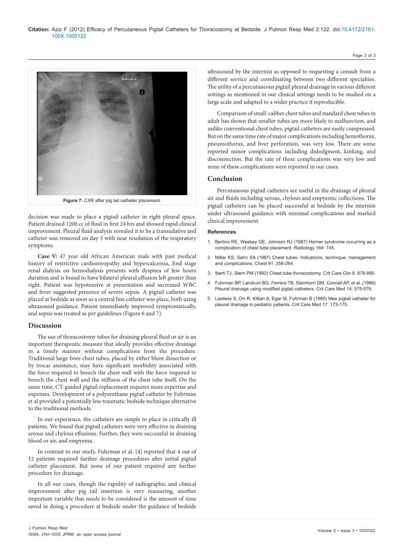
Case V: 47 year old African American male with past medical history of restrictive cardiomyopathy and hypercalcemia, End stage renal dialysis on hemodialysis presents with dyspnea of few hours duration and is found to have bilateral pleural effusion left greater than right. Patient was hypotensive at presentation and increased WBC and fever suggested presence of severe sepsis. A pigtail catheter was placed at bedside as soon as a central line catheter was place, both using ultrasound guidance. Patient immediately improved symptomatically, and sepsis was treated as per guidelines (Figure 6 and 7).
DiscussionThe use of thoracostomy tubes for draining pleural fluid or air is an
important therapeutic measure that ideally provides effective drainage in a timely manner without complications from the procedure. Traditional large-bore chest tubes, placed by either blunt dissection or by trocar assistance, may have significant morbidity associated with the force required to breech the chest wall with the force required to breech the chest wall and the stiffness of the chest tube itself. On the same time, CT-guided pigtail replacement requires more expertise and expenses. Development of a polyurethane pigtail catheter by Fuhrman et al provided a potentially less traumatic bedside technique alternative to the traditional methods.
In our experience, the catheters are simple to place in critically ill patients. We found that pigtail catheters were very effective in draining serous and chylous effusions. Further, they were successful in draining blood or air, and empyema.
In contrast to our study, Fuhrman et al. [4] reported that 4 out of 12 patients required further drainage procedures after initial pigtail catheter placement. But none of our patient required any further procedure for drainage.
In all our cases, though the rapidity of radiographic and clinical improvement after pig tail insertion is very reassuring, another important variable that needs to be considered is the amount of time saved in doing a procedure at bedside under the guidance of bedside
ultrasound by the internist as opposed to requesting a consult from a different service and coordinating between two different specialties. The utility of a percutaneous pigtail pleural drainage in various different settings as mentioned in our clinical settings needs to be studied on a large scale and adapted to a wider practice if reproducible.
Comparison of small-caliber chest tubes and standard chest tubes in adult has shown that smaller tubes are more likely to malfunction, and unlike conventional chest tubes, pigtail catheters are easily compressed. But on the same time rate of major complications including hemothorax, pneumothorax, and liver perforation, was very low. There are some reported minor complications including dislodgment, kinking, and disconnection. But the rate of these complications was very low and none of these complications were reported in our cases.
ConclusionPercutaneous pigtail catheters are useful in the drainage of pleural
air and fluids including serous, chylous and empyemic collections. The pigtail catheters can be placed successful at bedside by the internist under ultrasound guidance with minimal complications and marked clinical improvement.
References
1. Bertino RE, Wesbey GE, Johnson RJ (1987) Horner syndrome occurring as a complication of chest tube placement. Radiology 164: 745.
2. Miller KS, Sahn SA (1987) Chest tubes. Indications, technique, management and complications. Chest 91: 258-264.
3. Iberti TJ, Stern PM (1992) Chest tube thoracostomy. Crit Care Clin 8: 879-895.
4. Fuhrman BP, Landrum BG, Ferrara TB, Steinhorn DM, Connell AP, et al. (1986) Pleural drainage using modified pigtail catheters. Crit Care Med 14: 575-576.
5. Lawless S, Orr R, Killian A, Egar M, Fuhrman B (1989) New pigtail catheter for pleural drainage in pediatric patients. Crit Care Med 17: 173-175.
Figure 7: CXR after pig tail catheter placement.