Embed Size (px)
Citation preview

AXIOM® CONCEPTRADIOGRAPHIC EVALUATIONAT 1-YEAR FOLLOW-UP
INTERNAL DATA
Bone stability aroundAxiom® Implants

anthogyr
2
Since Branemark’s osseointegration principles1 were developed, crestal bone level alterations around implants are considered a reliable criterion to evaluate the successful outcome of a dental implant.
According to Albrektsson et al.2, X-rays of implants with an external hex design should show no peri-implant radiolucency, and the annual vertical bone loss should be less than 0.2 mm after the fi rst year of function. However, this study does not take into account the amount of crestal bone lost during the fi rst year. In 1981, Adell3 published a study in which he stated that more than 50% of the bone loss occurring over a 12-month period actually takes place within 3 months of loading. Two years later,4 he further stated that the greatest loss occurs during the fi rst 12 months following abutment placement.
In 2007, Misch et al. published a consensus paper published a consensus paperet al. published a consensus paperet al. 5 in which they defi ned implant quality of health criteria for rating implant success, survival, and failure. The James-Misch scale was modifi ed to criteria for rating implant success, survival, and failure. The James-Misch scale was modifi ed to four categories: success, satisfactory survival, compromised survival, and failure. An implant is four categories: success, satisfactory survival, compromised survival, and failure. An implant is considered a clinical success when radiographic peri-implant bone loss is less than 2 mm from considered a clinical success when radiographic peri-implant bone loss is less than 2 mm from initial surgery.
Two factors are most infl uential in reducing bone loss:Two factors are most infl uential in reducing bone loss:1. Tight implant-abutment junction to avoid bacterial colonization which results in soft tissue1. Tight implant-abutment junction to avoid bacterial colonization which results in soft tissue6
infl ammation. It has been demonstratedIt has been demonstrated7 that systems which feature a conical connection like that systems which feature a conical connection like the Axiom® implant system exhibit the lowest rate of microgaps (absence of microvements at the implant system exhibit the lowest rate of microgaps (absence of microvements at the implant-abutment interface under loading conditions) as compared to systems with a fl at-to-fl at implant-abutment interface under loading conditions) as compared to systems with a fl at-to-fl at connection. The very low rate of bacterial leakage in implant systems with a conical connection is connection. The very low rate of bacterial leakage in implant systems with a conical connection is well documented in the literature well documented in the literature 8,9.
2. Platform switching - This concept was introduced in the literature by Lazzara 2. Platform switching - This concept was introduced in the literature by Lazzara et al.et al.10 and Gardner11. As a matter of fact, platform switching seems to be an effective way to reduce the . As a matter of fact, platform switching seems to be an effective way to reduce the bone loss resulting from microgaps, and also to improve the predictibility of long-term treatment bone loss resulting from microgaps, and also to improve the predictibility of long-term treatment outcome by allowing preservation of peri-implant hard and soft tissue.outcome by allowing preservation of peri-implant hard and soft tissue.12,13,14,15,16
Several reviews of the literature and meta-analysesSeveral reviews of the literature and meta-analyses17,18 of comparative clinical studies of platform switched (PS) vs platform-matched (PM) implants show that a smaller amount of bone loss is seen switched (PS) vs platform-matched (PM) implants show that a smaller amount of bone loss is seen with PS implants (-0.20 mm vs -0.55 mm according to Atiehwith PS implants (-0.20 mm vs -0.55 mm according to Atieh18 ; -0.24 mm vs -0.86 mm according to Annibalito Annibali17).
Furthermore, Atieh et al.18 showed that a difference in implant and abutment diameters equal to or greater than 0.4 mm resulted in higher bone response.
This clinical study which involves 168 Axiom® implants evaluated at 1-year follow-up shows that excellent bone stability can be achieved through the use of an adequate connection system and platform switching.
INTRODUCTION

M U L T I C E N T E R C L I N I C A L S T U D I E S A T 1 - Y E A R F O L L O W - U P
3
MATERIALS AND METHODS
In order to evaluate the suitability of the surgical and prosthetic phases, practitioners were requested to fi ll in a questionnaire at each of the above mentioned stages.
Rating of implant success was performed after the fi rst year of function, according to the criteria defi ned by Misch5:1. No pain or tenderness upon function2. 0 mobility3. <2 mm radiographic bone loss from initial surgery4. No exudates history.
Implant failure5 was defi ned by any of the following:1. Pain on function2. Mobility3. Radiographic bone loss > 1/2 length of implant4. Uncontrolled exudates5. Implant no longer in mouth.
Changes in crestal bone level were measured by an independent dental surgeon using the Scion Image software. This software allows to compute pixel values and scale images. To achieve the highest accuracy, the software was calibrated using the implant length (the longest known distance reference on X-ray).
INVESTIGATIONAL CENTERS AND PATIENTSThis follow-up study was conducted in 10 investigational centers, some of which were already involvedin other follow-up studies. All practitioners were Axiom® implant system users.In total 84 patients have been involved in this study, 168 Axiom® implants were placed and followed during1 year.
STUDY DESIGNEach patient was evaluated at different stages:1. At subcrestal implant placement in a one-stage or two-stage surgery(according to surgeon preferences);2. At sutures removal (if necessary), 2 weeks after surgery;3. At loading, that is, 2-3 months post-surgery;4. At placement of the fi nal prosthesis;5. At 6-month and 1-year follow-ups.
RATING OF IMPLANT SUCCESS

anthogyr
4
G CLINICAL FOLLOW-UP
Table 1: Results of the clinical follow-up of Axiom® REG implants
Investigational centers 5Patients 17Implants followed for 1 year 18Bone gain 2Bone loss <2 mm 2Bone loss >2 mm 0Implants no longer in mouth 0
SUCCESS RATE 100 %
In 16 out of 18 implants, the X-rays taken at implantation and at 12-month follow-up showed no bone loss. Increase in bone density was found in 2 implants and bone loss was only 0.5 mm, which was considered a success according to the criteria defi ned in the literature.5
No device-related adverse events have been reported.
RESULTS (Table 1)
Seventeen patients recruited from 5 centers received a total of 18 implants (4.0 mm x 10.0 mm) which were placed in the posterior tooth region. All restored teeth were premolars or molars (Fig. 1). There were only single-tooth replacements and all of them were performed in one stage surgery.
PATIENTS
Fig. 1 - Restored teeth
035 36 37 46 47
2
4
6
8
10
Num
ber o
f im
plan
ts
Implantation site

M U L T I C E N T E R C L I N I C A L S T U D I E S A T 1 - Y E A R F O L L O W - U P
5
CLINICAL CASE courtesy of Dr. Carlos Francischone Jr, Brazil
ADDITIONAL RADIOGRAPHS
Fig 2: Initial status
Fig 4: Immediate postoperative X-ray Fig 6: 12 months later. Implant is stableand esthetic result is optimal
Fig 3: Implant placement
Fig 5: X-ray taken at 12 months postop with the crown in situ
X-rays of 3 cases followed for one year. No change in bone level.
Case 1:
Post-surgery
1-year follow-up
Case 2: Case 3:

anthogyr
6
Table 2: Results of the clinical follow-up of Axiom® PX implants
Investigational centers 4Patients 58Implants followed for 1 year 127Bone gain 1Bone loss <2 mm 7Bone loss >2 mm 3Implants no longer in mouth 1
SUCCESS RATE 96,8 %
All the implants but 4 were rated successful. These 4 implants were very challenging cases:
- One implant was lost in a female patient who said that she had placed excessive load on her tooth. The implant was replaced successfully.- In one patient treated with a 10 unit bridge supported by 5 implants placed in fresh sockets immediately after tooth extraction, one implant had 2 mm of marginal bone loss.- In one patient treated with a 4 unit bridge supported by 2 implants, one implant had 2 mm of marginal bone loss.- In a full upper arch restoration with immediate loading, one implant had 3 mm of marginal bone loss.
In the last 3 cases, bone loss did not adversely affect the stability of the restoration.In 7 implants, bone loss was less than 2 mm.
RESULTS (Table 2)
Fifty-eight patients recruited from 4 centers received a total of 127 implants: 49% of the implants supported single-tooth restorations, 34% supported complete dentures, and 17% supported bridges (Fig. 7).
In 71 cases, the implants were placed in fresh sockets immediately after tooth extraction.
PATIENTS
Fig 7: Percentage distribution of restorations
Full arch
One tooth
Several
49%34%
17%
C CLINICAL FOLLOW-UP

M U L T I C E N T E R C L I N I C A L S T U D I E S A T 1 - Y E A R F O L L O W - U P
7
CLINICAL CASE courtesy of Dr. Carlos Francischone Jr, Brazil
ADDITIONAL RADIOGRAPHS
Fig 8: Horizontal fractureof tooth number 11
Fig 11: Immediate loading
Fig 12,13 and 14: 12 months later. Esthetic result is optimal
Fig 10: Implant placement Fig 9: Atraumatic extraction
X-rays of 3 cases followed for one year. No change in bone level.
Case 1: Case 2: Case 3:
Post-surgery
1-year follow-up

anthogyr
8
Table 3: Results of the clinical follow-up of Axiom® 2.8 implants
Investigational centers 4Patients 11Implants followed for 1 year 23Bone gain 2Bone loss <2 mm 1Bone loss >2 mm 0Implants no longer in mouth 0
SUCCESS RATE 100 %
1 out of the 11 patients had 1 mm bone loss, which is considered a success according to the criteria defi ned in the literature.5 In the other 10 patients, no bone loss was noted. Increase in bone density even occurred in 2 patients. No adverse events have been reported.
RESULTS (Table 3)
Eleven patients with restricted mesial-distal space recruited from 4 centers received a total of 23 Axiom® 2.8 implants. In 50% of the patients, edentulism resulted from periodontal problems, in 25% from agenesis, and in the remaining patients, from trauma and cysts. Six implants were placed in fresh sockets immediately after tooth extraction. All the surgeries were performed in one stage. Restored teeth were mostly mandibular central incisors (Fig. 15).
PATIENTS
Fig 15: Restored teeth
12 13 22 3223 4131 42
1
2
3
4
5
6
7
8
Num
ber o
f im
plan
ts
Implantation site
B CLINICAL FOLLOW-UP
M U L T I C E N T E R C L I N I C A L S T U D I E S A T 1 - Y E A R F O L L O W - U P
9
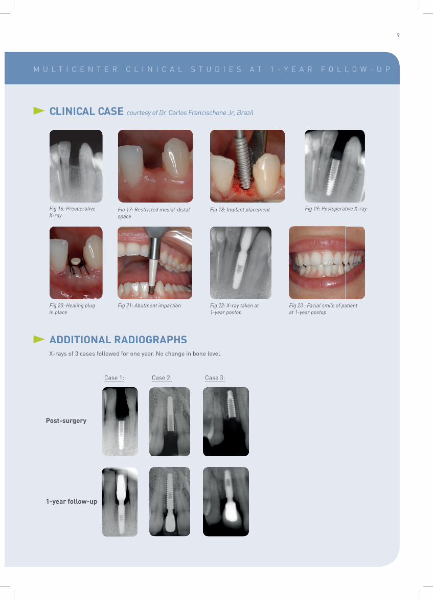
CLINICAL CASE courtesy of Dr. Carlos Francischone Jr, Brazil
ADDITIONAL RADIOGRAPHSX-rays of 3 cases followed for one year. No change in bone level.
Fig 16: PreoperativeX-ray
Fig 19: Postoperative X-ray
Fig 20: Healing plug in place
Fig 21: Abutment impaction Fig 22: X-ray taken at 1-year postop
Fig 23 : Facial smile of patientat 1-year postop
Fig 18: Implant placement Fig 17: Restricted mesial-distalspace
Case 1: Case 2: Case 3:
Post-surgery
1-year follow-up

anthogyr
10
Overall, from the 168 implants followed for one year, only 3 exhibited bone loss equal to or greater than 2 mm (Table 4). Within the limitation of this clinical follow-up, bone levels found confi rm that Axiom® implants have an excellent success rate at one year: 97.6 %.
Figures 25 and 26 show an increase in bone density and bone apposition induced by platform switching. This can be attributed to the excellent biocompatibility of the implant surface and the stable connection system. As far as this study goes, both have been found to promote long-term stability of Axiom® implants.
Moreover, mean bone loss in the whole series was -0.09 mm, much lower than the 2 mm loss which is considered by Misch5 as a criterion of success, and lower than the results published in the literature (-0.20 mm according to Atieh,18 and -0.24 mm according to Annibali17 for platform-switched implants) (Fig 24).
CONCLUSION
Table 4: Results of the clinical follow-up of Axiom® implants
G C B Total
Investigational centers 5 4 4 10Patients 17 58 11 84Implants followed for 1 year 18 127 23 168Bone gain 2 1 2 4Bone loss <2 mm 2 7 1 10Bone loss >2 mm 0 3 0 3Implants no longer in mouth 0 1 0 1
SUCCESS RATE 100 % 96,9 % 100 % 97,6 %
Fig 25: Postperative X-ray Fig 26: 1-year follow-up
X-rays of an Axiom® REG implant from the study group:
Bonegain
Consensus Misch 2007 Atieh Annibali
-2
-0.20 -0.24
0
-0.5
-1
-1.5
-2
-2.5
Axiom®
ConceptAxiom®
REGAxiom®
PXAxiom®
2.8
-0.03 -0.11 -0.04 -0.09
Bon
e lo
ss (m
m)
Fig 24: Mean bone loss observed during the Axiom® clinical follow-up study

M U L T I C E N T E R C L I N I C A L S T U D I E S A T 1 - Y E A R F O L L O W - U P
11
1. Branemark PI, Hansson BO, Adell R, Breine U, Lindström J, Hallen O et al. Osseointegrated implants in the treatment of the edentulous jaw. Experience from a 10-year period. Scand J Plastic Recontr Surg 1977 ; 16 : 1-132.
2. Albrektsson T, Zarb G, Worthington P, Eriksson AR. The long-term effi cacy of currently used dental implants: a review and proposed criteria of success. Int J Oral Maxillofac Implants 1986 ; 1 : 25.
3. Adell, et al. A 15 year study of osseointegrated implants in the treatment of the edentulous jaw, Int. J. Oral. Surg, 1981 : 10 : 387-416.
4. Adell, et al. Clinical results of osseointegrated implants supporting fi xed prostheses in edentulous jaws, J. Prosthet. Dent, 1983.
5. Misch et al. Implant Success, Survival, and Failure: The International Congress of Oral Implantologists (ICOI) Pisa Consensus Conference. Implant Dentistry 2008, vol 17 n°1. 5-15
6. Abrahamsson I, Berglundh T, Lindhe J. Soft tissue response to plaque formation at different implant systems.A comparative study in the dog. Clin Oral Implants Res 1998 ; 9 : 73-79.
7. Zipprich, Micromovements at the Implant-Abutment Interface: Measurement, Causes, and Consequences. Implantologie. (Vol. 15,2007 Issue 1, p. 31-46).
8. Do Nascimento et al. Leakage of saliva through the implant-abutment interface: in vitro evaluation of three different implant connections under unloaded and loaded conditions, Int J Oral Maxillofac Implants, 2012 May-Jun ; 27(3) : 551-60.
9. Assenza et al. Bacterial leakage in implants with different implant-abutment connections: an in vitro study,J Periodontol, 2012 Apr ; 83(4) : 491-7.
10. Lazzara RJ, Porter SS. Platform switching: a new concept in implant dentistry for controlling postrestorative crestal bone levels. Int J Periodontics Restorative Dent. 2006 Feb ; 26(1) : 9-17.
11. Gardner DM. Platform switching as a means to achieving implant esthetics. N Y State Dent J 2005 ; 71 : 34-37.
12. Cappiello M, Luongo R et al. Evaluation of peri-implant bone loss around platform-switched implants ;Int J Periodontics Restorative Dent. 2008 Aug; 28(4) : 345-55.
13. Luongo R, Guidone PC, Cochetto R. Hard and soft tissue responses to the platform-switched technique ;Int J Periodontics Restorative Dent.2008 Dec; 28(6) : 551-7.
14. Canullo et al. Platform switching and marginal bone-level alterations: the results of a randomized-controlled trial, Clin. Oral Impl. Res. 21, 2010 / 115–121.
15. Enkling et al. Effect of platform switching on peri-implant bone levels: a randomized clinical trial, Clin. Oral Impl. Res. 22, 2011 / 1185–1192.
16. Serrano-Sanchez et al. The infl uence of platform switching in dental implants. A literature review, Med Oral Patol Oral Cir Bucal. 2011 May 1 ; 16(3) : 400-5.
17. Annibali S, Bignozzi I, Cristalli MP. Peri-implant marginal bone level: a systematic review and meta-analysis of studies comparing platform switching versus conventionally restored implants. Journal of Clinical Periodontology.2012 Nov ; 39(11) : 1097-113
18. Atieh MA, Ibrahim HM, Atieh AH. Platform switching for marginal bone preservation around dental implants:a systematic review and meta-analysis. Journal of Periodontology. 2010 Oct;81(10):1350-66.
REFERENCES
Acknowledgements to all contributors to this study: Dr André Adan (Créteil, France), Dr Franck Azam (Aix les Bains,France), Dr Francis Bailly (Vienne, France), Dr Michel Bergoin (Challes les Eaux, France), Dr Charles Durif (Chambéry, France),Dr Pierre Esseyric (Chamonix, France), Dr Carlos Francischone Jr (Sao Paolo, Brazil), Dr Christian Legros (Limoges, France),Dr Bertrand Rousselet (Ambérieu en Bugey, France), Dr Jacques Vermeulen (Flumet, France).

simeda®
customized &
solutions
A
EF
D
IMPLANTEO®a
Pho
to c
redi
t: A
ntho
gyr
- A
ll ri
ghts
res
erve
d. N
on c
ontr
actu
al p
hoto
s.
C172
_GB
- 2
0140
4
2 237, Avenue André Lasquin74700 Sallanches - FrancePhone +33 (0)4 50 58 02 37 Fax +33 (0)4 50 93 78 60
www.anthogyr.com