Embed Size (px)
Citation preview

AD_________________
Award Number: W81XWH-09-1-0300 TITLE: The Mechanism of Action of Unique Small Molecules that Inhibit the Pim Protein Kinase Blocking Prostate Cancer Cell Growth PRINCIPAL INVESTIGATOR: Andrew S. Kraft, M.D. CONTRACTING ORGANIZATION: Medical University of South Carolina Charleston, SC 29425 REPORT DATE: May 2012 TYPE OF REPORT: Revised Final PREPARED FOR: U.S. Army Medical Research and Materiel Command Fort Detrick, Maryland 21702-5012 DISTRIBUTION STATEMENT: Approved for Public Release; Distribution Unlimited The views, opinions and/or findings contained in this report are those of the author(s) and should not be construed as an official Department of the Army position, policy or decision unless so designated by other documentation.

REPORT DOCUMENTATION PAGE Form Approved
OMB No. 0704-0188 Public reporting burden for this collection of information is estimated to average 1 hour per response, including the time for reviewing instructions, searching existing data sources, gathering and maintaining the data needed, and completing and reviewing this collection of information. Send comments regarding this burden estimate or any other aspect of this collection of information, including suggestions for reducing this burden to Department of Defense, Washington Headquarters Services, Directorate for Information Operations and Reports (0704-0188), 1215 Jefferson Davis Highway, Suite 1204, Arlington, VA 22202-4302. Respondents should be aware that notwithstanding any other provision of law, no person shall be subject to any penalty for failing to comply with a collection of information if it does not display a currently valid OMB control number. PLEASE DO NOT RETURN YOUR FORM TO THE ABOVE ADDRESS. 1. REPORT DATE May 2012
2. REPORT TYPE Revised Final
3. DATES COVERED 1 May 2009 - 30 April 2012
4. TITLE AND SUBTITLE The Mechanism of Action of Unique Small Molecules that Inhibit the Pim Protein Kinase Blocking Prostate Cancer Cell Growth
5a. CONTRACT NUMBER
5b. GRANT NUMBER W81XWH-09-1-0300
5c. PROGRAM ELEMENT NUMBER
6. AUTHOR(S)
5d. PROJECT NUMBER
Andrew S. Kraft, M.D.
5e. TASK NUMBER
E-Mail: [email protected]
5f. WORK UNIT NUMBER 7. PERFORMING ORGANIZATION NAME(S) AND ADDRESS(ES)
8. PERFORMING ORGANIZATION REPORT NUMBER
Medical University of South Carolina Charleston, SC 29425
9. SPONSORING / MONITORING AGENCY NAME(S) AND ADDRESS(ES) 10. SPONSOR/MONITOR’S ACRONYM(S) U.S. Army Medical Research and Materiel Command
Fort Detrick, Maryland 21702-5012 11. SPONSOR/MONITOR’S REPORT NUMBER(S) 12. DISTRIBUTION / AVAILABILITY STATEMENT Approved for public release; distribution unlimited 13. SUPPLEMENTARY NOTES 14. ABSTRACT: The Pim protein kinase is over expressed in prostate cancer. To clarify the role of this protein in regulating prostate cancer growth we have investigated its mechanism of action. We have examined the ability of small molecule inhibitors of this kinase to combine with other agents and have developed two rationally developed dual therapies. First, because overexpression of Bcl-2 family members is implicated in chemotherapeutic resistance in prostate cancer, we investigated the cooperative effects of Pim kinase inhibition with ABT-737, a small molecule antagonist of Bcl-2 family members. Strikingly, the addition of ABT-737 to Pim inhibitors triggered a robust apoptosis of prostate cancer cells in vitro and in vivo. Second, we demonstrate that inhibition of AKT in prostate cancer cell lines not only induces the expression of multiple RTKs, but increases the protein levels of serine threonine protein kinase Pim-1. Pim-1 activity is identified as essential in the feedback regulation of RTK levels by AKT inhibition. Both tissue culture and animal experiments demonstrate that the combination of AKT and Pim inhibitors provides synergistic inhibition of tumor growth. Thus Pim inhibitor has potential activity to treat this cancer.
15. SUBJECT TERMS Pim Kinases, Prostate Cancer, Cell Cycle, Kinase Inhibitors, SKP-2
16. SECURITY CLASSIFICATION OF: U
17. LIMITATION OF ABSTRACT
18. NUMBER OF PAGES
19a. NAME OF RESPONSIBLE PERSON USAMRMC
a. REPORT U
b. ABSTRACT U
c. THIS PAGE U
UU
82
19b. TELEPHONE NUMBER (include area code)
Standard Form 298 (Rev. 8-98) Prescribed by ANSI Std. Z39.18

Table of Contents
Page
Introduction…………………………………………………………….………..….. 4
Body………………………………………………………………………………….. 4
Key Research Accomplishments………………………………………….…….. 8
Reportable Outcomes……………………………………………………………… 9
Conclusion…………………………………………………………………………… 10
Appendix…………………………………………………………………………… 12
3
4
INTRODUCTION (Subject, purpose, and scope)
The overexpression of the serine/threonine Pim protein kinase in normal or cancerous prostate
cells stimulates the growth of these cells. Pim is over expressed in human prostate cancer, and
its level may parallel the onset of metastatic disease. In animal models the expression of the
c-Myc protein is associated with increased Pim protein levels. Thus, data from tissue culture,
human and animal prostate cancer implicates the Pim protein in prostate tumorigenesis. In this
application we suggest that Pim regulates the levels of the cell cycle inhibitor p27. We have
hypothesized that this decrease will coordinate with elevated c-Myc in tumor cells to drive tumor
growth. Our laboratory has developed novel benzylidene thiazolidine-2, 4 diones, (D5, SMI-4a)
that inhibit the activity of Pim kinase. These agents reverse Pim activity and allow the level of
p27 to rise. This protein will then function to inhibit tumor growth. The first goal of this grant
was to understand the mechanism by which Pim protein kinases regulate p27. We have
completed this work and published the mechanism in the Journal of Biological Chemistry (see
reportable outcomes) in which we detail the ability of Pim to phosphorylate both Skp-2 and
Cdc27. The second goal of this work is to determine the mechanism by which Pim controls the
levels of c-Myc and whether c-Myc content parallels the level of Pim protein kinase. We have
examined this question in a paper in the Proceedings of the National Academy of Sciences in
which we demonstrate that the knock-out of all Pim protein kinases decreases the levels of c-
Myc protein (see reportable outcomes). Finally, the third goal of this proposal was to evaluate
the anti-tumor activity of the Pim protein kinases and decipher whether inhibition of one or more
isoforms was necessary to inhibit tumor growth. We have completed this goal and submitted a
publication to Journal of Experimental Medicine (see reportable outcomes).
BODY
Our research efforts have focused on the Statement of Work, and in an attempt to bring together
data that will satisfy the needs of a peer reviewed publication. The results of these experiments
are documented in the enclosed paper and have been submitted to the Journal of Biologic
Chemistry. Rather than separating these tasks into individual goals for simplicity we have
combined this description.
Task 2(a, b) in year 1 and Task 1 and 2 (a, b) in year 2. (Reportable outcomes: J Biol Chem 285
(38): 29128-29137, 2010) we have found that the overexpression of active Pim1 protein kinase
but not kinase dead decreases the level of p27 protein. To evaluate the mechanism we have
focused our attention on the Skp2 protein which bind directly to p27 and activates the E3 ligase
which ubiquinates p27 leading to its degradation. We demonstrate that knocking down Pim-1
kinase increases Skp-2 and decreases p27 levels. Similarly, we find that inhibition of Pim
kinases with increasing doses of our small molecule Pim kinase inhibitor, SMI-4a [called in the
past D5], decreases the level of Skp-2 and increases p27 protein. In tissue culture,
overexpression of Pim-1 in normal mouse prostate epithelial cells increased Skp-2 levels while
decreasing p27 protein. Experiments carried out in which the half-life of the Skp-2 protein was
evaluated by adding cycloheximide to cells demonstrated that the expression of Pim-1 markedly
increased the half-life of Skp-2 protein. These results suggest that the regulation of Skp-2 by

5
Pim-1 decreases p27 levels. This effect is inhibited by SMI-4a, the Pim kinase inhibitors we
have developed.
To evaluate how Pim and Skp-2 may interact we examined whether these proteins could
coimmunoprecipitate. Both kinases active and dead Pims will bind directly to the Skp-2 protein.
Immunoprecipitating Pim-1 from serum stimulated cells demonstrates that Pim and Skp-2
associate in a cell cycle dependent manner. To evaluate how Pim-1 might affect the half-life of
the Skp-2 protein we evaluated whether Pim-1 could regulate the ubiquination of Skp-2. We
find that if a tagged Skp-2 and 6xHis-ubiquitin plasmids are transfected into cells along with
Pim-1 that the ubiquination of Skp-2 is inhibited, explaining the increased levels of this protein.
In addition, we find that Pim-1 can phosphorylate Skp-2 and the addition of our inhibitor, SMI-
4a, can block the Pim-1 to modify the Skp-2 protein. Together these results suggest that the Pim
protein kinase binds to Skp-2 and inhibits its ubiquination possibly in part secondary to the
ability of Pim-1 to phosphorylate Skp-2.
We have used GST fusion proteins to evaluate the phosphorylation of Skp2. Skp-2 is thought to
be phosphorylated on serine 64 and 72. We have defined a new phosphorylation site on this
protein that is modified by Pim-1. If this site is mutated in Skp-2 it partially decreases the
phosphorylation of this protein. This result suggests that Skp-2 is a target of Pim and that 417
and potentially other sites are phosphorylated. Expanding this experimental approach mutation
of S64 or S72 also inhibits Pim phosphorylation of Skp-2. Our small molecule Pim inhibitor,
SMI-4a, blocks the Pim-induced phosphorylation of this protein. To evaluate the potential
effects of these phosphorylations on the half-life of this protein each of these three sites was
mutated, transfected into cells, which were then treated with cycloheximide and the half-life of
the protein examined by Western blotting. We find that each of these phosphorylation mutants
shortens the half-life of these proteins. Another approach to examining the importance of these
phosphorylation sites is to cotransfect Skp-2 and p27. If Skp-2 is active then p27 levels will be
lowered. We find in Figure 13c that mutation of T417 and to a greater extent S64 but not S72
affects the level of p27. In conclusion, Pim phosphorylates Skp-2 on S64, S72, and T417 and
this phosphorylation regulates the half-life of Skp-2 and thus the levels of p27. Our Pim
inhibitors block this phosphorylation and thus would raise the levels of p27.
To discern how Pim might regulate Skp-2 ubiquination, we examined in detail the APC/C
complex either Cdc20 or Cdc27. This E3 ligase is regulated by. We find that the addition of
Pim-1 to cells dissociates the Cdc27 regulator protein from the APC/C complex, and knocking
Pim-1 down using siRNA increases the amount of this protein. We find that Pim-1 is capable of
phosphorylating Cdc27 but not Cdc20 both in vitro and when over expressed in cells.
Based on the interaction between and Pim-1 protein kinase and Skp-2 we have transfected either
one or both of these proteins into prostate cells and examined the levels of p27 and the location
of cells in the cell cycle. We find that cells expressing both proteins have the lowest levels of
p27, the fewest cells in G1 and the most cells in the G2 phase of the cell cycle. This result is
highly significant. Our small molecule inhibitor of Pim-1 (SMI-4a) can block the ability of Skp-
2 or serum to drive cells into the G2 phase of the cell cycle and stimulate an increase in p27.
We include a model for how the Pim kinase can regulate the levels of Skp-2. Pim-1 can
phosphorylate both Skp-2 and CDC27 and lead to their destruction. This combined effect can be

6
inhibited by SMI-4a. Our novel Pim inhibitor can be developed as a compound that increases
the levels of p27, blocks progression through the cell cycle, and inhibits tumor growth.
Task 3 completed in year 3. (Reportable outcome - Proceedings of the National Academy of
Sciences. 108:528-533, 2011) This task asked us to evaluate the interaction between the c-Myc
protein and Pim kinase using our unique small molecule Pim inhibitor, SMI-4a. To evaluate this
interaction, we have chosen to step back from the complicated prostate cancer models usually
employed in Pim research, and attempt to initially simplify this analysis using MEFs from
animals that are missing all of the 3 Pim kinase isoforms. Because there is no Pim kinase
activity the biology of these cells mimics the effect of a small molecule Pim inhibitor with no off
target effects. We find that the triple KO cells do not grow well but the addition to these cells of
Pim 3 to these cells stimulated their growth similar to normal. In contrast Pim-2 stimulated
intermediate growth and Pim-1 alone was not capable of stimulating the growth of these cells at
all.
These results are of importance to the prostate cancer field because immunohistochemistry of
human prostate cancers compared to normal paired samples from the same patient showed that
Pim-3 was over expressed in prostate tumors.
These TKO cells evidenced lower levels of ATP and higher amounts of AMP. The increased
AMP is shown to stimulate the AMP-dependent protein kinase (AMPK) in these cells (Fig. 3b).
Interestingly the over expression of Pim-3 is shown to reverse the levels of AMP:ATP, and to
inhibit AMPK. Additionally, Pim-3 is shown to elevate the levels of c-Myc, both from MEFs
derived from Pim-3 only mice and TKO MEFs that express Pim-3 after transduction. To
examine how Pim-3 elevated the levels of c-Myc we first did cycloheximide treatment and
release. Results demonstrate that TKO/Pim-3 cells synthesize more c-Myc at earlier time points.
When a polysome protocol was carried out c-Myc mRNA was found to associate with the
polysomes in TKO/Pim-3 and WT MEFs, but in TKO MEFs the c-Myc mRNA was found to be
associated with free ribosomes, suggesting that 5’ initiated translation was blocked in the
absence of the Pims.
We also demonstrate that Pim-3 is capable of elevating the levels of the PGC-1a. Even though
both c-Myc and Pim-3 increase the growth of TKO MEFs only Pim-3 is able markedly increase
the level of PGC-1α protein. Transduction of PGC-1α into TKO MEFs raised the levels of ATP
and lowered the level of phosphorylated AMPK, suggesting that the ability of Pim-3 to control
this enzyme is essential for its mechanism of action.
We therefore propose a model by which Pim-3 regulate PGC-1α, lowering the levels of AMP,
stimulating mTORC1 activity and increasing the synthesis of c-Myc stimulating the growth of
these fibroblasts. We are in the process of validating this model in prostate cancer cells but
initial results suggest that there is a significant degree of overlap. First, we find that the levels of
Pim-3 are elevated in human prostate cancer cells by IHC. Second, PC-3 and LNCaP cells
transfected with Pim-3 grow much better in serum or serum free conditions, suggesting that Pim-
3 may be controlling cellular ATP levels.

7
Task 3 to be completed in year 3. (Reportable outcome: Cancer Research, 2012 Jan 1:72(1):294-
303) This task asked us to evaluate the activity of specific Pim inhibitors. We have carried out a
study of the Pim inhibitor SMI-4a and published this in Cancer Research in 2012. This work is
summarized as follows. Because overexpression of Bcl-2 family members is implicated in
chemotherapeutic resistance in prostate cancer, we investigated the cooperative effects of Pim
kinase inhibition with ABT-737, a small molecule antagonist of Bcl-2 family members.
Strikingly, the addition of ABT-737 to Pim inhibitors triggered a robust apoptosis of prostate
cancer cells in vitro and in vivo. Pim inhibitors decreased levels of the Bcl-2 family member
Mcl-1, both by blocking 5'-cap dependent translation and decreasing protein half life. In
addition, Pim inhibition transcriptionally increased levels of the BH3 protein Noxa by activating
the unfolded protein response (UPR), lead to eIF-2α phosphorylation and increased expression of
CHOP. Increased levels of Noxa also inactivated the remaining levels of Mcl-1 protein activity.
Notably, these specific protein changes were essential to the apoptotic process because ABT-737
did not inhibit Mcl-1 protein activity and Mcl-1 overexpression blocked the apoptotic activity of
ABT-737. Our results therefore suggest that this combination treatment could be developed as a
potential therapy for human prostate cancer where overexpression of Pim kinases and anti-
apoptotic Bcl-2 family members drives tumor cell resistance to current anticancer therapies.
(Reportable outcome: Journal of Experimental Medicine submitted) Another approach to
treating prostate cancer is targeting prostate cancer treatment is to block the AKT signaling
pathway. Through mutation of PTEN, PI3 kinase, or AKT itself, this pathway is activated in
prostate cancer in 50-70% of patients. For this reason pharmaceutical companies have invested
in developing molecules that inhibit these enzymes. Importantly, we find that when AKT
inhibitors are used to treat prostate cancer that they cause a feedback stimulation of the levels of
cell surface receptor tyrosine kinases (RTKs). This increase in RTKs will then enhance AKT
stimulation, blunting the effect of these drugs. We find that the AKT inhibitor, GSK690693, not
only increases RTK levels but of importance also elevates the levels of Pim-1, but not Pim-2, or
Pim-3. Results demonstrate that the AKT inhibitor (GSK-690693) increases Pim-1
transcriptionally.
Importantly, we have discovered that inhibiting or decreasing the level of Pim-1, blocks the
feedback ability of AKT inhibitors to elevate the levels of RTKs. This can be shown either using
genetic knockouts, siRNAs or the small molecule Pim-1 inhibitor, SMI-4a. This may suggest
that inhibiting only Pim-1 will be sufficient to block the feedback increase in RTKs.
The mechanism by which Pim-1 regulates RTKs is unknown. Inhibitors of the mTORC1
activity do not block this increase suggesting that it is cap-independent .We find that both
GSK690693 and Pim-1 are able to stimulate translation from an internal ribosome entry site
(IRES). In this case we show that c-Met can be activated. This experiment involved cloning the
IRES sequence of c-Met in front of firefly luciferase and then examining whether Pim-1 could
regulate this sequence. Decreases in Pim-1 levels decreased firefly luciferase while increasing
Pim-1 increases this readout. Based on these results we have combined a Pim-1 and AKT
inhibitor and shown that they can synergistically inhibit cell growth on plastic, soft-agar growth
of prostate cancer cells, and block the subcutaneous growth of tumors in nude mice. These data
suggest that this combination by blocking feedback regulation can inhibit tumor growth and may
be a novel drug therapy for prostate cancer.

8
KEY RESEARCH ACCOMPLISHMENTS
Increases in Pim-1 kinase decreases Skp-2 levels and thus allows p27 protein to increase.
The changes in these proteins are inhibited by the small molecule Pim-1 inhibitor we
have developed.
Pim-1 binds to Skp-2 and inhibits its ubiquination and thus degradation.
Skp-2 is phosphorylated by Pim-1 and our small molecule Pim inhibitors block this
effect.
Threonine 417 in Skp-2 is identified as a new phosphorylation site in Skp-2
Serines 64 and 72 are mapped as Pim phosphorylation sites.
Pim-directed phosphorylation controls the half-life and activity of Skp-2.
Pim regulates the phosphorylation of Cdc27 to bind to the APC/C, and controls the
phosphorylation of this protein.
Pim-1 plus Skp-2 enhances transition through the cell cycle.
Pim kinase inhibitory compounds block the ability of cells to transverse the cell cycle.
Pim inhibitor treatment of prostate cancer decreased levels of Bcl-2 family member Mcl-
1.
Incubation with Pim inhibitors raised the levels of the Noxa protein.
Noxa protein levels were increased by an activation of unfolded protein response that led
to eIF-2a phosphorylation and increased expression of CHOP.
These induced protein changes sensitized prostate cancer cells to the pro-apoptotic action
of ABT-737, a small molecule that inhibits the activity of Bcl-2 family members.
In animal models, ABT-737 and the Pim inhibitor SMI-4a (D5) had synergistic activity in
killing prostate cancers implanted subcutaneously.
Inhibition of AKT in prostate cancer cells elevates the levels of both Pim-1, but not Pim-
2 or Pim-3, and cell surface receptor tyrosine kinases (RTKs).
Inhibition of Pim-1 with inhibitor, SMI-4a, or decreasing the level of Pim-1 protein
inhibited the ability of AKT inhibitors to induce RTKs.
AKT inhibition leads to the transcriptional up regulation of Pim-1.
Pim-1 or small molecule AKT inhibitors are able to increase the levels of RTKs by a cap-
independent mechanism.
The combination of a Pim-1 and AKT inhibitor is synergistic in killing prostate cancer
cells in tissue culture and in an animal model.
Knocking down Pim-1 or inhibiting it with small molecule Pim inhibitor SMI-4a blocked
mTORC1 activity.
Inhibiting Pim-1 led to increases in AMP kinase activity, a known mTORC1 inhibitor
Mouse embryo fibroblasts (MEFs) which were knock-out for all three Pims (TKO) grew
much more slowly than wild type and had elevated ratios of AMP:ATP
TKO MEFs had elevated AMPK activity.
These MEFs had decreased rates of protein synthesis and decreased levels of c-Myc
protein.
Elevating the levels of Pim-3 alone was sufficient to markedly increase growth, stimulate
increases in c-Myc protein levels, and elevate the levels of PGC-1α.
Transduction of PGC-1α was sufficient to elevate ATP and c-Myc protein.

9
REPORTABLE OUTCOMES
Abstracts
1. Cen B, Zemskova M, Beharry Z, Smith C D, Kraft AS. The Pim-1 protein kinase decreases
p27 protein half-life and increases prostate epithelial cell migration. AACR Annual Meeting
2009
2. Beharry Z, Lin Y, Mahajan S, Zemskova M, Xia Z, Smith C, Kraft AS. The Pim protein
kinases modulate mTOR activity by regulating the protein and mRNA levels of mTOR
pathway components and cellular AMP levels. AACR Special Conference in Cancer
Research-Metabolism and Cancer. September 2009, La Jolla, CA
3. Kraft AS, Cen B, Zemskova M, Beharry Z, Smith C. Potent protein kinase inhibitors block
Pim kinase mediated increase in prostate epithelial cell migration, regulation of p27 protein
half-life and secretion of hepatocyte growth factor. AACR Molecular Targets and Cancer
Therapeutics Conference, 2009
4. Kraft AS, Beharry Z, Lin Y, Mahajan S, Zemskova M, Xia Z, Smith C. Treatment with the
Pim protein kinase inhibitor SMI-4a enhances AMPK phosphorylation, decreases Raptor
levels, and blocks mTORC1 activity. AACR Molecular Targets and Cancer Therapeutics
Conference, 2009
5. Song JH, Kraft AS. Mechanisms of Therapy-Induced Senescence with ABT-737. AACR
102nd Annual Meeting, 2011
6. Song JH, Kraft AS. Pim Protein Kinase Induce the Unfolded Protein Response and
Sensitize Prostate Cancer Cells to Killing by ABT-737-Mediated Apoptosis. AACR Annual
Meeting, Advances in Prostate Cancer Research, 2012
7. Song JH, Kraft AS. Pim Kinase Inhibitors Sensitize Cancer Cells to Apoptosis Triggered by
Bcl-2 Family Inhibitor ABT-737. AACR Annual Meeting, Advances in Prostate Cancer
Research, 2012
8. Cen B, Sandeep M, Kraft AS. Overcoming Resistance to Inhibitors of the AKT Protein
Kinases by Targeting the Pim Protein Kinase Pathway. AACR Annual Meeting, Advances
in Prostate Cancer Research, 2012
Papers
1. Cen B, Sandeep M, Zemskova M, Beharry Z, Lin YW, Cramer SD, Lilly M, and Kraft AS.
Regulation of SKP2 Levels by the PIM-1 Protein Kinase. J Biol Chem 285 (38): 29128-
29137, 2010. PMID 20663873, PMCID: PMC2937943
2. Beharry Z, Mahajan S, Zemskova M, Lin Y-W, Tholanikunnel B, Xia Z, Smith CD, and
Kraft AS. The Pim protein kinases regulate energy metabolism and cell growth. Proceedings
of the National Academy of Sciences. 108:528-533, 2011, PMID:21187426, PMCID:
PMC3021022
3. Song JH, Kraft AS. Pim Kinase Inhibitors Sensitize Prostate Cancer Cells to Apoptosis
Triggered by Bcl-2 Family Inhibitor ABT-737. Cancer Research, 2012 Jan 1:72(1):294-303,
PMID: 22080570, PMCID: PMC3251634 (available 2013/1/1)
4. Cen B, Mahajan S, Wang W, and Kraft AS. Pim-1 Mediates the Elevation of Receptor
Tyrosine Kinases Induced by Small Molecule AKT Inhibitors in Prostate Cancer Cells.
Submitted Journal of Experimental Medicine 2012

10
CONCLUSIONS
We can conclude from this research that Pim remains an important target for treatment of human
prostate cancer. The Pim-1 protein kinase clearly plays a role in controlling the levels of p27 and
transition through the cell cycle. This work demonstrates that small molecule inhibitors of this
protein kinase, such as SMI-4a, will have activity as a treatment for prostate cancer.
Human prostate cancer cell lines and tumors have all three Pims, 1, 2 and 3. Further research
needs to qualify whether inhibition of one or all three Pims is needed to inhibit cell cycle
movement to a greater extent than inhibition of Pim-1 alone. Answering this question will
greatly assist in the development of targeted therapies to inhibit prostate cancer growth.
The serine/threonine Pim kinases are overexpressed in prostate cancers and promote cell growth
and survival. Here, we find that a novel Pim kinase inhibitor, SMI-4a, or Pim-1 siRNA blocked
the rapamycin-sensitive mammalian target of rapamycin (mTORC1) activity by stimulating the
phosphorylation and thus activating the mTORC1 negative regulator AMP-dependent protein
kinase (AMPK). Mouse embryonic fibro- blasts (MEFs) deficient for all three Pim kinases [triple
knockout (TKO) MEFs] demonstrated activated AMPK driven by elevated ratios of AMP:ATP
relative to wild-type MEFs. Consistent with these findings, TKO MEFs were found to grow
slowly in culture and have decreased rates of protein synthesis secondary to a diminished amount
of 5-cap–dependent translation. Pim-3 expression alone in TKO MEFs was sufficient to reverse
AMPK activation, increase protein synthesis, and drive MEF growth similar to wild type. Pim-3
expression was found to markedly increase the protein levels of both c-Myc and the peroxisome
proliferator-activated receptor gamma coactivator 1α (PGC-1α), enzymes capable of regulating
glycolysis and mitochondrial biogenesis, which were diminished in TKO MEFs. Overexpression
of PGC-1α in TKO MEFs elevated ATP levels and inhibited the activation of AMPK. These
results demonstrate the Pim kinase-mediated control of energy metabolism and thus regulation of
AMPK activity. We identify an important role for Pim-3 in modulating c-Myc and PGC-1α
protein levels and cell growth. We find by IHC of human samples that Pim-3 is elevated in
human prostate cancer, suggesting that these results will impact on human prostate cancer
growth.
The serine threonine Pim protein kinases are overexpressed in prostate cancers and promote cell
growth and survival. The PI3K/AKT pathway is activated in over 60% of human prostate
cancers, suggesting that compounds that inhibit this pathway may be useful for therapy.
However, accumulating evidence indicates that feedback regulation in response to inhibition of
PI3K/AKT signaling pathway may attenuate antitumor activity of inhibitors by increasing the
level of cell surface receptor tyrosine kinases (RTKs). We demonstrate that inhibition of AKT in
prostate cancer cell lines not only induces the expression of multiple RTKs, but increases the
protein levels of serine threonine protein kinase Pim-1. Pim-1 activity is identified as essential in
the feedback regulation of RTK levels by AKT inhibition. Knockdown of Pim-1 expression or
inhibition of Pim-1 activity with small molecules abrogates the induction of RTKs and
overexpression of Pim-1 increases RTK levels. Experiments using dual luciferase vectors
demonstrate that Pim-1 controls expression of c-Met and other RTKs at the translational level by
modulating IRES activity in a cap-independent manner. Both tissue culture and animal
experiments demonstrate that the combination of AKT and Pim inhibitors provides synergistic

11
inhibition of tumor growth. Our results demonstrate that Pim-1 is a novel mediator of resistance
to AKT inhibition, and that targeting Pim kinases significantly improves the efficacy of AKT
inhibitors in anticancer therapy.
So What -
The combination therapy of a Pim and AKT inhibitor could be brought into the clinic. This
work might suggest that inhibition of Pim-1 and not Pim-2 or 3 might be sufficient as dual
therapy. Alternatively, the observations in mouse embryo fibroblasts suggest that it is important
to inhibit all Pim kinases. This work suggests that Skp-2 levels and phosphorylation might act as
a biomarker of drug action. Our published work from this grant funding also demonstrates
additional possible combinations of Pim inhibitors and agents that block the Bcl-2 pathway.

12
APPENDIX
Abstracts
1. Cen B, Zemskova M, Beharry Z, Smith C D, Kraft AS. The Pim-1 protein kinase decreases
p27 protein half-life and increases prostate epithelial cell migration. AACR Annual Meeting
2009
2. Beharry Z, Lin Y, Mahajan S, Zemskova M, Xia Z, Smith C, Kraft AS. The Pim protein
kinases modulate mTOR activity by regulating the protein and mRNA levels of mTOR
pathway components and cellular AMP levels. AACR Special Conference in Cancer
Research-Metabolism and Cancer. September 2009, La Jolla, CA
3. Kraft AS, Cen B, Zemskova M, Beharry Z, Smith C. Potent protein kinase inhibitors block
Pim kinase mediated increase in prostate epithelial cell migration, regulation of p27 protein
half-life and secretion of hepatocyte growth factor. AACR Molecular Targets and Cancer
Therapeutics Conference, 2009
4. Kraft AS, Beharry Z, Lin Y, Mahajan S, Zemskova M, Xia Z, Smith C. Treatment with the
Pim protein kinase inhibitor SMI-4a enhances AMPK phosphorylation, decreases Raptor
levels, and blocks mTORC1 activity. AACR Molecular Targets and Cancer Therapeutics
Conference, 2009
5. Song JH, Kraft AS. Mechanisms of Therapy-Induced Senescence with ABT-737. AACR
102nd Annual Meeting, 2011
6. Song JH, Kraft AS. Pim Protein Kinase Induce the Unfolded Protein Response and
Sensitize Prostate Cancer Cells to Killing by ABT-737-Mediated Apoptosis. AACR Annual
Meeting, Advances in Prostate Cancer Research, 2012
7. Song JH, Kraft AS. Pim Kinase Inhibitors Sensitize Cancer Cells to Apoptosis Triggered by
Bcl-2 Family Inhibitor ABT-737. AACR Annual Meeting, Advances in Prostate Cancer
Research, 2012
8. Cen B, Sandeep M, Kraft AS. Overcoming Resistance to Inhibitors of the AKT Protein
Kinases by Targeting the Pim Protein Kinase Pathway. AACR Annual Meeting, Advances
in Prostate Cancer Research, 2012
Papers
1. Cen B, Sandeep M, Zemskova M, Beharry Z, Lin YW, Cramer SD, Lilly M, and Kraft AS.
Regulation of SKP2 Levels by the PIM-1 Protein Kinase. J Biol Chem 285 (38): 29128-
29137, 2010. PMID 20663873, PMCID: PMC2937943
2. Beharry Z, Mahajan S, Zemskova M, Lin Y-W, Tholanikunnel B, Xia Z, Smith CD, and
Kraft AS. The Pim protein kinases regulate energy metabolism and cell growth. Proceedings
of the National Academy of Sciences. 108:528-533, 2011, PMID:21187426, PMCID:
PMC3021022
3. Song JH, Kraft AS. Pim Kinase Inhibitors Sensitize Prostate Cancer Cells to Apoptosis
Triggered by Bcl-2 Family Inhibitor ABT-737. Cancer Research, 2012 Jan 1:72(1):294-303,
PMID: 22080570, PMCID: PMC3251634 (available 2013/1/1)
4. Cen B, Mahajan S, Wang W, and Kraft AS. Pim-1 Mediates the Elevation of Receptor
Tyrosine Kinases Induced by Small Molecule AKT Inhibitors in Prostate Cancer Cells.
Submitted Journal of Experimental Medicine 2012

The Pim-1 protein kinase decreases p27 protein half-life
and increases prostate epithelial cell migration
Bo Cen, Marina Zemskova, Zanna Beharry, Charles D. Smith, Andrew S. Kraft
Medical University of South Carolina, Charleston, SC
Abstract
Pim-1 proto-oncogene encodes a serine/threonine protein kinase that regulates apoptosis, cell
cycle progression, and transcription. The expression of this protein kinase is elevated in both
prostate intraepithelial neoplasia (PIN) and prostatic adenocarcinoma, suggesting an important
role for this protein kinase in prostate cancer growth and development. However, the mechanism
by which Pim-1 promotes cell proliferation and transformation is not fully understood. To
examine the role of kinase in regulating prostate cancer cell growth, we have overexpressed Pim
in mouse prostate epithelial cells (MPECs), a cell line that demonstrates stem cell characteristics.
We find that overexpression of Pim-1 markedly increases hepatocyte growth factor/ scatter factor
(HGF/SF) induced migration of MPECs. This increased migration is completely inhibited by two
novel small molecule Pim inhibitors 16a and 4a. Additionally we find that HGF/SF induced p27
upregulation in MPECs was strongly inhibited by the overexpression of Pim-1. Further studies
showed that, overexpression of Pim-1 did not alter the mRNA level of p27, but enhanced the
degradation and thus decreased the half-life of the p27 protein. The p27 degradation induced by
Pim-1 was cell cycle dependent. Consistent with this finding ubiquitylation assays examining
p27 demonstrate that Pim-1 increases this modification in vivo. We find that the Pim-1-mediated
ubiquitinylation is regulated by complex formation between Pim-1 and Skp2. Skp2 levels, as part
of the SCF protein complex, regulate the ubiquintylation and degradation of p27. Pim-1 does not
affect Skp2’s E3 ligase activity, but appears to inhibit the degradation of this protein. Pim-1
directly phosphorylates Skp2 in vitro, suggesting a mechanism for the stabilization of this
protein. Together our data demonstrate the complex pathway by which Pim-1 protein kinase
regulates p27 levels, and thus controls cell proliferation and possibly transformation.

The Pim protein kinases modulate mTOR activity by regulating the
protein and mRNA levels of mTOR pathway components
and cellular AMP levels
Zanna Beharry1, Ying-Wei Lin
2, Sandeep Mahajan
4, Marina Zemskova
3, Zuping Xia
1,
Charles Smith1,4
and Andrew S. Kraft4
1Departments of Pharmaceutical and Biomedical Sciences, South Carolina College of Pharmacy,
2Department of Pediatrics,
3Department of Cell and Molecular Pharmacology, and
4 The Hollings
Cancer Center, Medical University of South Carolina, Charleston, SC 29425
Abstract
We have identified a small molecule inhibitor of the Pim protein kinases, SMI-4a, and
found that the addition of this compound to cells blocks the phosphorylation of the
mTOR regulatory protein PRAS40 and subsequently the activity of the mTOR pathway
(Mol. Cancer. Ther. (2009) 8: 1473; Cancer Biol. Ther. (2009) 8: 846). Now we show
that the addition of SMI-4a to malignant cells increases the phosphorylation of AMPK
on Thr 172 in a LKB1-dependent manner, induces the phosphorylation of raptor on
Ser792, decreases the levels of Raptor protein, and inhibits mTORC1 activity.
Immunoprecipitation of mTOR from SMI-4a treated cells consistently showed lower
levels of bound Raptor and in vitro mTOR kinase assays showed a decreased ability to
phosphorylate 4E-BP1. Addition of proteasome inhibitors to these cells reversed the
decrease in Raptor protein levels, suggesting that Pim kinases prevent the ubiquitin-
mediated degradation of this protein. Knockdown of PIM-1 via siRNA in K562 leukemic
cells showed increased AMPK phosphorylation and decreased raptor protein levels,
further demonstrating an important role for Pim in regulating AMPK phosphorylation
and Raptor levels. Additionally, mouse embryo fibroblasts (MEFs) deficient for Pim-1,
Pim-2 and Pim-3 kinase (TKO MEFs) showed a significantly increased level of AMPK
phosphorylation compared to wild type MEFs. This was attributed to an increase in the
cellular level of AMP in TKO MEFs as measured by HPLC. Furthermore, the correlation
between increased AMPK phosphorylation and a lower level of raptor protein observed
with SMI-4a treatment or siRNA knockdown of Pim-1 in leukemic cells was also
observed in TKO MEFs, along with a decrease in mTOR protein level. Transfection of
individual Pim kinases into these TKO MEFs was able to reverse these effects,
decreasing AMPK phosphorylation, and increasing raptor and mTOR protein levels.
The cellular activity of mTORC1 was difficult to assess in TKO MEFs as we found
substantially lower protein levels of the mTORC1 substrates 4E-BP1 and p70S6K.
However, mTORC2 activity was unaffected by Pim kinase knockout as Akt was readily
phosphorylated upon serum stimulation of TKO MEFs. QT-PCR analysis of TKO MEFs
demonstrated that the decrease in protein levels of 4E-BP1 correlated with a decrease in
the mRNA encoding this protein. Our data demonstrate an important role for the Pim
family of protein kinases in regulating the mTOR pathway by affecting the level of the
pathway components through transcriptional and post-transcriptional mechanisms, as
well as regulation of cellular AMP levels.

Potent protein kinase inhibitors block Pim kinase-mediated increase in
prostate epithelial cell migration, regulation of p27 protein half-life,
and secretion of hepatocyte growth factor
Andrew S. Kraft, Bo Cen, Marina Zemskova, Zanna Beharry and Charles D. Smith
Medical University of SC Hollings Cancer Center, Charleston, SC.
Abstract
Pim-1 proto-oncogene encodes a serine/threonine protein kinasethat regulates apoptosis, cell
cycle progression, and transcription.The expression of this protein kinase is elevated in both
prostateintraepithelial neoplasia (PIN) and prostatic adenocarcinoma,suggesting an important
role for Pim-1 kinase in prostate cancerdevelopment and growth. To investigate the role of Pim-1
incontrolling tumor growth we have synthesized novel benzylidene-thiazolidine2,4-dione (J.
Med. Chem. (2009) 52:74) inhibitors of this kinase.The most potent members of this chemotype
have IC50s of 13 nMfor Pim-1. To examine the activity of these agents in prostatecancer we
have first set out to define the biochemical activityof Pim-1 in epithelial cells. We have
expressed Pim-1 in a mouseprostate epithelial cell (MPECs) line that demonstrates stemcell
characteristics. We find that Pim-1 expressing cells produceand secrete markedly increased
levels of hepatocyte growth factor/scatterfactor (HGF/SF), and this protein kinase stimulates
increasesin HGF/SF mRNA. Additionally, expression of Pim-1 markedly increasesHGF/SF
induced migration of MPECs. The contribution of Pim-1kinase to this biochemical pathway is
confirmed in murine embryonicfibroblasts (MEFs) that are deficient for all three Pim
proteinkinases (TKO) and evidence reduced level of HGF mRNA in TKOversus wild type
MEFs. The Pim-1-stimulated migration of MPECsis inhibited by two benzylidene-thiazolidine-2,
4-diones, SMI-4aand 16a, as well as known inhibitors of the HGF receptor, c-Met.HGF
treatment of MPECs induced p27 upregulation that could beinhibited by the expression of Pim-1,
thus allowing cell cycleprogression. Further studies showed that expression of Pim-1did not alter
the mRNA level of p27, but enhanced the cell cycle-dependentdegradation and thus decreased
the half-life of the p27 protein.Consistent with this finding, p27 ubiquitination assays showedthat
Pim-1 increases this modification in vivo. We found thatthe Pim-1-mediated ubiquitination is
regulated by complex formationbetween Pim-1 and Skp2, a protein component of the SCF
complex,which is known to regulate the ubiquitination and degradationof p27. Pim-1 does not
affect Skp2's E3 ligase activity, butappears to inhibit the degradation of Skp2 through
phosphorylation.Incubation of these cells with the Pim protein kinase inhibitor,SMI-4a,
decreased Skp2 expression and increased p27 and cyclinE expression. Together our data
demonstrates the complex pathwayby which Pim-1 protein kinase regulates HGF/SF and p27
levels,thus controlling cell migration, proliferation, and potentiallytransformation. Potent Pim
kinase inhibitors block these twosignaling pathways thus inhibiting prostate epithelial
cellmigration and growth.

Treatment with the Pim protein kinase inhibitor SMI-4a enhances AMPK
phosphorylation, decreases Raptor levels, and blocks mTORC1 activity
Andrew S. Kraft1, Zanna Beharry1, Ying-Wei Lin1, Sandeep Mahajan1, Marina Zemskova1,
Zuping Xia2 and Charles Smith1
1 Medical University of SC Hollings Cancer Center, Charleston, SC; 2 Medical University of SC
College of Pharmacy, Charleston, SC.
Abstract
We have identified a class of small molecule inhibitors of the Pim protein kinases, benzylidene
thiazolidine-2-4 diones (J.Med. Chem. (2009) 52:74) with the most potent members havingIC50s
of 13 nM for Pim-1 and 2.3 µM for Pim-2. Compounds in this chemotype demonstrated
selectivity of more than 2500-foldand 400-fold for Pim-1 or Pim-2 respectively while other
congenershad equivalent potency towards both isozymes. In vivo, these molecules inhibited Pim
kinase autophosphorylation and in amurine model inhibited the growth of subcutaneously
implanted murine adenocarcinoma JC cells. One of the members of this chemotype,SMI-4a, has
been shown to block the phosphorylation of the mTORregulatory protein PRAS40 and
subsequently the activity of themTOR pathway (Mol. Cancer. Ther. (2009) 8: 1473; Cancer
Biol.Ther. (2009) 8: 846). Now we show that the addition of SMI-4ato malignant cells increases
the phosphorylation of AMPK onThr 172 in a LKB1-dependent manner, induces the
phosphorylationo f Raptor on Ser792, decreases the levels of Raptor protein,and inhibits
mTORC1 activity. Immunoprecipitation of mTOR fromSMI-4a treated cells consistently showed
lower levels of bound Raptor and in vitro mTOR kinase assays from treated cells demonstrated a
decreased ability to phosphorylate 4E-BP1. Knockdown of PIM-1via siRNA in K562 leukemic
cells showed increased AMPK phosphorylation and decreased Raptor protein levels, further
demonstrating an important role for Pim kinase in regulating AMPK phosphorylationand Raptor
levels. Additionally, mouse embryo fibroblasts (MEFs)deficient for Pim-1, Pim-2 and Pim-3
kinase (TKO MEFs) showed a significantly increased level of AMPK phosphorylation compared
to wild type MEFs, which correlated with decreased mTORC1 activity and increased binding of
4E-BP1 with eIF-4E. The TKO MEFs grew significantly more slowly than wild type. The
decreased mTORC1activity correlated with an increase in the cellular level of AMP in TKO
MEFs. Furthermore, the correlation between increased AMPK phosphorylation and a lower level
of Raptor protein observed with SMI-4a treatment was also observed in TKO MEFs. Infectionof
TKO MEFs with lentiviruses expressing Pim 1 or Pim2 was able to reverse these effects,
decreasing AMPK phosphorylation, and increasing Raptor protein levels. The cellular activity of
mTORC1was difficult to assess in TKO MEFs as we found substantially lower protein levels of
the mTORC1 substrates 4E-BP1 and p70S6K.Akt was readily phosphorylated upon serum
stimulation of TKOMEFs and mTORC2 activity was unchanged. Given the role of Pim kinase in
regulating mTORC1 activity, we have combined SMI-4aand the mTOR inhibitor rapamycin
inducing synergistic blockade of this pathway.
Presentation Abstract
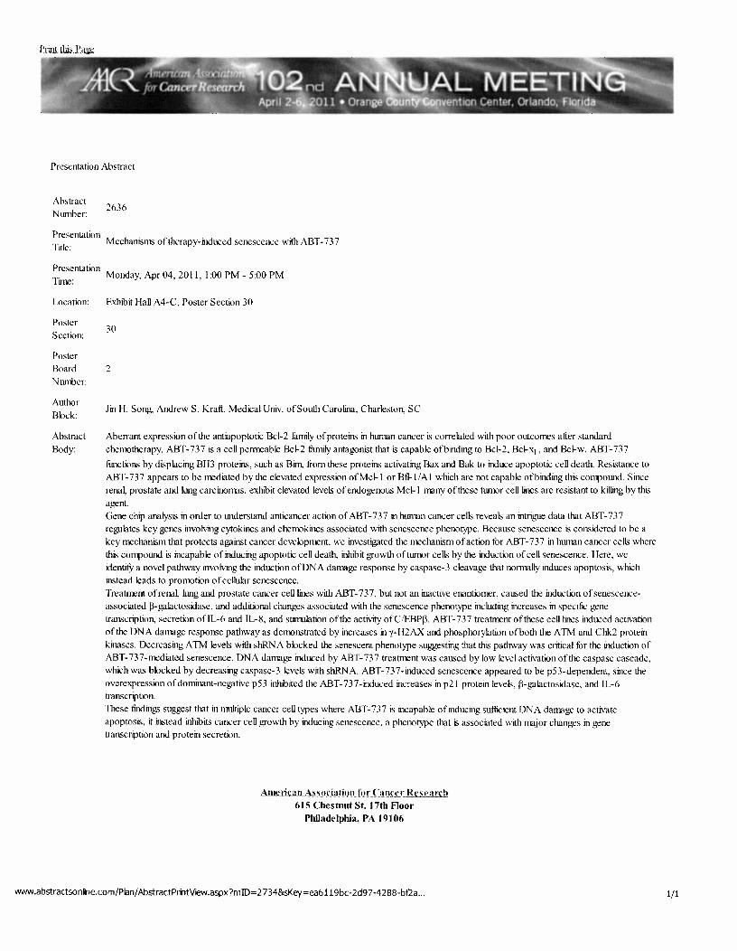
Abstract Nwnher:
2636
Presentation Title: Mechanism; of therapy-induced senescence with ABT-737
Presentation Time: Monday, Apr 04, 2011, I :00 PM- 5:00PM
Location:
Poster Section:
Poster
Board
NlllliJer:
Author
Block:
Abstract
Body:
Exhibit Hall A4-C, Poster Section 30
30
2
Jin H. Song, AndrewS. Kraft. Medical Univ. of South Carolina, Charleston, SC
Aberrant expression of the antiapoptotic Bcl-2 family of proteins in human cancer is correlated with poor outcomes after standard
chellKJtherapy. ABT-737 is a cell permeable Bcl-2 fumily antagonist that is capable ofbinding to Bcl-2, Bcl-xr_, and Bcl-w. ABT-737
fi.mctions by displacing BH3 proteins, such as Bim, from these proteins activating Bax and Bak to induce apoptotic cell death. Resistance to
ABT-73 7 appears to be mediated by the elevated expression ofMcl-1 or Btl- I /A I which are not capable ofbinding this compound. Since
rena~ prostate and lung carcinomas, exhibit elevated levels of endogenous Mcl-1 many of these tumor cell lines are resistant to killing by this
ah>ent. Gene chip analysis in order to tn1derstand anticancer action of ABT-737 in human cancer cells reveals an intrigue data that ABT-737 regulates key genes involving cytokines and chellKlkines associated with senescence phenotype. Because senescence is considered to be a
key mechanism that protects against cancer development, we investigated the mechanism of action for ABT-737 in human cancer cells where
this CO!TJIOund is incapable of inducing apoptotic cell death, inhibit growth oftuJOOr cells by the induction of cell senescence. Here, we
identil)r a novel pathway involving the induction ofDN A damage response by caspase-3 cleavage that normally induces apoptosis, which instead leads to prollKltion of cellular senescence.
Treatment of rena~ lung and prostate cancer cell lines with ABT-737, but not an inactive enantiomer, caused the induction of senescence
associated ~-galactosidase, and additional changes associated with the senescence phenotype including increases in specific gene transcription, secretion ofiL-6 and IL-8, and stimulation of the activity ofC/EBP~. ABT-737 treatment of these cell lines induced activation
of the DNA damage response pathway as dellKJnstrated by increases in y-H2AX and phosphorylation ofboth the A TM and Cbk2 protein
kinases. Decreasing A TM levels with shRN A blocked the senescent phenotype suggesting that this pathway was critical for the induction of ABT-737-mediated senescence. DNA damage induced by ABT-737 treatment was caused by low level activation of the caspase cascade,
which was blocked by decreasing caspase-3 levels with shRNA. ABT-737-induced senescence appeared to be p53-dependent, since the overexpression of dominant-negative p53 inhibited the ABT-737-induced increases in p21 protein levels, ~-h>alactosidase, and JL-6 transcription.
These findings suggest that in muhiple cancer cell types where ABT-737 is incapable of inducing sufficient DNA damage to activate apoptosis, it instead inhibits cancer cell growth by inducing senescence, a phenotype that is associated with major changes in gene transcription and protein secretion.
Ame!ican Association for Cancer Research 615 Chestnut St. 17th Floor
Philadelphia, PA 19106
'NWW .abstractsonfne.com/Pian/ AbstractPmtVteW.aspx?miD = 2734&sKey =ea6119bc-2d97 -4 288-bf2a ... 1/1

8/31/12 Abstract Prilt Vew
Prim thi.s Page
I : I I 1::. L . ...
Presentation Abstract
Abstract Nwroer:
LB-487
Presentation Title: Pim protein kinase inhibitors induce the tu1lolded protein response and sensitize prostate cancer cells to killing by ABT-737-rrediated apoptosis.
Presentation Tore: Wednesday, Apr 04,2012,8:00 AM -12:00 PM
Location:
Author Block:
Abstract Body:
McComlick Place West (Hall F), Poster Section 37
Jin H Song, AndrewS Kra(1. Medical University of South Carolina, Charleston, SC
Pun serine/threonine kinases contribute to prostate trnnorigenesis and therapeutic resistance, yet anticancer efficacy ofPim kinase inhibitors on prostate cancer i5 tu1known. We demonstrate for the first time that a genetically engineered decrease in Pim kinase levels or the addition ofPim kiimse small molecule inhibitors (e.g. SMI-4a) to hUimU prostate cancer cells. including LNCaP and PC-3, causes ER stress aod activates the unfolded protein response (UPR) stimulating increases in efF-2a phosphorylation, ATF-4, CHOP proteins, and cleavage ofXBP-1. Because (I) Bcl-2 titmi.Jy proteins reside on the ER lwren aod play an important role in regulation ofXBP- 1 splicing, (2) Bcl-2 titnlily rrembers are over expressed in prostate cancer to enhance Pim kinase anti-cancer activity, and (3) Pim kiimse inhibitors decrease Mel- I and increase Noxa protein, we treated prostate cancer cell lines with SM1-4a plus the Bcl-2 fumi.Jy antagonist ABT- 737, a small molecule antagonist ofBcl-2 liunily rrembers. Strikingly, the addition of ABT-737 to Pim inhibitors triggered a robust apoptosis of prostate cancer cells in vitro and in vivo. Pun inhibitors decreased levels of the Bcl-2 fittnily rrerroer Mcl-1, both by blocking 5'-cap dependent translation and decreasing protein halftife. Additionally, Pim inhibition transcriptionally increased levels of the BH3 protein Noxa by activating the UPR, lead to elF2a phosphorylation and increased expression of CHOP. Increased levels ofNoxa also inactivated the remaining levels ofMcl-1 protein activity. Notably, these specific protein changes were essential to the apoptotic process because ABT-737 did not inhibit Mel- I protein activity and Mcl-1 overexpression blocked the apoptotic activity of ABT-737. We find that the Pim kinase inhibitors and ABT- 73 7 are highly synergistic in killing prostate cancer both in vitro and when used together in a subcutaneous animal model of prostate cancer therapy. Our resuhs therefOre suggest that this combination treatment could be developed as a potential therapy for human prostate cancer where overexpression ofPin1 kinases and antiapoptotic Bcl-2 titmi.Jy rrembers drives nnmr cell resistance to current anticancer therapies.
American As~odation lor Can•·cr Rescan:!! 615 Chestnut St. 17th Floor
Philadelphia, PA 19106
www.abstractso n fne.com/PI3n/ AbstractPriltVew .aspx? mID= 2898&sKey =4 2 7 4eb3a-82f5-4ea9-b009 ... 1/1

Pim kinase inhibitors sensitize prostate cancer cells to apoptosis
triggered by Bcl-2 family inhibitor ABT-737
Song JH, Kraft AS.
Department of Biochemistry and Molecular Biology, Hollings Cancer Center, Medical
University of South Carolina, Charleston, South Carolina 29425, USA. [email protected]
Abstract
Pim serine/threonine kinases contribute to prostate tumorigenesis and therapeutic resistance, yet
Pim kinase inhibitors seem to have only limited effects on prostate cancer cell survival. Because
overexpression of Bcl-2 family members are implicated in chemotherapeutic resistance in
prostate cancer, we investigated the cooperative effects of Pim kinase inhibition with ABT-737,
a small molecule antagonist of Bcl-2 family members. Strikingly, the addition of ABT-737 to
Pim inhibitors triggered a robust apoptosis of prostate cancer cells in vitro and in vivo. Pim
inhibitors decreased levels of the Bcl-2 family member Mcl-1, both by blocking 5'-cap
dependent translation and decreasing protein half-life. In addition, Pim inhibition
transcriptionally increased levels of the BH3 protein Noxa by activating the unfolded protein
response (UPR), lead to eIF-2α phosphorylation and increased expression of CHOP. Increased
levels of Noxa also inactivated the remaining levels of Mcl-1 protein activity. Notably, these
specific protein changes were essential to the apoptotic process because ABT-737 did not inhibit
Mcl-1 protein activity and Mcl-1 overexpression blocked the apoptotic activity of ABT-737. Our
results therefore suggest that this combination treatment could be developed as a potential
therapy for human prostate cancer where overexpression of Pim kinases and antiapoptotic Bcl-2
family members drives tumor cell resistance to current anticancer therapies.

Overcoming Resistance to Inhibitors of the AKT Protein Kinases by
Targeting the Pim Protein Kinase Pathway
Bo Cen, Sandeep Mahajan, and Andrew S. Kraft, The Hollings Cancer Center at the Medical
University of South Carolina, Charleston, South Carolina 29425
Abstract
The AKT protein kinases are an important signal transduction target for the inhibition of prostate
cancer growth. AKT is activated in 50-80% of cancers secondary to deletions, mutations and loss
of heterozygosity of the PTEN phosphatase. Resistance to small molecule chemical inhibitors of
protein kinases in human patients involves the induction of alternative signal transduction
pathways. We demonstrate that the addition of the pan-AKT inhibitor GSK690693 to prostate
cancer cell lines, Du145, PC3 and PC3-LN4 induced marked up-regulation of multiple receptor
tyrosine kinases, including c-Met, Her3, IGF-IR, INSR and EphA2. Increases in these receptors
have the potential to block the growth inhibitory action of this AKT kinase inhibitor. In addition
to changes in these RTKs, GSK690693 also markedly increased the levels of the Pim-1 protein
kinase. Importantly, down regulation of Pim-1 either through using cells derived from
genetically engineered knock-out mice, knocking down the levels of Pim-1 with siRNAs or
treating cells with Pim-1 inhibitory small molecules, blocked the ability of GSK690693
treatment to induce increases in RTKs. In comparison, knockdown of wild type levels of Pim-1
was sufficient to lead to decreased expression of endogenous RTKs. The addition of GSK690693
and the Pim-1 inhibitor SMI-4a to PC3-LN4 cells caused a highly synergistic inhibition of
growth both in tissue culture and in soft agar assay. When these agents were administered to
immune-compromised mice that had been previously injected subcutaneously with PC3-LN4
cells, the combination markedly inhibited tumor growth where each agent given alone had only a
partial inhibitory effect. Based on these results we suggest that the Pim-1 protein kinase plays a
critical role in the induction of RTKs by AKT inhibitors. Thus combination therapy of prostate
cancer with a Pim and AKT inhibitor is likely to be a novel effective therapeutic strategy to
overcome potential resistance mechanisms to AKT kinase inhibitors.
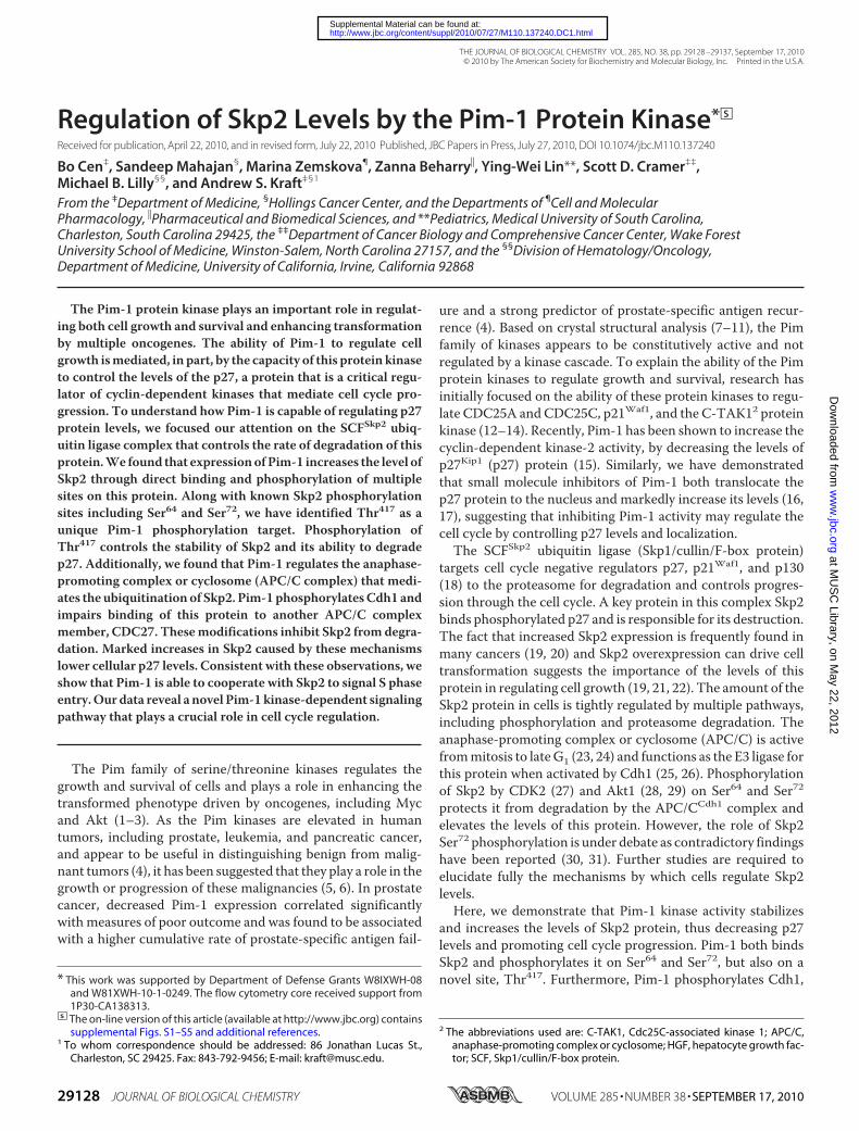
Regulation of Skp2 Levels by the Pim-1 Protein Kinase*□S
Received for publication, April 22, 2010, and in revised form, July 22, 2010 Published, JBC Papers in Press, July 27, 2010, DOI 10.1074/jbc.M110.137240
Bo Cen‡, Sandeep Mahajan§, Marina Zemskova¶, Zanna Beharry�, Ying-Wei Lin**, Scott D. Cramer‡‡,Michael B. Lilly§§, and Andrew S. Kraft‡§1
From the ‡Department of Medicine, §Hollings Cancer Center, and the Departments of ¶Cell and MolecularPharmacology, �Pharmaceutical and Biomedical Sciences, and **Pediatrics, Medical University of South Carolina,Charleston, South Carolina 29425, the ‡‡Department of Cancer Biology and Comprehensive Cancer Center, Wake ForestUniversity School of Medicine, Winston-Salem, North Carolina 27157, and the §§Division of Hematology/Oncology,Department of Medicine, University of California, Irvine, California 92868
The Pim-1 protein kinase plays an important role in regulat-ing both cell growth and survival and enhancing transformationby multiple oncogenes. The ability of Pim-1 to regulate cellgrowth ismediated, in part, by the capacity of this protein kinaseto control the levels of the p27, a protein that is a critical regu-lator of cyclin-dependent kinases that mediate cell cycle pro-gression. To understand how Pim-1 is capable of regulating p27protein levels, we focused our attention on the SCFSkp2 ubiq-uitin ligase complex that controls the rate of degradation of thisprotein.We found that expressionofPim-1 increases the level ofSkp2 through direct binding and phosphorylation of multiplesites on this protein. Along with known Skp2 phosphorylationsites including Ser64 and Ser72, we have identified Thr417 as aunique Pim-1 phosphorylation target. Phosphorylation ofThr417 controls the stability of Skp2 and its ability to degradep27. Additionally, we found that Pim-1 regulates the anaphase-promoting complex or cyclosome (APC/C complex) that medi-ates the ubiquitination of Skp2. Pim-1phosphorylatesCdh1 andimpairs binding of this protein to another APC/C complexmember, CDC27. Thesemodifications inhibit Skp2 fromdegra-dation. Marked increases in Skp2 caused by these mechanismslower cellular p27 levels. Consistent with these observations, weshow that Pim-1 is able to cooperate with Skp2 to signal S phaseentry.Our data reveal a novel Pim-1 kinase-dependent signalingpathway that plays a crucial role in cell cycle regulation.
The Pim family of serine/threonine kinases regulates thegrowth and survival of cells and plays a role in enhancing thetransformed phenotype driven by oncogenes, including Mycand Akt (1–3). As the Pim kinases are elevated in humantumors, including prostate, leukemia, and pancreatic cancer,and appear to be useful in distinguishing benign from malig-nant tumors (4), it has been suggested that they play a role in thegrowth or progression of these malignancies (5, 6). In prostatecancer, decreased Pim-1 expression correlated significantlywith measures of poor outcome and was found to be associatedwith a higher cumulative rate of prostate-specific antigen fail-
ure and a strong predictor of prostate-specific antigen recur-rence (4). Based on crystal structural analysis (7–11), the Pimfamily of kinases appears to be constitutively active and notregulated by a kinase cascade. To explain the ability of the Pimprotein kinases to regulate growth and survival, research hasinitially focused on the ability of these protein kinases to regu-late CDC25A and CDC25C, p21Waf1, and the C-TAK12 proteinkinase (12–14). Recently, Pim-1 has been shown to increase thecyclin-dependent kinase-2 activity, by decreasing the levels ofp27Kip1 (p27) protein (15). Similarly, we have demonstratedthat small molecule inhibitors of Pim-1 both translocate thep27 protein to the nucleus and markedly increase its levels (16,17), suggesting that inhibiting Pim-1 activity may regulate thecell cycle by controlling p27 levels and localization.The SCFSkp2 ubiquitin ligase (Skp1/cullin/F-box protein)
targets cell cycle negative regulators p27, p21Waf1, and p130(18) to the proteasome for degradation and controls progres-sion through the cell cycle. A key protein in this complex Skp2binds phosphorylated p27 and is responsible for its destruction.The fact that increased Skp2 expression is frequently found inmany cancers (19, 20) and Skp2 overexpression can drive celltransformation suggests the importance of the levels of thisprotein in regulating cell growth (19, 21, 22). The amount of theSkp2 protein in cells is tightly regulated by multiple pathways,including phosphorylation and proteasome degradation. Theanaphase-promoting complex or cyclosome (APC/C) is activefrommitosis to lateG1 (23, 24) and functions as the E3 ligase forthis protein when activated by Cdh1 (25, 26). Phosphorylationof Skp2 by CDK2 (27) and Akt1 (28, 29) on Ser64 and Ser72
protects it from degradation by the APC/CCdh1 complex andelevates the levels of this protein. However, the role of Skp2Ser72 phosphorylation is under debate as contradictory findingshave been reported (30, 31). Further studies are required toelucidate fully the mechanisms by which cells regulate Skp2levels.Here, we demonstrate that Pim-1 kinase activity stabilizes
and increases the levels of Skp2 protein, thus decreasing p27levels and promoting cell cycle progression. Pim-1 both bindsSkp2 and phosphorylates it on Ser64 and Ser72, but also on anovel site, Thr417. Furthermore, Pim-1 phosphorylates Cdh1,* This work was supported by Department of Defense Grants W8IXWH-08
and W81XWH-10-1-0249. The flow cytometry core received support from1P30-CA138313.
□S The on-line version of this article (available at http://www.jbc.org) containssupplemental Figs. S1–S5 and additional references.
1 To whom correspondence should be addressed: 86 Jonathan Lucas St.,Charleston, SC 29425. Fax: 843-792-9456; E-mail: [email protected].
2 The abbreviations used are: C-TAK1, Cdc25C-associated kinase 1; APC/C,anaphase-promoting complex or cyclosome; HGF, hepatocyte growth fac-tor; SCF, Skp1/cullin/F-box protein.
THE JOURNAL OF BIOLOGICAL CHEMISTRY VOL. 285, NO. 38, pp. 29128 –29137, September 17, 2010© 2010 by The American Society for Biochemistry and Molecular Biology, Inc. Printed in the U.S.A.
29128 JOURNAL OF BIOLOGICAL CHEMISTRY VOLUME 285 • NUMBER 38 • SEPTEMBER 17, 2010
at MU
SC
Library, on May 22, 2012
ww
w.jbc.org
Dow
nloaded from
http://www.jbc.org/content/suppl/2010/07/27/M110.137240.DC1.html Supplemental Material can be found at:

impairing its association with CDC27 and inhibiting APC/Cactivity, thus protecting Skp2 from degradation.
EXPERIMENTAL PROCEDURES
Antibodies, Drugs, and Reagents—Anti-Pim-1 (19F7) anti-body was produced and purified in this laboratory. Anti-cyclinE (HE12), anti-Met (25H2), anti-phospho-Met (D26), Myc tag(71D10), anti-AKT, anti-phospho-AKT (S473), and anti-polo-like kinase-1 antibodies were purchased from Cell SignalingTechnology. Anti-p27 (C19), anti-CDC27 (AF3.1), and anti-cyclin B1 (H-433) were from Santa Cruz Biotechnology. Anti-�-actin (AC-15), anti-FLAG M2, anti-HA (HA-7), and anti-�-tubulin (TUB 2.1) antibodies were from Sigma. Anti-Skp2 andanti-Cks1 antibodies were from Invitrogen/Zymed Laborato-ries Inc.. Anti-His tag antibody was from Qiagen. Anti-Cdh1(DH01) antibody was from Abcam. Anti-lamin B anti-body was from Calbiochem.Roscovitine and reagents for in vitro ubiquitination assay
were from Biomol. Cycloheximide, MG132, LY294002, wort-mannin, nocodazole, and thymidine were from Sigma.GSK690693 was provided by Glaxo Smith Kline.Recombinant human HGF was from Antigenix America.
Active GST-tagged Pim-1 was from SignalChem. Active His-tagged human Pim-1 was purified from Escherichia coli using aCalbiochemnickel-nitrilotriacetic acid column. GST andGST-Skp2 proteins were purified from E. coli using glutathione-Sepharose 4B resin (GE Healthcare).Plasmids—A Pim-1 siRNA plasmid and the control plasmid
were described previously (32). pGIPZ Pim-1 shRNA con-structs were from Open Biosystems.pCMV-Skp2 plasmid expressing FLAG-tagged Skp2 was
kindly provided by Dr. Liang Zhu (33). Site-directed mutantswere prepared using PCR based on this plasmid. HA-Cdh1 andHA-Cdc20 plasmids were described elsewhere (34). The Ubc3and ubiquitin plasmids have been previously described (35).The HA-Pim-1 and FLAG-Pim-1 constructs were generated bysubcloning murine Pim-1 cDNA into pcDNA3 vector, and theK67M (HA-tagged) mutant was constructed using PCR. AnN-terminally truncatedmutant (NT81) of Pim-1 was describedpreviously (36). Lentiviral expression constructs pLEX-Pim-1and pLEX-Skp2 was obtained by subcloning human Pim-1 andSkp2 cDNAs into pLEX vector (Open Biosystems). A humanPim-1 construct, pcDNA3-Pim-1, was described elsewhere(32).Cell Culture, Transfections, Transductions, and Cell Syn-
chronization—Cell lines were grown in RPMI 1640 medium(PC3) orDMEM(HeLa,HEK293T, Rat1, andmouse embryonicfibroblasts). The triple knock-out mouse of the Pim-1, -2, -3genes used to isolate mouse embryonic fibroblasts weredescribed previously (17). Mouse prostate epithelial cells wereisolated as described (37). HEK293T cells were transfected bythe calcium phosphate method, and HeLa cells were trans-fectedwith Lipofectamine 2000 reagent. Lentiviruses were pro-duced and transduced into Rat1 cells using kits from OpenBiosystems.For synchronization experiments, HeLa cells were treated
with 2 mM thymidine for 18 h, washed, and released into freshmedium for 9 h. Then, a second thymidine treatment was
applied to yield cells at the G1/S transition. Mitotic HeLa cellswere obtained by treating HeLa cells with 2 mM thymidine for24 h, washing, and releasing into freshmedium for 3 h. The cellswere then treated with 100 ng/ml nocodazole for 12 h.Ubiquitination Assays—In vitro p27 ubiquitination assays
were performed essentially as described (38). In brief, theSCFSkp2 complex was expressed and purified from insect cells(39) andmixedwith in vitro-translated 35S-labeled p27 that hadpreviously been incubatedwith cyclin E/Cdk2 alongwithmeth-ylated ubiquitin and ubiquitin aldehyde for 60min at 30 °C. Thereaction was stopped with 2� SDS sample buffer and run onpolyacrylamide gels. In vivo ubiquitination assays were per-formed as described (40). HEK293T cells were transfected withthe indicated plasmids for 24 h, treated with 10 �M MG132 for6 h, and lysed in denaturing buffer (6 M guanidine-HCl, 0.1 M
Na2HPO4/NaH2PO4, 10 mM imidazole). The cell extracts werethen incubated with nickel beads for 3 h, washed, and subjectedto immunoblot analysis.In Vitro and in Vivo Phosphorylation Assay—FLAG-Skp2 or
its mutants were immunoprecipitated with anti-FLAG anti-body from HEK293T cells. Immune complexes were washedthree times in radioimmune precipitation assay lysis buffer (150mM NaCl, 10 mM Tris-HCl, pH 7.5, 1% Nonidet P-40, 0.5%deoxycholate, 0.1%SDS), thenwashed twice in 1� kinase buffer(25 mM Tris-HCl, pH 7.5, 5 mM �-glycerophosphate, 2 mM
dithiothreitol, 0.1 mM Na3VO4, 10 mM MgCl2, 2 �M unlabeledATP) and incubated with 0.5 �g of recombinant active Pim-1kinase and 2 �Ci of [�-32P]ATP in 3 �l of total reaction bufferfor 30 min at 30 °C. Phosphorylation of Cdh1 or Cdc20 wasdetected using in vitro translated proteins produced by TNTCoupled Reticulcyte Lysate System (Promega). Reactions werestopped by washing twice in kinase buffer and boiling in 2�SDS loading buffer. Proteins were resolved by 9% SDS-PAGE,and 32P incorporation was detected by autoradiography. For invivo labeling experiments, HeLa cells were transfected with theindicated plasmids for 24 h, and the medium was changed tophosphate-free DMEM with 0.5% dialyzed FBS containing 200�Ciml�1 ortho-32PO4 for 4 h. Cells were lysed by radioimmuneprecipitation assay buffer for immunoprecipitation, and theimmune complexes were subjected to 9% SDS-PAGE followedby autoradiography analysis.Flow Cytometry—Cell cycle distribution was monitored by
FACS analysis of ethanol-fixed, propidium iodide-stainedcells on a Becton Dickinson FACSCalibur Analytical FlowCytometer.BrdU Incorporation Assay—Rat1 cells were seeded in 96-well
plates (3000 cells/well) and maintained as described in the fig-ure legends. An ELISA BrdU kit (Roche Applied Science) wasused to assay the cell cycle. Absorbance at 370 nm (referencewavelength 492 nm) was measured using a Molecular Devicesmicroplate reader.Densitometry Analysis—Densitometry was determined with
ImageJ version 1.42q software (National Institutes of Health)with normalization to the corresponding controls (�-actin orinput).Statistical Analysis—All assays were repeated at least three
times. The results of quantitative studies are reported asmean � S.D. Differences were analyzed by Student’s t test. p �
Pim-1 Regulates Skp2 Levels
SEPTEMBER 17, 2010 • VOLUME 285 • NUMBER 38 JOURNAL OF BIOLOGICAL CHEMISTRY 29129
at MU
SC
Library, on May 22, 2012
ww
w.jbc.org
Dow
nloaded from

0.05 was regarded as significant, and such differences are indi-cated in the figures.
RESULTS
Pim-1 Stabilizes Skp2 Protein—Overexpression inHeLa cellsof wild type Pim-1 but not a kinase-dead mutant, K67M, leadsto a decrease in the level of the p27 protein (Fig. 1A) without anychange in the mRNA level of this protein (supplementalFig. S2D). To evaluate the mechanism by which Pim-1 func-tions, we focused attention on the E3 ligase SCF complex that
targets p27 for proteasomal degra-dation and in particular the Skp2protein which is known to directlybind p27. Western blots demon-strate that transfection of the Pim-1kinase increases the levels of Skp2protein (Fig. 1A), while converselysiRNA or shRNA (supplementalFig. S1A) knockdown of endoge-nous Pim-1 expression reducesSkp2 levels. The interplay betweenthese two proteins is further dem-onstrated by the observation thattransfection of murine Pim-1 intoHeLa cells in which endogenousenzyme has been knocked downagain elevates the level of Skp2 (Fig.1A). Using two small molecule Pimkinase inhibitors, SMI-4a, whichhas demonstrated excellent selec-tivity (16, 17, 41), and a structurallyunrelated Pim kinase inhibitor,K00135 (27), treatment of bothHeLa cells (Fig. 1B) and PC3 pros-tate cancer cells (supplemental Fig.S1, B and C) causes a dose-depen-dent reduction of Skp2 proteinexpression and a concomitant risein p27. We and others have shownthat Pim-1 facilitates cell cycle pro-gression as overexpression of Pim-1promotes G1-S transition (15)whereas Pim kinase inhibitorcaused cell cycle arrest at G1 (16).Because the Akt protein kinase fam-ily is thought to control the levelof Skp2 (28, 29), we evaluatedwhether the PI3K inhibitor, wort-mannin or a pan-Akt inhibitor,GSK690693, had similar affects onSkp2 levels. However, no signifi-cant changes in the levels of Skp2were seen after treatment withthese reagents until the highestconcentrations tested (supple-mental Fig. S1, D and E). Interest-ingly, LY294002, which is both aPI3K and Pim-1 inhibitor (9),
reduced Skp2 expression (supplemental Fig. S1E).To test whether the effects of Pim-1 knockdown were cell
cycle-specific, we transfected HeLa cells with Pim-1 siRNA,blocked them in the G1/S boundary, and then released theminto the cell cycle and measured the Skp2 and p27 levels. Wefound that the siRNA knockdown of Pim-1 regulated these twoproteins throughout the cell cycle (Fig. 1C).To test the activity of Pim-1 in a different cellular system we
examined the role of Pim-1 overexpression in mouse prostateepithelial cells. These cells respond to hepatocyte growth factor
FIGURE 1. Regulation of Skp2 protein levels by Pim-1. A, HeLa cells were transiently transfected with cDNAsencoding green fluorescent protein (GFP), Pim-1, kinase-dead Pim-1 (K67M), or a siRNA to Pim-1 together withGFP, or Skp2, or Pim-1, or a scrambled sequence. Forty-eight h after transfection, extracts of these cells wereprobed on Western blots with the listed antibodies. B, HeLa cells were treated with various concentrations ofPim kinase inhibitor SMI-4a for 16 h, extracts were prepared, and immunoblotting was carried out with theidentified antibodies. C, HeLa cells were transfected with the indicated siRNA plasmids followed by a double-thymidine block treatment (see “Experimental Procedures”). Lysates were prepared at the indicated timepoints after release from the thymidine block and subjected to immunoblot analysis. Arrows indicate phos-phorylated and unphosphorylated forms of Cdh1. D, mouse prostate epithelial cells (MPECs) stably transfectedwith a control vector (pLNCX) or a human Pim-1-expressing plasmid were treated with HGF (50 ng/ml) for 24 hfollowed by immunoblot analysis. E, 24 h after transfection with expression plasmids (time 0), HEK293T cellswere incubated for the indicated times with cycloheximide (CHX, 100 �g/ml) followed by immunoblot analysiswith FLAG or �-actin antibodies. Densitometric analysis was performed using ImageJ software to quantify theexpression of Skp2. Skp2 band intensity was normalized to �-actin, then normalized to the t � 0 controls.
Pim-1 Regulates Skp2 Levels
29130 JOURNAL OF BIOLOGICAL CHEMISTRY VOLUME 285 • NUMBER 38 • SEPTEMBER 17, 2010
at MU
SC
Library, on May 22, 2012
ww
w.jbc.org
Dow
nloaded from

(HGF), a powerful mitogen and morphogen for epithelial andendothelial cells, through binding to its receptor the Met tyro-sine kinase (42, 43). The growth inhibitory activity of HGF oncancer cells is associated with up-regulation of p27 expression(44), mediated by down-regulation of Skp2 expression (45).Wefound that the HGF-induced p27 up-regulation is inhibited byPim-1 in mouse prostate epithelial cells (Fig. 1D). Similarresults were also obtained in Pim-overexpressing HeLa cellswhen they were treated with HGF (supplemental Fig. S1F).Finally, in HEK293T cells the coexpression of Pim-1, but not
kinase-dead Pim-1, K67M, or GFP, was able to induce a longerSkp2 half-life (Fig. 1E). Taken together, these experiments sug-gest that Pim-1 controls the levels of Skp2 and consequentlyregulates the amounts of p27 protein in cells.Pim-1 Binds Directly to Skp2 and Reduces Skp2 Ubiq-
uitination—We cotransfected HEK293 cells with FLAG-Skp2and either HA-Pim-1 or kinase-dead Pim-1 (HA-K67M)expression constructs. When cell lysates were subjected toimmunoprecipitation with HA antibody, we found that Pim-1and Skp2 are able to interact physically in cells irrespective ofthe Pim-1 kinase activity (Fig. 2A). In HEK293T cells that aretransfected then serum-starved and finally released into 15%serum, this interaction between Pim-1 and Skp2 occurs maxi-mally between hours 8 and 24 (Fig. 2B). We did not perform a
cell cycle analysis on these cells.However, knockdown of endoge-nous Pim-1 in HeLa cells appears toreduce Skp2 expression throughouta full cell cycle (Fig. 1C). Theseobservations suggest that Pim-1may regulate othermolecule(s) con-trolling Skp2 levels in vivo. Thisbinding is also seen in vitro in gluta-thione S-transferase (GST) pull-down experiments. RecombinantHis-tagged Pim-1 protein binds toSkp2; the binding of Pim-1 did notinterfere with the interactionbetween Skp2 and Ubc3, an E2ubiquitin enzyme that is known tointeract with the Skp2 protein(Fig. 2C). Furthermore, Pim-1 didnot interfere with the formation ofthe SCFSkp2 complex, from Skp2,Ubc3, and Rbx1 proteins (supple-mental Fig. S2A).
Like p27, Skp2 levels are regu-lated by ubiquitination and protea-some degradation (25, 26), suggest-ing that Pim-1 could decrease thelevels of Skp2 ubiquitination. Usingprotein extracts from HEK293Tcells transfected with Pim-1 andSkp2, we found that the presence ofactive but not kinase-dead Pim-1 issufficient to repress the ubiquitina-tion of the Skp2 protein markedly(Fig. 2D). Using this same approach,
consistent with the effect on Skp2, Pim-1 transfection wasfound to also increase p27 ubiquitination (supplemental Fig.S2C). This suggests that an increase in Skp2 levels is needed tomediate increased ubiquitination of p27 by Pim-1. Indeed, in anin vitro assay, the presence of Pim-1 did not directly influencep27 ubiquitination (supplemental Fig. S2B).Pim-1 Phosphorylates Skp2 onMultiple Sites—To determine
whether Skp2 was a substrate for Pim-1, purified His-taggedprotein kinase was incubated with immunoprecipitated FLAG-Skp2 in the presence of [�-32P]ATP. In this assay, Skp2 wasclearly phosphorylated, and this phosphorylation was de-creased by the addition of a small molecule Pim-1 inhibitor,SMI-4a (Fig. 3A). Pim-1 is known to phosphorylate thesequence R-X-R-L-S/T (46). Scanning the Skp2 sequence, weidentified a potential Pim-1 consensus site at the C terminus ofSkp2, Thr417, which is conserved from frog to humans (Fig. 3B).Mutation of this residue from threonine to alanine (T417A) ledto reduced Skp2 phosphorylation by Pim-1 in vitro, but did notcompletely abolish this modification (Fig. 3C). Previous studies(8–10) have demonstrated that Ser64 and Ser72 in Skp2 areCDK2 phosphorylation sites (27), and Ser72 can also be phos-phorylated by Akt1 (28, 29). Using GST-Pim-1 as a kinase,mutation of either Ser64 or Ser72 to Ala markedly decreasedSkp2 phosphorylation by Pim-1with both of these changes hav-
FIGURE 2. Pim-1 binds to Skp2. A, HEK293T cells were transfected with the indicated plasmids, protein immu-noprecipitated (IP) with HA antibody and immunoblotted with FLAG or Pim-1 antibody. Lysates of cells usedfor this assay are probed with identical antibodies. B, HEK293T cells were serum-starved (0.2%) for 48 h prior tothe addition of 15% FBS at 0 time. Cells were then harvested at the indicated time points, and coimmunprec-ipitation (co-IP) was performed. C, GST-Skp2 proteins were incubated overnight with His-tagged Pim-1 or Ubc3proteins purified from E. coli at 4 °C, washed with PBS, and subjected to immunoblot analysis (upper panel). GSTand GST-Skp2 were stained with Coomassie Brilliant Blue (lower panel). D, HEK293T cells were transfected withthe indicated plasmids, treated with 10 �M MG132 for 6 h, and ubiquitination as measured by binding tonickel-nitrilotriacetic acid (Ni-NTA) beads (see “Experimental Procedures”) followed by an immunoblot withanti-HA antibody. Immunoblot (IB) analysis was performed on total cell lysates from these HEK293T cells (twolower panels).
Pim-1 Regulates Skp2 Levels
SEPTEMBER 17, 2010 • VOLUME 285 • NUMBER 38 JOURNAL OF BIOLOGICAL CHEMISTRY 29131
at MU
SC
Library, on May 22, 2012
ww
w.jbc.org
Dow
nloaded from

ing a somewhat greater effect than the T417A mutation (Fig.3D), suggesting that each of these sites might also be a Pim-1target. It appears that phosphorylation of Ser64 and/or Ser72may be required for Thr417 phosphorylation to take placebecause mutation of either Ser64 or Ser72 almost completelyabolished Skp2 phosphorylation in this experiment. However, acomplete understanding of the relationship between these sitesrequires further studies.To test whether Pim-1 has a role in regulating Skp2 phos-
phorylation in vivo, HeLa cells were transfected with Pim-1and Skp2, metabolically labeled with orthophosphate, and thentreated with kinase inhibitors such as roscovitine (pan-CDKinhibitor), wortmannin (PI3K inhibitor), and SMI-4a (Pim-1inhibitor). Treatment with roscovitine and wortmanninreduced Skp2 phosphorylation in vivo. Overexpression ofPim-1 markedly increased Skp2 phosphorylation, and thisphosphorylation was inhibited by all three agents (Fig. 3E), sug-gesting that multiple kinases can play a role in regulating phos-phorylation of this protein.Skp2 is a degraded by theAPC/CCdh1 (25, 26) which is known
to have its highest activity from late mitosis to the G1 phase ofthe cell cycle (47). To test the impact of phosphorylation ofSkp2 on protein stability, we used HeLa cells that were releasedfrom a thymidine-nocodazole block in the G1 phase of the cell
cycle into media containing cycloheximide. Exit from mitosiswas monitored by the loss of histone H3 phospho-Ser10 immu-noreactivity and the degradation of cyclin B1 onWestern blots(Fig. 3F). Using this technique, we found that all three individ-ual Skp2 phosphorylation mutants were more efficientlydegraded than the wild type Skp2 protein (Fig. 3F), suggestingthat phosphorylation by protein kinases, including Pim-1, con-trols the rate of degradation of Skp2.We next examined the biological activity of wild type and
Skp2 phosphorylationmutants by transfecting them alongwithp27 into HeLa cells and then examining p27 levels by Westernblotting. We found that both the Skp2 T417A and S64Amutants decreased the ability of Skp2 to stimulate the degrada-tion of p27, but T417A retained some degrading activity (Fig.3G). In contrast, an aspartate mutation, T417D, that mimicsphosphorylation at this site was more efficient than the T417Aat degrading p27. Surprisingly, S72A mutation did not causeany detectable effect on p27 degradation (Fig. 3G).Pim-1 Impairs Cdh1 and CDC27 Interaction and Phosphor-
ylates Cdh1—Because Pim-1 regulates Skp2 ubiquitination, weexamined whether this enzyme might interact with compo-nents of the APC/C complex that are responsible for Skp2 deg-radation. In coimmunoprecipitation experiments done intransfected HEK293T cells, Pim-1 was found to complex with
FIGURE 3. Pim-1 phosphorylates Skp2. A, FLAG-Skp2 was immunoprecipitated from HEK293T cells, incubated with recombinant His-tagged Pim-1 for 30 minwith or without SMI-4a for an in vitro kinase assay (“Experimental Procedures”) followed by SDS-PAGE autoradiography (upper panel) and immunoblot analyses(lower panel). The phosphorylation of FLAG-Skp2 was quantified by densitometry from three independent experiments after normalization to input. B, C-terminal sequence of Skp2 contains a Pim-1 consensus site. C, GST-tagged Skp2 proteins or a T417A mutant was incubated with recombinant GST-Pim-1 and[�-32P]ATP for 30 min, and subjected to SDS-PAGE followed by autoradiography. The phosphorylation of GST-Skp2 was quantified by densitometry from threeindependent experiments with normalization to Coomassie Blue staining. D, wild type (WT) FLAG-Skp2 and its mutants T417A, S64A, S72A, and S64A/S72Awere immunoprecipitated from HEK293T cells, incubated with recombinant GST-tagged Pim-1 and [�-32P]ATP for 30 min, followed by SDS-PAGE autoradiog-raphy (upper panel) and immunoblot analysis (lower panel). The phosphorylation of FLAG-Skp2 was quantified by densitometry from three independentexperiments following normalization to the level of protein input. E, HeLa cells were pretreated with roscovitine (20 �M), wortmannin (1 �M), or SMI-4a (10 �M)for 1 h, transfected with human Pim-1 and Skp2, and labeled with 32Pi followed by FLAG immunoprecipitation, autoradiography (upper panel), and FLAG/Pim-1immunoblots (two lower panels). The phosphorylation of FLAG-Skp2 was quantified by densitometry from three independent experiments along with nor-malization to Skp2 expression. F, HeLa cells were transfected with the indicated Skp2 constructs and synchronized in M phase by mitotic shake-off of cellsobtained after release from a thymidine-nocodazole block. The cells were then replated and allowed to progress through the cell cycle in the presence ofcycloheximide (100 �g/ml). Immunoblot analysis was performed at specific time points using antibodies to cyclin B and phosphohistone H3 Ser10 (p-H3 (S10))as controls. Densitometric analysis was performed using ImageJ software to quantify the expression of Skp2. Skp2 band intensity was normalized to �-actinand then normalized to the t � 0 controls. G, HEK293T cells were transfected with a FLAG-tagged p27 Skp2 construct or a GFP control. Expression of exogenousp27 and Skp2 is measured by immunoblotting.
Pim-1 Regulates Skp2 Levels
29132 JOURNAL OF BIOLOGICAL CHEMISTRY VOLUME 285 • NUMBER 38 • SEPTEMBER 17, 2010
at MU
SC
Library, on May 22, 2012
ww
w.jbc.org
Dow
nloaded from

either Cdh1 or CDC20, two well known activators of APC/C(supplemental Fig. S3A). However, Pim-1 did not physicallyimpair the interaction between Cdh1 or CDC20 and Skp2(supplemental Fig. S3B). Using the same methodology, incontrast, we found that Pim-1 could impair the interactionbetween Cdh1 and CDC27, another APC/C component, in aphosphorylation-dependent manner (Fig. 4A). The twokinase-dead Pim-1 mutants, K67M and NT81 (36), are alsoable to form a complex with Cdh1, but only wild type Pim-1was capable of reducing the interaction between Cdh1 andCDC27 (Fig. 4A). Additionally, incubation with the Pim-1inhibitor, SMI-4a (supplemental Fig. S4C) or treatment withsiRNA to knock down endogenous Pim-1 expression (Fig.4B) increased the Cdh1/CDC27 interaction.Our results are consistent with previous findings that dem-
onstrate that phosphorylation of Cdh1 dissociates this proteinfrom the APC/C complex (48, 49). Cdh1 is hyperphosphory-lated in vivo during S, G2, and M phases, and this phosphory-lation causes an electrophoretic mobility shift on SDS-poly-acrylamide gels (Fig. 5A) (48, 50, 51). To explore the role of
Pim-1 in phosphorylation of Cdh1 further, we first reprobedthe same membrane used in Fig. 1C with antibodies againstCdh1 and CDC27. In cells treated with Pim-1 siRNA, Cdh1displayed higher mobility at 0, 4, 8, and 12 h compared withthose in control cells (Fig. 1C), suggesting that phosphorylationwas reduced. To confirm this finding, we knocked down endog-enous Pim-1 expression in HeLa cells using shRNA and thensubjected these cells to a double-thymidine block. The cellswere then released from the block (for cell cycle analysis, seeFig. 5A), and immunoblotting was performed to examine Cdh1phosphorylation. Phosphorylation of endogenous Cdh1 at 8 hafter release from a double-thymidine block was dramaticallyreduced in Pim-1 knockdown cells as judged by the proteinmobility shift (Fig. 4C).Additionally, we found that recombinant Pim-1 was capable
of phosphorylating in vitro translated Cdh1 but not CDC20(Fig. 4D). Cdh1 is heavily phosphorylated by CDKs in vivo. Toexamine whether Pim-1 can also phosphorylate Cdh1 in vivo,we first treated HeLa cells with the CDK inhibitor roscovitineor Pim inhibitor SMI-4a and then labeled them with 32Pi.
FIGURE 4. Pim-1 kinase phosphorylates Cdh1 and impairs its binding to CDC27. A, HEK293T cells were transfected with HA-Cdh-1, Pim-1, or kinase-deadPim-1 K67M or NT81, immunoprecipitated (IP) with HA antibody followed by Western blotting with antibodies to CDC27 and Pim-1. Lysates from these cellswere immunoblotted (IB) with antibody as shown. B, HeLa cells were cotransfected with HA-Cdh1 and Pim-1 or scrambled siRNA plasmids before harvestingfor coimmunoprecipitation analysis. Levels of transfected proteins in lysates were monitored by immunoblotting. C, HeLa cells were transfected with theindicated shRNA plasmids followed by a double-thymidine block. After release from the block cell, lysates were prepared at 8 and 16 h and subjected toimmunoblot analysis. The arrows indicate phosphorylated and unphosphorylated Cdh-1. D, in vitro translated HA-tagged Cdh1 or Cdc20 was incubated withrecombinant Pim-1 and [�-32P]ATP in an in vitro kinase assay. Autoradiography (upper panel) and immunoblot (lower panel) analyses were performed. Thephosphorylation of Cdh1 or Cdc20 was quantified by densitometry from three independent experiments after normalization to the loaded protein. E, HeLacells were transfected with HA-Cdh1 and human Pim-1, and the kinase inhibitors roscovitine (20 �M) and SMI-4a (10 �M) were added 1 h before labeling with32Pi. HA-Cdh-1 was immunoprecipitated, and autoradiography (top panel) and immunoblot analysis were performed (second panel). The phosphorylation ofCdh1 was quantified by densitometry from three independent experiments with normalization to Cdh1 expression (HA). A coimmunoprecipitation experi-ment was performed to monitor Cdh1 and CDC27 interaction under the same experimental conditions (lower three panels). F, HEK293T cells were transfectedwith FLAG-Skp2, myc-Cdh-1, and HA-tagged WT and kinase-dead (K67M) Pim kinase, and the extracts were immunoblotted with the specified antibodies.G, HeLa cells were transiently transfected with a siRNA to Pim-1 or a scrambled sequence. Forty-eight h after transfection, extracts of these cells were probedon Western blots with the listed antibodies (left two lanes). HeLa cells were treated with 20 �M Pim kinase inhibitor SMI-4a or K00135 for 16 h, extracts wereprepared, and immunoblotting was carried out with the identified antibodies (right three lanes).
Pim-1 Regulates Skp2 Levels
SEPTEMBER 17, 2010 • VOLUME 285 • NUMBER 38 JOURNAL OF BIOLOGICAL CHEMISTRY 29133
at MU
SC
Library, on May 22, 2012
ww
w.jbc.org
Dow
nloaded from

Pim-1 Regulates Skp2 Levels
29134 JOURNAL OF BIOLOGICAL CHEMISTRY VOLUME 285 • NUMBER 38 • SEPTEMBER 17, 2010
at MU
SC
Library, on May 22, 2012
ww
w.jbc.org
Dow
nloaded from

Roscovitine or SMI-4a treatment reduced Cdh1 phosphoryla-tion, and overexpression of Pim-1 in the presence of roscovitinereversed the roscovitine effect, although the combination ofroscovitine and SMI-4a treatment further decreased Cdh1phosphorylation (Fig. 4E). We performed coimmunoprecipita-tion experiments to monitor the Cdh1/CDC27 interactionsunder these conditions. Consistently, roscovitine treatmentincreased the Cdh1/CDC27 interaction whereas overexpres-sion of Pim-1 in the presence of roscovitine suppressed thiseffect. Combined treatment with roscovitine and SMI-4a fur-ther increased the Cdh1/CDC27 interaction compared withroscovitine or SMI-4a alone (Fig. 4E).Because the APC/C activity to degrade Skp2 can be activated
by Cdh1 overexpression (25, 26), we tested the ability of Pim-1to reverse this effect. We found that coexpression of wild-typePim-1, but not its mutant, K67M, is capable of blocking Cdh1-mediated degradation of Skp2 (Fig. 4F).However, data in Fig. 4Fcannot distinguish whether Pim-1 blocks degradation of Skp2by acting on Cdh1 or Skp2 or both.Because Pim-1 expression impairs APC/CCdh1 activity, we
examined whether other knownAPC/CCdh1 substrates are reg-ulated by Pim-1 expression. Knockdown of endogenous Pim-1expression or suppression of Pim-1 kinase activity with Pimkinase inhibitors SMI-4a orK00135 inHeLa cells led to reducedprotein expression of both polo-like kinase-1 and CDK subunit1 (Cks1), two proteins known to be regulated by APC/CCdh1
(Fig. 4G). This finding further demonstrates that Pim-1 is anegative regulator of APC/CCdh1 activity.Pim-1 Is Required for Skp2 to Signal Cell Cycle S Phase Entry—
Based on the activities of Pim-1, we have attempted to correlatethe levels of this enzyme with other cell cycle regulatory com-ponents. HeLa cells were released from a double-thymidineblock, cell cycle progression was monitored by FACS analysis(Fig. 5A), and the expression patterns of Skp2, p27, Cdh1,CDC27, cyclin B1, and cyclin A were measured by Westernblotting (Fig. 5B). We found that Pim-1 levels were very high atS (4 h) and G2/M phases (8 h) of the cell cycle. Lower Pim-1expressionwas seen at G1 and theG1/S boundary (0, 16, 20, and24 h; Figs. 5A and 1C). Because Pim-1 is a constitutively activekinase (7–11), this expression pattern of Pim-1 should repre-sent its activity profile during cell cycle progression. Interest-ingly, but not surprisingly, Pim-1 activity coincides with Skp2expression (Fig. 5A) and inversely correlates with Cdh1 activity(47, 52) during the cell cycle (Fig. 5A). Because Skp2 is known tohave the ability to induce S phase in quiescent fibroblasts (40,53), we determined whether the Skp2/Pim-1 interaction isimportant for S phase progression. Investigation of this ques-tion was carried out using Rat1 cells because they were able toundergo complete cell cycle blockade at G0/G1 upon serumstarvation. As judged by FACS analysis, we found that in theabsence of serum addition, overexpressed Skp2 or Pim-1 eachstimulates S phase entry (Fig. 5B), and coexpression of Skp2 and
Pim-1 further enhances the S phase entry of these cells (Fig. 5B).Conversely, as revealed by both FACS analysis and BrdU incor-poration, treatment with a small molecule Pim inhibitor, SMI-4a, reduced Skp2-induced S phase progression (Fig. 5C) andimpaired serum-induced S phase entry (Fig. 5C). Anotherstructurally unrelated small molecule Pim inhibitor, K00135(54), displayed a similar effect (supplemental Fig. S5A). Theseobservations suggest that Pim kinases are required along withSkp2 to allow cells to exit from quiescence.
DISCUSSION
The data presented suggest the novel observation that thePim-1 protein kinase through a dual mechanism can regulatethe levels and hence the activity of Skp2. Pim-1 is capable ofbinding and phosphorylating Skp2 and stabilizing protein lev-els, but does not affect the interaction of Skp2with the E2 ligaseUbc3. Conversely, both siRNA and small molecule Pim-1inhibitors decrease Skp2 levels and phosphorylation. Skp2 isphosphorylated by CDK2 at Ser64 and Ser72 (27) and by Akt1 atSer72 to stabilize this protein (28, 29). Pim-1 appears capable ofphosphorylating Skp2 at these two sites (Fig. 3), as well as aunique site in the C terminus, Thr417, that is highly conservedthroughout the animal kingdom, including humans and mice.Phosphorylation of this site is required for maximal Skp2 activ-ity and stabilization of Skp2 protein levels in vivo (Fig. 3). In theprostate cancer cell line PC3 that contains an activated Akt, asmall molecule Pim inhibitor SMI-4a but not wortmannin orthe Akt inhibitor GSK690693 decreased the levels of Skp2.LY294002, which inhibits bothAkt and Pim, displayed an effectsimilar to that of SMI-4a, suggesting that in this cell line thePims are essential for the regulation of Skp2 levels. Unlike Akt(28, 29), Pim-1 kinase did not appear to regulate Skp2 subcel-lular localization (supplemental Fig. S4). The Pim kinases sharemultiple similarities with AKT (1, 55, 56). It is possible that therelative abundance of each of these Skp2-phosphorylatingkinases may decide which is essential to the control of Skp2levels. It is quite surprising that our Skp2 S72A mutant did notlose p27 degradation activity compared with the wild type Skp2(Fig. 3G) because two previous studies demonstrated that thisvery same Skp2mutant completely lost ubiquitin ligase activity(28, 29). However, another two recent reports confirmed ourfinding (30, 31). The half-life of this mutant was indeed shorterthan that of wild-type Skp2 (Fig. 3F), consistent with previousreports (27–29).The degradation of Skp2 is regulated by APC/CCdh1 complex
(25, 26) which preferentially associates with non-phospho-Ser64 formof Skp2 (27). Pim-1 kinase activity does not affect thebinding of Cdh1 to total Skp2 (supplemental Fig. S3B), but doesimpair the interaction between Cdh1 and CDC27 (Fig. 4A),Interaction with CDC27/APC3 protein allows Cdh1 to activatethe APC/C (57). Although Cdh1 is inhibited by both the Emi-1protein and multiple phosphorylations initiated in part by
FIGURE 5. Pim-1 is required for Skp2 to signal cell cycle S phase entry. A, HeLa cells were treated with a double-thymidine block and released into freshmedium. Cells were then harvested at the indicated time points and subjected to FACS (left panel) and immunoblot analysis (right panel). The arrow denotes thePim-1 signal. * indicates a nonspecific signal. B, Rat1 cells were transduced with a lentivirus carrying the indicated cDNAs. Cells were maintained in low serumconditions (0.2%) for 48 h before harvested for FACS (upper panel) and immunoblot (lower panel) analyses. Percent of S phase cells was compared with vectorcontrol, except where indicated by a bracket. C, the experiment was performed as in B except that SMI-4a (5 �M) was added 3 h before a 20% FBS stimulation(16 h). S phase induction was also determined by BrdU incorporation assay. Brackets indicate comparison of with and without SMI-4a treatment.
Pim-1 Regulates Skp2 Levels
SEPTEMBER 17, 2010 • VOLUME 285 • NUMBER 38 JOURNAL OF BIOLOGICAL CHEMISTRY 29135
at MU
SC
Library, on May 22, 2012
ww
w.jbc.org
Dow
nloaded from

cyclin A-CDK2 and cyclin B1-CDK1 (47, 52), it has been pro-posed that an additional kinase may play a role (58). Here, wedemonstrate that Cdh1 is a phosphorylation target of Pim-1(Fig. 4, D and E) and the knockdown of Pim-1 with siRNAreduces Cdh1 phosphorylation during S, G2, and M phases(Figs. 1C and 4C), demonstrating the critical involvement ofPim-1 in tightly controlled Cdh1 phosphorylation during thecell cycle. It remains unknown whether Pim-1 and CDKsshare some phosphorylation sites on Cdh1. Further studies arerequired to determine the precise Pim-1 sites and how thesetwo different types of kinases cooperate to control Cdh1 activ-ity. Interestingly, the levels of Pim-1 protein are correlated withCdh1 phosphorylation during cell cycle progression as highPim-1 expression and high Cdh1 phosphorylation were seenduring S, G2, and M phases, and the opposite occurred duringthe G1 phase (Fig. 5A). Given the role of Cdh1 in regulatingmitosis, this may explain why Pim-1 is not only required forSkp2 to signal S phase entry (Fig. 5), but also plays a critical rolein G2/M phase regulation. Consistent with this hypothesis,mouse embryo fibroblasts that are knocked out for all three Pimkinase isoforms display increased number of cells in the G2/Mphase of the cell cycle (supplemental Fig. S5). These observa-tions are in concert with previous discoveries suggesting thatPim-1 functions in mitosis (59–61). Therefore, the Pim-1kinase regulates Skp2 levels through the Pim-1 kinase activity,reduces APC/CCdh1 E3 ligase activity, and thus protects Skp2from degradation (Fig. 4F).The Pim-1 protein kinase is abnormally elevated in human
cancers, regulated by growth factors, and collaborates withother oncogenes to induce cell transformation (1, 2, 5, 6). The
ability of this enzyme to modulate the activity of both theSCFSkp2 and APC/CCdh1 (Fig. 6) and thus control p27 levels islikely to be essential to the biological activities of this proteinkinase.
Acknowledgments—We thank Dr. Liang Zhu (Albert Einstein Collegeof Medicine, Yeshiva University) for providing the Skp2 expressionconstruct and Dr. Xuedong Liu (University of Colorado-Boulder) forproviding the active Skp2 complex for the in vitro ubiquitinationassay.
REFERENCES1. Hammerman, P. S., Fox, C. J., Birnbaum, M. J., and Thompson, C. B.
(2005) Blood 105, 4477–44832. Ellwood-Yen, K., Graeber, T. G., Wongvipat, J., Iruela-Arispe, M. L.,
Zhang, J., Matusik, R., Thomas, G. V., and Sawyers, C. L. (2003) CancerCell 4, 223–238
3. Zippo, A., De Robertis, A., Serafini, R., and Oliviero, S. (2007) Nat. CellBiol. 9, 932–944
4. Dhanasekaran, S. M., Barrette, T. R., Ghosh, D., Shah, R., Varambally, S.,Kurachi, K., Pienta, K. J., Rubin, M. A., and Chinnaiyan, A. M. (2001)Nature 412, 822–826
5. Shah, N., Pang, B., Yeoh, K. G., Thorn, S., Chen, C. S., Lilly, M. B., andSalto-Tellez, M. (2008) Eur. J. Cancer 44, 2144–2151
6. Speers, C., Tsimelzon, A., Sexton, K., Herrick, A. M., Gutierrez, C., Cul-hane, A., Quackenbush, J., Hilsenbeck, S., Chang, J., and Brown, P. (2009)Clin. Cancer Res. 15, 6327–6340
7. Bullock, A. N., Debreczeni, J., Amos, A. L., Knapp, S., and Turk, B. E.(2005) J. Biol. Chem. 280, 41675–41682
8. Kumar, A.,Mandiyan, V., Suzuki, Y., Zhang, C., Rice, J., Tsai, J., Artis, D. R.,Ibrahim, P., and Bremer, R. (2005) J. Mol. Biol. 348, 183–193
9. Jacobs, M. D., Black, J., Futer, O., Swenson, L., Hare, B., Fleming, M., andSaxena, K. (2005) J. Biol. Chem. 280, 13728–13734
10. Qian, K. C., Wang, L., Hickey, E. R., Studts, J., Barringer, K., Peng, C.,Kronkaitis, A., Li, J., White, A., Mische, S., and Farmer, B. (2005) J. Biol.Chem. 280, 6130–6137
11. Bullock, A. N., Russo, S., Amos, A., Pagano, N., Bregman, H., Debreczeni,J. E., Lee, W. H., von Delft, F., Meggers, E., and Knapp, S. (2009) PLoS One4, e7112
12. Mochizuki, T., Kitanaka, C., Noguchi, K., Muramatsu, T., Asai, A., andKuchino, Y. (1999) J. Biol. Chem. 274, 18659–18666
13. Bachmann, M., Hennemann, H., Xing, P. X., Hoffmann, I., and Moroy, T.(2004) J. Biol. Chem. 279, 48319–48328
14. Wang, Z., Bhattacharya, N., Mixter, P. F., Wei, W., Sedivy, J., andMagnu-son, N. S. (2002) Biochim. Biophys. Acta 1593, 45–55
15. Morishita, D., Katayama, R., Sekimizu, K., Tsuruo, T., and Fujita, N. (2008)Cancer Res. 68, 5076–5085
16. Beharry, Z., Zemskova,M.,Mahajan, S., Zhang, F., Ma, J., Xia, Z., Lilly, M.,Smith, C. D., and Kraft, A. S. (2009)Mol. Cancer Ther. 8, 1473–1483
17. Lin, Y.W., Beharry, Z.M., Hill, E. G., Song, J. H.,Wang,W., Xia, Z., Zhang,Z., Aplan, P. D., Aster, J. C., Smith, C. D., and Kraft, A. S. (2009) Blood 115,824–833
18. Cardozo, T., and Pagano, M. (2004) Nat. Rev. Mol. Cell Biol. 5, 739–75119. Gstaiger,M., Jordan, R., Lim,M., Catzavelos, C., Mestan, J., Slingerland, J.,
and Krek, W. (2001) Proc. Natl. Acad. Sci. U.S.A. 98, 5043–504820. Signoretti, S., DiMarcotullio, L., Richardson, A., Ramaswamy, S., Isaac, B.,
Rue, M., Monti, F., Loda, M., and Pagano, M. (2002) J. Clin. Invest. 110,633–641
21. Latres, E., Chiarle, R., Schulman, B. A., Pavletich, N. P., Pellicer, A., Ing-hirami, G., and Pagano, M. (2001) Proc. Natl. Acad. Sci. U.S.A. 98,2515–2520
22. Shim, E. H., Johnson, L., Noh, H. L., Kim, Y. J., Sun, H., Zeiss, C., andZhang, H. (2003) Cancer Res. 63, 1583–1588
23. Reed, S. I. (2003) Nat. Rev. Mol. Cell Biol. 4, 855–86424. Nakayama, K. I., and Nakayama, K. (2006) Nat. Rev. Cancer 6, 369–381
FIGURE 6. Model of Pim-1 regulation on Skp2 degradation. Nonphosphor-ylated Skp2 binds to E3 ligase APC/CCdh1 and gets ubiquitinated (Ub) followedby proteasome-mediated degradation. Pim-1 kinase phosphorylates Skp2 onmultiple sites: Ser64, Ser72, and Thr417. Furthermore, the phosphorylation ofCdh1 by Pim-1 reduces Cdh1 and APC/C interaction. Both Pim-1 actions resultin decreased Skp2 ubiquitination and consequently increased Skp2 stability.
Pim-1 Regulates Skp2 Levels
29136 JOURNAL OF BIOLOGICAL CHEMISTRY VOLUME 285 • NUMBER 38 • SEPTEMBER 17, 2010
at MU
SC
Library, on May 22, 2012
ww
w.jbc.org
Dow
nloaded from

25. Bashir, T., Dorrello,N.V., Amador, V., Guardavaccaro,D., and Pagano,M.(2004) Nature 428, 190–193
26. Wei,W., Ayad, N. G.,Wan, Y., Zhang, G. J., Kirschner, M.W., and Kaelin,W. G., Jr. (2004) Nature 428, 194–198
27. Rodier, G., Coulombe, P., Tanguay, P. L., Boutonnet, C., and Meloche, S.(2008) EMBO J. 27, 679–691
28. Gao, D., Inuzuka, H., Tseng, A., Chin, R. Y., Toker, A., andWei,W. (2009)Nat. Cell Biol. 11, 397–408
29. Lin, H. K., Wang, G., Chen, Z., Teruya-Feldstein, J., Liu, Y., Chan, C. H.,Yang,W. L., Erdjument-Bromage, H., Nakayama, K. I., Nimer, S., Tempst,P., and Pandolfi, P. P. (2009) Nat. Cell Biol. 11, 420–432
30. Bashir, T., Pagan, J. K., Busino, L., and Pagano, M. (2010) Cell Cycle 9,971–974
31. Boutonnet, C., Tanguay, P. L., Julien, C., Rodier, G., Coulombe, P., andMeloche, S. (2010) Cell Cycle 9, 975–979
32. Zemskova, M., Sahakian, E., Bashkirova, S., and Lilly, M. (2008) J. Biol.Chem. 283, 20635–20644
33. Ji, P., Jiang, H., Rekhtman, K., Bloom, J., Ichetovkin, M., Pagano, M., andZhu, L. (2004)Mol. Cell 16, 47–58
34. Biggs, J. R., Peterson, L. F., Zhang, Y., Kraft, A. S., and Zhang, D. E. (2006)Mol. Cell. Biol. 26, 7420–7429
35. Cen, B., Li, H., and Weinstein, I. B. (2009) J. Biol. Chem. 284, 5265–527636. Aho, T. L., Sandholm, J., Peltola, K. J., Mankonen, H. P., Lilly, M., and
Koskinen, P. J. (2004) FEBS Lett. 571, 43–4937. Barclay, W. W., and Cramer, S. D. (2005) Prostate 63, 291–29838. Carrano, A. C., Eytan, E., Hershko, A., and Pagano, M. (1999) Nat. Cell
Biol. 1, 193–19939. Wang, W., Ungermannova, D., Chen, L., and Liu, X. (2003) J. Biol. Chem.
278, 32390–3239640. Kim, S. Y., Herbst, A., Tworkowski, K. A., Salghetti, S. E., andTansey,W. P.
(2003)Mol. Cell 11, 1177–118841. Xia, Z., Knaak, C., Ma, J., Beharry, Z. M., McInnes, C., Wang, W., Kraft,
A. S., and Smith, C. D. (2009) J. Med. Chem. 52, 74–8642. Balkovetz, D. F., and Lipschutz, J. H. (1999) Int. Rev. Cytol. 186, 225–26043. Bottaro, D. P., Rubin, J. S., Faletto, D. L., Chan,A.M., Kmiecik, T. E., Vande
Woude, G. F., and Aaronson, S. A. (1991) Science 251, 802–80444. Nagahara, H., Vocero-Akbani, A. M., Snyder, E. L., Ho, A., Latham, D. G.,
Lissy, N. A., Becker-Hapak, M., Ezhevsky, S. A., and Dowdy, S. F. (1998)Nat. Med. 4, 1449–1452
45. Zhang, H., Ozaki, I., Mizuta, T., Yoshimura, T., Matsuhashi, S., Hisatomi,A., Tadano, J., Sakai, T., andYamamoto, K. (2003)Hepatology38, 305–313
46. Bachmann, M., and Moroy, T. (2005) Int. J. Biochem. Cell Biol. 37,726–730
47. Peters, J. M. (2006) Nat. Rev. Mol. Cell Biol. 7, 644–65648. Jaspersen, S. L., Charles, J. F., and Morgan, D. O. (1999) Curr. Biol. 9,
227–23649. Visintin, R., Craig, K., Hwang, E. S., Prinz, S., Tyers, M., and Amon, A.
(1998)Mol. Cell 2, 709–71850. Zachariae, W., Schwab, M., Nasmyth, K., and Seufert, W. (1998) Science
282, 1721–172451. Kramer, E. R., Scheuringer, N., Podtelejnikov, A. V.,Mann,M., and Peters,
J. M. (2000)Mol. Biol. Cell 11, 1555–156952. van Leuken, R., Clijsters, L., and Wolthuis, R. (2008) Biochim. Biophys.
Acta 1786, 49–5953. Sutterluty, H., Chatelain, E.,Marti, A.,Wirbelauer, C., Senften,M.,Muller,
U., and Krek, W. (1999) Nat. Cell Biol. 1, 207–21454. Pogacic, V., Bullock, A. N., Fedorov, O., Filippakopoulos, P., Gasser, C.,
Biondi, A., Meyer-Monard, S., Knapp, S., and Schwaller, J. (2007) CancerRes. 67, 6916–6924
55. Choudhary, C., Olsen, J. V., Brandts, C., Cox, J., Reddy, P. N., Bohmer,F. D., Gerke, V., Schmidt-Arras, D. E., Berdel, W. E., Muller-Tidow, C.,Mann, M., and Serve, H. (2009)Mol. Cell 36, 326–339
56. Amaravadi, R., andThompson, C. B. (2005) J. Clin. Invest. 115, 2618–262457. Kraft, C., Vodermaier, H. C., Maurer-Stroh, S., Eisenhaber, F., and Peters,
J. M. (2005)Mol. Cell 18, 543–55358. Hall, M. C., Warren, E. N., and Borchers, C. H. (2004) Cell Cycle 3,
1278–128459. Bhattacharya, N., Wang, Z., Davitt, C., McKenzie, I. F., Xing, P. X., and
Magnuson, N. S. (2002) Chromosoma 111, 80–9560. Roh, M., Gary, B., Song, C., Said-Al-Naief, N., Tousson, A., Kraft, A.,
Eltoum, I. E., and Abdulkadir, S. A. (2003) Cancer Res. 63, 8079–808461. Roh, M., Song, C., Kim, J., and Abdulkadir, S. A. (2005) J. Biol. Chem. 280,
40568–40577
Pim-1 Regulates Skp2 Levels
SEPTEMBER 17, 2010 • VOLUME 285 • NUMBER 38 JOURNAL OF BIOLOGICAL CHEMISTRY 29137
at MU
SC
Library, on May 22, 2012
ww
w.jbc.org
Dow
nloaded from

The Pim protein kinases regulate energymetabolism and cell growthZanna Beharrya, Sandeep Mahajanb, Marina Zemskovac, Ying-Wei Lind, Baby G. Tholanikunnelb, Zuping Xiaa,Charles D. Smitha,b, and Andrew S. Kraftb,1
aDepartment of Pharmaceutical and Biomedical Sciences, South Carolina College of Pharmacy, bHollings Cancer Center, cDepartment of Cell andMolecular Pharmacology, and dDepartment of Pediatrics, Medical University of South Carolina, Charleston, SC 29425
Edited by Peter K. Vogt, The Scripps Research Institute, La Jolla, CA, and approved November 19, 2010 (received for review September 3, 2010)
The serine/threonine Pim kinases are overexpressed in solidcancers and hematologic malignancies and promote cell growthand survival. Here, we find that a novel Pim kinase inhibitor,SMI-4a, or Pim-1 siRNA blocked the rapamycin-sensitive mamma-lian target of rapamycin (mTORC1) activity by stimulating the phos-phorylation and thus activating the mTORC1 negative regulatorAMP-dependent protein kinase (AMPK). Mouse embryonic fibro-blasts (MEFs) deficient for all three Pim kinases [triple knockout(TKO) MEFs] demonstrated activated AMPK driven by elevatedratios of AMP∶ATP relative to wild-type MEFs. Consistent withthese findings, TKO MEFs were found to grow slowly in cultureand have decreased rates of protein synthesis secondary to adiminished amount of 5′-cap–dependent translation. Pim-3 expres-sion alone in TKO MEFs was sufficient to reverse AMPK activation,increase protein synthesis, and drive MEF growth similar to wildtype. Pim-3 expression was found to markedly increase the proteinlevels of both c-Myc and the peroxisome proliferator-activatedreceptor gamma coactivator 1α (PGC-1α), enzymes capable ofregulating glycolysis and mitochondrial biogenesis, which werediminished in TKO MEFs. Overexpression of PGC-1α in TKO MEFselevated ATP levels and inhibited the activation of AMPK. Theseresults demonstrate the Pim kinase-mediated control of energymetabolism and thus regulation of AMPK activity. We identifyan important role for Pim-3 in modulating c-Myc and PGC-1α pro-tein levels and cell growth.
LKB1 ∣ mitochondria ∣ mTOR ∣ 4EBP1
The Pim serine/threonine kinases include three isoforms, Pim-1, Pim-2, and Pim-3, that are implicated in the growth and
progression of hematological malignancies, prostate cancer,and, in the case of Pim-3, in precancerous and cancerous lesionsof the pancreas, liver, colon, and stomach (1–5). Pim-1 and Pim-2have been shown to cooperate with c-Myc in inducing lymphoma(6), and prostate cancer (7), and in the absence of Pim-1 andPim-2, Pim-3 is activated in c-Myc-induced lymphomas (8). Themechanisms suggested to explain this Pim–Myc synergism includePim-mediated stabilization of c-Myc protein (9) and regulationof gene transcription via Pim-1 phosphorylation of histone H3at active sites of c-Myc transcription (10). Other Pim kinase sub-strates that suggest these enzymes play a role in cell cycle progres-sion and antiapoptosis include BAD, Bcl-2, Bcl-xL (11, 12),p27Kip1 (13), and Cdc25A (14).
Recently, Pim kinases have been suggested to promote the ac-tivity of the rapamycin-sensitive mammalian target of rapamycin(mTORC1) (15–17). mTORC1 is a serine/threonine kinase thatregulates cell growth and metabolism (18). The mTORC1 com-plex, composed of mTOR, raptor, GβL, and PRAS40, promotesprotein synthesis by phosphorylating 4EBP1, thus stimulating itsdissociation from the translational regulator eukaryotic initiationfactor 4E (eIF4E) (17) allowing for cap-dependent translation.mTORC1 activity is regulated by a cascade of enzymes includingLKB1, AMP-dependent protein kinase (AMPK), and TSC1 and 2(19). AMPK senses the cellular energy status and is activated viaLKB1-mediated phosphorylation when there is a decline in ATP
levels and concomitant rise in AMP levels; i.e., high AMP∶ATPratio (20). Activated AMPK down-regulates the energeticallydemanding process of protein synthesis by inhibiting mTORC1activity through phosphorylating TSC2 and raptor (20). Themechanisms by which Pim kinase stimulates mTORC1 appearcomplex and include 4EBP1, eIF4E (16, 21–23), and PRAS40phosphorylation (15).
Because of the importance of the Pim kinase signal transduc-tion pathway in the progression of various cancers, multiplegroups have developed small-molecule inhibitors of this kinasefamily (24–28). We have identified unique benzylidene-thiazoli-dine-2-4-diones (23, 29) that inhibit Pim kinase activity in vitroat nanomolar concentrations, and in culture induce apoptosisof human leukemic cells (30) and synerigize with rapamycin todownregulate 4EBP1 phosphorylation and inhibit cell growth(29). Taking advantage of these inhibitors, siRNA, and geneticallyengineered Pim-deficient cells, we have discovered a unique rolefor Pim-3 in regulating mTORC1 activity through modulation ofATP levels by the induction of c-Myc and the transcriptionalcoactivator and master regulator of mitochondrial biogenesis per-oxisome proliferator-activated receptor gamma coactivator 1α(PGC-1α).
ResultsPim Kinase Negatively Regulates AMPK.To examine the mechanismsby which Pim kinase can regulate the mTORC1 pathway, thehuman erythroleukemia cell line K562 was incubated with thethiazolidinedione Pim kinase inhibitor SMI-4a (23), and thephosphorylation of AMPK was studied. AMPK activation resultsin the phosphorylation of raptor and TSC2 and thus inhibitsmTORC1 activity (20, 31). Pim kinase inhibition with SMI-4a in-duced the activation of AMPK as determined by phosphorylationof AMPKα at Thr172, and the AMPK targets acetyl-CoA carbox-ylase (ACC) at Ser79 and raptor at Ser792 and inhibition ofmTORC1 activity as determined by decreased phosphorylationof the mTORC1 targets S6K and 4EBP1 (Fig. 1 A and B). Ad-ditionally, knockdown of Pim-1 levels with a targeted siRNA in-creased AMPK phosphorylation (Fig. 1C), suggesting that Pim-1negatively regulates the phosphorylation of this enzyme. Becausethe LKB1 kinase is known to activate AMPK via phosphorylationat Thr172 (32) and loss of LKB1 activity is frequently associatedwith the transformed phenotype (32), we examined the abilityof SMI-4a and SMI-16a, another Pim kinase inhibitor, (29) toregulate AMPK phosphorylation in a panel of LKB1-containing(H358, H661) and deficient (H23, H460, A549) lung cancer cell
Author contributions: Z.B. and A.S.K. designed research; Z.B., S.M., M.Z., Y.-W.L., andB.G.T. performed research; Z.B., S.M., M.Z., Y.-W.L., B.G.T., Z.X., and C.D.S. contributednew reagents/analytic tools; Z.B., S.M., M.Z., B.G.T., and A.S.K. analyzed data; and Z.B.and A.S.K. wrote the paper.
The authors declare no conflict of interest.
This article is a PNAS Direct Submission.1To whom correspondence should be addressed. E-mail: [email protected].
This article contains supporting information online at www.pnas.org/lookup/suppl/doi:10.1073/pnas.1013214108/-/DCSupplemental.
528–533 ∣ PNAS ∣ January 11, 2011 ∣ vol. 108 ∣ no. 2 www.pnas.org/cgi/doi/10.1073/pnas.1013214108
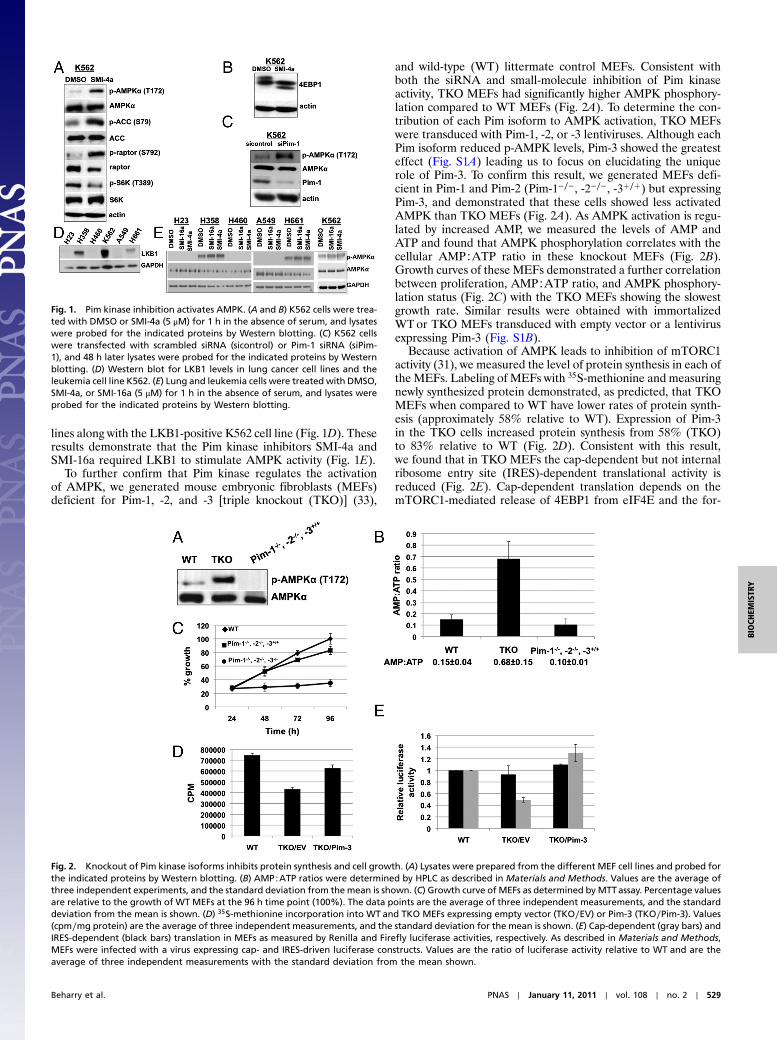
lines along with the LKB1-positive K562 cell line (Fig. 1D). Theseresults demonstrate that the Pim kinase inhibitors SMI-4a andSMI-16a required LKB1 to stimulate AMPK activity (Fig. 1E).
To further confirm that Pim kinase regulates the activationof AMPK, we generated mouse embryonic fibroblasts (MEFs)deficient for Pim-1, -2, and -3 [triple knockout (TKO)] (33),
and wild-type (WT) littermate control MEFs. Consistent withboth the siRNA and small-molecule inhibition of Pim kinaseactivity, TKO MEFs had significantly higher AMPK phosphory-lation compared to WT MEFs (Fig. 2A). To determine the con-tribution of each Pim isoform to AMPK activation, TKO MEFswere transduced with Pim-1, -2, or -3 lentiviruses. Although eachPim isoform reduced p-AMPK levels, Pim-3 showed the greatesteffect (Fig. S1A) leading us to focus on elucidating the uniquerole of Pim-3. To confirm this result, we generated MEFs defi-cient in Pim-1 and Pim-2 (Pim-1−∕−, -2−∕−, -3þ∕þ) but expressingPim-3, and demonstrated that these cells showed less activatedAMPK than TKO MEFs (Fig. 2A). As AMPK activation is regu-lated by increased AMP, we measured the levels of AMP andATP and found that AMPK phosphorylation correlates with thecellular AMP∶ATP ratio in these knockout MEFs (Fig. 2B).Growth curves of these MEFs demonstrated a further correlationbetween proliferation, AMP∶ATP ratio, and AMPK phosphory-lation status (Fig. 2C) with the TKO MEFs showing the slowestgrowth rate. Similar results were obtained with immortalizedWTor TKO MEFs transduced with empty vector or a lentivirusexpressing Pim-3 (Fig. S1B).
Because activation of AMPK leads to inhibition of mTORC1activity (31), we measured the level of protein synthesis in each ofthe MEFs. Labeling of MEFs with 35S-methionine and measuringnewly synthesized protein demonstrated, as predicted, that TKOMEFs when compared to WT have lower rates of protein synth-esis (approximately 58% relative to WT). Expression of Pim-3in the TKO cells increased protein synthesis from 58% (TKO)to 83% relative to WT (Fig. 2D). Consistent with this result,we found that in TKO MEFs the cap-dependent but not internalribosome entry site (IRES)-dependent translational activity isreduced (Fig. 2E). Cap-dependent translation depends on themTORC1-mediated release of 4EBP1 from eIF4E and the for-
Fig. 1. Pim kinase inhibition activates AMPK. (A and B) K562 cells were trea-ted with DMSO or SMI-4a (5 μM) for 1 h in the absence of serum, and lysateswere probed for the indicated proteins by Western blotting. (C) K562 cellswere transfected with scrambled siRNA (sicontrol) or Pim-1 siRNA (siPim-1), and 48 h later lysates were probed for the indicated proteins by Westernblotting. (D) Western blot for LKB1 levels in lung cancer cell lines and theleukemia cell line K562. (E) Lung and leukemia cells were treated with DMSO,SMI-4a, or SMI-16a (5 μM) for 1 h in the absence of serum, and lysates wereprobed for the indicated proteins by Western blotting.
Fig. 2. Knockout of Pim kinase isoforms inhibits protein synthesis and cell growth. (A) Lysates were prepared from the different MEF cell lines and probed forthe indicated proteins by Western blotting. (B) AMP∶ATP ratios were determined by HPLC as described in Materials and Methods. Values are the average ofthree independent experiments, and the standard deviation from the mean is shown. (C) Growth curve of MEFs as determined byMTTassay. Percentage valuesare relative to the growth of WT MEFs at the 96 h time point (100%). The data points are the average of three independent measurements, and the standarddeviation from the mean is shown. (D) 35S-methionine incorporation into WT and TKO MEFs expressing empty vector (TKO∕EV) or Pim-3 (TKO∕Pim-3). Values(cpm∕mg protein) are the average of three independent measurements, and the standard deviation for the mean is shown. (E) Cap-dependent (gray bars) andIRES-dependent (black bars) translation in MEFs as measured by Renilla and Firefly luciferase activities, respectively. As described in Materials and Methods,MEFs were infected with a virus expressing cap- and IRES-driven luciferase constructs. Values are the ratio of luciferase activity relative to WT and are theaverage of three independent measurements with the standard deviation from the mean shown.
Beharry et al. PNAS ∣ January 11, 2011 ∣ vol. 108 ∣ no. 2 ∣ 529
BIOCH
EMISTR
Y

mation of the eIF4F complex. Using m7-GTP beads in a pull-down assay (34), we found that in TKO MEFs eIF4E is highlybound to 4EBP1, whereas eIF4G binding is lost and thus the abil-ity of eIF4E to promote translation is inhibited (Fig. S1C). Theseresults demonstrate that reduced Pim kinase activity correlateswith increased AMP∶ATP ratio, activation of AMPK, inhibitionof mTORC1 activity, and reduced overall protein synthesis.
Pim-3 Regulates c-Myc Levels. In comparison to TKO MEFs, Pim-3–expressing cells demonstrated increased 5′-cap-dependentprotein synthesis and growth similar to WT MEFs. Becausethe levels of the c-Myc protein are controlled by 5′-cap-depen-dent transcription, and c-Myc is important in the regulation ofboth cell growth and overall cellular metabolism, we examinedthe levels of c-Myc in MEFs from each genotype. Western blotsdemonstrate that the expression of Pim-3 either in primary MEFs(Fig. 3A) or after transduction into TKO MEFs (Fig. 3B) mark-edly increased c-Myc protein. Similar to p-AMPK (Fig. S1A),neither Pim-1 nor Pim-2 was able to induce the significant levelsof c-Myc protein observed with Pim-3 overexpression in TKOMEFs (Fig. 3B). Furthermore, expression of Pim-1 or Pim-2 inPim-3–only MEFs led to a decrease in c-Myc protein levels(Fig. S1D), suggesting the possibility that the Pim kinases couldcompete for substrates or interact directly. Because of themarked difference in c-Myc protein levels in MEFs containingPim-3 only, we tested two additional MEF cell lines generatedfrom different embryos of the same genotype and again observedincreased c-Myc protein levels (Fig. S2B). To determine whetherthe increased c-Myc level in Pim-3–expressing cells is unique toMEFs in culture, we measured the c-Myc levels in spleen lysatesof 4-mo-old WT, TKO, and Pim-1−∕−, -2−∕−, -3þ∕þ mice and againfound the highest level of c-Myc protein in the Pim-3–onlygenotype (Fig. S2C). Because deletion of one Pim might grosslyelevate the level of another, in this case Pim-3, we measured thelevel of Pim-3 mRNA in the different MEF genotypes but did notfind a significant difference (Fig. S2A). These results suggestedthat Pim-3 might modulate c-Myc translation.
To determine the effect of Pim-3 on c-Myc translation, we trea-ted MEFs with cycloheximide until c-Myc protein was completelydegraded, washed out the cycloheximide, and then monitored byWestern blotting the rate of increase in c-Myc protein over time.To make this comparison possible, 2.5 times more protein fromTKOMEFs was loaded on these SDS gels. TKOMEFs showed adelay in protein synthesis with only 53 and 43% as much c-Mycprotein synthesis in the first 15 min when compared to WT andTKO∕Pim-3 MEFS, respectively (Fig. 3C). This result is consis-tent with the reduced protein synthesis in the TKO MEFs(Fig. 2D). Because the overall translational efficiency in cells isreflected by changes in the polysome/monosome ratio, we deter-mined the polysome profile using cytosolic extracts of WT, TKO,and TKO∕Pim-3 cells. TKO cells showed a significant reductionin heavy polysomes and a corresponding increase in free ribo-some subunits (Fig. 3D Upper). However, expression of Pim-3in TKO cells resulted in a significant increase in heavy polysomes.c-Myc mRNA showed redistribution from heavy toward lighterpolysomes and monosomes and free subunits in TKO cells rela-tive to WT (Fig. 3D Lower). This shift was reversed by Pim-3expression, suggesting that Pim-3 is capable of controlling thetranslation of c-Myc mRNA (Fig. 3D Lower). Because Pim-1and -2 have been shown to increase the stability of c-Myc (9),we examined changes in c-Myc protein stability in TKO andTKO∕Pim-3 MEFs after cycloheximide treatment but foundno significant difference in c-Myc half-life in the Pim-3–contain-ing cells (Fig. S2D). Finally, we found that the expression ofc-Myc in TKO MEFs led to a decrease in AMPK activation(Fig. S2E) consistent with the ability of c-Myc to stimulate cellgrowth.
Pim-3 and c-Myc Regulate PGC-1α Levels. The differences in thegrowth rate between the TKO and Pim-3 only MEFs couldpossibly be explained by the Pim-3–mediated increased c-Myc be-cause the latter is known to control multiple factors that regulatecell growth and metabolism (35, 36). Therefore, we compared thegrowth rate of TKOMEFs stably expressing empty vector, Pim-3,or c-Myc to WT MEFs. The TKO∕c-Myc MEFs were able to
Fig. 3. Pim-3 elevates c-Myc levels. (A) c-Myc protein levels in each MEF genotype as determined byWestern blotting. (B) TKOMEFs were infected with emptyvector (EV), Pim-1, -2, or -3 lentiviruses, and 48 h later lysates were probed for c-Myc levels and compared toWTMEFs. (C) TKOMEFs expressing EVor Pim-3 weretreated for 90 min with cycloheximide (CHX, 10 μM), the media replaced, and lysates probed for c-Myc levels by Western blotting. Densitometry analysis wasperformed, and the values at the 15 min time point relative to DMSO are shown. To obtain a relatively equal amount of c-Myc protein at the 90 min time pointwith DMSO, ∼2.5-fold more TKO∕EV protein lysate was loaded relative to TKO∕Pim-3. (D) Ribosome fractions of WTand TKOMEFs expressing EVor Pim-3 wereprepared by sucrose gradient (see Materials and Methods), and the level of c-Myc mRNA associated with each fraction was determined by PCR.
530 ∣ www.pnas.org/cgi/doi/10.1073/pnas.1013214108 Beharry et al.

grow in the absence of Pim kinases but did not reach the samedensity in 96 h (Fig. 4A). The shRNA-mediated knockdown ofc-Myc in TKO∕Pim-3 MEFs did not completely inhibit cell pro-liferation (Fig. 4B). Together, these results suggest that Pim-3and c-Myc do not have completely overlapping biologic activities.To understand how Pim-3 decreases the AMP∶ATP ratio andinhibits AMPK phosphorylation, we measured the levels ofPGC-1α. PGC-1α activates a wide variety of transcription factorsthat result in increased mitochondrial biogenesis and oxidativephosphorylation (37). Increased expression of PGC-1α can leadto elevations in ATP levels (38), whereas PGC-1α knockout leadsto reduced ATP levels in murine hearts (39). PGC-1α expressionand PGC-1α–dependent gene expression are induced by chemicalactivation of AMPK, and AMPK directly phosphorylates PGC-1α, leading to increased transcriptional activity (40–42). Wefound that the levels of PGC-1α mRNA and protein were greatlyreduced in TKO MEFs, highest in Pim-3–only MEFs, and inter-mediate in WT cells (Fig. 4C and D). To examine the contribu-tions of Pim-3 and c-Myc in regulating PGC-1α levels, we infectedTKO MEFs with lentiviruses expressing c-Myc or Pim-3 andfound that Pim-3 induced marked increases in PGC-1α mRNA(12-fold) and protein; the effect of c-Myc alone was a 4-foldincrease in mRNA, and the increase in protein was quantitatedat only 10% that of Pim-3 (Fig. 4D, E).
The above results suggest that the increased AMP∶ATP ratioin TKO MEFs may be attributed to low ATP levels due to de-creased PGC-1α protein, thus leading to AMPK activation. Toexamine whether overexpression of PGC-1α in TKO MEFswas sufficient to reduce p-AMPK by increasing the level of cel-lular ATP, we transduced TKOMEFs with a lentivirus expressingPGC-1α. Western blots and biochemical analysis demonstratethat PGC-1α expression in TKO MEFs decreased the level ofp-AMPK (Fig. 5A) and increased the levels of ATP (Fig. 5B),leading to decreased 4EBP1 binding to eIF4E while increasing
eIF4G association with the eIF4E protein (Fig. S3). In contrast,PGC-1α expression in TKO MEFs showed little effect on c-Myclevels (Fig. 5A). Thus, Pim-3, by controlling the levels of bothc-Myc and PGC-1α, is able to impact on AMPK phosphorylation,mTORC1 activity, 5′-cap-dependent translation, and ultimatelycell growth (Fig. 5C).
DiscussionThe combined approach of genetic knockout, RNAi, and small-molecule inhibition implicate the Pim kinases in regulating theAMP∶ATP ratio and energy metabolism. These effects lead tothe modulation of the mTORC1 pathway by AMPK and the con-trol of cell growth. In leukemic cells, the pan-Pim kinase inhibitorSMI-4a stimulated the phosphorylation and activation of AMPK,whereas in TKO MEFs the ratio of AMP∶ATP was markedlyincreased and AMPK was activated. Because AMPK is a negativeregulator of mTORC1, we found in leukemic cells treated withSMI-4a and in TKOMEFs that mTORC1 activity is inhibited andcap-dependent translation is significantly decreased. In MEFs,the expression of Pim-3 alone could reverse these processes, low-ering the AMP∶ATP ratio, decreasing the activation of AMPK,and increasing cap-dependent translation, all resulting in cellulargrowth rates comparable to WT MEFs. The differences betweenthe TKO and Pim-3–only MEFs could be explained in part bythe Pim-3–mediated increased c-Myc because the latter controlsmultiple transcription factors that regulate cell growth andmetabolism (35, 36). Infection of TKO MEFS with a lentivirusexpressing c-Myc increased the growth of these cells but didnot duplicate the growth curve of Pim-3–expressing MEFs.
In muscle and fat tissue, the ability of activated AMPK tomaintain an energy balance is achieved in part by stimulatingPGC-1α (41). The ability of PGC-1α to coactivate multipletranscription factors makes this protein a master regulator ofmitochondrial biogenesis (43). Considering this link between
Fig. 4. Pim-3 and c-Myc affect PGC-1α levels. (A) Growth curve of TKO MEFs expressing empty vector (EV), c-Myc, or Pim-3 as determined by an MTT assay.Percentage values are relative to the value of WT MEFs at the 96 h time point (100%). The data points are the average of three independent measurements,and the standard deviation from the mean is shown. (B) TKO∕Pim-3 MEFs were infected with nontargeting shRNA (shctl) or c-Myc targeting shRNA (shMyc)lentiviruses. Equal numbers of shctl and shMyc cells were plated 48 h postinfection, and after an additional 72 h viability was determined by an MTT assay andrepresented as a percent absorbance (%Abs) with shctl set at 100%. The data points are the average of three independent measurements, and the standarddeviation from the mean is shown. (Inset) Lysates were prepared at 120 h postinfection and probed for the indicated proteins by Western blotting. (C) PGC-1αprotein levels in MEFs as determined byWestern blotting. (D) PGC-1αmRNA levels in primary MEFs (WT, TKO, Pim-1−∕−, -2−∕−, -3þ∕þ) or TKOMEFs infected withEV, c-Myc, or Pim-3 lentiviruses as determined by QT-PCR 48 h after infection. Values are the average of three independent measurements, and the standarddeviation from the mean is shown. (E) PGC-1α protein levels as determined by Western blotting in TKO MEFs 48 h postinfection with EV, c-Myc, or Pim-3.
Beharry et al. PNAS ∣ January 11, 2011 ∣ vol. 108 ∣ no. 2 ∣ 531
BIOCH
EMISTR
Y

AMPK and PGC-1α in the sensing and regulation of the cell’senergy status, the levels of PGC-1α were investigated and foundto be significantly lower in TKO MEFs. In comparison, Pim-3–containing MEFs showed increased levels of PGC-1α relativeto WT. Therefore, in the case of the TKO MEFs, chronic AMPKactivation coupled with drastically reduced levels of PGC-1αprotein resulted in an elevated AMP∶ATP ratio. Accordingly,infection of TKO MEFs with a lentivirus expressing PGC-1αwas shown to increase ATP levels and decrease AMPK activation.The increased PGC-1α levels in Pim-3–only MEFs cannot beattributed solely to increased c-Myc because TKO∕c-Myc MEFsshowed lower levels of PGC-1α mRNA and protein relative toTKO∕Pim-3 MEFs. This suggests the possibility that Pim-3 andc-Myc could cooperate in regulating PGC-1α levels inMEFs. Thiscooperation may extend beyond transcription/translation becausePGC-1α levels and activity are regulated by multiple posttransla-tional mechanisms (37).
Pim-3 is the least-studied kinase of the Pim family; however, ithas been linked to the development and progression of colon andpancreatic cancers (2–4, 44). Despite the high sequence identityand overlapping substrate specificity of the Pim kinases, Pim-3expression alone is shown to overcome at least some of the defectsfound in the loss of both Pim-1 and Pim-2, including growth rate.Additionally, the knockout of Pim-1 and -2 and the expression ofPim-3 only led to a marked increase in c-Myc protein relative toWT MEFs. The observation that the transduction of Pim-1 or -2into MEFs containing Pim-3 suppressed c-Myc levels suggestedthe possibility that individual Pim isoforms may regulate eachother either directly or through substrate competition. This posesthe question of whether Pim isoforms either individually or actingin concert regulate different biological processes and under whatcellular circumstances. The question of the activity of Pim iso-forms is of importance to the design of small-molecule inhibitorstargeting these kinases and their use in the treatment of diseases,including cancer. Both Pim-1 and -2 are known to enhance c-Myc–induced transformation (6, 12) and phosphorylate and stabilizec-Myc protein, leading to increased transcriptional activity (9).In the MEFs used in this study, Pim-3 expression alone enhancedcap-dependent translation, increased c-Myc levels without chan-ging the protein’s stability, and increased the cell growth rate.Because elevated levels of both Pim-3 and c-Myc are found in
gastrointestinal cancers, our results suggest the possibility thatPim-3 might enhance the growth of these tumor cells in part byregulating c-Myc levels, thus highlighting the potential utility ofPim-3 targeted inhibitors.
Materials and MethodsCell Culture. MEFs were derived from 14.5-d-old embryos and were geno-typed as described (45). For stable cell lines, TKO MEFs were transducedwith lentiviruses encoding empty vector, PIM-1, Pim-2, Pim-3, or c-Myc andselected with puromycin (4 μg∕mL).
Construction of Lentiviral Vectors. The open reading frames of PIM-1 (human,33 kDa isoform), PIM-2 (mouse), Pim-3 (mouse), c-Myc (mouse), and PGC-1α(human, a gift from Young-In Chi, Department of Molecular and CellularBiochemistry, University of Kentucky, Lexington, KY) were amplified by PCRfrom full-length cDNA clones and subcloned into the AgeI-MluI sites ofpLex-MCS lentiviral vector (Open Biosystems). Methods for preparation oflentiviral stocks are detailed in SI Materials and Methods.
Quantitative RT-PCR (QT-PCR). Total RNA was isolated from MEFs usingthe RNeasy kit (Qiagen) according to the manufacturer’s protocol. Thefirst-strand cDNA was synthesized using Superscript first-strand synthesiskit and Oligo (dT) primer (Invitrogen).
Biochemical Analysis. K562 cells were transfected with scrambled siRNA orsiPim-1 (ON-TARGETplus SMARTpool, Thermo Scientific) using Lipofecta-mine™2000 (Invitrogen) according to the manufacturer’s protocol, and 48 hposttransfection lysates were prepared. Cell growth was measured using the3-(4,5-dimethylthiazol-2-yl)-2,5-diphenyl tetrazolium bromide (MTT) assay.ATP, ADP, and AMP were measured by HPLC as described previously (46),and ATP was also measured using the ATP Bioluminescence Assay Kit HS II(Roche) with 105 cells. eIF4E was captured on m7-GTP sepharose (GE Life-sciences) from WT and TKO MEFs lysate and bound 4EBP1 and eIF4G deter-mined by Western blotting.
35S-Methionine Incorporation. Cells were serum starved for 1 h in methionine-free medium (Invitrogen), followed by labeling with 100 mCi of35S-methionine∕mL. Lysates and labeled proteins were precipitated withtrichloroacetic acid on glass microfiber filters (Whatman) using vacuumfiltration, and 35S-incorporation was counted.
Cap- vs. IRES-Dependent Translation. A bicistronic retroviral vector, pMSCV∕rLuc-pol IRES-fLuc (a gift from Peter B. Bitterman, Department of Medicine,University of Minnesota, Minneapolis, MN), was used to produce viralparticles for infecting WT, TKO and TKO∕Pim-3 MEFs. Cells were collected
Fig. 5. Expression of PGC-1α restores the AMP∶ATP ratio in TKO MEFs. (A) PGC-1α overexpression in TKO MEFs reduces AMPK activation. Lysates were pre-pared from TKO MEFs 48 h after transduction with empty vector (EV) or PGC-1α lentiviruses, and protein levels compared by Western blotting. (B) ATP levelsdetermined in lysates from Fig. 5A as described in Materials and Methods. Values are the average of three independent measurements, and the standarddeviation from the mean is shown. (C) Schematic summary of biologic changes observed in TKO MEFs expressing Pim-3.
532 ∣ www.pnas.org/cgi/doi/10.1073/pnas.1013214108 Beharry et al.

48 hr postinfection and Renilla/Firefly luciferase activities were quantifiedusing the dual-luciferase reporter assay system (Promega) and a luminometeraccording to the manufacturer’s instructions.
Polysome Profile Analysis. Sucrose density gradient centrifugation wasemployed to separate the ribosome fractions as described previously (47).c-Myc mRNA level in each fraction was measured by PCR.
ACKNOWLEDGMENTS.We thank YefimManevich and Robin Muise-Helmericks(Medical University of South Carolina) for technical assistance. We aregrateful to Anton Berns (Netherlands Cancer Institute) and Paul B. Rothman(University of Iowa Medical Center) for the generous gift of Pim TKO mice.We also thank Vortex Biotechnology for providing the Pim kinase inhibitorsused in this study. This work was supported in part by Department of DefenseGrant W81XWH-08-PCRP-IDA, and the shared resources are supported bythe Hollings Cancer Center.
1. Shah N, et al. (2008) Potential roles for the PIM1 kinase in human cancer—Amolecularand therapeutic appraisal. Eur J Cancer 44:2144–2151.
2. Fujii C, et al. (2005) Aberrant expression of serine/threonine kinase Pim-3 in hepato-cellular carcinoma development and its role in the proliferation of human hepatomacell lines. Int J Cancer 114:209–218.
3. Li YY, et al. (2006) Pim-3, a proto-oncogene with serine/threonine kinase activity, isaberrantly expressed in human pancreatic cancer and phosphorylates bad to blockbad-mediated apoptosis in human pancreatic cancer cell lines. Cancer Res66:6741–6747.
4. Popivanova BK, et al. (2007) Proto-oncogene, Pim-3 with serine/threonine kinaseactivity, is aberrantly expressed in human colon cancer cells and can preventBad-mediated apoptosis. Cancer Sci 98:321–328.
5. White E (2003) The pims and outs of survival signaling: Role for the Pim-2 proteinkinase in the suppression of apoptosis by cytokines. Genes Dev 17:1813–1816.
6. Allen JD, Verhoeven E, Domen J, van der Valk M, Berns A (1997) Pim-2 transgeneinduces lymphoid tumors, exhibiting potent synergy with c-myc. Oncogene15:1133–1141.
7. Wang J, et al. (2010) Pim1 kinase synergizes with c-MYC to induce advanced prostatecarcinoma. Oncogene 29:2477–2487.
8. Mikkers H, et al. (2002) High-throughput retroviral tagging to identify components ofspecific signaling pathways in cancer. Nat Genet 32:153–159.
9. Zhang Y, Wang Z, Li X, Magnuson NS (2008) Pim kinase-dependent inhibition of c-Mycdegradation. Oncogene 27:4809–4819.
10. Zippo A, De Robertis A, Serafini R, Oliviero S (2007) PIM1-dependent phosphorylationof histone H3 at serine 10 is required for MYC-dependent transcriptional activationand oncogenic transformation. Nat Cell Biol 9:932–944.
11. Macdonald A, et al. (2006) Pim kinases phosphorylate multiple sites on Bad andpromote 14-3-3 binding and dissociation from Bcl-XL. BMC Cell Biol 7:1.
12. Shirogane T, et al. (1999) Synergistic roles for Pim-1 and c-Myc in STAT3-mediated cellcycle progression and antiapoptosis. Immunity 11:709–719.
13. Morishita D, Katayama R, Sekimizu K, Tsuruo T, Fujita N (2008) Pim kinases promotecell cycle progression by phosphorylating and down-regulating p27Kip1 at thetranscriptional and posttranscriptional levels. Cancer Res 68:5076–5085.
14. Bachmann M, et al. (2006) The oncogenic serine/threonine kinase Pim-1 directlyphosphorylates and activates the G2/M specific phosphatase Cdc25C. Int J BiochemCell Biol 38:430–443.
15. Zhang F, et al. (2009) PIM1 protein kinase regulates PRAS40 phosphorylation andmTOR activity in FDCP1 cells. Cancer Biol Ther 8:846–853.
16. Hammerman PS, Fox CJ, Birnbaum MJ, Thompson CB (2005) Pim and Akt oncogenesare independent regulators of hematopoietic cell growth and survival. Blood105:4477–4483.
17. Laplante M, Sabatini DM (2009) mTOR signaling at a glance. J Cell Sci 122:3589–3594.18. Gibbons JJ, Abraham RT, Yu K (2009) Mammalian target of rapamycin: Discovery of
rapamycin reveals a signaling pathway important for normal and cancer cell growth.Semin Oncol 36:S3–S17.
19. Bai X, Jiang Y (2010) Key factors in mTOR regulation. Cell Mol Life Sci 67:239–253.20. Shaw RJ (2009) LKB1 and AMP-activated protein kinase control of mTOR signalling
and growth. Acta Physiol 196:65–80.21. Chen WW, Chan DC, Donald C, Lilly MB, Kraft AS (2005) Pim family kinases enhance
tumor growth of prostate cancer cells. Mol Cancer Res 3:443–451.22. Lilly M, Kraft A (1997) Enforced expression of the Mr 33,000 Pim-1 kinase enhances
factor-independent survival and inhibits apoptosis in murine myeloid cells. CancerRes 57:5348–5355.
23. Xia Z, et al. (2009) Synthesis and evaluation of novel inhibitors of Pim-1 and Pim-2protein kinases. J Med Chem 52:74–86.
24. Pogacic V, et al. (2007) Structural analysis identifies imidazo[1,2-b]pyridazines as PIMkinase inhibitors with in vitro antileukemic activity. Cancer Res 67:6916–6924.
25. Akue-Gedu R, et al. (2009) Synthesis, kinase inhibitory potencies, and in vitroantiproliferative evaluation of new Pim kinase inhibitors. J Med Chem 52:6369–6381.
26. Chen LS, Redkar S, Bearss D, Wierda WG, Gandhi V (2009) Pim kinase inhibitor,SGI-1776, induces apoptosis in CLL lymphocytes. Blood 114:4150–4157.
27. Grey R, et al. (2009) Structure-based design of 3-aryl-6-amino-triazolo[4,3-b]pyridazineinhibitors of Pim-1 kinase. Bioorg Med Chem Lett 19:3019–3022.
28. Tong Y, et al. (2008) Isoxazolo[3,4-b]quinoline-3,4(1H,9H)-diones as unique, potentand selective inhibitors for Pim-1 and Pim-2 kinases: Chemistry, biological activities,and molecular modeling. Bioorg Med Chem Lett 18:5206–5208.
29. Beharry Z, et al. (2009) Novel benzylidene-thiazolidine-2,4-diones inhibit Pim proteinkinase activity and induce cell cycle arrest in leukemia and prostate cancer cells. MolCancer Ther 8:1473–1483.
30. Lin Y, et al. (2009) A small molecule inhibitor of Pim protein kinases blocks the growthof precursor T-cell lymphoblastic leukemia/lymphoma. Blood 115:824–833.
31. Gwinn DM, et al. (2008) AMPK phosphorylation of raptor mediates a metaboliccheckpoint. Mol Cell 30:214–226.
32. Hezel AF, Bardeesy N (2008) LKB1; linking cell structure and tumor suppression.Oncogene 27:6908–6919.
33. Mikkers H, et al. (2004) Mice deficient for all PIM kinases display reduced body sizeand impaired responses to hematopoietic growth factors.Mol Cell Biol 24:6104–6115.
34. Connolly E, Braunstein S, Formenti S, Schneider RJ (2006) Hypoxia inhibits proteinsynthesis through a 4E-BP1 and elongation factor 2 kinase pathway controlled bymTOR and uncoupled in breast cancer cells. Mol Cell Biol 26:3955–3965.
35. Liu YC, et al. (2008) Global regulation of nucleotide biosynthetic genes by c-Myc. PLoSOne 3–e2722.
36. Li F, et al. (2005) Myc stimulates nuclearly encodedmitochondrial genes andmitochon-drial biogenesis. Mol Cell Biol 25:6225–6234.
37. Canto C, Auwerx J (2009) PGC-1alpha, SIRT1 and AMPK, an energy sensing networkthat controls energy expenditure. Curr Opin Lipidol 20:98–105.
38. Liang H, Bai Y, Li Y, Richardson A, Ward WF (2007) PGC-1alpha-induced mitochondrialalterations in 3T3 fibroblast cells. Ann NY Acad Sci 1100:264–279.
39. Arany Z, et al. (2005) Transcriptional coactivator PGC-1 alpha controls the energy stateand contractile function of cardiac muscle. Cell Metab 1:259–271.
40. Suwa M, Nakano H, Kumagai S (2003) Effects of chronic AICAR treatment on fibercomposition, enzyme activity, UCP3, and PGC-1 in rat muscles. J Appl Physiol95:960–968.
41. Jäger S, Handschin C, St.-Pierre J, Spiegelman BM (2007) AMP-activated protein kinase(AMPK) action in skeletal muscle via direct phosphorylation of PGC-1α. Proc Natl AcadSci USA 104:12017–12022.
42. Irrcher I, Ljubicic V, Kirwan AF, Hood DA (2008) AMP-activated protein kinase-regulated activation of the PGC-1alpha promoter in skeletal muscle cells. PLoS One3:e3614.
43. Finley LW, Haigis MC (2009) The coordination of nuclear and mitochondrial commu-nication during aging and calorie restriction. Ageing Res Rev 8:173–188.
44. Li YY, Wu Y, Tsuneyama K, Baba T, Mukaida N (2009) Essential contribution ofEts-1 to constitutive Pim-3 expression in human pancreatic cancer cells. Cancer Sci100:396–404.
45. Xu J (2005) Preparation, culture, and immortalization of mouse embryonic fibroblasts.Curr Protoc Mol Biol Chap 28: Unit 28.1.
46. Hahn-Windgassen A, et al. (2005) Akt activates the mammalian target of rapamycinby regulating cellular ATP level and AMPK activity. J Biol Chem 280:32081–32089.
47. Ishimaru D, et al. (2009) Regulation of Bcl-2 expression by HuR in HL60 leukemia cellsand A431 carcinoma cells. Mol Cancer Res 7:1354–1366.
Beharry et al. PNAS ∣ January 11, 2011 ∣ vol. 108 ∣ no. 2 ∣ 533
BIOCH
EMISTR
Y
Therapeutics, Targets, and Chemical Biology
Pim Kinase Inhibitors Sensitize Prostate Cancer Cells toApoptosis Triggered by Bcl-2 Family Inhibitor ABT-737
Jin H. Song1,3 and Andrew S. Kraft2,3
AbstractPim serine/threonine kinases contribute to prostate tumorigenesis and therapeutic resistance, yet Pim kinase
inhibitors seem to have only limited effects onprostate cancer cell survival. Because overexpression of Bcl-2 familymembers are implicated in chemotherapeutic resistance in prostate cancer, we investigated the cooperativeeffects of Pimkinase inhibitionwith ABT-737, a smallmolecule antagonist of Bcl-2 familymembers. Strikingly, theaddition of ABT-737 to Pim inhibitors triggered a robust apoptosis of prostate cancer cells in vitro and in vivo. Piminhibitors decreased levels of the Bcl-2 family member Mcl-1, both by blocking 50-cap dependent translation anddecreasing protein half life. In addition, Pim inhibition transcriptionally increased levels of the BH3 protein Noxaby activating the unfolded protein response (UPR), lead to eIF-2a phosphorylation and increased expression ofCHOP. Increased levels of Noxa also inactivated the remaining levels of Mcl-1 protein activity. Notably, thesespecific protein changes were essential to the apoptotic process because ABT-737 did not inhibit Mcl-1 proteinactivity and Mcl-1 overexpression blocked the apoptotic activity of ABT-737. Our results therefore suggest thatthis combination treatment could be developed as a potential therapy for human prostate cancer whereoverexpression of Pim kinases and antiapoptotic Bcl-2 family members drives tumor cell resistance to currentanticancer therapies. Cancer Res; 72(1); 294–303. �2011 AACR.
Introduction
The Pim family of serine threonine protein kinases plays acritical but unexplicated role in the growth and progression ofhuman prostate cancer (1). This enzyme family is overex-pressed in human prostate cancer compared with benignbiopsies (2) and enhanced levels of nuclear Pim-2 in tumorcells have been associated with a higher risk of prostate-specific antigen (PSA) recurrence and with perineural invasionof the prostate gland (3). Moderate to strong cytoplasmicstaining of Pim-1 was seen in tumors of 68% of patients witha Gleason score of 7 or higher (4). Pim-1 is overexpressed inhigh grade prostate intraepithelial neoplasia, and Pim stainingmay be helpful in differentiating benign glands from intrae-pithelial neoplasia (2). We have previously reported that theexpression of Pim-1 in prostate cancer cells confers a growthadvantage on these tumor cells (5). Patients with high levels of
Pim-1 expression in the prostate are at significantly greater riskfor developing metastatic cancer (6). In animals, overexpres-sion of the c-Myc protein in the prostate induces neoplasia andis associated with increased Pim protein kinase levels (7). In asubrenal capsular assay for prostate regeneration, expressionof Pim-1 kinase promotes c-Myc-driven prostate carcinogen-esis (8). These studies suggest that the Pimprotein kinasesmaybe a target to inhibit prostate cancer growth.
Through chemical library screening (9), we have identifiedthe chemotype of benzylidene-thiazolidine-2,4-diones as aspecific Pim protein kinase inhibitor and focused on SMI-4a,a pan isotype inhibitor, as a potential lead compound. SMI-4a induces both apoptosis and cell-cycle arrest in a widevariety of leukemic cell lines (10). In addition, oral admin-istration of SMI-4a to mice is well tolerated and causesmoderate inhibition of leukemic growth when cells aregrown as tumor xenografts (11). When administered toprostate cancer cells, Pim inhibitors induce a cell-cycle blockbut are only moderately inhibitory to growth (10). Althoughthe mechanism of action of this agent has not been fullyevaluated, we have shown that application of SMI-4a to bothleukemia and prostate cells inhibits mammalian target ofrapamycin complex 1 (mTORC1) activity and 4E-BP1 phos-phorylation (10, 12), possibly by activating the AMP-depen-dent protein kinase (AMPK). Application of Pim inhibitorsstimulates a fall in ATP levels and increases the concentra-tion of AMP (12). Activated AMPK phosphorylates raptorand TSC2 to block mTORC1 activity (13). AMPK activationby Pim inhibition requires phosphorylation of Thr-172 byLKB (12), suggesting that control of energy supplies by SMI-4a could be an essential part of its activity.
Authors' Affiliations: Departments of 1Biochemistry and Molecular Biol-ogy, and 2Medicine, and 3the Hollings Cancer Center, the MedicalUniversity of South Carolina, Charleston, South Carolina
Note: Supplementary data for this article are available at Cancer ResearchOnline (http://cancerres.aacrjournals.org/).
Corresponding Authors: Jin H. Song, Department of Biochemistry andMolecular Biology, Medical University of South Carolina, 86 JonathanLucas Street, Charleston, SC 29425. Phone: 843-792-9366; Fax: 843-792-3200; E-mail: [email protected]; and Andrew S. Kraft, Department ofMedicine, Medical University of South Carolina, 86 Jonathan Lucas Street,Charleston, SC 29425. Phone: 843-792-8284; Fax: 843-792-3200; E-mail:[email protected]
doi: 10.1158/0008-5472.CAN-11-3240
�2011 American Association for Cancer Research.
CancerResearch
Cancer Res; 72(1) January 1, 2012294
American Association for Cancer Research Copyright © 2012 on May 22, 2012cancerres.aacrjournals.orgDownloaded from
Published OnlineFirst November 11, 2011; DOI:10.1158/0008-5472.CAN-11-3240

The resistance of prostate cancers to undergo apoptosiswhen challenged with various chemotherapeutic agents couldbe due to the overexpression of antiapoptotic members of theBcl-2 protein family. Studies have shown that metastaticprostate cancer and castration-refractory tumors are positivelyassociated with Bcl-2 overexpression (14), so the moderatesensitivity of prostate cancer cells to Pim kinase inhibitorscould be due to deregulated expression of pro- and antiapop-totic Bcl-2 proteins. ABT-737, a small molecule BH3 mimeticthat interacts tightly with Bcl-2/Bcl-xL/Bcl-w and inhibits theiractivity, has the potential to overcome this resistance (15).However, this agent does not bind to Mcl-1 and Bfl-1 proteins.Research by us and others shows that Mcl-1 can constrain theproapoptotic activity of ABT-737 (16). Recent reports showedthat that ABT-737 acquires intrinsic resistance to apoptosis byincreasing Mcl-1 binding with Bim (17), and ABT-737 resis-tance is associated with reduced levels of Bcl-2:Bim hetero-dimers (18) and high Mcl-1 expression (16, 19). Although ABT-737 has generated anticancer activity in small cell lung cancer,leukemia, and multiple myeloma, prostate cancer cells in vitroexhibit a poor response to the proapoptotic action of ABT-737because the Mcl-1 protein is widely expressed at high levels inprostate cancer cell lines and inhibits the proapoptotic func-tion of ABT-737.We have shown that ABT-737 as a single agenthas little proapoptotic activity in prostate cancer cells (20, 21).However, the combination of ABT-737 and a Pim inhibitor ishighly synergistic in inducing apoptotic cell death. We inves-tigated the ABT-737/Pim inhibitor synergy, with particularfocus on the mechanism by which Pim inhibitors regulateapoptotic pathways. Thus, we suggest a rationale for this novelcombination therapy.
Materials and Methods
Cell lines, cell culture, and chemicalsProstate cancer cell lines LNCaP, PC-3, DU-145, and 22Rv1
were purchased from the ATCC. Cells were grown in DMEM orRPMI1640 supplemented with 10% FBS, 2 mmol/L Glutamaxand 1% antibiotics (Invitrogen) as previously described (21).Subconfluent cells were treated with Pim inhibitors or vehiclein the absence of FBS. (Z)-5-(3-Trifluoromethylbenzylidene)thiazolidine-2,4-dione (referred to as SMI-4a) and [Z,E]-5-[4-ethylbenzylidine]-2-thioxothiazolidin-4-one (referred to as10058-F4) were from Calbiochem. For animal experiment,SMI-4a was prepared as we reported previously (9). K00135was purchased from BioFocus. 8-(4-Hydroxylphenyl)-2-[(dimethlamino)methyl][1]benzothieno-[3,2-d]pyrimidin-4(3H)-one (referred to as Pimi-14j; ref. 22) and ABT-737 were agift of Abbott Laboratories. Other chemicals of analytic gradewere purchased from EMD Chemicals and Sigma-Aldrich.
Short hairpin RNAs and plasmidsThe Arrest-In Lentiviral expression system (Open Biosys-
tems was used to establish a LNCaP cell line harboring smallhairpin microRNAs (shRNA) as described previously (12, 20).Lentiviruses pGIPZ shRNAmir against human Pim-1(RHS4531-NM_002648), Pim-2 (RHS4531-NM_006875), andPim-3 (RHS4531-NM_001001852), and a nonsilencing control
(RHS4348) were purchased from Open Biosystems. PC-3 cellswere transfected with pcDNA3.1-HA-Bcl-2 (23) and pcDNA3-Bcl-2 (AddGene) by LipofectAMINE2000 (Invitrogen) and thentransfectants were selected and grown in 1 mg/mL of G418(Sigma-Aldrich).
Tumor growth in vivoXenografts bearing prostate tumors were generated by
injection of LNCaP cells (5 � 106) in the flanks of the maleNU/NU nudemice (Charles River). After tumors were grown toat least 100 mm3 (�1 week after implantation), 36 mice wererandomly divided into 4 different treatment groups: Group 1(6 mice), vehicle only (30% propylene glycol, 5% Tween-80, 65%of 5% dextrose in water, pH 4–5); Group 2 (12 mice), 60 mg/kgSMI-4a twice daily treatments (BID); Group 3 (6 mice),50 mg/kg ABT-737 once a day (QD); and Group 4 (12 mice),combination treatment with SMI-4a (BID) and ABT-737 (QD).Mice received oral gavages for SMI-4a or/and intraperitonealinjection for ABT-737. Treatment was begun on day 8 andadministered 5 of 7 days each week for 3 weeks. The growth ofthe subcutaneous tumors was measured twice each week, andmouse body weight was determined on days 0 and 21. Tumorsize was calculated using the equation (L � W2)/2. TheInstitutional Animal Care and Use Committee at the MedicalUniversity of South Carolina approved these animal experi-ments. For the immunohistochmistry of xenograft tumortissues, tissue slices were processed to generate 5 mm tissueslides. Sections were stained with H&E, mouse monoclonalantibody to human Mcl-1 (Santa Cruz Biotechnology), andrabbit antibody to cleaved caspase-3 (Cell Signaling Technol-ogy) according to the manufacturer's protocol for theseproducts.
Quantitative real time PCR, immunoblotting, andbiochemical analysis
Quantitative real time PCR (qT-PCR) and immunoblotanalyses were carried out as previously reported (20) withslight modification as described in the Supplementary Meth-ods. Methods for the 7-Methyl-GTP cap binding assay and35S-methinone incorporation were reported previously (12)and are further described in the Supplementary Methods.
Results
Inhibition of Bcl-2 like proteins with ABT-737 synergizeswith SMI-4a to induce apoptosis
SMI-4a, a small molecule Pim kinase inhibitor, has preclin-ical efficacy in lymphoid and myeloid leukemia (11) but thehuman prostate cancer cell lines LNCaP, PC-3, and 22Rv1 onlyrespond modestly to SMI-4a treatment (Supplementary Fig.S1A). To showwhether the response to Pim inhibitors was Pimspecific, we knocked down the expression of all 3 Pims, 1, 2, and3, using shRNAs. The knockdown of each of these individualPimkinases in LNCaP cells, as shown by qT-PCR, decreased thegrowth of these human prostate cancer cells (SupplementaryFig. S2). Because Bcl-2 protein family expression is associatedwith resistance of prostate cancer to standard chemotherapy,and a higher frequency of Bcl-2 expression is seen in recurrent
Synergy between Pim Kinase Inhibitor and ABT-737
www.aacrjournals.org Cancer Res; 72(1) January 1, 2012 295
American Association for Cancer Research Copyright © 2012 on May 22, 2012cancerres.aacrjournals.orgDownloaded from
Published OnlineFirst November 11, 2011; DOI:10.1158/0008-5472.CAN-11-3240

tumors after castration (24, 25), we examined whether com-bination therapy with a small molecule BH3 mimetic ABT-737and a Pim inhibitor would provide an enhanced apoptoticresponse. ABT-737 binds and induces apoptosis by inhibitingthe activity of Bcl-2, Bcl-xL, and Bcl-w. However, this com-pound is incapable of binding to Bcl-2 like the protein Mcl-1,and increased expression of this protein in numerous cancercell types was sufficient to inhibit the activity of ABT-737 (21).ABT-737 treatment of LNCaP cells alone induced only a smallpercentage of cell death, but when combined with SMI-4a, ahigh degree of synergy in inducing cell death occurred (Fig. 1A).ABT-737 treatment alone induced slight cleavage of PARP-1(Fig. 1B), a marker of apoptosis, but when combined with SMI-4amarked cleavage of this proteinwas seen. Similar synergisticresults were obtained with another structurally different Piminhibitor Pimi-14j (22), which blocks Pim activity in vitro in thelow nanomolar range (Fig. 1A). Combination therapy with thisPimkinase inhibitor andABT-737 induced enhanced caspase-3and PARP-1 cleavage (Fig. 1B). A robust induction of cell deathby these 2 agents was also seen in PC-3 cells, another humanprostate cancer cell line (Supplementary Fig. S3). A third Pimkinase inhibitor K00135 was also synergistic with ABT-737(Supplementary Fig. S3). Moreover, this combination over-came resistance to cell death in human prostate cancer cellsoverexpressing Bcl-2 (Fig. 1C).
Pim kinase inhibitors downregulate Mcl-1 proteinexpression by lowering global protein synthesis
To determine a potential mechanism by which Pim inhibi-tors synergize with ABT-737 to kill prostate cancer cells, we
examined the ability of SMI-4a to regulate the levels of BH3 andBcl-2 family member proteins important in controlling apo-ptotic cell death.Western blot results (Fig. 2A) show that eitherinhibition of Pim kinases with SMI-4a or knockdown of Pimkinases with shRNAs induced a marked decrease in Mcl-1 andan increase in Noxa protein. These results are significantbecause Noxa is capable of binding and inhibiting the anti-apoptotic activity of Mcl-1 and thereby overcomes ABT-737resistance. In LNCaP cells, the levels of Mcl-1 expression werereduced by SMI-4a treatment in a time- and dose-dependentmanner (Fig. 2B). In addition, in the PC-3 cell line a decrease inthe Mcl-1 protein levels was induced by SMI-4a treatment(Supplementary Fig. S4A) and 2 additional small molecule Pimkinase inhibitors, K00135 and Pimi-14j (Supplementary Fig.S4B). No significant change in the levels of MCL-1 mRNAoccurred after the treatment with the Pim inhibitor SMI-4a(Supplementary Fig. S4C). A further suggestion that Pim kinaseplays a role in regulating Mcl-1 protein levels comes from theevaluation of mouse embryo fibroblasts (MEF) derived frommice that were genetically engineered to knock out all 3 Pimproteins (TKO; ref. 12). After 35S-methionine labeling, theseMEFs showed a lower level of the Mcl-1 protein (Supplemen-tary Fig. S4D; ref. 12). Given that MCL-1 mRNA levels wereunaffected by Pim inhibitors, we next labeled LNCaP cells with35S-methionine followed by immunoprecipitation and exam-ined theMcl-1 synthesis. Our results show that SMI-4a inhibitsthe translation of this protein (Fig. 2C). In addition, 35Smethionine-labeled whole cell extracts showed that this drugtreatment suppressed total protein synthesis in LNCaP cells(Fig. 2D). These results are identical to those previously
Figure 1. Bcl-2 inhibitor ABT-737 synergizes with small molecule inhibitors of Pim protein kinases to overcome cell resistance mediated by Bcl-2overexpression. A, LNCaP cells were treated with DMSO, ABT-737 (3 mmol/L), SMI-4a (10 mmol/L), Pimi-14j (10 mmol/L), or combinations for 16 hours.The percentage of cell death was evaluated using trypan blue staining assay (mean � SD, n ¼ 6). B, whole cell extracts from the above treatmentswere analyzed by immunoblotting with the antibodies shown. C, PC-3 cells were transfected with HA-tagged Bcl-2 plasmid and individual clones selectedwith 1 mg/mL G418 treatment. Parental PC-3 (P) cells and selected individual clones were treated with DMSO, SMI-4a, ABT-737 or combinationsof both agents at the indicated doses. The percentage of viable cells after 24 hours of treatment was determined by MTT assay (mean � SD, n ¼ 4).
Song and Kraft
Cancer Res; 72(1) January 1, 2012 Cancer Research296
American Association for Cancer Research Copyright © 2012 on May 22, 2012cancerres.aacrjournals.orgDownloaded from
Published OnlineFirst November 11, 2011; DOI:10.1158/0008-5472.CAN-11-3240
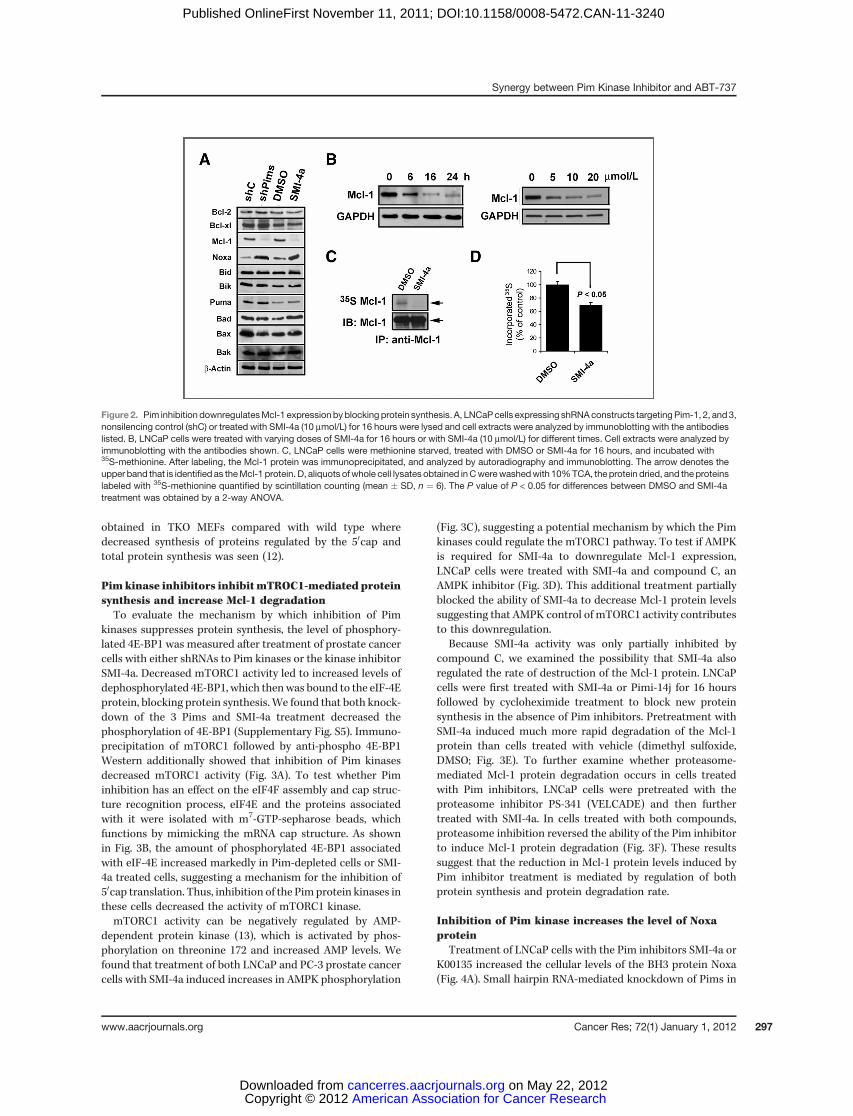
obtained in TKO MEFs compared with wild type wheredecreased synthesis of proteins regulated by the 50cap andtotal protein synthesis was seen (12).
Pimkinase inhibitors inhibitmTROC1-mediated proteinsynthesis and increase Mcl-1 degradationTo evaluate the mechanism by which inhibition of Pim
kinases suppresses protein synthesis, the level of phosphory-lated 4E-BP1 was measured after treatment of prostate cancercells with either shRNAs to Pim kinases or the kinase inhibitorSMI-4a. Decreased mTORC1 activity led to increased levels ofdephosphorylated 4E-BP1, which thenwas bound to the eIF-4Eprotein, blocking protein synthesis. We found that both knock-down of the 3 Pims and SMI-4a treatment decreased thephosphorylation of 4E-BP1 (Supplementary Fig. S5). Immuno-precipitation of mTORC1 followed by anti-phospho 4E-BP1Western additionally showed that inhibition of Pim kinasesdecreased mTORC1 activity (Fig. 3A). To test whether Piminhibition has an effect on the eIF4F assembly and cap struc-ture recognition process, eIF4E and the proteins associatedwith it were isolated with m7-GTP-sepharose beads, whichfunctions by mimicking the mRNA cap structure. As shownin Fig. 3B, the amount of phosphorylated 4E-BP1 associatedwith eIF-4E increased markedly in Pim-depleted cells or SMI-4a treated cells, suggesting a mechanism for the inhibition of50cap translation. Thus, inhibition of the Pimprotein kinases inthese cells decreased the activity of mTORC1 kinase.mTORC1 activity can be negatively regulated by AMP-
dependent protein kinase (13), which is activated by phos-phorylation on threonine 172 and increased AMP levels. Wefound that treatment of both LNCaP and PC-3 prostate cancercells with SMI-4a induced increases in AMPK phosphorylation
(Fig. 3C), suggesting a potential mechanism by which the Pimkinases could regulate the mTORC1 pathway. To test if AMPKis required for SMI-4a to downregulate Mcl-1 expression,LNCaP cells were treated with SMI-4a and compound C, anAMPK inhibitor (Fig. 3D). This additional treatment partiallyblocked the ability of SMI-4a to decrease Mcl-1 protein levelssuggesting that AMPK control of mTORC1 activity contributesto this downregulation.
Because SMI-4a activity was only partially inhibited bycompound C, we examined the possibility that SMI-4a alsoregulated the rate of destruction of the Mcl-1 protein. LNCaPcells were first treated with SMI-4a or Pimi-14j for 16 hoursfollowed by cycloheximide treatment to block new proteinsynthesis in the absence of Pim inhibitors. Pretreatment withSMI-4a induced much more rapid degradation of the Mcl-1protein than cells treated with vehicle (dimethyl sulfoxide,DMSO; Fig. 3E). To further examine whether proteasome-mediated Mcl-1 protein degradation occurs in cells treatedwith Pim inhibitors, LNCaP cells were pretreated with theproteasome inhibitor PS-341 (VELCADE) and then furthertreated with SMI-4a. In cells treated with both compounds,proteasome inhibition reversed the ability of the Pim inhibitorto induce Mcl-1 protein degradation (Fig. 3F). These resultssuggest that the reduction in Mcl-1 protein levels induced byPim inhibitor treatment is mediated by regulation of bothprotein synthesis and protein degradation rate.
Inhibition of Pim kinase increases the level of Noxaprotein
Treatment of LNCaP cells with the Pim inhibitors SMI-4a orK00135 increased the cellular levels of the BH3 protein Noxa(Fig. 4A). Small hairpin RNA-mediated knockdown of Pims in
Figure 2. Pim inhibition downregulatesMcl-1 expressionbyblocking protein synthesis. A, LNCaPcells expressing shRNAconstructs targetingPim-1, 2, and3,nonsilencing control (shC) or treated with SMI-4a (10 mmol/L) for 16 hours were lysed and cell extracts were analyzed by immunoblotting with the antibodieslisted. B, LNCaP cells were treated with varying doses of SMI-4a for 16 hours or with SMI-4a (10 mmol/L) for different times. Cell extracts were analyzed byimmunoblotting with the antibodies shown. C, LNCaP cells were methionine starved, treated with DMSO or SMI-4a for 16 hours, and incubated with35S-methionine. After labeling, the Mcl-1 protein was immunoprecipitated, and analyzed by autoradiography and immunoblotting. The arrow denotes theupper band that is identifiedas theMcl-1 protein. D, aliquots ofwhole cell lysates obtained inCwerewashedwith 10%TCA, the protein dried, and the proteinslabeled with 35S-methionine quantified by scintillation counting (mean � SD, n ¼ 6). The P value of P < 0.05 for differences between DMSO and SMI-4atreatment was obtained by a 2-way ANOVA.
Synergy between Pim Kinase Inhibitor and ABT-737
www.aacrjournals.org Cancer Res; 72(1) January 1, 2012 297
American Association for Cancer Research Copyright © 2012 on May 22, 2012cancerres.aacrjournals.orgDownloaded from
Published OnlineFirst November 11, 2011; DOI:10.1158/0008-5472.CAN-11-3240

LNCaP cells was also sufficient to increase Noxa protein levels(Supplementary Fig. S6A). The Noxa protein specifically bindsand inhibits the Mcl-1 protein's antiapoptotic activity. Immu-noprecipitation shows that the Pim inhibitor-increased Noxaprotein bound to Mcl-1 (Fig. 4B). As seen on Western blot, thePim inhibitors concurrently decreased the overall levels of theMcl-1 protein (Fig. 4C). To determine how Pim inhibitors mightregulate Noxa protein levels, we examined the unfolded proteinresponse (UPR). In the condition of an energy deficit, activationof the endoplasmic reticulum stress response is known tostimulate the UPR by eIF-2a phosphorylation and subsequentincreases in the protein levels of the ATF4 and CHOP transcrip-tion factors (39). ATF4 induces increases in the NoxamRNA andprotein (26, 27). To clarify whether induction of Noxa by Piminhibitors is associatedwith the eIF-2a armofUPR signaling, wetreated LNCaP cells and evaluatedprotein levels of these factors.We found that SMI-4a treatment stimulates the phosphoryla-tionof eIF-2a (Fig. 4C) and induces increases inATF4andCHOPprotein in a dose dependent fashion (Fig. 4D and E) similarly toknown activators of this pathway, tunicamycin (Tm) andthasigargen (Tg; Supplementary Fig. 6B).
Pim inhibitors induce UPR followed by ER stressOn the basis of activation of eIF-2a phosphorylation, we
examined whether Pim inhibitors also induces other arms of
the ER stress pathway by assessing changes in ATF6 andXBP-1 transcriptional activity. XBP-1 mRNA is increased byATF6 and spliced by IRE1 in response to ER stress, resultingin production of a transcription factor that can induce genesassociated with the UPR (28). QT-PCR analysis showed thatthe induction of ATF6 mRNA and XBP-1 mRNA occurs inresponse to Pim inhibitor treatment (Fig. 5A). The splicedversion of XBP-1 is seen in this PCR assay as a larger band.This assay confirmed that SMI-4a treatment of LNCaP cells,when compared with the Tunicamycin control, is capable ofinducing the splicing of XBP-1 in a dose-dependent fashion(Fig. 5B). Pim inhibitor-induced splicing was seen in PC-3,Du145, and C4–2B cell lines (Supplementary Fig. S7A–C).Moreover, we found that SMI-4a did not increase XBP-1splicing in MEFs lacking all Pim isoforms (TKO) MEF cells(Supplementary Fig. S7D) and knockdown of Pims in LNCaPcells with shRNAs decreased the ability of Pim inhibitors tocause XBP-1 splicing (Supplementary Fig. S7E). Activation ofXBP-1 is known to lead to transcription of glucose-regulatedprotein 78 (grp78/BiP) and its splice variant grp78va. Wefound that both grp78/Bip and grp78va transcripts weresignificantly increased by treatment of LNCaP cells withSMI-4a or K00135 (Fig. 5C). This data show that inhibition ofPim kinase pathways is sufficient to activate a second arm ofthe UPR response.
Figure 3. Pim inhibitor SMI-4a promotesMcl-1 degradation by inhibitingmTORC1 signaling. A, LNCaP cells stably expressing shRNAconstructs against Pim-1, 2, and 3 (shPims) or nonsilencing control (shC) or treated with SMI-4a for 16 hours were lysed with CHAPS buffer and resulting lysates were subjected toimmunoprecipitation (IP) using an antibody to mTOR and the immunoprecipitates probed with Abs to mTOR, 4E-BP1, and phosho-4E-BP1 (Thr37/46). Celllysates (25 mg) were also immunoblotted as a control (Input). B, cell extracts isolated after treatments described in A were incubated with m7-GTP beadsto pull down50capcomponents. Afterwashing, proteins bound to thebeadswereprobedby immunoblottingwith antibodies to eIF4E and4E-BP1.Cell lysates(25 mg) were also immunoblotted as a control (Input). C, LNCaP and PC-3 cells were treated with either DMSO or SMI-4a (10 mmol/L) for 6 or 18 hours.Cellular extracts were analyzed by immunoblotting with antibodies shown. D, LNCaP cells were exposed to 5 mmol/L of the AMPK inhibitor CompoundC (Cpd C) prior to treatment with either DMSO or SMI-4a (10 mmol/L) for 16 hours. Cell extracts were analyzed by immunoblotting with antibodies shown.E, LNCaP cells were treated with DMSO or SMI-4a for 16 hours followed by cycloheximide (CHX) and cell samples were collected at the times indicated.Cell lysates were analyzed by immunoblotting with antibodies shown. The graph is based on densitometry scanning (NIH ImageJ software) of the aboveimmunoblots. F, LNCaP cells were pretreated with the proteasome inhibitor PS-341 (100 nmol/L) for 1 hour and then treated with either DMSO orSMI-4a (10 mmol/L) for 16 hours. Cell extracts were analyzed by immunoblotting with antibodies shown.
Song and Kraft
Cancer Res; 72(1) January 1, 2012 Cancer Research298
American Association for Cancer Research Copyright © 2012 on May 22, 2012cancerres.aacrjournals.orgDownloaded from
Published OnlineFirst November 11, 2011; DOI:10.1158/0008-5472.CAN-11-3240
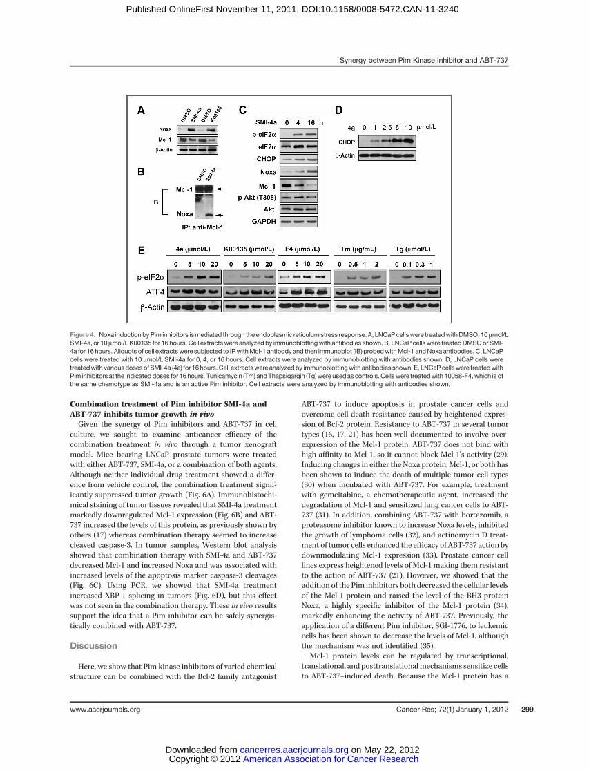
Combination treatment of Pim inhibitor SMI-4a andABT-737 inhibits tumor growth in vivoGiven the synergy of Pim inhibitors and ABT-737 in cell
culture, we sought to examine anticancer efficacy of thecombination treatment in vivo through a tumor xenograftmodel. Mice bearing LNCaP prostate tumors were treatedwith either ABT-737, SMI-4a, or a combination of both agents.Although neither individual drug treatment showed a differ-ence from vehicle control, the combination treatment signif-icantly suppressed tumor growth (Fig. 6A). Immunohistochi-mical staining of tumor tissues revealed that SMI-4a treatmentmarkedly downregulated Mcl-1 expression (Fig. 6B) and ABT-737 increased the levels of this protein, as previously shown byothers (17) whereas combination therapy seemed to increasecleaved caspase-3. In tumor samples, Western blot analysisshowed that combination therapy with SMI-4a and ABT-737decreased Mcl-1 and increased Noxa and was associated withincreased levels of the apoptosis marker caspase-3 cleavages(Fig. 6C). Using PCR, we showed that SMI-4a treatmentincreased XBP-1 splicing in tumors (Fig. 6D), but this effectwas not seen in the combination therapy. These in vivo resultssupport the idea that a Pim inhibitor can be safely synergis-tically combined with ABT-737.
Discussion
Here, we show that Pim kinase inhibitors of varied chemicalstructure can be combined with the Bcl-2 family antagonist
ABT-737 to induce apoptosis in prostate cancer cells andovercome cell death resistance caused by heightened expres-sion of Bcl-2 protein. Resistance to ABT-737 in several tumortypes (16, 17, 21) has been well documented to involve over-expression of the Mcl-1 protein. ABT-737 does not bind withhigh affinity to Mcl-1, so it cannot block Mcl-1's activity (29).Inducing changes in either the Noxa protein,Mcl-1, or both hasbeen shown to induce the death of multiple tumor cell types(30) when incubated with ABT-737. For example, treatmentwith gemcitabine, a chemotherapeutic agent, increased thedegradation of Mcl-1 and sensitized lung cancer cells to ABT-737 (31). In addition, combining ABT-737 with bortezomib, aproteasome inhibitor known to increase Noxa levels, inhibitedthe growth of lymphoma cells (32), and actinomycin D treat-ment of tumor cells enhanced the efficacy of ABT-737 action bydownmodulating Mcl-1 expression (33). Prostate cancer celllines express heightened levels of Mcl-1 making them resistantto the action of ABT-737 (21). However, we showed that theaddition of the Pim inhibitors both decreased the cellular levelsof the Mcl-1 protein and raised the level of the BH3 proteinNoxa, a highly specific inhibitor of the Mcl-1 protein (34),markedly enhancing the activity of ABT-737. Previously, theapplication of a different Pim inhibitor, SGI-1776, to leukemiccells has been shown to decrease the levels of Mcl-1, althoughthe mechanism was not identified (35).
Mcl-1 protein levels can be regulated by transcriptional,translational, and posttranslationalmechanisms sensitize cellsto ABT-737–induced death. Because the Mcl-1 protein has a
Figure 4. Noxa induction byPim inhibitors ismediated through the endoplasmic reticulum stress response. A, LNCaP cellswere treatedwithDMSO, 10mmol/LSMI-4a, or 10 mmol/L K00135 for 16 hours. Cell extractswere analyzed by immunoblottingwith antibodies shown. B, LNCaP cells were treatedDMSOor SMI-4a for 16 hours. Aliquots of cell extracts were subjected to IPwithMcl-1 antibody and then immunoblot (IB) probedwithMcl-1 andNoxa antibodies. C, LNCaPcells were treated with 10 mmol/L SMI-4a for 0, 4, or 16 hours. Cell extracts were analyzed by immunoblotting with antibodies shown. D, LNCaP cells weretreatedwith variousdoses of SMI-4a (4a) for 16 hours. Cell extractswere analyzedby immunoblottingwith antibodies shown. E, LNCaPcellswere treatedwithPim inhibitors at the indicated doses for 16hours. Tunicamycin (Tm) andThapsigargin (Tg)were usedas controls. Cellswere treatedwith 10058-F4,which is ofthe same chemotype as SMI-4a and is an active Pim inhibitor. Cell extracts were analyzed by immunoblotting with antibodies shown.
Synergy between Pim Kinase Inhibitor and ABT-737
www.aacrjournals.org Cancer Res; 72(1) January 1, 2012 299
American Association for Cancer Research Copyright © 2012 on May 22, 2012cancerres.aacrjournals.orgDownloaded from
Published OnlineFirst November 11, 2011; DOI:10.1158/0008-5472.CAN-11-3240
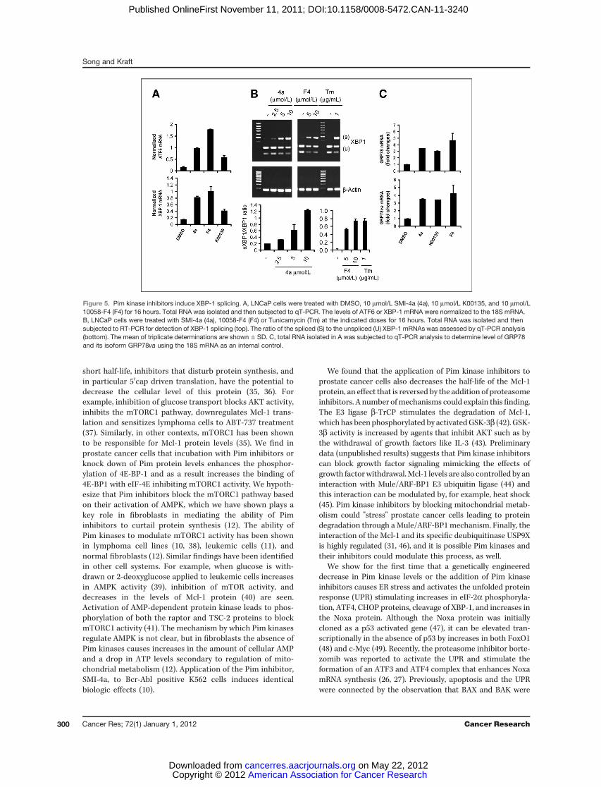
short half-life, inhibitors that disturb protein synthesis, andin particular 50cap driven translation, have the potential todecrease the cellular level of this protein (35, 36). Forexample, inhibition of glucose transport blocks AKT activity,inhibits the mTORC1 pathway, downregulates Mcl-1 trans-lation and sensitizes lymphoma cells to ABT-737 treatment(37). Similarly, in other contexts, mTORC1 has been shownto be responsible for Mcl-1 protein levels (35). We find inprostate cancer cells that incubation with Pim inhibitors orknock down of Pim protein levels enhances the phosphor-ylation of 4E-BP-1 and as a result increases the binding of4E-BP1 with eIF-4E inhibiting mTORC1 activity. We hypoth-esize that Pim inhibitors block the mTORC1 pathway basedon their activation of AMPK, which we have shown plays akey role in fibroblasts in mediating the ability of Piminhibitors to curtail protein synthesis (12). The ability ofPim kinases to modulate mTORC1 activity has been shownin lymphoma cell lines (10, 38), leukemic cells (11), andnormal fibroblasts (12). Similar findings have been identifiedin other cell systems. For example, when glucose is with-drawn or 2-deoxyglucose applied to leukemic cells increasesin AMPK activity (39), inhibition of mTOR activity, anddecreases in the levels of Mcl-1 protein (40) are seen.Activation of AMP-dependent protein kinase leads to phos-phorylation of both the raptor and TSC-2 proteins to blockmTORC1 activity (41). The mechanism by which Pim kinasesregulate AMPK is not clear, but in fibroblasts the absence ofPim kinases causes increases in the amount of cellular AMPand a drop in ATP levels secondary to regulation of mito-chondrial metabolism (12). Application of the Pim inhibitor,SMI-4a, to Bcr-Abl positive K562 cells induces identicalbiologic effects (10).
We found that the application of Pim kinase inhibitors toprostate cancer cells also decreases the half-life of the Mcl-1protein, an effect that is reversed by the addition of proteasomeinhibitors. A number ofmechanisms could explain this finding.The E3 ligase b-TrCP stimulates the degradation of Mcl-1,which has been phosphorylated by activatedGSK-3b (42). GSK-3b activity is increased by agents that inhibit AKT such as bythe withdrawal of growth factors like IL-3 (43). Preliminarydata (unpublished results) suggests that Pim kinase inhibitorscan block growth factor signaling mimicking the effects ofgrowth factorwithdrawal.Mcl-1 levels are also controlled by aninteraction with Mule/ARF-BP1 E3 ubiquitin ligase (44) andthis interaction can be modulated by, for example, heat shock(45). Pim kinase inhibitors by blocking mitochondrial metab-olism could "stress" prostate cancer cells leading to proteindegradation through aMule/ARF-BP1 mechanism. Finally, theinteraction of the Mcl-1 and its specific deubiquitinase USP9Xis highly regulated (31, 46), and it is possible Pim kinases andtheir inhibitors could modulate this process, as well.
We show for the first time that a genetically engineereddecrease in Pim kinase levels or the addition of Pim kinaseinhibitors causes ER stress and activates the unfolded proteinresponse (UPR) stimulating increases in eIF-2a phosphoryla-tion, ATF4, CHOP proteins, cleavage of XBP-1, and increases inthe Noxa protein. Although the Noxa protein was initiallycloned as a p53 activated gene (47), it can be elevated tran-scriptionally in the absence of p53 by increases in both FoxO1(48) and c-Myc (49). Recently, the proteasome inhibitor borte-zomib was reported to activate the UPR and stimulate theformation of an ATF3 and ATF4 complex that enhances NoxamRNA synthesis (26, 27). Previously, apoptosis and the UPRwere connected by the observation that BAX and BAK were
Figure 5. Pim kinase inhibitors induce XBP-1 splicing. A, LNCaP cells were treated with DMSO, 10 mmol/L SMI-4a (4a), 10 mmol/L K00135, and 10 mmol/L10058-F4 (F4) for 16 hours. Total RNA was isolated and then subjected to qT-PCR. The levels of ATF6 or XBP-1 mRNA were normalized to the 18S mRNA.B, LNCaP cells were treated with SMI-4a (4a), 10058-F4 (F4) or Tunicamycin (Tm) at the indicated doses for 16 hours. Total RNA was isolated and thensubjected to RT-PCR for detection of XBP-1 splicing (top). The ratio of the spliced (S) to the unspliced (U) XBP-1 mRNAs was assessed by qT-PCR analysis(bottom). The mean of triplicate determinations are shown � SD. C, total RNA isolated in A was subjected to qT-PCR analysis to determine level of GRP78and its isoform GRP78va using the 18S mRNA as an internal control.
Song and Kraft
Cancer Res; 72(1) January 1, 2012 Cancer Research300
American Association for Cancer Research Copyright © 2012 on May 22, 2012cancerres.aacrjournals.orgDownloaded from
Published OnlineFirst November 11, 2011; DOI:10.1158/0008-5472.CAN-11-3240

needed to induce the UPR by inositol-requiring-enzyme 1a(IRE-1a; ref. 50). IRE-1a executes site-specific cleavage of XBP-1mRNA to producemore stable transcript XBP-1 that encodesa potent transcriptional activator of UPR target genes (28).Dose dependent increases in XBP-1 splicing by Pim inhibitorsconfirm our finding that Pim inhibition drives cells to activateER stress. Compared with wild MEF cells, XBP-1 splicing wasnot increased in Pim knock-out (TKO) MEF cells, suggestingagain the potential role of Pim in regulating this pathway.Additional experiments will be necessary to determine theexact biochemical mechanism by which inhibition of the Pimkinases modulates the UPR.On the basis of the cell culturefindings that inhibition of Pim
with SMI-4a increases Noxa and decreases Mcl-1 protein levelsgreatly enhancing the apoptotic activity of ABT-737, we com-bined these agents in an in vivo animal experiment. Resultsshow that single agent therapy had little anticancer activitywhereas combination treatment inhibited the growth ofLNCaP cells grown subcutaneously. Immunohistochemistryand Western blots show that this combined therapy activatesthe UPR and induces apoptosis of these tumor cells (Fig. 6). Onthe basis of their ability to regulate protein synthesis and the
UPR (see model Fig. 6E), these results suggest that Pim kinaseinhibitors could be used in the clinic to enhance the anticanceractivity of ABT-737.
Disclosure of Potential Conflicts of Interest
A.S. Kraft owns shares in Vortex Biotechnology.
Acknowledgments
The authors thank Abbott Laboratories and Dr. Joel Leverson for providingABT-737 and Pim inhibitor Pimi-14j. Bcl-2 expression vector was kindly providedby Dr. Timothy C. Chambers at the University of Arkansas for Medical Sciences(Little Rock, AR). Members of the Andrew S. Kraft and Charles D. Smithlaboratories (Medical University of South Carolina) have been very kind inproviding research reagents, materials, and technical assistance.
Grant Support
This work was supported by the Department of Defense Grant W81XWH-08-PCRP-IDA and the NIH 1P30-CA138313.
The costs of publication of this article were defrayed in part by the payment ofpage charges. This article must therefore be hereby marked advertisement inaccordance with 18 U.S.C. Section 1734 solely to indicate this fact.
Received September 27, 2011; revisedNovember 4, 2011; accepted November 4,2011; published OnlineFirst November 11, 2011.
Figure6. Tumor growth in vivo is inhibitedbycombination treatment of ABT-737andSMI-4a.A,micewere subcutaneously inoculatedwith LNCaPcells. Tumorvolume was monitored during the following treatments: vehicle (V), ABT-737 (ABT; 50 mg/kg; i.p., QD), SMI-4a (4a; 60 mg/kg, oral gavage, BID) or acombination of SMI-4a and ABT-737 (ABT plus 4a) for 16 days (mean � SEM). B, immunohistochemical analysis of Mcl-1, cleaved caspase-3 (casp-3) andH&E staining. C, Western blot analysis of Noxa, Mcl-1, cleaved casp-3, b-actin, and glyceraldehydes-3-phosphate dehydrogenase (GAPDH). Fiverepresentative tumors for each treatment were examined. D, qT-PCR analysis of XBP-1 splicing in subcutaneous tumors of treatment groups. Fiverepresentative tumors for each treatment group are shown. E, proposed mechanistic model by which Pim inhibitor synergizes with ABT-737 to induceapoptosis. Pim inhibitors reduceMcl-1protein translation throughactivationofAMPK toblock themTORC1-mediated 50cap-dependentprotein translation. Inaddition, this treatment activates the UPR signaling to induce Noxa, thereby promoting increased Mcl-1 turnover.
Synergy between Pim Kinase Inhibitor and ABT-737
www.aacrjournals.org Cancer Res; 72(1) January 1, 2012 301
American Association for Cancer Research Copyright © 2012 on May 22, 2012cancerres.aacrjournals.orgDownloaded from
Published OnlineFirst November 11, 2011; DOI:10.1158/0008-5472.CAN-11-3240

References1. Dhanasekaran SM, Barrette TR, Ghosh D, Shah R, Varambally S,
Kurachi K, et al. Delineation of prognostic biomarkers in prostatecancer. Nature 2001;412:822–6.
2. Cibull TL, Jones TD, Li L, Eble JN, Ann Baldridge L, Malott SR, et al.Overexpression of Pim-1 during progression of prostatic adenocar-cinoma. J Clin Path 2006;59:285–8.
3. Dai H, Li R, Wheeler T, Diaz de Vivar A, Frolov A, Tahir S, et al. Pim-2upregulation: biological implications associatedwith disease progres-sion and perinueral invasion in prostate cancer. Prostate 2005;65:276–86.
4. Valdman A, Fang X, Pang ST, Ekman P, Egevad L. Pim-1 expression inprostatic intraepithelial neoplasia and human prostate cancer. Pros-tate 2004;60:367–71.
5. Chen WW, Chan DC, Donald C, Lilly MB, Kraft AS. Pim family kinasesenhance tumor growth of prostate cancer cells. Mol Cancer Res2005;3:443–51.
6. Shah N, Pang B, Yeoh KG, Thorn S, Chen CS, Lilly MB, et al. Potentialroles for the PIM1 kinase in human cancer - a molecular and thera-peutic appraisal. Eur J Cancer 2008;44:2144–51.
7. Valdman A, Fang X, Pang ST, Ekman P, Egevad L. Pim-1 expression inprostatic intraepithelial neoplasia and human prostate cancer. Pros-tate 2004;60:367–71.
8. Zhang X, Lee C, Ng PY, Rubin M, Shabsigh A, Buttyan R. Prostaticneoplasia in transgenic mice with prostate-directed overexpression ofthe c-myc oncoprotein. Prostate 2000;43:278–85.
9. Xia Z,KnaakC,Ma J,Beharry ZM,McInnesC,WangW, et al. Synthesisand evaluation of novel inhibitors of Pim-1 and Pim-2 protein kinases.J Med Chem 2009;52:74–86.
10. Beharry Z, Zemskova M, Mahajan S, Zhang F, Ma J, Xia Z, et al. Novelbenzylidene-thiazolidine-2,4-diones inhibit Pim protein kinase activityand induce cell cycle arrest in leukemia and prostate cancer cells. MolCancer Ther 2009;8:1473–83.
11. Lin YW, Beharry ZM, Hill EG, Song JH, Wang W, Xia Z, et al. A smallmolecule inhibitor of Pim protein kinases blocks the growth of pre-cursor T-cell lymphoblastic leukemia/lymphoma. Blood 2010;115:824–33.
12. Beharry Z, Mahajan S, Zemskova M, Lin YW, Tholanikunnel BG, Xia Z,et al. The Pim protein kinases regulate energy metabolism and cellgrowth. Proc Natl Acad Sci U S A 2011;108:528–33.
13. Shaw RJ. LKB1 and AMP-activated protein kinase control of mTORsignalling and growth. Acta Physiol (Oxf) 2009;196:65–80.
14. LiuAY,CoreyE,BladouF, LangePH,VessellaRL. Prostatic cell lineagemarkers: emergence of BCL2 þcells of human prostate cancer xeno-graft LuCaP 23 following castration. Int J Cancer 1996;65:85–9.
15. Fesik SW. Promoting apoptosis as a strategy for cancer drug discov-ery. Nat Rev Cancer 2005;5:876–85.
16. van Delft MF, Wei AH, Mason KD, Vandenberg CJ, Chen L, CzabotarPE, et al. The BH3 mimetic ABT-737 targets selective Bcl-2 proteinsand efficiently induces apoptosis via Bak/Bax if Mcl-1 is neutralized.Cancer Cell 2006;10:389–99.
17. YeciesD,CarlsonNE, Deng J, Letai A. Acquired resistance to ABT-737in lymphoma cells that up-regulate MCL-1 and BFL-1. Blood;115:3304–13.
18. Hann CL, Daniel VC, Sugar EA, Dobromilskaya I, Murphy SC, Cope L,et al. Therapeutic efficacy of ABT-737, a selective inhibitor of BCL-2, insmall cell lung cancer. Cancer Res 2008;68:2321–8.
19. Certo M, Del Gaizo Moore V, Nishino M, Wei G, Korsmeyer S, Arm-strong SA, et al. Mitochondria primed by death signals determinecellular addiction to antiapoptotic BCL-2 family members. Cancer Cell2006;9:351–65.
20. Song JH, Kandasamy K, Zemskova M, Lin YW, Kraft AS. The BH3mimetic ABT-737 induces cancer cell senescence. Cancer Res2011;71:506–15.
21. Song JH, Kandasamy K, Kraft AS. ABT-737 induces expression of thedeath receptor 5 and sensitizes human cancer cells to TRAIL-inducedapoptosis. J Biol Chem 2008;283:25003–13.
22. Tao ZF, Hasvold LA, Leverson JD, Han EK, Guan R, Johnson EF, et al.Discovery of 3H-benzo[4,5]thieno[3,2-d]pyrimidin-4-ones as potent,highly selective, and orally bioavailable inhibitors of the human pro-
tooncogene proviral insertion site in moloney murine leukemia virus(PIM) kinases. J Med Chem 2009;52:6621–36.
23. Upreti M, Chu R, Galitovskaya E, Smart SK, Chambers TC. Key role forBak activation and Bak-Bax interaction in the apoptotic response tovinblastine. Mol Cancer Ther 2008;7:2224–32.
24. McDonnell TJ, Troncoso P, Brisbay SM, Logothetis C, Chung LW,Hsieh JT, et al. Expression of the protooncogene bcl-2 in the prostateand its associationwith emergenceof androgen-independent prostatecancer. Cancer Res 1992;52:6940–4.
25. Banerjee PP, Banerjee S, Brown TR. Bcl-2 protein expression corre-lates with cell survival and androgen independence in rat prostaticlobes. Endocrinology 2002;143:1825–32.
26. Wang Q, Mora-Jensen H, Weniger MA, Perez-Galan P, Wolford C, HaiT, et al. ERAD inhibitors integrate ER stress with an epigenetic mech-anism to activate BH3-only protein NOXA in cancer cells. Proc NatlAcad Sci U S A 2009;106:2200–5.
27. Armstrong JL, Flockhart R, Veal GJ, Lovat PE, Redfern CP. Regulationof endoplasmic reticulum stress-induced cell death by ATF4 in neu-roectodermal tumor cells. J Biol Chem 2011;285:6091–100.
28. Yoshida H, Matsui T, Yamamoto A, Okada T, Mori K. XBP1 mRNA isinduced by ATF6 and spliced by IRE1 in response to ER stress toproduce a highly active transcription factor. Cell 2001;107:881–91.
29. Lin X, Morgan-Lappe S, Huang X, Li L, Zakula DM, Vernetti LA, et al.`Seed' analysis of off-target siRNAs reveals an essential role ofMcl-1 inresistance to the small-molecule Bcl-2/Bcl-XL inhibitor ABT-737.Oncogene 2007;26:3972–9.
30. Chen S, Dai Y, Harada H, Dent P, Grant S. Mcl-1 down-regulationpotentiates ABT-737 lethality by cooperatively inducingBak activationand Bax translocation. Cancer Res 2007;67:782–91.
31. Zhang C, Cai TY, Zhu H, Yang LQ, Jiang H, Dong XW, et al. Synergisticantitumor activity of gemcitabine and ABT-737 in vitro and in vivothrough disrupting the interaction of USP9X and Mcl-1. Mol CancerTher 2011;10:1264–75.
32. Paoluzzi L, Gonen M, Bhagat G, Furman RR, Gardner JR, Scotto L,et al. The BH3-only mimetic ABT-737 synergizes the antineoplasticactivity of proteasome inhibitors in lymphoid malignancies. Blood2008;112:2906–16.
33. Olberding KE, Wang X, Zhu Y, Pan J, Rai SN, Li C. Actinomycin Dsynergistically enhances the efficacy of the BH3 mimetic ABT-737 bydownregulating Mcl-1 expression. Cancer Bio Ther 2011;10:918–29.
34. Chen L, Willis SN, Wei A, Smith BJ, Fletcher JI, Hinds MG, et al.Differential targeting of prosurvival Bcl-2 proteins by their BH3-onlyligands allows complementary apoptotic function. Mol Cell 2005;17:393–403.
35. Chen LS, Redkar S, Taverna P, Cortes JE, Gandhi V. Mechanisms ofcytotoxicity to Pim kinase inhibitor, SGI-1776, in acute myeloid leu-kemia. Blood 2011;118:693–702.
36. Hsieh AC, Costa M, Zollo O, Davis C, Feldman ME, Testa JR, et al.Genetic dissection of the oncogenicmTORpathway reveals druggableaddiction to translational control via 4EBP-eIF4E. Cancer Cell 2010;17:249–61.
37. Coloff JL, Macintyre AN, Nichols AG, Liu T, Gallo CA, Plas DR, et al.Akt-dependent glucose metabolism promotes mcl-1 synthesis tomaintain cell survival and resistance to bcl-2 inhibition. Cancer Res2011;71:5204–13.
38. Schatz JH, Oricchio E, Wolfe AL, Jiang M, Linkov I, Maragulia J, et al.Targeting cap-dependent translation blocks converging survival sig-nals byAKT andPIMkinases in lymphoma. JExpMed2011;208:1799–807.
39. Zhong D, Liu X, Schafer-Hales K, Marcus AI, Khuri FR, Sun SY, et al. 2-Deoxyglucose induces Akt phosphorylation via a mechanism inde-pendent of LKB1/AMP-activated protein kinase signaling activation orglycolysis inhibition. Mol Cancer Ther 2008;7:809–17.
40. Pradelli LA, BeneteauM, Chauvin C, JacquinMA,Marchetti S,Munoz-Pinedo C, et al. Glycolysis inhibition sensitizes tumor cells to deathreceptors-induced apoptosis byAMPkinase activation leading toMcl-1 block in translation. Oncogene 2010;29:1641–52.
41. Corradetti MN, Inoki K, Bardeesy N, DePinho RA, Guan KL. Regulationof the TSC pathway by LKB1: evidence of a molecular link between
Song and Kraft
Cancer Res; 72(1) January 1, 2012 Cancer Research302
American Association for Cancer Research Copyright © 2012 on May 22, 2012cancerres.aacrjournals.orgDownloaded from
Published OnlineFirst November 11, 2011; DOI:10.1158/0008-5472.CAN-11-3240

tuberous sclerosis complex and Peutz-Jeghers syndrome. Genes Dev2004;18:1533–8.
42. Ding Q, He X, Hsu JM, Xia W, Chen CT, Li LY, et al. Degradation ofMcl-1 by beta-TrCP mediates glycogen synthase kinase 3-inducedtumor suppression and chemosensitization. Mol Cell Bio 2007;27:4006–17.
43. Maurer U, Charvet C, Wagman AS, Dejardin E, Green DR. Glycogensynthase kinase-3 regulates mitochondrial outer membrane permea-bilization and apoptosis by destabilization of MCL-1. Mol Cell2006;21:749–60.
44. Zhong Q, Gao W, Du F, Wang X. Mule/ARF-BP1, a BH3-only E3ubiquitin ligase, catalyzes the polyubiquitination of Mcl-1 and regu-lates apoptosis. Cell 2005;121:1085–95.
45. Stankiewicz AR, Livingstone AM, Mohseni N, Mosser DD. Regulationof heat-induced apoptosis by Mcl-1 degradation and its inhibition byHsp70. Cell Death Diff 2009;16:638–47.
46. Schwickart M, Huang X, Lill JR, Liu J, Ferrando R, French DM, et al.Deubiquitinase USP9X stabilizes MCL1 and promotes tumour cellsurvival. Nature 2010;463:103–7.
47. Oda E, Ohki R, Murasawa H, Nemoto J, Shibue T, Yamashita T, et al.Noxa, a BH3-only member of the Bcl-2 family and candidate mediatorof p53-induced apoptosis. Science 2000;288:1053–8.
48. Valis K, Prochazka L, Boura E, Chladova J, Obsil T, Rohlena J, et al.Hippo/Mst1 stimulates transcription of the proapoptotic mediatorNOXA in a FoxO1-dependent manner. Cancer Res 2011;71:946–54.
49. Nikiforov MA, Riblett M, Tang WH, Gratchouck V, Zhuang D, Fernan-dez Y, et al. Tumor cell-selective regulation of NOXA by c-MYC inresponse to proteasome inhibition. ProcNatl AcadSci USA2007;104:19488–93.
50. Hetz C, Bernasconi P, Fisher J, Lee AH, BassikMC, AntonssonB, et al.Proapoptotic BAX and BAK modulate the unfolded protein responseby a direct interaction with IRE1alpha. Science 2006;312:572–6.
Synergy between Pim Kinase Inhibitor and ABT-737
www.aacrjournals.org Cancer Res; 72(1) January 1, 2012 303
American Association for Cancer Research Copyright © 2012 on May 22, 2012cancerres.aacrjournals.orgDownloaded from
Published OnlineFirst November 11, 2011; DOI:10.1158/0008-5472.CAN-11-3240

Pim-1 Mediates the Elevation of Receptor Tyrosine Kinases Induced
by Small Molecule AKT Inhibitors in Prostate Cancer Cells
Bo Cen1*, Sandeep Mahajan2, Wenxue Wang2, and Andrew S. Kraft1,2*
1Department of Medicine, Medical University of South Carolina, Charleston, SC 29425,
and the 2Hollings Cancer Center, Medical University of South Carolina, Charleston, SC
29425
*To whom correspondence should be addressed:
1) Bo Cen, Department of Medicine, Medical University of South Carolina, 86 Jonathan
Lucas Street, Charleston, SC 29425. Tel: 843-792-9366; Fax: 843-792-3200; Email:
[email protected]. (Communicating author)
2) Andrew S. Kraft, Department of Medicine, Hollings Cancer Center, Medical University
of South Carolina, 86 Jonathan Lucas Street, Charleston, SC 29425. Tel: 843-792-8284;
Fax: 843-792-9456; Email: [email protected].
Running Head: Pim-1 regulates expression of RTKs
Abbreviations used: IRES, internal ribosome entry site; RTK, receptor tyrosine kinase;
siRNA, small interfering RNA; UTR, untranslated region.
Total character count (including spaces): ~ 35,000
Abstract
The PI3K/AKT pathway is hyper-activated in many types of cancer but effective
therapeutic targeting has proven difficult. The antitumor activity of AKT inhibitors is
attenuated by upregulation of receptor tyrosine kinases (RTKs) through a feedback
mechanism, but the molecular mechanisms underlying this upregulation are not yet fully
understood. Here, we demonstrate that knockdown of Pim-1 expression or inhibition of
Pim-1 activity with small molecule inhibitors curtails the AKT inhibitor-induced
upregulation of RTKs in prostate cancer cells. Although Pim kinases have been
implicated in cap-dependent translational control, we find that in the context of AKT
inhibition the expression of RTKs is controlled by Pim-1 in a cap-independent manner
through regulation of internal ribosome entry site activity. Inhibition or downregulation of
AKT enhanced the expression of Pim-1 kinase as well as the RTKs, providing further
evidence of the close relationship of these two pathways and potential compensatory
mechanisms. Combination of a Pim inhibitor with an AKT inhibitor resulted in a highly
synergistic inhibition of prostate tumor growth both in vitro and in a mouse model.
Pim-1 is thus a novel mediator of resistance to AKT inhibition, and targeting of this
protein kinase may significantly improve the efficacy of AKT inhibitors in anticancer
therapy.

Introduction
The PI3K/AKT pathway is commonly activated in human cancer and controls cellular
processes that contribute to the initiation and maintenance of cancer (Vivanco and
Sawyers, 2002). It is activated in 40% of primary and 70% of metastatic prostate
cancers secondary to mutations or deletions in the phosphatase and tensin homolog
(Suzuki et al., 1998; Vivanco and Sawyers, 2002; Yoshimoto et al., 2007). Activation of
the pathway can be associated with mutations in the PI3K catalytic subunit P110 alpha
(Vivanco and Sawyers, 2002); point mutations in the regulatory subunit of PI3K and in
each of the three AKT isoforms (Bellacosa et al., 2005; Vivanco and Sawyers, 2002);
and activation of receptor tyrosine kinases (RTKs) by mutation (e.g., EGFR), or gene
amplification (e.g., HER2), which can result in activation of downstream PI3K/AKT
(Hynes and Lane, 2005; Vivanco and Sawyers, 2002). Multiple small molecules
inhibitors have been developed that target PI3K/mTOR or AKT (LoPiccolo et al., 2008)
but the efficacy of these drugs is compromised by the stimulation of compensatory
signaling pathways that have the potential to enhance tumor growth (Chakrabarty et al.,
2012; Chandarlapaty et al., 2011; Muranen et al., 2012). There is accumulating
evidence that inhibition of the PI3K/AKT pathway can lead to adaptive resistance due to
upregulation and activation of numerous RTKs (Chakrabarty et al., 2012; Chandarlapaty
et al., 2011; Muranen et al., 2012). The mechanism underlying the AKT inhibition-
induced upregulation of some of these RTKs, including HER3, INSR, and IGF1R, has
been shown to involve FOXO transcription factors (Chandarlapaty et al., 2011);
however, these transcription factors do not appear to be involved in the AKT inhibition-

induced upregulation of other RTKs, including MET, HER2, and RET (Chandarlapaty et
al., 2011).
The Pim family of serine/threonine kinases regulates cell survival pathways and has
been implicated in the progression of several human cancers, including prostate cancer
(Dhanasekaran et al., 2001). Clinically, the expression of the Pim kinases are elevated
in human prostate cancer (Dhanasekaran et al., 2001), in which the PI3K/AKT pathway
is activated, and the levels of Pim correlate with survival of patients with certain
subtypes of human lymphoma (Hsi et al., 2008), suggesting that the Pim kinases could
play an important role in regulating tumor growth and, potentially, patient survival. As
the Pim kinases have overlapping activity with AKT with both regulating apoptosis, cell-
cycle progression, and cellular metabolism (Amaravadi and Thompson, 2005; Sussman,
2009) and AKT and the Pim kinases share substrates in common (Amaravadi and
Thompson, 2005; Sussman, 2009) it has been suggested that Pim could play an
important role in the activation of AKT (Muraski et al., 2007). The activity of AKT kinases
is regulated by membrane recruitment and phosphorylation. In contrast, Pim kinases
are thought to be constitutively active with changes in Pim kinase levels, and thus
activity, being regulated for the most part by transcriptional and translational
mechanisms (Amaravadi and Thompson, 2005; Nawijn et al., 2011). Reciprocal
regulation of AKT and Pim-1 levels is suggested by the report that forced expression of
nuclear-targeted AKT induces Pim-1 and either expression of a dominant-negative Pim-
1 or genetic deletion of the enzyme increased AKT expression and phospho-AKT levels
in cardiomyocytes (Muraski et al., 2007). Coexpression of AKT and Pim in the mouse

hematopoietic system has been shown to lead to additive increases in cell size, survival,
and an enhanced rate of tumor formation (Hammerman et al., 2005).
Here, we demonstrate that inhibition of AKT leads to transcriptional induction of the
Pim-1 protein kinase. In turn, Pim-1 can regulate the activation of RTKs by a unique
cap-independent stimulation of the internal ribosome entry site (IRES) translation. A
combination of small molecule AKT and Pim kinase inhibitors acts synergistically to
block prostate tumor growth in vitro and in vivo, suggesting that simultaneous targeting
of these two pathways could have a significant impact on cancer therapy.

RESULTS AND DISCUSSION
AKT inhibition induces Pim-1 expression
Treatment of the highly metastatic prostate cancer PC3-LN4 cells with the pan-AKT
inhibitor GSK690693 markedly increased the levels of Pim-1 kinase protein in a time-
and concentration-dependent fashion (Fig. 1, A and B) but had a minimal effect on the
expression of Pim-3 protein and reduced the levels of Pim-2 (Fig. 1 C). Similar results
were obtained using another AKT inhibitor, MK2206 (Fig. 1 C) and upon GSK690693
treatment of the prostate cancer cell line DU145, the breast cancer cell line BT474, and
HeLa cells (Unpublished data). The effect of GSK690693 on Pim-1 was not secondary
to an off-target effect as knockdown in PC3-LN4 cells of all three AKTs with small
interfering RNAs (siRNAs) increased the levels of Pim-1 protein (Fig. 1 D).
Treatment of PC3-LN4 cells with GSK690693 and MK2206 resulted in elevations in the
levels of Pim-1, but not Pim-2 or Pim-3 mRNA (Fig. 1 E). Similarly, treatment of PC3-
LN4 cells with siRNAs directed at AKT1, AKT2, and AKT3 also resulted in the elevation
of Pim-1 mRNA (Fig. 1 F), further suggesting that AKT controls the levels of this mRNA.
To further determine whether GSK690693 regulates the transcription of the Pim-1 gene,
a 3.0 kb promoter fragment of the Pim-1 promoter was cloned upstream of a luciferase
reporter and this construct was transfected into PC3-LN4 cells. Addition of GSK690693
increased the activity of this promoter (Fig. 1 G). Taken together, we identify a
previously unknown feedback regulation of Pim-1 expression resulting from AKT
inhibition/downregulation. The recognition of this compensatory mechanism is

potentially important to understanding the biological role of the Pim-1 and the
development of unique cancer therapeutic strategies involving Pim-1 and AKT.
Upregulation of Pim-1 is required for AKT inhibitor-associated induction of RTKs
Consistent with the previous reports of upregulation of these RTKs in response to AKT
inhibition by MK2206 (Chandarlapaty et al., 2011), treatment of cells with GSK690693
increased the protein levels of multiple RTKs, including MET, EPHA2, RON, EGFR,
HER2, HER3, INSR, and IGF1R (Fig. S1 A). Induction of HER3, INSR, and IGF1R
transcript levels were seen with MK2206 treatment and this appeared to be mediated by
FOXO transcription factors (Chandarlapaty et al., 2011). However, mRNA levels of
other RTKs e.g. MET and HER2 were not induced by AKT inhibitors (Chandarlapaty et
al., 2011). Thus, it remains unknown how AKT inhibition induces the expression of
these RTKs.
GSK690693 also completely blocked the phosphorylation of two well-known AKT
substrates, GSK3β and PRAS40, demonstrating the effectiveness of this compound at
inhibiting AKT activity in these cells (Fig. S1 B) and caused the paradoxical
hyperphosphorylation of AKT at its two regulatory sites (Thr308 and Ser473) (Fig. S1 B),
a common property of ATP-competitive AKT inhibitors and considered to be a marker of
inhibition of its kinase activity (Okuzumi et al., 2009).
To determine if Pim-1 plays an important regulatory role in the ability of AKT inhibitors
to modulate RTKs, we first determined the effects of Pim-directed siRNAs and small
molecules inhibitors on protein levels. The use of siRNA directed at Pim-1 demonstrated
that a forced reduction in Pim-1 levels markedly reduced the ability of GSK690693 to
elevate the protein levels of multiple RTKs, including MET and EPHA2, as well as HER3,
HER2, INSR, and IGF1R (Fig. 2 A). GSK690693 upregulation of RTK protein levels
also was reduced in both PC3-LN4 and the breast cancer cell line BT474 (Fig. 2 B) by
the addition of SMI-4a, a small molecule Pim kinase inhibitor (Xia et al., 2009).
The results of phospho-RTK antibody array analysis revealed that treatment of PC3-
LN4 cells with GSK690693 for 24 h increased the tyrosine phosphorylation of the RTKs
tested in the assay, i.e., MET, EPHA2, HER2, INSR, and EGFR (Fig. S2). This change
in RTK phosphorylation is consistent with the AKT-inhibitor-induced increases in the
protein levels of the RTKs; however, it cannot be ruled out that GSK690693 stimulates
RTK phosphorylation through an alternative mechanism (Chandarlapaty et al., 2011).
Treatment with the small molecule Pim-1 kinase inhibitor SMI-4a blocked the
GSK690693-induced RTK-phosphorylation of the RTKs (Fig. S2), demonstrating that
inhibition of Pim reverses the activity of this AKT inhibitor. To further evaluate the role
of Pim-1 in regulating ATK-inhibitor induced upregulation of RTKs, embryonic fibroblasts
were isolated from mice that are genetically engineered to knock-out expression of all
three Pim isoforms (TKO). GSK690693 treatment of the cells increased the levels of the
RTKs tested, i.e., MET, HER3, IGF1R, and EPHA2 protein in wild-type MEFs, but not in
the Pim kinase-deficient cells (Fig. 2 C). Our data suggests that Pim-1 mediates the
upregulation of a number of RTKs i.e. MET, EPHA2, HER2, HER3, IGF1R, and INSR,
in an AKT-inhibitor induced manner suggesting that blocking Pim-1 activity could
function in tumor cells to inhibit this feedback mechanism.
Pim-1 controls RTK expression under physiological conditions

To test whether Pim-1 affects the baseline level of RTK proteins in tumor cells, we
treated PC3-LN4 and BT474 cells with three different Pim kinase inhibitors, SMI-4a,
SMI-16a (Xia et al., 2009), and K00135 (Pogacic et al., 2007). Treatment with these
inhibitors decreased the protein levels of the RTKs tested, i.e., MET, EPHA2, and HER3,
in both cell lines (Fig. 2 D). Similarly, siRNA targeting of Pim-1 decreased the levels of
MET and HER3 as well as HER2 and EGFR protein in PC3-LN4 cells (Fig. 2 E).
Conversely, overexpression of human Pim-1 in PC3-LN4 increased the levels of the
RTKs tested, i.e., MET, HER3, and EPHA2 as well as HER2 and EGFR protein (Fig. 2
E). These findings demonstrate that Pim-1 is capable of regulating RTK expression
under physiological conditions.
Pim-1 regulates RTK expression through cap-independent translation
Expression of human Pim-1 in PC3-LN4 cells did not affect the levels of RTK mRNAs
(Fig. S3, A and B) or the half-life of the RTKs (Fig. S3 C), suggesting that Pim-1 may
control the levels of these proteins through a translational mechanism. Pim-2 has been
shown to control cap-dependent translation independent of mTORC1 through indirect
phosphorylation of 4E-BP1, and this activity is not shared with Pim-1 (Fox et al., 2005;
Hammerman et al., 2005). To define the role of cap-dependent translation in the
mechanism regulating RTK increase by AKT inhibitors, GSK690693 was combined with
two potent inhibitors of mTORC1/mTORC2, PP242 and AZD8055 (Chresta et al., 2010;
Feldman et al., 2009), which are both inhibitors of cap-dependent translation. The
phosphorylation of 4E-BP1 was markedly reduced and the phosphorylation of eIF2α
was increased when these inhibitors were combined with the GSK690693 treatment
(Fig. 3 A), suggesting that 5’-cap dependent translation is suppressed. Similarly,

treatment with either PP242 or AZD8055 in combination with GSK690693 strongly
reduced the binding of eIF4G and increased the binding of 4E-BP1 to m7GTP-
sepharose, while GSK690693 alone had only a minor effect (Fig. S4). However, as
seen on western blotting, even though these compounds markedly inhibited cap-
dependent translation, they did not affect the GSK690693-induced elevation of the
levels of either Pim-1 protein or of the RTKs tested, i.e., MET, EPHA2, HER3, HER2,
and IGF1R (Fig. 3 A). These results were consistent with the results of a recent study
(Thoreen et al., 2012) in which other investigators utilized treatment with Torin 1, an
ATP competitive mTOR inhibitor, to identify mRNAs whose translation is controlled by
mTORC1. This group identified MET, IGF1R, and INSR as mRNAs that do not depend
on mTORC1 activity for their translation (Thoreen et al., 2012). Together, these data
suggest that the upregulation of the RTKs induced by AKT inhibitors could be controlled,
at least in part, by a cap-independent mechanism.
To determine whether GSK690693 can regulate IRES-mediated translation, we
transduced PC3-LN4 cells with a dual vector containing the SV-40 promoter driving the
production of Renilla luciferase protein and an IRES controlling the level of firefly
luciferase and quantitated the ratio of firefly to Renilla luciferase. Using the cells
transduced with this vector, we found that treatment with GSK690693 induced an
increase in IRES activity in constructs containing either cellular, i.e., HIF1α, Myc, and
VEGF (Lang et al., 2002), or viral, i.e., cricket paralysis virus, CrPV, and hepatitis C
virus, HCV (Landry et al., 2009) IRES sequences (Fig. 3 B). Our results are consistent
with the hypothesis put forward by Muranen and colleagues that inhibition of

PI3K/mTOR could lead to enhanced cap-independent translation mediated by an IRES
(Muranen et al., 2012).
To examine whether the translation of RTKs may use a cap-independent mechanism,
we first determined whether the MET 5’ untranslated region (UTR) contained an IRES
that could be stimulated by either GSK690693 or Pim-1. The MET 5’UTR is relatively
long (408 nt) and is GC-rich (Gambarotta et al., 1994), which are two common
properties of IRES-containing 5’UTRs. The 5’UTR of MET was cloned and inserted in
front of firefly luciferase in the dicistronic vector pRF (Stoneley et al., 1998). Upon
transfection of PC3-LN4 cells, the presence of the MET 5’UTR sequence increased the
expression of downstream firefly luciferase relative to Renilla by 38-fold compared to
the negative control vector (Fig. 3 C), suggesting that it could function as an IRES. In
comparison, the IRESs of encephalomyocarditis virus (EMCV), HIF1α, and VEGF
produced 18-, 9-, and 13-fold increases, respectively. In PC3LN4 cells transfected with
the pRF vector containing the MET IRES, overexpression of Pim-1 resulted in an
increase in firefly luciferase as compared to control treatment. (Fig. 3 C). Collectively,
these results indicated that Pim-1 can potentially regulate translation of MET in a cap-
independent fashion.
To determine whether the MET 5’UTR is sufficient to drive translation by acting
as an IRES and to rule out the possibility of a cryptic promoter in the 5’UTR of MET, we
in vitro transcribed the pRF vector containing the MET IRES capped the dicistronic
mRNA, and then transfected this mRNA directly into PC3-LN4 cells. Insertion of the
MET or VEGF 5’UTR resulted in a 7- or 5-fold increase in the firefly/Renilla ratio,
respectively, demonstrating that these sequences could function as an IRES. In

comparison, when a dual luciferase construct containing the viral EMCV IRES was
transcribed, capped, and transduced into these cells, the firefly/Renilla ratio increased
by 114-fold (Fig. 3 D). Thus, in comparison to a viral IRES, both the MET and VEGF
sequences were relatively weak stimulators of protein translation. Besides MET, other
RTKs including IGF1R have been reported to have IRES elements in their 5’UTRs
(Giraud et al., 2001). As shown in Fig. 3 E, the IRES activity of the 5’UTR of IGF1R
represented by the firefly/Renilla ratio was increased on treatment of the cells with
GSK690693 or Pim-1 overexpression and, conversely, was decreased on knockdown of
endogenous Pim-1 protein levels. Additionally, knockdown of Pim-1 in PC3-LN4 cells
led to a reduction of IRES activities of viral, CrPVand HCV, and cellular, HIF1α and Myc,
IRESs (Fig. 3 F). This data suggests that Pim-1 could be a more general regulator of
IRES-mediated translation. This concept is further supported by our finding that the
upregulation of proteins whose translation can be controlled by an IRES-mediated
mechanism under cellular stress, Bcl-2, Myc, VEGF, and HIF1α, is stimulated by
GSK690693 and requires Pim-1 expression (Fig. 3 G).
Combination treatment with an AKT and a Pim inhibitor synergistically blocks
prostate tumor growth in vitro and in vivo
As a preliminary test of whether combined inhibition of AKT and Pim kinases might
provide synergistic antitumor efficacy, we tested the effects of the inhibitors on the
proliferation of PC3-LN4 cells in vitro. Treatment of PC3-LN4 cells with the Pim inhibitor
SMI-4a in combination with the AKT inhibitor GSK690693 resulted in a synergistic
enhancement of the inhibition of proliferation as demonstrated by combination index of
less than 0.5 (Fig. 4 A and unpublished data). Treatment of PC3-LN4 cells with SMI-4a

in combination with GSK690693 also resulted in a markedly greater reduction in both
the numbers and the size of colonies seen in a soft-agar colony formation assay than
when either drug was used alone (Fig. 4 B).
To test the activity of these agents in vivo, PC3-LN4 cells were injected into mice and
treated with GSK690693 alone, SMI-4a alone, or both drugs in combination on a daily
basis for 21 days starting at 15 days after tumor implantation. When used alone,
treatment these drugs caused a modest inhibition of tumor growth whereas the
combined treatment resulted in a markedly greater inhibition of tumor growth (Fig. 4 C).
As shown in Fig. 4 D, immunoblot analysis of lysates of tumors harvested at the
termination of the experiment on day 36 indicated that the levels of MET, EPHA2, and
HER3 protein were upregulated in the tumors from mice treated with GSK690693 alone
as compared with the tumors from mice treated with vehicle alone (Fig. 4 D). This
upregulation of the RTKs was reduced in the tumors from mice treated with a
combination of GSK690693 and SMI-4a (Fig. 4 D).
The results of these experiments provide insights into the mechanisms underlying the
compensatory interplay between AKT and Pim-1 in the regulation of tumor cell behavior
influenced by the expression of RTKs. They suggest a model in which reduction in AKT
activity is associated with an increase in the levels of Pim-1 protein kinase that occurs
through a transcriptional mechanism. This increase in Pim-1 kinase is associated, in
turn, with promotion of the expression of RTKs through a cap-independent mechanism.
Downregulation of Pim-1 either by genetic means or by small molecule inhibitors blocks
the feedback elevation in RTKs associated with inhibition of AKT (Fig. 4 E). Likewise
inhibitors of Pim synergize with small molecule AKT inhibitors to inhibit the growth of

prostate cancer cells. Because both these kinase pathways are highly activated in
human prostate cancer, dual inhibitor treatment of these tumors could be a particularly
attractive strategy.

MATERIALS AND METHODS
Antibodies, drugs, and reagents. Anti-Pim-1 (Cat#3247), anti-MET (Cat#3127), anti-
RON (Cat#2654), Anti-EGFR (Cat#2646), anti-IGF1R (Cat#3018), anti-INSR
(Cat#3020), anti-AKT (pan, Cat#4691), anti-phospho-AKT (S473, Cat#4058), anti-
phospho-AKT (T308, Cat#3127), anti-AKT1 (Cat#2938), anti-AKT2 (Cat#3063), anti-
AKT3 (Cat#3788), anti-PRAS40 (Cat#2610), anti-phospho-PRAS40 (Cat#2997), anti-
GSK3β (Cat#9315), anti-phospho-GSK3β (Cat#9322), anti-phospho-eIF2α (Cat#3398),
anti-eIF2 α (Cat#2103) , anti-phospho-4E-BP1 (Cat#2855), anti-4E-BP1 (Cat#9452),
anti-phospho-S6 (Cat#2215), anti-S6 (Cat#2317), anti-Pim-2 (Cat#4730), anti-Pim-3
(Cat#4165), and anti-Myc (Cat#9402) antibodies were purchased from Cell Signaling
Technology. Anti-EPHA2 (Cat#sc-924), anti-HER3 (Cat#sc-7390), anti-HIF1α (Cat#sc-
10790), anti-VEGF (Cat#sc-7269) antibodies were obtained from Santa Cruz
Biotechnology. Anti-Bcl-2 antibody was from BD Transduction Laboratories
(Cat#610539). Anti-β-actin (Cat#A3854) and anti-GAPDH (Cat#G9295) antibodies were
bought from Sigma.
GSK690693 was kindly provided by GlaxoSmithKline for in vitro and in vivo studies.
MK2206, PP242, AZD8055 were purchased from Selleck Biochemicals.
Plasmids and siRNAs. A Pim-1 siRNA plasmid and the control plasmid were described
previously (Zemskova et al., 2008). A second Pim-1 siRNA plasmid was made by using
the following primers and restriction sites BamHI and HindIII of the pSilence3.1H1puro
vector: F,
GATCCGATATGGTGTGTGGAGATATTCAAGAGATATCTCCACACACCATATCTTTTT

TGGAAA; R,
AGCTTTTCCAAAAAAGATATGGTGTGTGGAGATATCTCTTGAATATCTCCACACACC
ATATCG. A scrambled sequence was obtained based on this sequence to construct a
negative control plasmid. A human Pim-1 construct, pcDNA3-Pim-1 was described
elsewhere (Zemskova et al., 2008). Dicistronic luciferase plasmids containing CrPV and
HCV IRES were described (Landry et al., 2009). Dicistronic luciferase plasmids
containing HIF, VEGF and Myc IRES and the control plasmid pRF were described
previously (Lang et al., 2002). A dicistronic luciferase plasmid containing the IGF1R
IRES was described elsewhere (Meng et al., 2005).
siRNA for Pim-1 and a negative control were from Dharmacon. siRNA for AKT1/2 was
were from Cell Signaling Technology. siRNA for AKT3 was from Origene.
Immunoblotting. Cells were harvested in lysis buffer A consisting of 50 mM Tris pH 7.4,
150 mM NaCl, 1% NP-40, 5 mM EDTA. Following 30 min incubation in lysis buffer at
4°C, lysates were cleared by centrifugation at 13k RPM 10 min 4°C, then protein
concentrations were determined by DC Protein Assay (BioRad, Hercules, CA).
7-Methyl-GTP cap binding assay. After treatment, 5 × 10⁵ cells were washed in
phosphate-buffered saline then resuspended in lysis buffer A. After centrifugation
(16,000 g for 10 min at 4 °C), 200 µg of protein was applied to 20 μl of m7-GTP-
sepharose 4B beads (GE Healthcare ) and incubated for 3 h at 4 °C. The beads were
then washed with lysis buffer A three times followed by boiling in Laemmli sample buffer.
Cell Culture and transfections. Cell lines were grown in RPMI (PC3-LN4, DU145,
BT474) or DMEM (Hela, MEFs) in 5% CO2. The mouse embryo fibroblasts (MEFs)

which were triple knock-out for all Pim genes were previously described (2). Cells were
transfected with lipofectamine 2000 reagent for both plasmids and siRNAs according to
manufacturer’s instructions.
Real-time PCR analyses. SYBR Green reactions were done using a BioRad iQ5 real-
time quantitative PCR system. For data analysis, raw counts were normalized to the
housekeeping gene average for the same time point and condition (ΔCt). Counts are
reported as fold change relative to the untreated control (2–ΔΔCt). All primers were
designed and synthesized by Integrated DNA Technologies (IDT). The following is a list
of primers used: MET-F, GCC CAA ACC ATT TCA ACT GAG; MET-R, ACC TGT TAT
TGT GCT CCC AC. EPHA2-F, ACT GCC AGT GTC AGC ATC AAC C; EPHA2-R,
GTG ACC TCG TAC TTC CAC ACT C. HER3-F, CGA TGC TGA GAA CCA ATA CCA
G; HER3-R, ATA GCC TGT CAC TTC TCG AAT C. INSR-F, GGA AGT TAC GTC TGA
TTC GAG G; INSR-R, TGA GTG ATG GTG AGG TTG TG. IGF1R-F, CCT GCA CAA
CTC CAT CTT CGT G; IGF1R-R, CGG TGA TGT TGT AGG TGT CTG C. EGFR-F,
AAG CCA TAT GAC GGA ATC CC; EGFR-R, GGA ACT TTG GGC GAC TAT CTG.
Pim-1-F, CGA CAT CAA GGA CGA AAA CAT C; Pim-1-R, ACT CTG GAG GGC TAT
ACA CTC. AKT1-F, TGG ACT ACC TGC ACT CGG AGA A; AKT1-R, GTG CCG CAA
AAG GTC TTC ATG G. AKT2-F, CAT CCT CAT GGA AGA GAT CCG C; AKT2-R, GAG
GAA GAA CCT GTG CTC CAT G. AKT3-F, CGG AAA GAT TGT GTA CCG TGA TC;
AKT3-R: CTT CAT GGT GGC TGC ATC TGT G. β-Actin-F, CAT TGC TGA CAG GAT
GCA GAA GG; β-ACTIN-R, TGC TGG AAG GTG GAC AGT GAG G.
Soft-agar colony formation assays. The soft-agar assay was performed on 6-well
plates in duplicate. For each well, 5,000 cells were mixed thoroughly in cell growth

medium containing 0.7% agarose (Cat#A9045, Sigma) and GSK690693 or SMI-4a.
Cells were then plated onto bottom layers prepared with 1% agarose in regular medium.
Medium containing GSK690693 or SMI-4a was added to each well every four days. The
assays were terminated after 21 days and colonies were stained with crystal violet and
counted under a microscope.
Phospho-RTK arrays. Human Phospho-RTK arrays (R&D Systems) were utilized
according to the manufacturer’s instructions. Briefly, cells were washed with cold PBS
and lysed in 1% Igepal CA-630 lysis buffer and 500 μg of lysates incubated with blocked
membranes overnight at 4°C. Membranes were subsequently washed and exposed to
chemiluminescent reagent and exposed to X-ray film. Quantification was performed by
densitometry using ImageJ software.
In vitro transcription and RNA transfection. The mRNAs were purified with MEGA
clear kit (Ambion), quantified spectrophotometrically and their qualities were verified on
a denaturing agarose gel. RNA transfection was performed with TransIT®-mRNA
Transfection Kit (Mirus) according to the manufacturer's suggestion. An aliquot of 1 μg
of capped mRNAs and 2 μl of TransIT mRNA reagent together with 1 μl of mRNA boost
reagent were used to transfect 80% confluent cells grown in 12-well plates. At 16 h after
transfection cells were harvested and lysed for luciferase activity assay.
Luciferase Assays. Firefly luciferase and Renilla luciferase activities were measured in
a luminometer (Model TD 20/20; Turner Designs) using the reagents provided with the
dual luciferase reporter kit (Promega). Transfection efficiency was normalized to the
corresponding Renilla luciferase activity for each construct. When a dicistronic vector

was used, transfection efficiency was corrected by normalizing the data to the β-
galactosidase activity from a cotransfected plasmid carrying this enzyme.
Cell Proliferation Measurement. Cells were plated in 96-well plates at 3000 cells/well
in 100 μl of 10% FBS-containing medium. After 72 hr incubation, the medium was
replaced with 0.2% FBS medium with GSK690693, SMI-4a or DMSO. Cells viability was
measured using a MTT assay. The absorbance was read at 590 nm with a reference
filter of 620 nm. The Combination index was calculated using the software CalcuSyn
version2.1 (Biosoft).
Animal Experiments. 4-6 week old nu/nu nude male mice were obtained from Charles
River Laboratories and maintained in pressurized ventilated caging. All studies were
performed in compliance with institutional guidelines under an IACUC approved protocol
(MUSC#3081). For efficacy studies, mice with well-established tumors were selected
and randomized approximately fourteen days post-implantation (Size > 150mm3); PC3-
LN4 xenograft tumors were established in nude mice by subcutaneously injecting 5 ×
106 cells suspended in PBS into the right flank. Mice were treated with vehicle,
GSK690693, or SMI-4a, or GSK690693 + SMI-4a at the indicated doses. GSK690693
was dissolved in 30% propylene glycol, 5% Tween-80, 65% of 5% dextrose in water(pH
4–5), and administered intraperitoneally daily while SMI-4a was dissolved in the same
solvent and administered by oral gavage twice daily. Tumor dimensions were measured
with a caliper and tumor volumes calculated (tumor volume (mm3) = (length × width2)/2.).
Statistical Analysis- The results of quantitative studies are reported as mean ±SD or
mean ±SEM (for animal experiments).

Online supplemental materials. Contains additional data supporting the concepts
presented in this paper. Fig. S1 shows upregulation of RTKs induced by GSK690693 as
well as effect of GSK690693 on phosphorylation of PRAS40, GSK3β, and AKT. Fig. S2
presents results of phospho-RTK array. Fig. S3 shows Pim-1 controls neither mRNA
expression of RTKs nor half-life of RTKs. Fig. S4 shows 7-Methyl-GTP cap binding
analysis of cells treated with GSK690693, PP242, and AZD8055.

ACKNOWLEDGEMENTS. This work is supported by NIH Grant 1K01DK085196 (to
B.C.), NIH/NCRR Grant UL1RR029882, DOD W81XWH-09-1-0300 (to A.S.K.), DOD
W81XWH-10-1-0249 (to A.S.K.), and in part by pilot research funding, Hollings Cancer
Center’s Cancer Center Support Grant P30 CA138313 at the Medical University of
South Carolina. We thank members of the Kraft laboratory, including Drs. Zanna
Beharry for critical reading of the manuscript, Jin Song for helping to carry out
experiments, and Marina Zemskova for providing the luciferase construct containing the
human Pim-1 promoter. We are grateful to GlaxoSmithKline for supplying GSK690693.
Dicistronic plasmids were kindly provided by Drs. Scott Blume, University of Alabama at
Birmingham, Birmingham, AL (IGF1R); Gregory Goodall, Institute of Medical and
Veterinary Science, Adelaide SA 5000, Australia (HIF, c-Myc, and VEGF); Gregg
Johannes, Drexel University, Philadelphia, PA (EMCV); and Robert Gemmill, Medical
University of South Carolina, Charleston, SC (CrPV and HCV).

REFERENCES
Amaravadi, R., and C.B. Thompson. 2005. The survival kinases Akt and Pim as potential pharmacological targets. J Clin Invest 115:2618-2624.
Bellacosa, A., C.C. Kumar, A. Di Cristofano, and J.R. Testa. 2005. Activation of AKT kinases in cancer: implications for therapeutic targeting. Adv Cancer Res 94:29-86.
Chakrabarty, A., V. Sanchez, M.G. Kuba, C. Rinehart, and C.L. Arteaga. 2012. Feedback upregulation of HER3 (ErbB3) expression and activity attenuates antitumor effect of PI3K inhibitors. Proc Natl Acad Sci U S A 109:2718-2723.
Chandarlapaty, S., A. Sawai, M. Scaltriti, V. Rodrik-Outmezguine, O. Grbovic-Huezo, V. Serra, P.K. Majumder, J. Baselga, and N. Rosen. 2011. AKT inhibition relieves feedback suppression of receptor tyrosine kinase expression and activity. Cancer Cell 19:58-71.
Chresta, C.M., B.R. Davies, I. Hickson, T. Harding, S. Cosulich, S.E. Critchlow, J.P. Vincent, R. Ellston, D. Jones, P. Sini, D. James, Z. Howard, P. Dudley, G. Hughes, L. Smith, S. Maguire, M. Hummersone, K. Malagu, K. Menear, R. Jenkins, M. Jacobsen, G.C. Smith, S. Guichard, and M. Pass. 2010. AZD8055 is a potent, selective, and orally bioavailable ATP-competitive mammalian target of rapamycin kinase inhibitor with in vitro and in vivo antitumor activity. Cancer Res 70:288-298.
Dhanasekaran, S.M., T.R. Barrette, D. Ghosh, R. Shah, S. Varambally, K. Kurachi, K.J. Pienta, M.A. Rubin, and A.M. Chinnaiyan. 2001. Delineation of prognostic biomarkers in prostate cancer. Nature 412:822-826.
Feldman, M.E., B. Apsel, A. Uotila, R. Loewith, Z.A. Knight, D. Ruggero, and K.M. Shokat. 2009. Active-site inhibitors of mTOR target rapamycin-resistant outputs of mTORC1 and mTORC2. PLoS Biol 7:e38.
Fox, C.J., P.S. Hammerman, and C.B. Thompson. 2005. The Pim kinases control rapamycin-resistant T cell survival and activation. J Exp Med 201:259-266.
Gambarotta, G., S. Pistoi, S. Giordano, P.M. Comoglio, and C. Santoro. 1994. Structure and inducible regulation of the human MET promoter. J Biol Chem 269:12852-12857.
Giraud, S., A. Greco, M. Brink, J.J. Diaz, and P. Delafontaine. 2001. Translation initiation of the insulin-like growth factor I receptor mRNA is mediated by an internal ribosome entry site. J Biol Chem 276:5668-5675.
Hammerman, P.S., C.J. Fox, M.J. Birnbaum, and C.B. Thompson. 2005. Pim and Akt oncogenes are independent regulators of hematopoietic cell growth and survival. Blood 105:4477-4483.
Hsi, E.D., S.H. Jung, R. Lai, J.L. Johnson, J.R. Cook, D. Jones, S. Devos, B.D. Cheson, L.E. Damon, and J. Said. 2008. Ki67 and PIM1 expression predict outcome in mantle cell lymphoma treated with high dose therapy, stem cell transplantation and rituximab: a Cancer and Leukemia Group B 59909 correlative science study. Leuk Lymphoma 49:2081-2090.
Hynes, N.E., and H.A. Lane. 2005. ERBB receptors and cancer: the complexity of targeted inhibitors. Nat Rev Cancer 5:341-354.
Landry, D.M., M.I. Hertz, and S.R. Thompson. 2009. RPS25 is essential for translation initiation by the Dicistroviridae and hepatitis C viral IRESs. Genes Dev 23:2753-2764.
Lang, K.J., A. Kappel, and G.J. Goodall. 2002. Hypoxia-inducible factor-1alpha mRNA contains an internal ribosome entry site that allows efficient translation during normoxia and hypoxia. Mol Biol Cell 13:1792-1801.
LoPiccolo, J., G.M. Blumenthal, W.B. Bernstein, and P.A. Dennis. 2008. Targeting the PI3K/Akt/mTOR pathway: effective combinations and clinical considerations. Drug Resist Updat 11:32-50.
Meng, Z., P.H. King, L.B. Nabors, N.L. Jackson, C.Y. Chen, P.D. Emanuel, and S.W. Blume. 2005. The ELAV RNA-stability factor HuR binds the 5'-untranslated region of the human IGF-IR transcript and

differentially represses cap-dependent and IRES-mediated translation. Nucleic Acids Res 33:2962-2979.
Muranen, T., L.M. Selfors, D.T. Worster, M.P. Iwanicki, L. Song, F.C. Morales, S. Gao, G.B. Mills, and J.S. Brugge. 2012. Inhibition of PI3K/mTOR Leads to Adaptive Resistance in Matrix-Attached Cancer Cells. Cancer Cell 21:227-239.
Muraski, J.A., M. Rota, Y. Misao, J. Fransioli, C. Cottage, N. Gude, G. Esposito, F. Delucchi, M. Arcarese, R. Alvarez, S. Siddiqi, G.N. Emmanuel, W. Wu, K. Fischer, J.J. Martindale, C.C. Glembotski, A. Leri, J. Kajstura, N. Magnuson, A. Berns, R.M. Beretta, S.R. Houser, E.M. Schaefer, P. Anversa, and M.A. Sussman. 2007. Pim-1 regulates cardiomyocyte survival downstream of Akt. Nat Med 13:1467-1475.
Nawijn, M.C., A. Alendar, and A. Berns. 2011. For better or for worse: the role of Pim oncogenes in tumorigenesis. Nat Rev Cancer 11:23-34.
Okuzumi, T., D. Fiedler, C. Zhang, D.C. Gray, B. Aizenstein, R. Hoffman, and K.M. Shokat. 2009. Inhibitor hijacking of Akt activation. Nat Chem Biol 5:484-493.
Pogacic, V., A.N. Bullock, O. Fedorov, P. Filippakopoulos, C. Gasser, A. Biondi, S. Meyer-Monard, S. Knapp, and J. Schwaller. 2007. Structural analysis identifies imidazo[1,2-b]pyridazines as PIM kinase inhibitors with in vitro antileukemic activity. Cancer Res 67:6916-6924.
Stoneley, M., F.E. Paulin, J.P. Le Quesne, S.A. Chappell, and A.E. Willis. 1998. C-Myc 5' untranslated region contains an internal ribosome entry segment. Oncogene 16:423-428.
Sussman, M.A. 2009. Mitochondrial integrity: preservation through Akt/Pim-1 kinase signaling in the cardiomyocyte. Expert Rev Cardiovasc Ther 7:929-938.
Suzuki, H., D. Freije, D.R. Nusskern, K. Okami, P. Cairns, D. Sidransky, W.B. Isaacs, and G.S. Bova. 1998. Interfocal heterogeneity of PTEN/MMAC1 gene alterations in multiple metastatic prostate cancer tissues. Cancer Res 58:204-209.
Thoreen, C.C., L. Chantranupong, H.R. Keys, T. Wang, N.S. Gray, and D.M. Sabatini. 2012. A unifying model for mTORC1-mediated regulation of mRNA translation. Nature 485:109-113.
Vivanco, I., and C.L. Sawyers. 2002. The phosphatidylinositol 3-Kinase AKT pathway in human cancer. Nat Rev Cancer 2:489-501.
Xia, Z., C. Knaak, J. Ma, Z.M. Beharry, C. McInnes, W. Wang, A.S. Kraft, and C.D. Smith. 2009. Synthesis and evaluation of novel inhibitors of Pim-1 and Pim-2 protein kinases. J Med Chem 52:74-86.
Yoshimoto, M., I.W. Cunha, R.A. Coudry, F.P. Fonseca, C.H. Torres, F.A. Soares, and J.A. Squire. 2007. FISH analysis of 107 prostate cancers shows that PTEN genomic deletion is associated with poor clinical outcome. Br J Cancer 97:678-685.
Zemskova, M., E. Sahakian, S. Bashkirova, and M. Lilly. 2008. The PIM1 kinase is a critical component of a survival pathway activated by docetaxel and promotes survival of docetaxel-treated prostate cancer cells. J Biol Chem 283:20635-20644.

Figure Legends
Fig. 1. AKT inhibition induces expression of Pim-1. PC3-LN4 cells were treated with (A)
5 μM GSK690693 for the times indicated, (B) increasing doses of GSK690693 as
indicated for 24 h, (C) 5 μM GSK690693 or 2 μM MK2206 for 24 h, and (D) siRNAs
against AKT1, AKT2, and AKT3 or a negative control siRNA for 72 h. Whole cell lysates
were subjected to immunoblot analyses with the indicated antibodies. (E) Cells as in (C)
were harvested and total RNA was isolated. Real-time qPCR analyses were performed
with Pim-1, Pim-2, Pim-3-specific primers. Results were normalized to the expression of
β-actin. (F) PC3-LN4 cells were treated with siRNAs against AKT1,2 (siA1,2), AKT3
(siA3), AKT1,2,3 (siA1,2,3), or a nontargeting control siRNA (siC) for 72 h and then
RNA isolated, and real-time qPCR with indicated primers perfomred. (G) PC3-LN4 cells
were transfected with a luciferase reporter containing a 3.0 kb human Pim-1 promoter.
After 24 h, cells were treated with DMSO or two different doses of GSK690693 (GSK)
as indicated for additional 24 h before harvesting for luciferase assays. Results were
normalized to Renilla luciferase activity by a co-transfected plasmid carrying this
enzyme. Data in A, B, C, and D are representative of at least three independent
experiments. Data in E, F, and G are mean ±SD of three independent experiments.
Fig. 2. Pim-1 is required for elevated expression of RTKs induced by AKT inhibition.
Immunoblot analyses were carried out with the indicated antibodies in (A) PC3-LN4
cells were treated with two different siRNAs (1 & 2) against Pim-1 as well as a
nontargeting control siRNA (two left lanes) for 48 h followed by adding GSK690693 (5
μM) for additional 24 h, (B) PC3-LN4 cells and BT474 cells were treated with
GSK690693 (GSK, 5 μM), or SMI-4a (4a, 10 μM), or the combination of the two
compounds for 24 h, (C) Wild-type (WT), Pim kinase triple-knockout (TKO) murine
embryonic fibroblast cells were treated with 5 μM GSK690693 for 24 h, (D) PC3-LN4
cells and BT474 cells were treated with three different Pim inhibitors SMI-4a (4a, 10 μM),
or SMI-16a (16a, 10 μM), or K00135 (K, 5 μM) for 24 h, (E) PC3-LN4 cells were
transfected with a nontargeting control siRNA, siRNA against Pim-1, an empty vector, or
a Pim-1 expressing plasmid for 72 h. Data are representative of at least two
independent experiments.
Fig. 3. Pim-1 regulates RTK translation through controlling IRES activity. (A) PC3-LN4
cells were treated with GSK690693 (5 μM) alone, or in combination with PP242 (2 μM)
or AZD8055 (1 μM) for 24 h, and immunoblotting performed. (B) Dicistronic luciferase
plasmids containing viral (CrPV and HCV) or cellular (HIF1α, VEGF, and Myc) IRESs
were transfected into PC3-LN4 cells. GSK690693 (5 μM) was added 6 h after
transfection for additional 24 h. Luciferase activities were determined. (C) Dicistronic
plasmids pRF, pR-EMCV-F, pR-HIF-F, pR-VEGF-F, and pR-MET-F were transfected
into PC3-LN4 cells. A Pim-1 expressing plasmid was cotransfected with pR-MET-F as
indicated. GSK690693 (GSK, 5 μM) was added 6 h after transfection as indicated.
Luciferase activities were determined 24 h after transfection. (D) Capped,
polyadenylated dicistronic mRNAs as indicated were transfected into PC3-LN4 cells.
Luciferase activities were determined 16 h after transfection. The ratios of firefly/Renilla
activities are shown relative to the ratio for RF, which was given a value of 1. (E) A
dicistronic plasmid containing IGF1R IRES was transfected into PC3-LN4 cells. A Pim-1
expressing plasmid or siRNA against Pim-1 was cotransfected. GSK690693 (GSK, 5
μM) was added 6 h after transfection. Luciferase activities were determined 48 h after

transfection. (F) Dicistronic luciferase plasmids containing viral (CrPV and HCV) or
cellular (HIF1α and Myc) IRESs were transfected into PC3-LN4 cells together with
siRNA against Pim-1 or a nontargeting control siRNA as indicated. Luciferase activities
were determined 48h after transfection. (G) PC3-LN4 cells were treated with two
different siRNAs (1 & 2) against Pim-1 as well as a nontargeting control siRNA (two left
lanes) for 48h followed by adding GSK690693 (5 μM) for additional 24h as indicated.
Whole cell lysates were subjected to immunoblot analyses with the indicated antibodies.
Data in A and G are representative of at least two independent experiments. Data in B,
C, D, E, and F are mean ±SD of four independent experiments and are shown as the
ratio of firefly/Renilla luciferase activities.
Fig. 4. Combined inhibition of AKT and Pim demonstrates synergistic antitumor activity.
(A) PC3-LN4 cells were treated with increasing doses of GSK690693 and SMI-4a (4a)
as indicated in media containing 0.2% serum for 72 h followed by a MTT assay. Similar
results were obtained from three independent experiments. One representative
experiment is shown. (B) PC3-LN4 cells were plated in 10% serum and 0.7% agarose-
containing medium with 10 μM of GSK690693 or 4a alone or in combination. Colonies
were stained with crystal violet and counted after 21 days. Data are mean ±SD of three
independent experiments. Bar, 200 µM. (C) Nu/Nu nude mice bearing PC3-LN4 tumors
were randomized into four groups: vehicle, GSK690693 (30 mg/kg i.p. daily), 4a (60
mg/kg oral twice/day), and the combination of GSK690693 and 4a. Tumor size was
measured every three days. The results are presented as the mean tumor volume
±SEM (n = 6 mice/group). (D) Immunoblot analyses of tumors in (C) with the indicated
antibodies. Tumors were harvested 6 h after the last dose of therapy on Day 36. The

numbers above each lane represent individual tumors of that treatment group. (E) A
model for feedback upregulation of RTK expression mediated by Pim-1 kinase. AKTi,
AKT inhibitors; Pimi, Pim inhibitors.
Supplemental Figure Legends
Fig.S1. AKT inhibition induces expression of RTKs. (A) PC3-LN4 cells were treated with
GSK690693 as indicated for 24 h. Whole cell lysates were subjected to immunoblot
analyses with the indicated antibodies. (B) Extracts from (A) were probed by Western
blotting for the AKT substrates and AKT phosphorylation. Data are representative of at
least two independent experiments.
Fig.S2. Phospho-RTK array analysis of cells treated with AKT and Pim inhibitors. PC3-
LN4 cells were treated with GSK690693 (GSK, 5 μM), or SMI-4a (4a, 10 μM), or the
combination of the two compounds for 24 h. 500 μg of whole cell lysates was applied to
each blot for phospho-RTK array analyses. Data are representative of two independent
experiments.
Fig.S3. Pim-1 does not control RTK mRNA expression or protein half-life. (A) A Pim-1
expressing construct or a vector control was transfected into PC3-LN4 cells for 48 h
before RNA isolation and real-time qPCR was carried out. (B) A siRNA against Pim-1 or
a nontargrting control siRNA was transfected into PC3-LN4 cells for 72h. RNA was
isolated and real-time qPCR performed. Data in A and B are mean ±SD of three
independent experiments. (C) A Pim-1 expressing construct or the vector control was
transfected into PC3-LN4 cells for 48h before cycloheximide (CHX, 100 μg/ml) was

added. Cells were harvested at the indicated time points for immunoblot analyses with
the indicated antibodies. Data are representative of two independent experiments.
Fig.S4. mTORC inhibitors suppress cap-dependent initiation. PC3-LN4 cells were
treated with GSK690693 (5 μM), PP242 (2 μM), and AZD8055 (1 μM) for 24 h. Cell
lysates were incubated with m7-GTP-Sepharose. Elutes and whole cell lysates (WCL)
were subject to immunoblot analyses with the indicated antibodies. Data are
representative of three independent experiments.
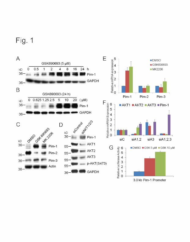
Fig. 1
A
kD 0 36-
36-
B
GSK690693 (5 t.~M)
0.5 2 4 8 16 24 h
•- W Pim-1
GAPDH
GSK690693 (24 h)
kD 36-
0 0.625 1.25 2.5 5 10 20 ( IJM)
Pim-1
36-------- GAPDH
c
kD 36- Pim-1
36_ . Pim-2
D kD 36-
;,.. r:V"' ~0 ~ 0(;' f .(.)
~ ~
Pim-1
55---AKT1
55--AKT2
AKT3 55-
E
Pim-3
Actin 55--p-AKT(S473)
36---GAPDH
G
Pim-1
• DMSO
• GSK690693
• MK2206
Pim-2 Pim-3
siC siA1 ,2 siA3 siA1 ,2,3
3.0 kb Pim-1 Promoter

Fig. 2
A GSK690693: + + + B siPim-1: 2 PC3-LN4 BT474 kD - --MET ., .,
130- '<t '<t - 0 + 0 + 130- EPHA2 (/) :.:: :.:: (/) :.:: :.:: :2 (/) ., (/) :2 (/) ., (/) 0 0 '<t 0 0 0 '<t 0
130- - HER3 1~~-- - MET
HER2 HER2
97- -- IGF1R ~-
HER3
97-- -- - INSR -Actin 36-
36-
----Actin E
" 36- -~ ~ :--c D ~0 ~" ~0(.i Q.~
WT TKO PC3-LN4 BT474 kD - ~· MET 130-GSK690693: - + + 0 0 .. HER2
(/) ., (/)
kD :2 :2 ., • HER3 - MET
., (0 ., (0 :.:: 130- 0 '<t :.:: 0 '<t
kD - MET 130- EPHA2 130--130-
HER3 130-- - • EPHA2 130-130-
IGF1R --- HER3 36- Pim-1 130- .. --- Actin Actin
36 Actin 36- 36-

Fig. 3
A GSK690693
0 (/)
:::!: 0
kD 15-
15-36-
36-1
B
56
D DMSO
• GSK690693
CrPV HCV HIF1a Myc VEGF
36- p-eiF2a{S51) D E 36-36-
130-
13D-
13()-97-
55- -
Pim-1
MET
EPHA2
HER3
HER2
IGF1R
p-AKT {5473)
55 • • --AKT{pan)
36_ ----.. -SGAPDH
looo ·c ., a:: ~00 +---------;;: 0
~ 10 ~ ~ Q; a::
F g ·c 1.4 ., ~ 1.2
~ 1
.Q 0.8
~ 0.6 ., .<:: 0.4 o; 0.2 Q; a:: 0
RF MET VEGF EMCV
o siC • siPim-1
CrPV HCV HIF1a Myc
G GSK690693: + + + siPim-1 : 2
36- Pim-1
55-Myc
28- - - Bcl-2
VEGF 15-
130-- - - ~ - HIF1a 55-kD - ..., - - Actin

Fig. 4
A
0 • 0
c
E
B
1 l
- Vehicle
~GSK
15 18 21 24 27 30 33 36 Day
OMSO GSK 4a GSK +
D Vehicle GSK
1 2 3 1 2 3 kO
130- _ ____ _
GSK + 4a
1 2 3 ----130- -·------
4a
2 3
130- -. ... - - -~-- ~ -- -
4a
MET
EPHA2
HER3
36-
PRAS40
Pim-1
36 ..... ~~--~~ .... .__. ..... ~tin
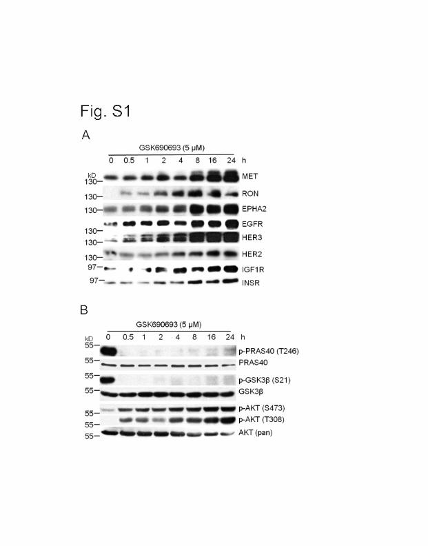
Fig. S1
A GSK690693 (5 IJM)
0 0 .5 1 2 4 8 16 24 h
kD 130-
13Q- . ..
97-
MET
RON
EPHA2
HER2
• ... ---- IGF1R 97- __
- - ---- INSR
8 GSK690693 (5 IJM)
kO 0 0.5 1 2 4 8 16 24 h 55-
p-PRAS40 (T246)
55---------PRAS40
55--
55--.. - p-GSK3J3 (S21 )
& & - GSK313 55- _ _____ _., p-AKT (S473)
55- .._ - p-AKT (T308) 55_.._._ ____ _ _ AKT (pan)

Fig. 82
• DMSO • ft • HER2 MET
EGFR-,&/ GSK •• -• • •
• •
4a -" ••• GSK+ 4a
•
_, . . .. •
INSR EPHAl
t I • .. . l'. . • • .. • • •

Fig. 83 A
6
MET HER3 INSR IGF1 R EPHA2 Pim-1
B c: DsiC • siPim-1 0 'ii)
~ 1.5 c. >< Cl)
c( 1 z
0:: E Cl)
.2: 0.5 (ij (ij 0::
0
MET HER3 INSR IGF1R EPHA2 Pim-1
c GFP
CHX: 0 0.5 1 2 4 6
kD 13o----
13Q- - -
36-
~~-------------
Pim-1
0 0.5 1 2 4 6 h
--- - MET
----- EPHA2 Pim-1
............. AKT (pan)
-.--~~- Actin

Fig. S4
GSK690693: PP242:
+ + + + +
AZD8055: + + kD ._ ______ eiF4E a.
1~ ~ 15- -._ --.... .,._.,._ 4E-BP1 t;> ,.._
220--- -- - E eiF4G
..J (.)
4E-BP1 ~
eiF4G


![Untitled-1 [] · YT.. 0300 YT.. 0300 YT.. 0300 YT.. 0300. Title: Untitled-1 Author: Eyup Created Date: 4/4/2016 11:28:30 AM](https://img.dokumen.tips/doc/110x75/5f24e2450a7e2c6cc2663645/untitled-1-yt-0300-yt-0300-yt-0300-yt-0300-title-untitled-1-author.jpg)


