Embed Size (px)
Citation preview

Autophagic flux without a block differentiatesvaricella-zoster virus infection from herpessimplex virus infectionErin M. Buckinghama, John E. Carpentera, Wallen Jacksona, Leigh Zerbonib, Ann M. Arvinb, and Charles Grosea,1
aVirology Laboratory, Department of Pediatrics, University of Iowa Children’s Hospital, Iowa City, IA 52242; and bDepartments of Pediatrics and Microbiologyand Immunology, Stanford University School of Medicine, Stanford, CA 94305
Edited by Elliott Kieff, Harvard Medical School and Brigham and Women’s Hospital, Boston, MA, and approved December 5, 2014 (received for reviewSeptember 17, 2014)
Autophagy is a process by which misfolded and damaged proteinsare sequestered into autophagosomes, before degradation in andrecycling from lysosomes. We have extensively studied the role ofautophagy in varicella-zoster virus (VZV) infection, and haveobserved that vesicular cells are filled with >100 autophagosomesthat are easily detectable after immunolabeling for the LC3 pro-tein. To confirm our hypothesis that increased autophagosomeformation was not secondary to a block, we examined all condi-tions of VZV infection as well as carrying out two assessments ofautophagic flux. We first investigated autophagy in human skinxenografts in the severe combined immunodeficiency (SCID)mouse model of VZV pathogenesis, and observed that autopha-gosomes were abundant in infected human skin tissues. We nextinvestigated autophagy following infection with sonically pre-pared cell-free virus in cultured cells. Under these conditions,autophagy was detected in a majority of infected cells, but wasmuch less than that seen after an infected-cell inoculum. In otherwords, inoculation with lower-titered cell-free virus did not reflectthe level of stress to the VZV-infected cell that was seen afterinoculation of human skin in the SCID mouse model or monolayerswith higher-titered infected cells. Finally, we investigated VZV-induced autophagic flux by two different methods (radiolabelingproteins and a dual-colored LC3 plasmid); both showed no evi-dence of a block in autophagy. Overall, therefore, autophagywithin a VZV-infected cell was remarkably different from autoph-agy within an HSV-infected cell, whose genome contains twomodifiers of autophagy, ICP34.5 and US11, not present in VZV.
autophagy | autophagosome | SCID-mouse | ICP34.5 | Epstein–Barr virus
VZV induces macroautophagy (hereafter referred to asautophagy) in skin cells within the typical exanthem associ-
ated with either primary VZV infection (varicella or chickenpox)or VZV reactivation (herpes zoster or shingles). During priorstudies, the extent of autophagy was gauged by enumeration ofautophagosomes by both 2D and 3D microscopy (1, 2). Theusual number of autophagosomes seen by 3D animation was 100per infected cell, but sometimes approached 200 per cell. Incontrast, a typical nonstressed cell usually has fewer than 4autophagosomes (3, 4). When monolayers were inoculated withVZV-infected cells, the traditional method for VZV infection,autophagy was again easily seen after enumeration of autophago-somes and immunoblotting for the LC3-phosphatidylethanolamineconjugate (LC3-II). Again these results suggested that autophagicflux was present during VZV infection in cultured cells.As part of a more extensive assessment of autophagy after
VZV-induced cellular stress, we have now investigated autoph-agy in infected human skin xenografts from the SCID mousemodel of VZV infection. This model is the most accurate rep-resentation of the skin manifestation of varicella in the humanhost (5, 6). Finally, we addressed an important point about thenature of VZV-induced autophagy. Because the number ofautophagosomes seen in the human vesicle cells from varicella
and herpes zoster patients is so high, the question has arisenwhether there is a late block in the maturation of autophago-somes to autolysosomes. In this report, we demonstrate that (i)autophagy induced by VZV infection is related to the overallstress to the cell, namely, a higher inoculum leads to greaterautophagy; and that (ii) autophagosomes induced during VZVinfection mature into autolysosomes without an obvious blockbefore final maturation. The autophagic flux assay results con-firm that VZV infection induces autophagy that proceeds tocompletion, possibly allowing the cell to alleviate the cellularstress caused by the viral infection (7). In previous work, weshowed that inhibition of autophagy led to a significant decreasein VZV titer. Overall, therefore, autophagy within a VZV-infectedcell is remarkably different from autophagy within an HSV-infected cell, an alphaherpesvirus whose genome contains twomodifiers of autophagy, ICP34.5 and US11 (8–13). In contrast,autophagy appears to be proviral in the life cycle of VZV.
ResultsAutophagy in VZV-Infected Human Skin Xenografts in the SCIDMouse. VZV is renowned for its specificity to a limited numberof human cell lines. Because of this restriction to human tissues,there is no animal model that completely mimics the humandisease varicella. However, the SCID mouse model for VZVinfection has provided the best alternative system to study themanifestations of VZV replication in the skin. In this model,human fetal skin xenografts are inserted beneath the skin of the
Significance
Varicella-zoster virus (VZV) is an important pathogen, whichcauses varicella and herpes zoster in humans. In general,there are similarities in virus-host interactions between thealphaherpesviruses. One notable exception is the responseto autophagy. VZV infection induces autophagy. This is incontrast to herpes simplex virus (HSV), which has two genesthat inhibit autophagy, ICP34.5 and US11; neither is presentin the smaller VZV genome. In this study, we found that VZV-induced autophagic flux was not blocked. These resultsreinforce prior observations showing a proviral effect ofautophagy on VZV infectivity and spread. These VZV findingsalso exhibit similarities with recent data about a requirementfor early phase autophagy during Epstein–Barr virus infection,a phylogenetically distant gammaherpesvirus.
Author contributions: E.M.B., J.E.C., L.Z., A.M.A., and C.G. designed research; E.M.B., J.E.C.,W.J., and L.Z. performed research; E.M.B., J.E.C., and C.G. analyzed data; and E.M.B., J.E.C.,L.Z., A.M.A., and C.G. wrote the paper.
The authors declare no conflict of interest.
This article is a PNAS Direct Submission.
Freely available online through the PNAS open access option.1To whom correspondence should be addressed. Email: [email protected].
This article contains supporting information online at www.pnas.org/lookup/suppl/doi:10.1073/pnas.1417878112/-/DCSupplemental.
256–261 | PNAS | January 6, 2015 | vol. 112 | no. 1 www.pnas.org/cgi/doi/10.1073/pnas.1417878112
Dow
nloa
ded
by g
uest
on
Janu
ary
4, 2
020

severe combined immunodeficiency (SCID) mouse (14). In turn,replicate xenografts are inoculated with VZV-infected cells andthen harvested at 7, 14, and 21 d after infection. This modelsystem allows for the study of VZV infection of a differentiatedhuman tissue in the context of no adaptive immune response.The pathology is remarkably similar to that seen in biopsies ofvesicles during human varicella (5). To confirm and extend ourautophagy investigations, therefore, we examined skin xenograftsinfected with VZV in vivo.VZV-infected human skin was removed 21 d post infection
and sections were stained with a monoclonal antibody to VZVglycoprotein E (gE), to identify infected areas of the tissue, aswell as rabbit polyclonal antibody against LC3, to identifyautophagosomes. The sections were examined by brightfield andconfocal fluorescence microscopy. Images of complete serialsections are shown in Fig. 1. The brightfield image (Fig. 1A)shows evidence of major disruption of the tissue interior due toviral pathology. The fluorescent image (Fig. 1B) shows extensiveVZV gE (red) expression throughout the tissue section, as wellas LC3 expression, indicative of autophagosomes (green).Examination of these fluorescently stained tissues at higher
power, and analysis of Z stacks assembled into 3D projectionsusing the Imaris imaging program confirmed that VZV infectionin the SCID mouse model was associated with abundantautophagy. Fig. 2 A–F shows several representative images fromthese xenografts, with many LC3-positive puncta (green) seen inand near VZV-infected (red) cells. Mock infection of humantissue in the SCID mouse was also performed and analyzed byconfocal microscopy. The skin tissue was intact (Fig. 2G) and
there was very minimal LC3 staining present (Fig. 2H) and noVZV gE expression (Fig. 2I). In summary, abundant autophagywas evident in the VZV-infected skin xenograft.
Comparison of Autophagy in the VZV-Infected Skin Xenografts withHuman Vesicles. In early experiments, we documented the pres-ence of autophagosomes in skin vesicles from immunocompetentchildren with varicella or adults with herpes zoster (15, 16). Toconfirm that the autophagy seen in the VZV-infected skinxenografts in SCID mice, which lack adaptive immune responses,recapitulated the response seen in human skin vesicles fromchildren with varicella, we performed a direct comparison. Ascan be seen by viewing Fig. 3 A and B, the pattern and distri-bution of LC3-positive (green) autophagosomes were similar inthe two samples. Together, these data again demonstrated thevalidity of the SCID mouse model to recapitulate pathogenesisin human skin infected VZV during varicella or herpes zoster.
Autophagy After High Input Cell-Free Virus Inoculation. VZV as-sembly in infected cells is an extremely labile process. In contrastto HSV, complete enveloped virions are not released frominfected cells. Instead, VZV-infected cells are sonically disruptedto release what is called cell-free virus. The titer of this product isvery low; the estimated particle to PFU ratio was 40,000:1 (17).To expand our knowledge about the role of input inoculum onthe subsequent induction of autophagy in VZV-infected cells, wepostulated that inoculation of cells with cell-free virus would leadto lower levels of autophagy than seen in the human vesicle or
Fig. 1. VZV induces extensive LC3 puncta indicative of autophagosomes inhuman skin cells in the xenografts in the SCID mouse model. VZV-infectedhuman skin xenografts at 21 dpi were immunolabeled with antibodies toVZV gE (red) and to LC3 (green), as well as the Hoescht 33342 DNA stain (blue).Brightfield (A) and fluorescent (B) images of successive, whole sections ofhuman skin xenografts showed extensive disruption of the tissue with prom-inent VZV gE expression as well as LC3 staining of autophagosomes. Areas oftissue disruption caused by VZV infection in A are noted by arrows.
Fig. 2. VZV infection induces extensive autophagosome formation in hu-man skin xenografts. (A–F) Sections from 21 dpi human skin xenografts wereimmunolabeled with MAb 3B3 (anti-VZV gE, red) and anti-LC3 antibody[Santa Cruz (A–E) or Epitomics (F), green] primary antibodies as well asH33342 (DNA stain, blue). Z-stack images were analyzed using the Imarissoftware to create 3D reconstructions that show LC3-positive autophago-somes as green spots and nuclei as blue spots. Abundant autophagosomesare noted in both samples. gE staining (red) is displayed flat to show diffuseand abundant staining in infected cells, often most highly concentrated inthe ER/Golgi. Images shown are from either 400× total magnification (A andB) or 630× total magnification (C–F). (G–I) These three images each showa single fluorescent channel of the same 2D confocal image of mock infectedhuman skin implants from a SCID-hu mouse at 630× total magnification.Mock infection showed no gE staining (I) and minimal LC3 staining (H).
Buckingham et al. PNAS | January 6, 2015 | vol. 112 | no. 1 | 257
MICRO
BIOLO
GY
Dow
nloa
ded
by g
uest
on
Janu
ary
4, 2
020
after inoculation with infected cells. In an initial set of experi-ments, the inoculum was a relatively high input of cell-free virus.MRC-5 fibroblasts were infected with 1600 pfu per 10 cm2.
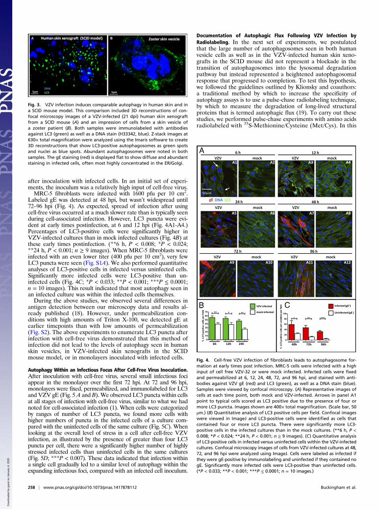
Labeled gE was detected at 48 hpi, but wasn’t widespread until72–96 hpi (Fig. 4). As expected, spread of infection after usingcell-free virus occurred at a much slower rate than is typically seenduring cell-associated infection. However, LC3 puncta were evi-dent at early times postinfection, at 6 and 12 hpi (Fig. 4A1-A4.)Percentages of LC3-positive cells were significantly higher inVZV-infected cultures than in mock infected cultures (Fig. 4B) atthese early times postinfection. (**6 h, P < 0.008; *P < 0.024;**24 h, P < 0.001; n ≥ 9 images). When MRC-5 fibroblasts wereinfected with an even lower titer (400 pfu per 10 cm2), very fewLC3 puncta were seen (Fig. S1A). We also performed quantitativeanalyses of LC3-positive cells in infected versus uninfected cells.Significantly more infected cells were LC3-positive than un-infected cells (Fig. 4C; *P < 0.033; **P < 0.001; ***P ≤ 0.0001;n = 10 images). This result indicated that most autophagy seen inan infected culture was within the infected cells themselves.During the above studies, we observed several differences in
antigen detection between our microscopy data and results al-ready published (18). However, under permeabilization con-ditions with high amounts of Triton X-100, we detected gE atearlier timepoints than with low amounts of permeabilization(Fig. S2). The above experiments to enumerate LC3 puncta afterinfection with cell-free virus demonstrated that this method ofinfection did not lead to the levels of autophagy seen in humanskin vesicles, in VZV-infected skin xenografts in the SCIDmouse model, or in monolayers inoculated with infected cells.
Autophagy Within an Infectious Focus After Cell-Free Virus Inoculation.After inoculation with cell-free virus, several small infectious fociappear in the monolayer over the first 72 hpi. At 72 and 96 hpi,monolayers were fixed, permeabilized, and immunolabeled for LC3and VZV gE (Fig. 5 A and B). We observed LC3 puncta within cellsat all stages of infection with cell-free virus, similar to what we hadnoted for cell-associated infection (1). When cells were categorizedby ranges of number of LC3 puncta, we found more cells withhigher numbers of puncta in the infected cells of a culture com-pared with the uninfected cells of the same culture (Fig. 5C). Whenlooking at the overall level of stress in a cell after cell-free VZVinfection, as illustrated by the presence of greater than four LC3puncta per cell, there were a significantly higher number of highlystressed infected cells than uninfected cells in the same cultures(Fig. 5D; ***P < 0.007). These data indicated that infection withina single cell gradually led to a similar level of autophagy within theexpanding infectious foci, compared with an infected cell inoculum.
Documentation of Autophagic Flux Following VZV Infection byRadiolabeling. In the next set of experiments, we postulatedthat the large number of autophagosomes seen in both humanvesicle cells as well as in the VZV-infected human skin xeno-grafts in the SCID mouse did not represent a blockade in thetransition of autophagosomes into the lysosomal degradationpathway but instead represented a heightened autophagosomalresponse that progressed to completion. To test this hypothesis,we followed the guidelines outlined by Klionsky and coauthors:a traditional method by which to increase the specificity ofautophagy assays is to use a pulse-chase radiolabeling technique,by which to measure the degradation of long-lived structuralproteins that is termed autophagic flux (19). To carry out thesestudies, we performed pulse-chase experiments with amino acidsradiolabeled with 35S-Methionine/Cysteine (Met/Cys). In this
Fig. 3. VZV infection induces comparable autophagy in human skin and ina SCID mouse model. This comparison included 3D reconstructions of con-focal microscopy images of a VZV-infected (21 dpi) human skin xenograftfrom a SCID mouse (A) and an impression of cells from a skin vesicle ofa zoster patient (B). Both samples were immunolabeled with antibodiesagainst LC3 (green) as well as a DNA stain (H33342, blue). Z-stack images at630× total magnification were analyzed using the Imaris software to create3D reconstructions that show LC3-positive autophagosomes as green spotsand nuclei as blue spots. Abundant autophagosomes were noted in bothsamples. The gE staining (red) is displayed flat to show diffuse and abundantstaining in infected cells, often most highly concentrated in the ER/Golgi.
Fig. 4. Cell-free VZV infection of fibroblasts leads to autophagosome for-mation at early times post infection. MRC-5 cells were infected with a highinput of cell free VZV-32 or were mock infected. Infected cells were fixedand permeabilized at 6, 12, 24, 48, 72, and 96 hpi, and stained with anti-bodies against VZV gE (red) and LC3 (green), as well as a DNA stain (blue).Samples were viewed by confocal microscopy. (A) Representative images ofcells at each time point, both mock and VZV-infected. Arrows in panel A1point to typical cells scored as LC3 positive due to the presence of four ormore LC3 puncta. Images shown are 400× total magnification. (Scale bar, 50μm.) (B) Quantitative analysis of LC3 positive cells per field. Confocal imageswere viewed in ImageJ and LC3-positive cells were identified as cells thatcontained four or more LC3 puncta. There were significantly more LC3-positive cells in the infected cultures than in the mock cultures. (**6 h, P <0.008; *P < 0.024; **24 h, P < 0.001; n ≥ 9 images). (C) Quantitative analysisof LC3-positive cells in infected versus uninfected cells within the VZV-infectedcultures. Confocal microscopy images of cells from VZV-infected cultures at 48,72, and 96 hpi were analyzed using ImageJ. Cells were labeled as infected ifthey were gE-positive by immunolabeling and uninfected if they contained nogE. Significantly more infected cells were LC3-positive than uninfected cells.(*P < 0.033; **P < 0.001; ***P ≤ 0.0001; n = 10 images.)
258 | www.pnas.org/cgi/doi/10.1073/pnas.1417878112 Buckingham et al.
Dow
nloa
ded
by g
uest
on
Janu
ary
4, 2
020

experiment, monolayers of uninfected cells and VZV-infectedcells were pulse labeled for 2 h and then chased with unlabeledmedium for 24 h. By scintillation counting, we determined that∼35% of the radiolabeled Met/Cys was taken up by the cellsduring the pulse period and that ∼7% of that amount was ex-pelled into the medium in the first 2 h of chase. This percentagecorresponded to degradation of short-lived proteins by protea-somes. Another 7% of the absorbed fraction was expelled intothe medium during the chase period from 2–24 h, correspondingto degradation of longer lived structural proteins.To determine the contribution of autophagy to the degrada-
tion of longer lived proteins, Klionsky at al (19). recommendedthat half of the monolayers for each experiment receive treat-ment with 3-methyladenine (3-MA), an inhibitor of class I andclass III phosphoinositide 3-kinase (PI3K) (20–22). Given thatover 80% of the radiolabel was retained in the cells during thechase, we chose to measure the degradation rate by densitometryof lysates removed and saved during the chase period. In general,protein bands became lighter during the chase period due todegradation (Fig. 6). This effect was quantitated using meanpixel values measured with ImageJ. The difference betweenuntreated cells and 3-MA-treated cells was revealed by sub-tracting the pulse value from each chase value to arrive ata “darkness” value. As expected, the degradation rate in un-infected fibroblast cells (Fig. 6A, lanes 3–5 vs. lanes 6–8) was
insensitive to 3-MA treatment, indicating that the base rate of−0.3 darkness/h is primarily proteasomal. VZV-infected cellsgenerated a higher degradation rate (−0.64 darkness/h) than un-infected cells (Fig. 6B). Treatment with 3-MA dramatically low-ered the degradation rate of VZV infected cells (to −0.13darkness/h; Fig. 6B, lanes 3–5, without 3-MA vs. lanes 6, 7, 8 with3-MA), a result indicating that the majority of the degradation oflonger-lived proteins in VZV-infected cells was due to autophagy.
Documentation of Autophagic Flux with a Dual-Colored LC3 Plasmid.To confirm and expand the above results, we obtained themRFP-GFP tandem fluorescent-tagged LC3 plasmid (ptfLC3).This plasmid contains both red and green fluorescent proteinstagged to the LC3 protein. During the process of autophagy,autophagosomes fuse with lysosomes to form autolysosomes,which provides an acidic environment and proteases for thedegradation of the contents of the autophagosome. The autoly-sosome is distinguished from an autophagolysosome, which isa compartment that is specific to the process of xenophogy (23).It has been found that the fluorescent signal from GFP isquenched in an acidic environment of the lysosome, whereas thesignal from mRFP remains intact (24). Thus, in autophago-somes, there will be yellow signals (overlap of red and greenfluorescence) whereas the green signal is lost as the autopha-gosome fuses with the more acidic lysosome (24). Therefore,autolysosomes appear with red fluorescent LC3.MRC-5 fibroblasts were transfected with the tandem fluores-
cent tagged LC3 plasmid (ptfLC3) and subsequently inoculatedwith VZV-infected cells at 48 h posttransfection. At 72 h post-infection (hpi), cells were fixed and stained with an antibodyagainst VZV gE (white). Samples were examined by confocalmicroscopy and Z-stacks of images were analyzed using Imarissoftware for 3D reconstruction. A representative 3D image re-construction is shown in Fig. 7. LC3 puncta were visible ininfected cells. Spheres that are green and red (yellow arrow)represent autophagosomes that have not yet fused with lyso-somes. Spheres that are red represent autolysosomes. Manyimages were analyzed with similar patterns of the presence ofboth red puncta as well as green and red puncta in infected cells,indicating that autophagic flux was intact in VZV-infected cells.These data confirmed our previous results that autophagy pro-ceeded to completion in VZV infection. VZV infection did notcause a block in autophagic flux.
DiscussionThe role of autophagy during HSV-1 infection has been found toinvolve two viral proteins, ICP34.5 and US11. The initial dis-covery about the role of ICP34.5 was brought to the forefront byOrvedahl et al., who demonstrated that this neurovirulenceprotein bound to the autophagy protein Beclin 1, the mammalianortholog of yeast Atg6 (9). Beclin 1 interacts with severalautophagy cofactors, to regulate and promote formation of theBeclin 1–Vps34–Vps15 complexes (22, 25). A domain withinamino acids 68–87 of ICP34.5 interacts with Beclin 1 and therebyinhibits its ability to induce autophagy (9). Deletion of theICP34.5 gene from the HSV long repeat region leads to in-creased autophagy in infected animals and decreased neuro-virulence (9, 26). Subsequently, Lussignol et al. investigated therole of HSV US11 and confirmed that US11 also inhibitedautophagy but via a mechanism independent of inhibition ofBeclin 1 (13). What is important for interpretation of our study ofVZV-induced autophagy is the fact that very few alphaherpesvirusgenomes except for HSV-1 and HSV-2 have the ICP34.5 gene (27–30). In addition, many such as VZV lack an ortholog of US11 (31).VZV with the smallest herpesvirus genome, therefore, has dis-carded US11 (or never acquired it) and never captured the ICP34.5/GADD34-homolog gene during its evolution (32).However, the VZV genome has retained the HSV ICP0
ortholog, ORF61. Infection of differentiated skin cells withintheir tissue microenvironment as observed using xenografts inSCID mice has revealed a dramatic up-regulation of the type I
Fig. 5. Individual cells within a focus of infection after cell-free VZV in-fection exhibited LC3 puncta similar to cells infected with cell-associatedVZV. MRC-5 cells were infected with a high-input of cell-free VZV-32 andfixed at 72 and 96 hpi. (A) Infected cells at 72 hpi (VZV gE; red) are apparentin the representative image at 400×. (B) Higher magnification (800×) imagesshowing LC3 (green) in the area outlined by the white rectangle in panel A.Representative LC3 puncta indicative of autophagosomes are indicated bysolid white arrows; similar puncta in a newly infected cell are indicated bya dashed white line. (C) Stacked bar chart showing the percentage of cellswith a given number of LC3 puncta for both uninfected cells and infectedcells from cultures infected with cell free VZV for 72–96 h. Blue colors in-dicate levels of puncta in unstressed cells, whereas red and pink colors in-dicate levels of puncta in stressed cells. (D) Different presentation of datafrom panel C, showing percent of stressed cells (cells that contain four ormore LC3 puncta) in populations of uninfected (no gE staining) or infected(gE positive) cells. Cells infected with cell-free VZV exhibited more LC3puncta indicative of autophagosomes than uninfected cells (***P < 0.007)and at levels similar to cells infected with cell-associated virus (1).
Buckingham et al. PNAS | January 6, 2015 | vol. 112 | no. 1 | 259
MICRO
BIOLO
GY
Dow
nloa
ded
by g
uest
on
Janu
ary
4, 2
020

IFN response in uninfected cells surrounding the VZV lesion,associated with STAT1 activation and enhanced numbers ofpromyelocytic protein (PML) nuclear bodies (33, 34). In con-trast, VZV-infected cells show disruption of these innate anti-viral mechanisms in an ORF61-dependent manner. Conversely,VZV infection induces STAT3 activation, leading to survivinexpression, which is necessary for viral replication; inhibiting thispathway severely impairs VZV replication in skin (35). Thefinding that autophagic flux is maintained in VZV-infected skincells suggests that this process may also represent a virus-cellinteraction that has important proviral effects in vivo.We had previously investigated VZV autophagic flux by
measurement of stress-inducible intracellular p62/SQSTM1protein levels in infected cells, and showed that VZV-inducedautophagy was completed, leading to the degradation of p62/SQSTM1 (15). To confirm and extend our observations, wemeasured autophagic flux by a traditional pulse-chase assay usingradiolabeled amino acids. This assay measures the turnover oflong-lived proteins leading to a determination of autophagic flux.It is a well established methodology that is highly quantifiable,and is a recommended method in the “Guidelines for the useand interpretation of assays for monitoring autophagy” byKlionsky and colleagues (36). This assay showed that long-livedproteins were degraded by VZV-induced autophagy.
We also investigated autophagic flux in VZV-infected cells usingthe mRFP-GFP tandem fluorescent-tagged LC3 plasmid (ptfLC3)created by Yoshimori and colleagues (24). This innovative plas-mid has been used by other groups studying autophagy, todemonstrate the completion of autophagy by the formation ofautolysosomes, such as in a study of HIV from Levine and col-leagues (37). They used the ptfLC3 plasmid to show that siRNAknockdown of GAPR-1, a newly identified inhibitor of autoph-agy, increased autophagosome formation by causing an increasein autophagic flux rather than a block in autophagosome matu-ration. Another group studied of the role of autophagy inNewcastle Disease Virus (NDV) infection (38). This studyshowed that NDV infection induces autophagic flux in two dif-ferent kinds of chicken cells. Also, the previously mentionedstudy by Lussignol et al. used ptfLC3-transfected HeLa cells toshow that the HSV-1 protein US11 blocked autophagic flux toa similar degree as HSV-1 ICP34.5 (13). All of these studiestogether highlight the utility of the tandem fluorescent LC3 plas-mid in the study of autophagic flux in virus systems. Once again,VZV is found to have a very different relationship with autophagythan the closely related alphaherpesvirus HSV-1, which has beenshown to inhibit autophagy in several ways, including blockingautophagosome maturation (9, 12). Instead, the level of inductionof autophagy by VZV is similar to that of known chemical inducersof autophagy, such as trehalose and tunicamycin (1).Finally, the discussion about the proviral effects of autophagy
during herpesvirus infection can now be extended beyondalphaherpesviruses based on an insightful report on the gamma-1herpesvirus, Epstein–Barr virus (EBV) (39). Although EBVblocks autophagic flux during its replication cycle, the autophagicmachinery is then usurped by EBV for transport of viral par-ticles. Specifically, early phases of autophagic flux are requiredfor the production of infectious EBV particles, whereas the latephase of autophagic flux (autolysosome formation) was blocked.Via this strategy, EBV redirects the expanding autophagic ma-chinery that is reacting to the stress induced by viral infectiontoward the production of EBV particles. Thus, in a broadersense, autophagy is proviral in that autophagic components areused during the EBV replication cycle. As cited above, VZVbeing the herpesvirus with the smallest lacks the viral genes thatinhibit the late phases of flux. Otherwise stated, of the 8 genes inthe HSV-1 genome (152 kbp) not found in the VZV genome(124 kbp), two inhibit autophagy. Instead, VZV accommodatesits replication cycle within an infected cell and its titer, similar to
Fig. 6. Autophagic flux during VZV infection of fibro-blasts. VZV-infected monolayers (by cell-associated virus)were pulse labeled with 35S-labeled Met/Cys and thenlysates saved at chase times 6, 12, and 24 h with andwithout 3-MA treatment. Reduced lysates at all pulse andchase time points were electrophoresed, transferred toPVDF membranes, and exposed to film. The darkness ofa selected area of each lane was measured with ImageJ andplotted relative to the pulse value. The procedure was du-plicated three times. (A) Uninfected MRC-5 cells. 3-MAtreatment made little difference to the overall rate ofdegradation in uninfected cells. The area used to measurepixel density is shown as a box. (B) VZV-infected cells. 3-MAtreatment significantly decreased protein degradation inVZV-infected cells, indicating increased autophagic flux ininfected cells.
Fig. 7. Analysis of autophagic fluxwith a tandem red/green fluorescent protein-tagged LC3 plasmid. MRC-5 cells transfectedwith the tandem fluorescent taggedLC3 plasmid were inoculated with VZV-infected cells for 72 h then immunola-beled. Samples were examined by confocal microscopy, and analyzed with Imarissoftware for 3D reconstruction. LC3 puncta are illustrated by spheres that aregreen and red (yellowarrow), showing LC3 expression in autophagosomes, or redonly (red arrow), showing LC3 expression in autolysosomes, where GFP fluores-cence has been extinguished. Nuclei (blue), VZV gE (white). (Scale bar, 5 μm.)
260 | www.pnas.org/cgi/doi/10.1073/pnas.1417878112 Buckingham et al.
Dow
nloa
ded
by g
uest
on
Janu
ary
4, 2
020

EBV, declines when autophagy is inhibited with either siRNA or3-methyladenine (3-MA) (1). Thus, autophagy provides a linkbetween two of the most phylogenetically divergent humanherpesviruses: alpha VZV and gamma EBV.
Materials and MethodsMore detailed experimental information is provided in SI Materialsand Methods.
Viruses and Cells. VZV-32 is a low passage laboratory strain; its genomehas been completely sequenced and falls within European clade 1 of VZVgenotypes (40). Skin explants were infected with the parental rOka strain asin ref. 5. MRC-5 human fibroblast cells and MeWo strain of human mela-noma cells were grown as in Grose and Brunel (41).
Generation of Cell-Free VZV or Uninfected Cell Sonicate. Three 25-cm2 MeWomonolayers were infected at 1:8 infected: uninfected ratio and incubated at32 °C for 72 h. The infected monolayers were scraped into 2 mL of SPA media(0.2 M sucrose, 0.01 M sodium phosphate, 1% BSA pH 7.4) and sonicated onice for 20 s. The sonicate was centrifuged at 300 × g for 10 min at 4 °C. Thepellet was discarded and the supernatant was diluted with 75 mL of com-plete MEM and added to 24 wells on six-well plates (3 mL per well).
VZV Infection of Human Skin Xenografts. Construction of human skin xeno-grafts in SCID mice and subsequent inoculation with VZV or mock-infectedcells was done as described (5, 42).
Primary and Secondary Antibody Reagents. Primary antibodies required forthis study included the previously described VZV-specific murine monoclonalantibody (MAb) 3B3 and 370 (gE; ORF68; 1:1,000). Also used was a rabbitpolyclonal antibody to MAP1LC3B (1:200: sc-28266, Santa Cruz Bio-technology), and a rabbit MAb anti-LC3A/B (1:1,000; 2057-1, Epitomics).Secondary antibodies used were AlexaFluor 488 and 546 fluoroprobesconjugated to goat anti-rabbit IgG or goat anti-mouse IgG F(ab’)2 frag-ment (1:1,250; Invitrogen).
Imaging Protocols. Samples of infected and uninfected cells were immuno-labeled and prepared for confocal microscopy by methods described pre-viously (1, 2, 16).
Transfections of Cells with Tandem Fluorescent LC3 Plasmid. MRC-5 fibroblastswere transfected with the tandem fluorescent tagged LC3 plasmid (ptfLC3,Plasmid#21074 from Addgene.org; created by T. Yoshimori), as in ref. 24.After 24 h, the monolayer was inoculated with VZV-infected cells.
Protein Degradation Measurement by Pulse-Chase Radiolabeling. The basicexperimental protocol is described in the autophagy literature (19) and ourprocedure is modeled on that used by Isler et al. (43). The 3-MA method-ology has been described (1).
ACKNOWLEDGMENTS. We thank the University of Iowa Central MicroscopyResearch Facility for use of imaging equipment (NIH Grant 1S10RR025439-01).This work was supported by NIH Grant AI89716.
1. Buckingham EM, Carpenter JE, Jackson W, Grose C (2014) Autophagy and the effectsof its inhibition on varicella-zoster virus glycoprotein biosynthesis and infectivity.J Virol 88(2):890–902.
2. Jackson W, Yamada M, Moninger T, Grose C (2013) Visualization and quantitation ofabundant macroautophagy in virus-infected cells by confocal three-dimensionalfluorescence imaging. J Virol Methods 193(1):244–250.
3. Kabeya Y, et al. (2000) LC3, a mammalian homologue of yeast Apg8p, is localized inautophagosome membranes after processing. EMBO J 19(21):5720–5728.
4. Tra T, et al. (2011) Autophagy in human embryonic stem cells. PLoS ONE 6(11):e27485.5. Moffat JF, et al. (1998) Attenuation of the vaccine Oka strain of varicella-zoster virus
and role of glycoprotein C in alphaherpesvirus virulence demonstrated in the SCID-humouse. J Virol 72(2):965–974.
6. Ku CC, Besser J, Abendroth A, Grose C, Arvin AM (2005) Varicella-Zoster virus path-ogenesis and immunobiology: New concepts emerging from investigations with theSCIDhu mouse model. J Virol 79(5):2651–2658.
7. Carpenter JE, Grose C (2014) Varicella-zoster virus glycoprotein expression differen-tially induces the unfolded protein response in infected cells. Front Microbiol 5:322.
8. Chou J, Kern ER, Whitley RJ, Roizman B (1990) Mapping of herpes simplex virus-1neurovirulence to gamma 134.5, a gene nonessential for growth in culture. Science250(4985):1262–1266.
9. Orvedahl A, et al. (2007) HSV-1 ICP34.5 confers neurovirulence by targeting the Beclin1 autophagy protein. Cell Host Microbe 1(1):23–35.
10. Alexander DE, Ward SL, Mizushima N, Levine B, Leib DA (2007) Analysis of the role ofautophagy in replication of herpes simplex virus in cell culture. J Virol 81(22):12128–12134.
11. English L, et al. (2009) Autophagy enhances the presentation of endogenous viralantigens on MHC class I molecules during HSV-1 infection. Nat Immunol 10(5):480–487.
12. Gobeil PA, Leib DA (2012) Herpes simplex virus γ34.5 interferes with autophagosomematuration and antigen presentation in dendritic cells. MBio 3(5):e00267–e12.
13. Lussignol M, et al. (2013) The herpes simplex virus 1 Us11 protein inhibits autophagythrough its interaction with the protein kinase PKR. J Virol 87(2):859–871.
14. Zerboni L, Sen N, Oliver SL, Arvin AM (2014) Molecular mechanisms of varicella zostervirus pathogenesis. Nat Rev Microbiol 12(3):197–210.
15. Takahashi MN, et al. (2009) Varicella-zoster virus infection induces autophagy in bothcultured cells and human skin vesicles. J Virol 83(11):5466–5476.
16. Carpenter JE, Jackson W, Benetti L, Grose C (2011) Autophagosome formation duringvaricella-zoster virus infection following endoplasmic reticulum stress and the un-folded protein response. J Virol 85(18):9414–9424.
17. Carpenter JE, Henderson EP, Grose C (2009) Enumeration of an extremely high par-ticle-to-PFU ratio for Varicella-zoster virus. J Virol 83(13):6917–6921.
18. Reichelt M, Brady J, Arvin AM (2009) The replication cycle of varicella-zoster virus:Analysis of the kinetics of viral protein expression, genome synthesis, and virion as-sembly at the single-cell level. J Virol 83(8):3904–3918.
19. Klionsky DJ (2008) Getting into the flow. Autophagy 4(2):139–140.20. Wu YT, et al. (2010) Dual role of 3-methyladenine in modulation of autophagy via
different temporal patterns of inhibition on class I and III phosphoinositide 3-kinase.J Biol Chem 285(14):10850–10861.
21. Seglen PO, Gordon PB (1982) 3-Methyladenine: Specific inhibitor of autophagic/ly-sosomal protein degradation in isolated rat hepatocytes. Proc Natl Acad Sci USA 79(6):1889–1892.
22. Funderburk SF, Wang QJ, Yue Z (2010) The Beclin 1-VPS34 complex—at the crossroadsof autophagy and beyond. Trends Cell Biol 20(6):355–362.
23. Klionsky DJ, Eskelinen EL, Deretic V (2014) Autophagosomes, phagosomes, autoly-sosomes, phagolysosomes, autophagolysosomes... wait, I’m confused. Autophagy10(4):549–551.
24. Kimura S, Noda T, Yoshimori T (2007) Dissection of the autophagosomematuration processby a novel reporter protein, tandem fluorescent-tagged LC3. Autophagy 3(5):452–460.
25. Furuya N, Yu J, Byfield M, Pattingre S, Levine B (2005) The evolutionarily conserveddomain of Beclin 1 is required for Vps34 binding, autophagy and tumor suppressorfunction. Autophagy 1(1):46–52.
26. Bolovan CA, Sawtell NM, Thompson RL (1994) ICP34.5 mutants of herpes simplex virustype 1 strain 17syn+ are attenuated for neurovirulence in mice and for replication inconfluent primary mouse embryo cell cultures. J Virol 68(1):48–55.
27. Davison AJ, Scott JE (1986) The complete DNA sequence of varicella-zoster virus. J GenVirol 67(Pt 9):1759–1816.
28. Ravi V, Kennedy PG, MacLean AR (1998) Functional analysis of the herpes simplexvirus type 2 strain HG52 RL1 gene: The intron plays no role in virulence. J Gen Virol79(Pt 7):1613–1617.
29. Tang S, Guo N, Patel A, Krause PR (2013) Herpes simplex virus 2 expresses a novel formof ICP34.5, a major viral neurovirulence factor, through regulated alternative splicing.J Virol 87(10):5820–5830.
30. Guliani S, et al. (2002) Macropodid herpesvirus 1 encodes genes for both thymidylatesynthase and ICP34.5. Virus Genes 24(3):207–213.
31. Cohen JI (2010) The varicella-zoster virus genome. Curr Top Microbiol Immunol 342:1–14.
32. Grose C (2012) Pangaea and the Out-of-Africa Model of Varicella-Zoster Virus Evo-lution and Phylogeography. J Virol 86(18):9558–9565.
33. Ku CC, et al. (2004) Varicella-zoster virus transfer to skin by T Cells and modulation ofviral replication by epidermal cell interferon-alpha. J Exp Med 200(7):917–925.
34. Wang L, et al. (2011) Disruption of PML nuclear bodies is mediated by ORF61 SUMO-interacting motifs and required for varicella-zoster virus pathogenesis in skin. PLoSPathog 7(8):e1002157.
35. Sen N, et al. (2012) Signal transducer and activator of transcription 3 (STAT3) andsurvivin induction by varicella-zoster virus promote replication and skin pathogenesis.Proc Natl Acad Sci USA 109(2):600–605.
36. Klionsky DJ, et al. (2012) Guidelines for the use and interpretation of assays formonitoring autophagy. Autophagy 8(4):445–544.
37. Shoji-Kawata S, et al. (2013) Identification of a candidate therapeutic autophagy-inducing peptide. Nature 494(7436):201–206.
38. Sun Y, et al. (2014) Autophagy benefits the replication of Newcastle disease virus inchicken cells and tissues. J Virol 88(1):525–537.
39. Granato M, et al. (2014) Epstein-barr virus blocks the autophagic flux and appro-priates the autophagic machinery to enhance viral replication. J Virol 88(21):12715–12726.
40. Peters GA, et al. (2006) A full-genome phylogenetic analysis of varicella-zoster virusreveals a novel origin of replication-based genotyping scheme and evidence of re-combination between major circulating clades. J Virol 80(19):9850–9860.
41. Grose C, Brunel PA (1978) Varicella-zoster virus: Isolation and propagation in humanmelanoma cells at 36 and 32 degrees C. Infect Immun 19(1):199–203.
42. Moffat JF, et al. (1998) The ORF47 and ORF66 putative protein kinases of varicella-zoster virus determine tropism for human T cells and skin in the SCID-hu mouse. ProcNatl Acad Sci USA 95(20):11969–11974.
43. Isler JA, Skalet AH, Alwine JC (2005) Human cytomegalovirus infection activates andregulates the unfolded protein response. J Virol 79(11):6890–6899.
Buckingham et al. PNAS | January 6, 2015 | vol. 112 | no. 1 | 261
MICRO
BIOLO
GY
Dow
nloa
ded
by g
uest
on
Janu
ary
4, 2
020