Embed Size (px)
Citation preview

Autonomic
nervovous system

Parasympathicusrest or digest
Sympathicusfight or flight
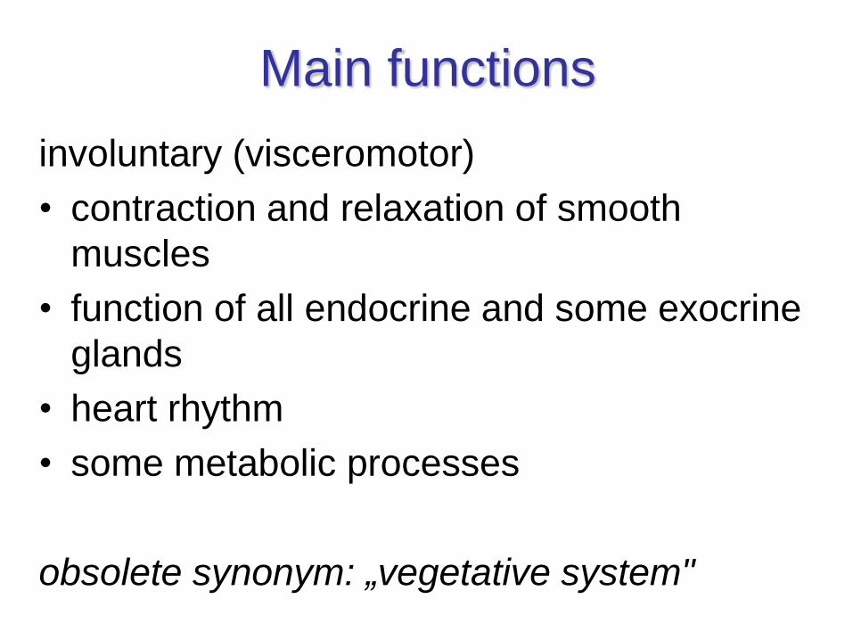
Main functions
involuntary (visceromotor)
• contraction and relaxation of smooth
muscles
• function of all endocrine and some exocrine
glands
• heart rhythm
• some metabolic processes
obsolete synonym: „vegetative system"

Classification of ANS
• sympathetic system
– fight or flight
• parasympathetic system
– rest or digest
• enteric system

Types of impulses conducted
by fibers of ANS
• nuclei in CNS → visceromotor fibers →autonomic ganglia (integration of information from CNS and ANS)
• free nerve endings → viscerosensory fibers→ ggl. spinale or ggl. n. VII, IX, X
– are not functional part of ANS !!!
– mechanoreceptors, chemoreceptors
– afferent fibers of reflectory pathways (coughing, defecation, vasomotor…)
– visceral pain (e.g. colic, angina)

parasympathetic part sympathetic part


Medicaments influencing ANS
+• Sympathomimetics
– diect: adrenaline, noradrenaline, dopamine, dobutamine, isoprenaline
– selective
– indirect
• Parasympathomimetics– acetylcholine, pilocarpine, karbachole, physoostigmine, organophosphates
-• Sympatholytics: alfa- and beta blockers
– a phentolamine, prazosine, yohimbine, ergotamine
– atenolole, propranolole, labetanole, pindolole, bopindolole
• Parasympatholytics– atropine, scopolamine, ipratropium
– contraindication: glaucoma (with closed angle), hyperplasia of prostate, paralytic ileus

receptor tissue effect
a1majority of vascular smooth muscle cells contraction (vascular resistance)
m. dilatator pupillae contraction (mydriasis)
uterus contraction
penis, glandulae vesiculosae ejaculation
GIT - sphincters contraction
a2presynaptic receptors in synpases inhibition of mediator releasing
trombocyti stimulation of aggregation
1 heart positive chrono-, dromo-, bathmo-,
inotropic effect
juxtaglomerulal cells of kidneys b-cells of pancreas release of renine
B-cells of insulae pancreaticae release of insuline
2smooth muscle cells of bronchi, vessels, longitdudinal
layer in intesine, uterus
relaxation
liver stimulation of glycogenolysis
striated muscles shivering ( uptake K+)
3lipocytes lipolysis
D1 smooth muscle cells relaxation of splanchnic vessels
D2 nerve endings modification of mediators release
Sympathetic part - stimulation of receptors

• vasoconstriction of skin, mucous andsplanchnic vessels, minimal in coronary andcerebral circulation, higher peripheral resistance,higher blood pressure → following bradycardia(both local and peripheral)
• mydriasis (contraction of m. dilatator pupillae),reduction of intraocular pressure (elevatedreabsorption and reduced production of humoraquosus by means of vasoconstriction ofvessels in corpus ciliare)
• contraction of pregnant uterus
• ejaculation
• contraction of m. sphincter vesicae
Stimulation of a1 receptor

• (presynaptic) reduction of noradrenaline
release (mainly in CNS)
• stimulation of thrombocytes aggregation
• vasoconstriction in local application,
otherwise by stimulation of central
receptors → reduced tonus of sympathetic
part and blood pressure = hypotensive
effect by central mechanism
Stimulation of a2 receptor

heart:
• frequency (chronotropy) - SA node
• automatism (bathmotropy) - AV node,
ventricles
• contractility (inotropy)
• conduction of speed (dromotropy)
• oxygen consumption
kidneys:
• secretion of renin (start of RAA system)
Stimulation of 1 receptor

• vasodilatation in skeletal muscles („preparation
to flight or fight"), diastolic blood pressure
• bronchodilatation
• relaxation of uterus (indication in threatening
premature delivery)
• relaxation of intestinal wall (+ α2)
• slowing of intestinal passage
• relaxation of urinary bladder wall
• glycogenolysis → elevated glycaemia, elevated
insulin secretion
• tremor of skeletal muscles
Stimulation of 2 receptor

• lipolysis
Stimulation of 3 receptor

Parasympathetic part – cholinergic receptors
Muscarine (M) and nicotine (N) receptors
Receptor Localization G protein Receptor stimulation
activates following actions
M1 Nerves + IP3, DAG cascade
M2 Heart, nerves, smooth
muscles
+ production of cAMP
M3 Glands, smooth muscles + IP3, DAG cascade
M4 CNS? + production of cAMP
M5 CNS? + IP3, DAG cascade
NM Neuromuscular plate - Opening of Na+/K+ canal
and depolarization
NN Receptors in ganglia - Opening of Na+/K+ canal
and depolarization

Parasympathetic part
Stimulation of muscarine receptor (M)Organ Part of organ Effect
Eye M. sphincter pupillae Contraction – miosis
M. ciliaris Contraction – accommodation, vision at near
Heart SA node frequency (negative chronotropic)
Atria contractility (negative inotropic)
AV node conductive speed (negative dromotropic)
prolonged refractory phase
Ventricles contractility (negative inotropic)
Vessels Dilatation (EDRF=NO)
RT Smooth muscles cells of bronchi Bronchoconstriction
Glands Stimulace
GIT Motility motility
Sphincters Relaxation
Glands secretion
Urinary bladder M. sphincter vesicae + m. trigoni vesicae Relaxation
M. detrusor Contraction
Glands Sweat, salivatory, lacrimal, nasopharyngeal secretion

depends on prevailing if certain organ innervation
• Vessels (arteriolae) are innervated mainly by sympathetic part stimulation of N-receptors in ganglia elevated transmission of impulse in postganglinoic neuron of sympathetic and followed activation of sympathetic receptors (α1) in corresponding effector cell elevation of blood pressure
• Heart (atria) + GIT – parasympathetic tonus is prevailing tonus stimulation of N-receptors in ganglia elevated transmission of impulse in postganglinoic neuron of parasympathetic and activation of M receptors elevated motility of GIT
• Stimulation of suprarenal glands – release of adrenaline and noradrenaline clonus up to spasm of striated muscles
Parasympathetic part
Stimulation of nicotine receptor (N)

Homotropic anf heterotropic inhibition
individual and mutual inhibition of S and PS


SYMPATHETIC
PART
„thoracolumbar system“


Truncus sympathicus
• ganglion trunci sympathici (21-25) =
paravertebral ganglia
• rr. intergaglionares
• rr. communicantes albus + griseus
• in front of vertebral column, on lateral sides
of vertebrae within fascia
• spatium parapharyngeum (paraviscerale),
mediastinum posterius, retroperitoneum

Ganglion cervicale superius
• rr. comunicantes grisei do C1-4 (+ n. XII)
• n. jugularis do n. IX a n. X.
• n. caroticus internus → plexus caroticus internus– nn. caroticotympanici
– n. petrosus profundus → (ggl. pterygopalatinum)
– plexus ophthalmicus → (ggl. ciliare)
→ rr. orbitales → m. orbitalis + mm. tarsales
• n. caroticus externus → plexus caroticus externus– plexus a. meningeae mediae → (ggl. oticum)
– plexus a. facialis → (ggl. submandibulare)
• nn. laryngopharyngei → plexus pharyngeus
• n. cardiacus cervicalis superior → plexus cardiacus


Ganglion cervicale medium
• rr. comunicantes grisei into C5-6
• (branches to plexus thyroideus inferior)
• n. cardiacus cervicalis medius → plexus
cardiacus
• ansa subclavia – loop to ggl.
cervicothoracicum / stellatum in front of
arteria subclavia

Ganglion cervicothoracicum /
stellatum
= ggl. cervicale inferius + thoracicum primum (90%)
← rr. communicantes albi from C8-T3
• rr. communicantes grisei into C7-T3
• plexus subclavius
• n. vertebralis → plexus vertebalis
• n. cardiacus cervicalis inferior → plexus cardiacus

Claude Bernard-Horner´s syndrome
• Johann Friedrich Horner (1831–1886)
ophthalmologist, Switzerland
• Claude Bernard (1813–1878)
physiologist, France

Claude Bernard-Horner´s syndrome
• miosis ( anisocoria)
• ptosis
• anhidrosis
• enophthalmus
disturbance of cervical sympathetic system

Claude Bernard-Horner´s syndrome
• in children (inborn Horner‘s syndrome)
sometimes leads to a difference in eye color
between the two eyes = heterochromia
• mnemonics „Horny PAMELa"
for Ptosis, Anhidrosis, Miosis, Enophthalmos
and Loss of ciliospinal reflex
• ciliospinal reflex = dilation of the ipsilateral
pupil on painful stimulation of the skin at the
side of the neck

CBH sy• First-order neuron disorder:
central lesions that involve the hypothalamospinal pathway (e.g. transsection of the cervical spinal cord)
• Second-order neuron disorder:
preganglionic lesions (e.g. compression of the sympathetic chain by a lung tumor)
• Third-order neuron disorder:
postganglionic lesions at the level of the internal carotid artery (e.g. a tumor in the sinus cavernosus)

Ganglia thoracica
10 pairs of ganglia (90 %) ← rr. communicantes albi
• rr. communicantes grisei into nn. intercostales
• rr. vaculares → plexus aorticus thoracicus
• nn. cardiaci thoracici from T2-T4(5)
• rr. pulmonales thoracici from T2-4
• rr. oesophageales
• n. splanchnicus thoracicus major from T5(6)-9 → ggll. coeliaca
• n. splanchnicus thoracicus minor from T10,11 → ggll. coeliaca
• n. splanchnicus thoracicus imus from T12 → ggll. aorticorenalia
ganglia thoracica splanchnica are inserted in nn. splanchnici on their way


Ganglia lumbalia
4-5 pairs of ganglia ← rr. communicantes albi
from L1-3 into upper 3 ganglia
• rr. communicantes grisei → nn. spinales
lumbales
• rr. vasculares → plexus aa. lumbalium
• n. splanchnici lumbales 1-3 → plexus
aorticus abdominalis
• n. splanchnici lumbales 4-5 → plexus
hypogastricus superior

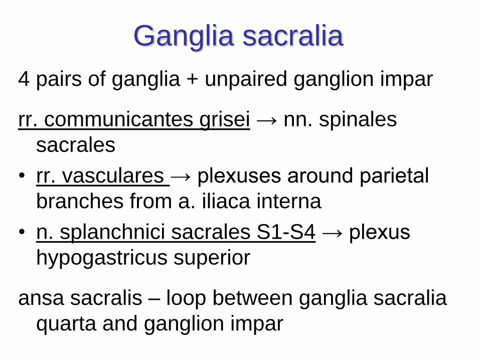
Ganglia sacralia
4 pairs of ganglia + unpaired ganglion impar
rr. communicantes grisei → nn. spinales
sacrales
• rr. vasculares → plexuses around parietal
branches from a. iliaca interna
• n. splanchnici sacrales S1-S4 → plexus
hypogastricus superior
ansa sacralis – loop between ganglia sacralia
quarta and ganglion impar

Plexus aorticus abdominalis
sympathetic fibers: nn. splanchnici thoracici (major, minor, imus), lumbales 1-3
parasympathetic fibers: rr. coeliaci nn. vagorum
mixed plexus around aorta abdominalis + prevertebral ganglia
• paired ggl. coelicum + ggl. aorticorenale
• unpaired ggl . mesentericum sup. + inf.
→ along arteries → homonymous plexuses
• stomach → oral majority of rectum (enteric system), pancreas, liver
• spleen, suprarenal glands, kidneys, ureters, testes♂ / ovaries♀, uterine tubes (1/2) ♀

ggl. coelicum
ggl. aorticorenale
ggl. mesentericum sup.
ggl. mesentericum inf.

Plexus aorticus abdominalis
continuation
• plexus hypogastricus superior (pure sympathetic (from bifurcatio across promontorium)
→ n. hypogastricus dx. + sin. → plexus hypogastricus inferior s. pelvicus (mixed plexus) → pelvic organs except ovaries♀, uterine tubes (1/2)♀, fundus uteri♀ and urinary bladder
• plexus iliacus dx. + sin. (pure sympathetic)→ lower limb


PARASYMPATHICUS
„kraniosakrální systém “


Parasympathetic part
= „craniosacral system“
• nuclei of cranial nerves
– preganglionic part of ncll. accessorii n. III Edinger-Westphal
– ncl. salivatorius superior (VII.)
– ncl. salivatorius inferior (IX.)
– ncl. posterior n. X
• ncl. intermediolateralis S2-4
ganglia situated within skull or within organs walls
ganglia situated close to effector organs


Gerenal schme of
parasympathetic ganglion
• radix parasympatica
• radix sympathica
• radix sensoria

Ganglion ciliare Schacheri
• orbit
• dorsally to bulbus oculi and laterally to n. opticus
AF-PS: preganglionic part of ncll. accessorii n. III
Edinger-Westphal → n.III → ramus ganglionaris
ciliaris
AF-S: ncl. intermediolateralis C8-T1 → ggl. cervicale
superius → n. et plexus caroticus internus → plexus
ophthalmicus (not synapsed)
EF: nn. ciliares breves (mixed) → m. ciliaris, m.
sphincter pupillae, m. dilatator pupillae, m. tarsalis
sup. + inf. (m. orbitalis)

Ganglion ciliare Schacheri


Ganglion pterygopalatinum Meckeli
• fossa pterygopalatina, below n. maxillaris
AF-PS: ncl. salivatorius superior (VII.) → n. VII → n. intermedius → n. petrosus major → n. canalis pterygoidedi Vidii (mixed) →
AF-S: ncl. intermediolateralis C8-T1 → ggl. cervicale superius → n. et plexus caroticus internus → n. petrosus profundus → n. canalis pterygoidedi Vidii (mixed) → (not synapsed in ganglion)
EF: → n. zygomaticus → r. communicans lacrimalis→ gl. lacrimalis
EF: → rr. nasales posteriores → gll. nasales
EF: → nn. palatini major + minores → gll. palatinae
EF: → n. pharyngeus → gll. nasopharyngeae

Ganglion submandibulare Langleyi
• trigonum submandibulare
• at crossing of n. lingualis and ductus
submandibularis
AF-PS: ncl. salivatorius superior (VII) → n. intermedius → n. VII → chorda tympani → n. lingualis (from n.V3) → r. communicans lingualis
AF-PS: ncl. intermediolateralis C8-T1 → ggl. cervicale superius → n. et plexus caroticus externus → plexus a. facialis (not synapsed)
EF: n. lingualis → gl. sublingualis + gll. linguales
EF: rr. glandulares → gl. submandibularis


Ganglion oticum Arnoldi
• fossa infratemporalis, medially to n. mandibularis
AF-PS: ncl. salivatorius inferior (IX.) → n. IX → n.
tympanicus → plexus tympanicus → n. petrosus
minor
AF-S: ncl. intermediolateralis C8-T1 → ggl. cervicale
superius → n. et plexus caroticus externus → plexus
a. meningeae mediae (not synapsed)
EF: r. communicans auriculotemporalsi (mixed) → n.
auriculotemporalis → gl. parotidea
→ r. communicans buccalis (mixed) → n. buccalis →
gll. buccales


Plexus hypogastricus inferior s. pelvicusmixed plexus
AF-PS: nn. splanchnici pelvici S2-4 (obsoletely nn. erigentes)
AF-S: truncus sympathicus → plexus aorticus abdominalis → plexus hypogastricus superior → nn. hypogastrici
AF-S: truncus sympathicus → ganglia sacralia → nn. splanchnici sacrales
• pelvic organs except ovaries♀, uterine tubes (1/2)♀, fundus of uterus♀ and fundus of urinary bladder
EF (mixed): → plexus rectalis (aboral minority of rectum)
→ parasympathetic fibers ascend as orally as Cannon-Bőhm´spoint = hindgut
→ plexus prostaticus + deferentialis♂/ uterovaginalis♀
→ plexus vesicalis- m. sphincter urethrae (nucleus n. pudendi Onufi in spinal cord segments
S2-4)
→ n. cavernosus penis ♂ / clitoridis ♀ (erectile bodies)


Paraganglia
• chromafine (paraganglia sympathica)
– paraganglion aorticum abdominale Zuckerkandli
– glomus coccygeum Luschkae
– glomus jugulare, tympanicum…
• without chromafine reaction (former
paraganglia parasympathica)
– baro- a chemoreceptors
– glomus caroticum
– glomus supracardiacum (aorticum)

Enteric system
cardia of stomach → upper margin of m. sphincter ani internus, biliary ducts and gallblader, pancreas
• plexus myentericus Auerbachi
• plexus submucosus Meissneri
• ganglia within the intestinal wall
• fibers
– visceromotor sympathetic + parasympathetic
– viscerosensory via both systems + reflectory ones
• Cajal interstitial cells
– pacemaker of intestinal muscle layers

CNS
• highest autonomic center = hypothalamus
• controled by limbic system (insula)
• nuclei influenced by reticular formation
(reflexes)


![MUSIC SCORES - Andrea Angelini...Diffusa est gratia - Propter veritatem - Vultum tuum William Byrd (c.1540-1623)Superius [Soprano] Medius [Alto] Contratenor [Tenor 1]](https://img.dokumen.tips/doc/110x75/60e5d162560c6e541272ff22/music-scores-andrea-diffusa-est-gratia-propter-veritatem-vultum-tuum-william.jpg)