Embed Size (px)
Citation preview

GIOVANNI BATTISTA FOGAZZI
CLINICAL AND RESEARCH LABORATORY ON URINARY SEDIMENT
U.O. DI NEFROLOGIA E DIALISI
FONDAZIONE IRCCS CA’ GRANDA OSPEDALE MAGGIORE POLICLINICO
MILANO- ITALY
AUTOMATED ANALYSIS OF
URINE SEDIMENT
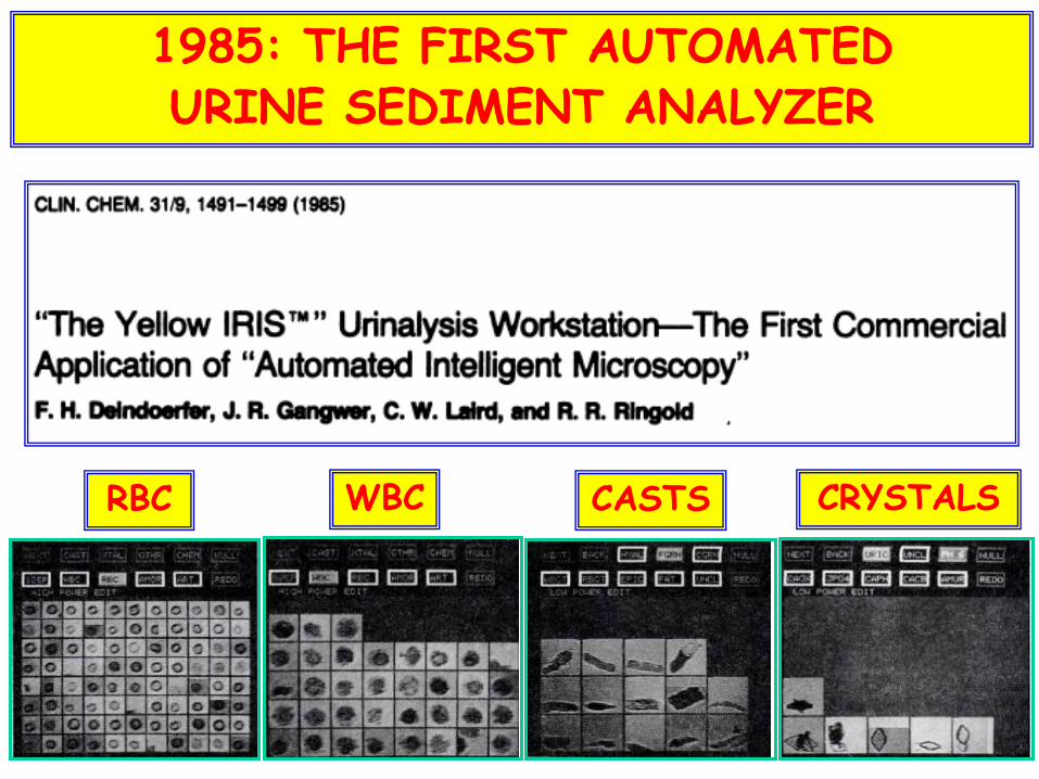
1985: THE FIRST AUTOMATED URINE SEDIMENT ANALYZER
RBC WBC CASTS CRYSTALS

Workshop 2: STRATEGIES IN URINALYSIS
Prof Dolphe Kutter
“Automation of urinalysis: possibilities and problems”
3 JULY 1995: 11th IFCC EUROPEAN CONGRESS OF CLINICAL CHEMISTRY,
TAMPERE (FINLAND)
During the discussion that followed, a representative of an
international company stated:
“Our company has decided to stop investing in this sector
because the technology is not assisting us any further, we feel
at a standstill…”

• In the developed world, automated urine sediment analyzers are in use in all large laboratories
• Three types of instruments are on the market, each one being based on its own technology:
• Automated intelligent microscopy (iQ200, Beckmann)
• Flow cytometry (UF-1000i, Sysmex)
• Cuvette-based microscopy (UriSed/sediMAX, 77 Elektronika/A. Menarini Diagnostics)
TODAY, 20 YEARS LATER
AUTOMATED INTELLIGENT MICROSCOPY: iQ200

• An automated microscope is focalized on a planar flow cell, in which the particles flow as a sheet, being sandwiched between two layers of an enveloping fluid
• A stroboscopic lamp, firing 24 bursts/second, stops the motion of the particles passing through the camera
• The stopped motion view is observed through magnifying lenses
• The images are collected by a videocamera

• A very high number of images/sample is taken
• For each particle, the background is removed in order to better identify and show the particle
• Each particle is analyzed by a neural network which contains 26,000 reference images
• Each particle is isolated within
one image, which is then
inserted in one particle category
RBC

EXAMPLE OF IMAGES SUPPLIED BY iQ200 (URIC ACID)
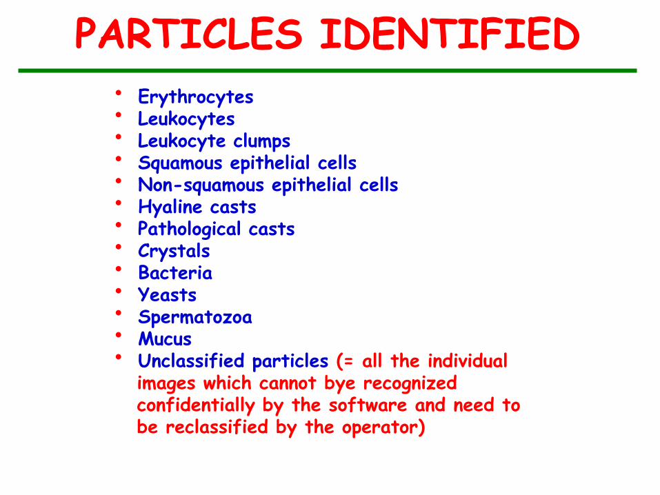
PARTICLES IDENTIFIED
• Erythrocytes • Leukocytes • Leukocyte clumps • Squamous epithelial cells • Non-squamous epithelial cells • Hyaline casts • Pathological casts • Crystals • Bacteria • Yeasts • Spermatozoa • Mucus • Unclassified particles (= all the individual images which cannot bye recognized confidentially by the software and need to be reclassified by the operator)
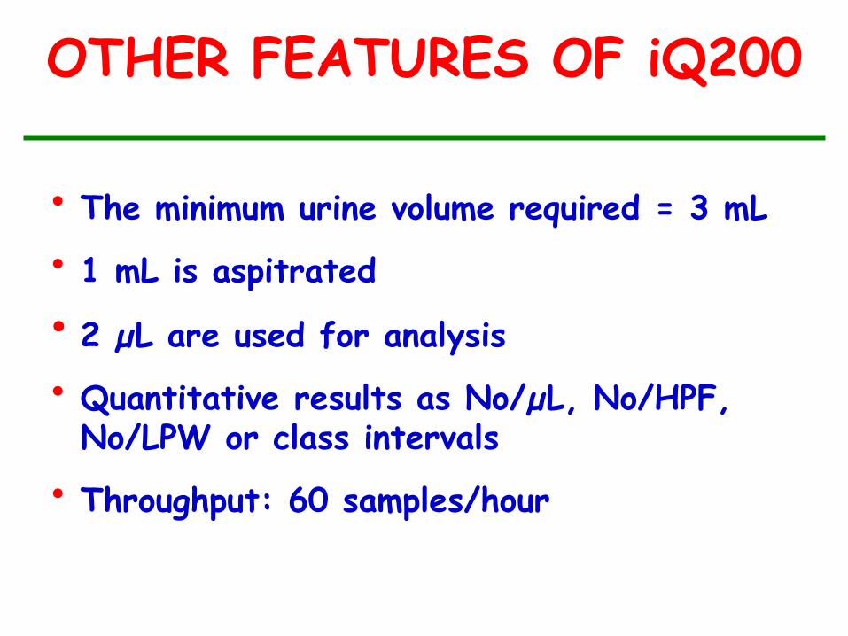
OTHER FEATURES OF iQ200
• The minimum urine volume required = 3 mL
• 1 mL is aspitrated
• 2 µL are used for analysis
• Quantitative results as No/µL, No/HPF, No/LPW or class intervals
• Throughput: 60 samples/hour

FLOW CYTOMETRY: UF-1000i
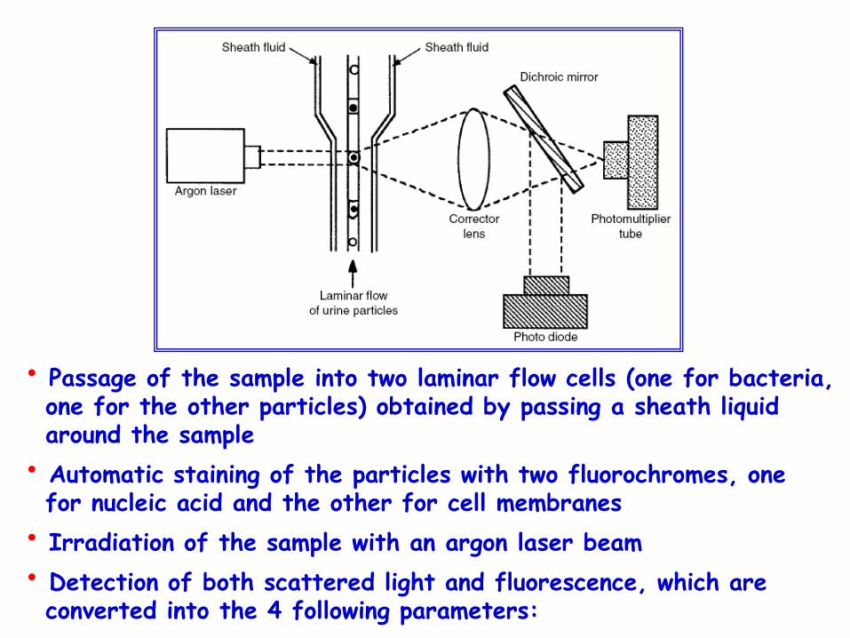
• Passage of the sample into two laminar flow cells (one for bacteria, one for the other particles) obtained by passing a sheath liquid around the sample
• Automatic staining of the particles with two fluorochromes, one for nucleic acid and the other for cell membranes
• Irradiation of the sample with an argon laser beam
• Detection of both scattered light and fluorescence, which are converted into the 4 following parameters:
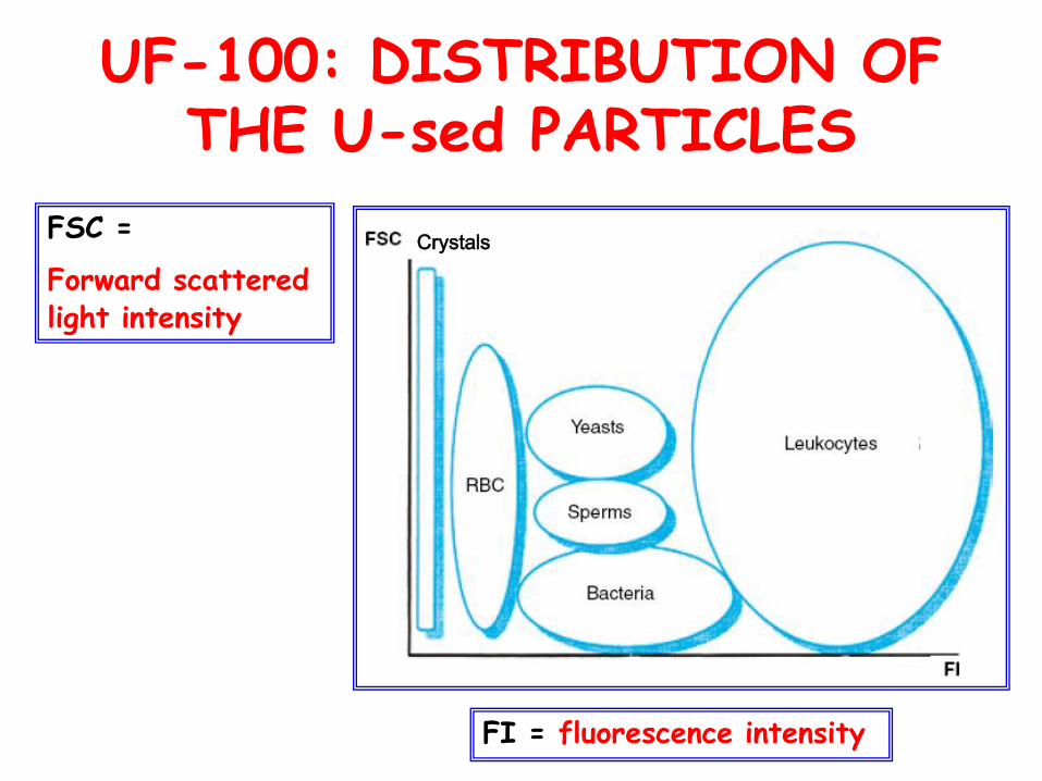
UF-100: DISTRIBUTION OF THE U-sed PARTICLES
FSC =
Forward scattered light intensity
FI = fluorescence intensity
Crystals

Flw =
Fluorescence pulse width
Fscw = forward scattered light pulse width
UF-100: DISTRIBUTION OF THE U-sed PARTICLES
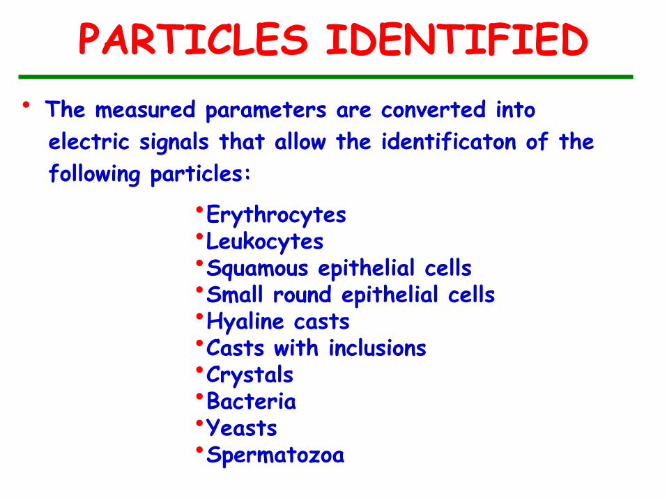
PARTICLES IDENTIFIED
• The measured parameters are converted into
electric signals that allow the identificaton of the
following particles:
•Erythrocytes •Leukocytes •Squamous epithelial cells •Small round epithelial cells •Hyaline casts •Casts with inclusions •Crystals •Bacteria •Yeasts •Spermatozoa

EXAMPLE OF REPORT (1)

EXAMPLE OF REPORT (2)

OTHER FEATURES OF UF 1000i
• The urine volume required = 0.8-1.2 mL
• 9 µL are used for analysis
• Quantitative results as No/µL & No/HPF
• Throughput: 100 samples/hour
CUVETTE-BASED MICROSCOPY:
UriSed/sediMAX
• A walk-away automatic urine sediment analyzer, which has been developed since 2008 by 77 Elektronika, Budapest Kft, Hungary (and distributed as sediMAX in several European countries by A.Menarini Diagnostics, Florence, Italy)
• It supplies B/W images of particles within whole fields of view
• These are similar to the microscopic fields seen with manual microscopy
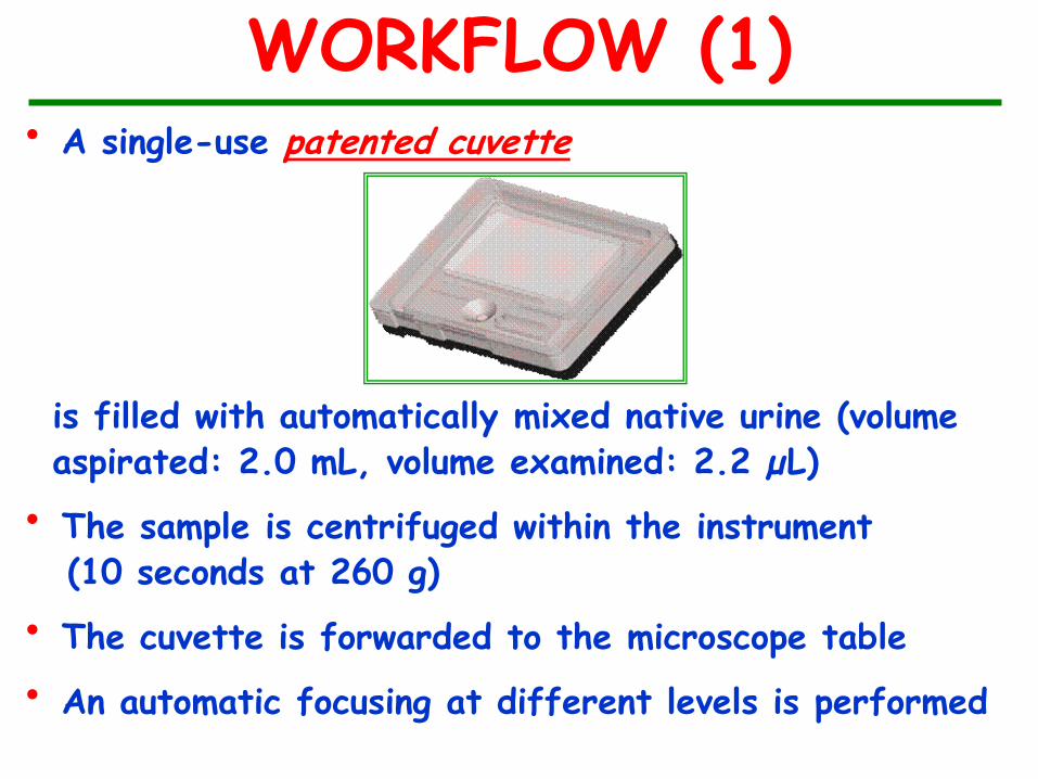
WORKFLOW (1) • A single-use patented cuvette
is filled with automatically mixed native urine (volume aspirated: 2.0 mL, volume examined: 2.2 µL)
• The sample is centrifuged within the instrument (10 seconds at 260 g)
• The cuvette is forwarded to the microscope table
• An automatic focusing at different levels is performed

WORKFLOW (2)
• A built-in camera takes a digital image of each field
of view (magnification: ~400x)
• For each sample 15 images are taken
• Identification and quantitation of the particles (as
No/µL or No/HPF) is carried out by Auto Image
Evaluation Module (AIEM), a complex artificial
neural network structure which has specifically been
developed for the instrument
• Throughput: 100 samples/hour
PARTICLES IDENTIFIED (1)
• Erythrocytes • Leukocytes • Squamous epithelial cells • Non-squamous epithelial cells • Hyaline casts • Pathological casts • Crystals: CaOx, UA, struvite • Bacteria • Yeasts • Spermatozoa • Mucus
PARTICLES IDENTIFIED (2)
• Other particles which might be present in the
whole field of view but are not recognized by
the instrument may be identified by the
operator
• Due to this unique feature, urinary profiles
- and the clinical diagnoses associated with
them - can be identified (see the three
following examples)

WHOLE FIELD OF VIEW: Many WBCS and bacteria
URINARY TRACT INFECTION
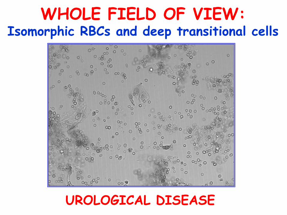
WHOLE FIELD OF VIEW: Isomorphic RBCs and deep transitional cells
UROLOGICAL DISEASE

WHOLE FIELD OF VIEW: Dysmorphic RBCs and fatty particles
NEPHROTIC SEDIMENT
sediMAX DEVELOPMENTS OVER TIME
• sediMAX
• sediMAX 2
• sediMAX LITE (semi-automated)
• sediMAX conTRUST
Supplies both bright field and phase
contrast microscopy images (a further
progress in automated urinary sediment
examination)

sediMAX conTRUST
Bright field Phase contrast

CONCLUSIONS
AUTOMATED Used ANALYZERS: ADVANTAGES
• Walk-away instruments
• Examine high numbers of samples in short time
• Require small volumes of urine
• Abolish the problems caused by centrifugation
• Achieve acceptable accuracy for some particles (RBCs, WBCs, squamous epithelial cells)
• Supply quantitative results with small variation coefficients
• Leave time for the manual examination of the more complex samples
AUTOMATED Used ANALYZERS: LIMITATIONS
• Include in one category only renal tubular epithelial cells and transitional epithelial cells, which have totally different clinical implications
• Underestimate casts, of which, in addition, they can identify only hyaline and “non hyaline” (or “pathologic”) subtypes
• Identify only a few types of crystals
• Miss lipids completely
• For all tese reasons not yet qualified to investigate complex renal and non-renal samples

AUTOMATED Used ANALYZERS: THEIR PLACE IN LABS
• They supply an acceptable accuracy for the negative samples and those with minor changes, which represent the vast majority of samples examined in central labs
• Therefore, they are very useful/recommended for labs with >100 samples/day
• Their utility is greatly increased if, for selected cases, their use is integrated with manual microscopy performed in a proper way by motivated and trained personnel

THANK YOU FOR YOUR KIND ATTENTION