Embed Size (px)
Citation preview
AUTOIMMUNE PANCREATITIS AND IGG-4 RELATED DISEASE
Prasad Kulkarni, MD FACGAssoc Professor of Medicine USFDirector, GI Endoscopy James A Haley VA Hospital, Tampa FL
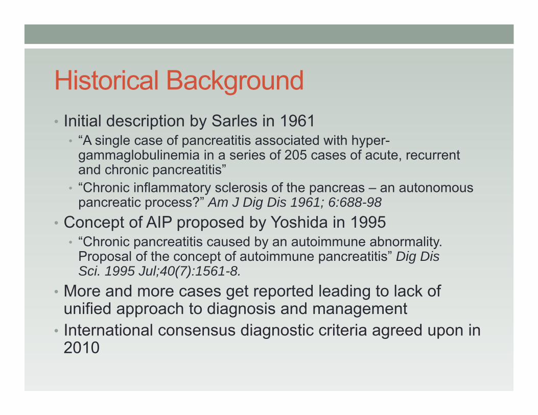
Historical Background• Initial description by Sarles in 1961
• “A single case of pancreatitis associated with hyper-gammaglobulinemia in a series of 205 cases of acute, recurrent and chronic pancreatitis”
• “Chronic inflammatory sclerosis of the pancreas – an autonomous pancreatic process?” Am J Dig Dis 1961; 6:688-98
• Concept of AIP proposed by Yoshida in 1995• “Chronic pancreatitis caused by an autoimmune abnormality.
Proposal of the concept of autoimmune pancreatitis” Dig Dis Sci. 1995 Jul;40(7):1561-8.
• More and more cases get reported leading to lack of unified approach to diagnosis and management
• International consensus diagnostic criteria agreed upon in 2010
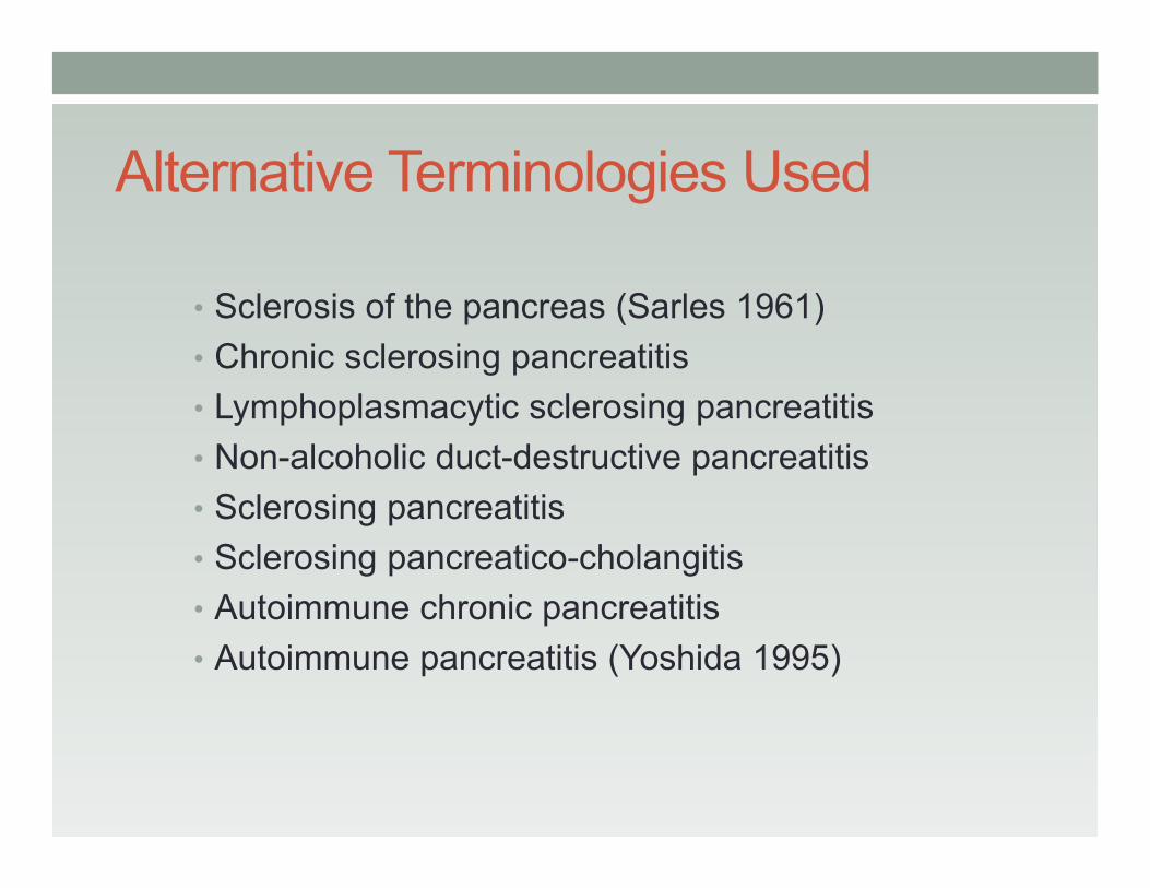
Alternative Terminologies Used
• Sclerosis of the pancreas (Sarles 1961)• Chronic sclerosing pancreatitis• Lymphoplasmacytic sclerosing pancreatitis• Non-alcoholic duct-destructive pancreatitis• Sclerosing pancreatitis• Sclerosing pancreatico-cholangitis• Autoimmune chronic pancreatitis • Autoimmune pancreatitis (Yoshida 1995)
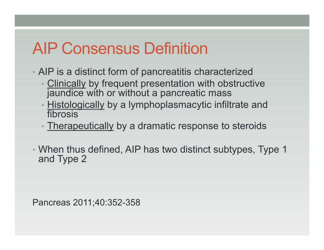
AIP Consensus Definition• AIP is a distinct form of pancreatitis characterized
• Clinically by frequent presentation with obstructive jaundice with or without a pancreatic mass
• Histologically by a lymphoplasmacytic infiltrate and fibrosis
• Therapeutically by a dramatic response to steroids
• When thus defined, AIP has two distinct subtypes, Type 1 and Type 2
Pancreas 2011;40:352-358
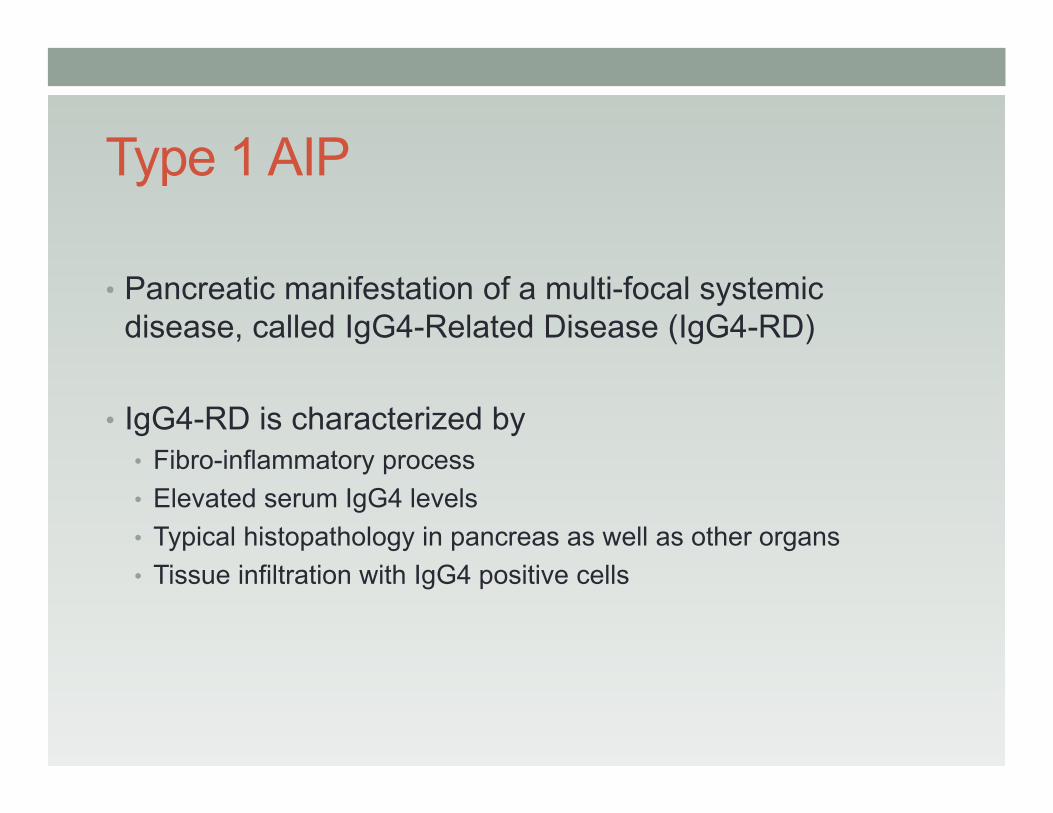
Type 1 AIP
• Pancreatic manifestation of a multi-focal systemic disease, called IgG4-Related Disease (IgG4-RD)
• IgG4-RD is characterized by• Fibro-inflammatory process• Elevated serum IgG4 levels• Typical histopathology in pancreas as well as other organs• Tissue infiltration with IgG4 positive cells
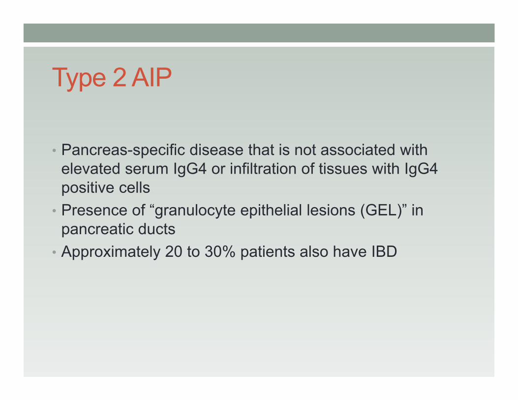
Type 2 AIP
• Pancreas-specific disease that is not associated with elevated serum IgG4 or infiltration of tissues with IgG4 positive cells
• Presence of “granulocyte epithelial lesions (GEL)” in pancreatic ducts
• Approximately 20 to 30% patients also have IBD
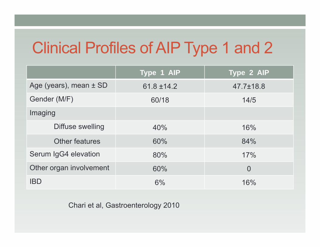
Clinical Profiles of AIP Type 1 and 2Type 1 AIP Type 2 AIP
Age (years), mean ± SD 61.8 ±14.2 47.7±18.8
Gender (M/F) 60/18 14/5
Imaging
Diffuse swelling 40% 16%
Other features 60% 84%
Serum IgG4 elevation 80% 17%
Other organ involvement 60% 0
IBD 6% 16%
Chari et al, Gastroenterology 2010
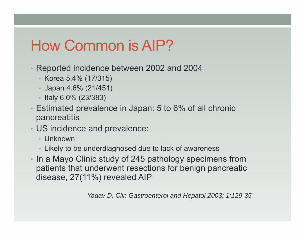
How Common is AIP?• Reported incidence between 2002 and 2004
• Korea 5.4% (17/315)• Japan 4.6% (21/451)• Italy 6.0% (23/383)
• Estimated prevalence in Japan: 5 to 6% of all chronic pancreatitis
• US incidence and prevalence: • Unknown• Likely to be underdiagnosed due to lack of awareness
• In a Mayo Clinic study of 245 pathology specimens from patients that underwent resections for benign pancreatic disease, 27(11%) revealed AIP
Yadav D. Clin Gastroenterol and Hepatol 2003; 1:129-35
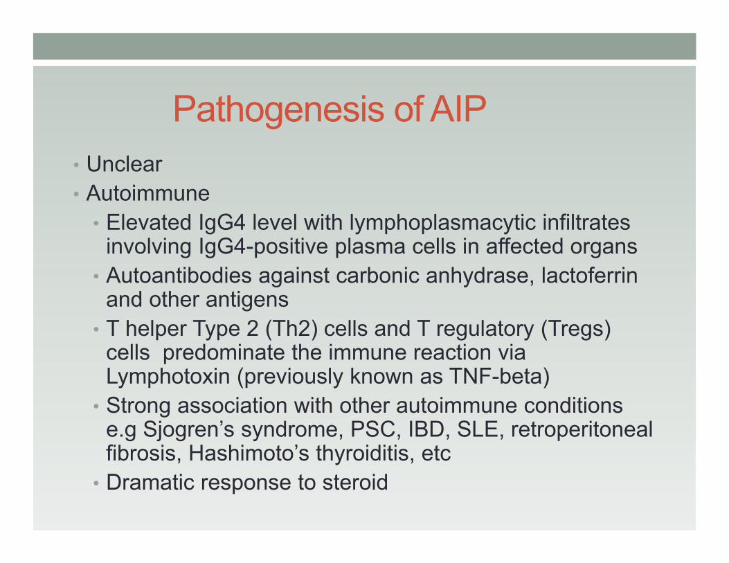
Pathogenesis of AIP• Unclear• Autoimmune
• Elevated IgG4 level with lymphoplasmacytic infiltrates involving IgG4-positive plasma cells in affected organs
• Autoantibodies against carbonic anhydrase, lactoferrinand other antigens
• T helper Type 2 (Th2) cells and T regulatory (Tregs) cells predominate the immune reaction via Lymphotoxin (previously known as TNF-beta)
• Strong association with other autoimmune conditions e.g Sjogren’s syndrome, PSC, IBD, SLE, retroperitoneal fibrosis, Hashimoto’s thyroiditis, etc
• Dramatic response to steroid
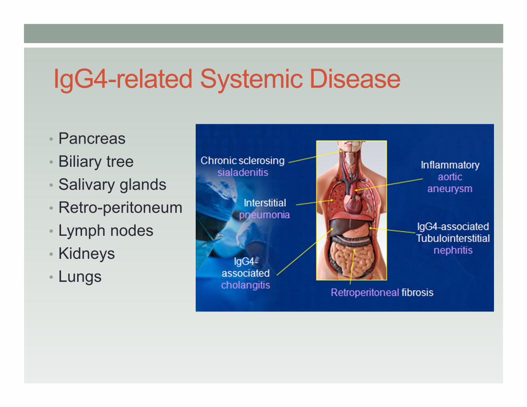
IgG4-related Systemic Disease
• Pancreas• Biliary tree• Salivary glands• Retro-peritoneum• Lymph nodes• Kidneys• Lungs
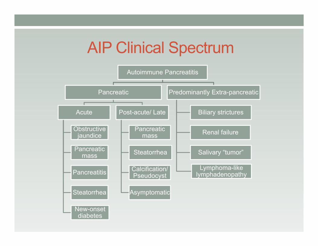
AIP Clinical SpectrumAutoimmune Pancreatitis
Pancreatic
Acute
Obstructive jaundice
Pancreatic mass
Pancreatitis
Steatorrhea
New-onset diabetes
Post-acute/ Late
Pancreatic mass
Steatorrhea
Calcification/ Pseudocyst
Asymptomatic
Predominantly Extra-pancreatic
Biliary strictures
Renal failure
Salivary “tumor”
Lymphoma-like lymphadenopathy
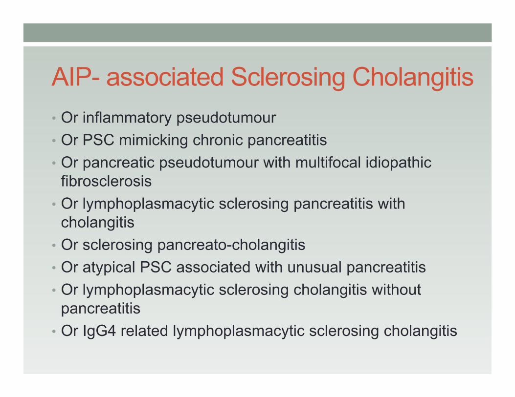
AIP- associated Sclerosing Cholangitis• Or inflammatory pseudotumour• Or PSC mimicking chronic pancreatitis• Or pancreatic pseudotumour with multifocal idiopathic
fibrosclerosis• Or lymphoplasmacytic sclerosing pancreatitis with
cholangitis• Or sclerosing pancreato-cholangitis• Or atypical PSC associated with unusual pancreatitis• Or lymphoplasmacytic sclerosing cholangitis without
pancreatitis• Or IgG4 related lymphoplasmacytic sclerosing cholangitis
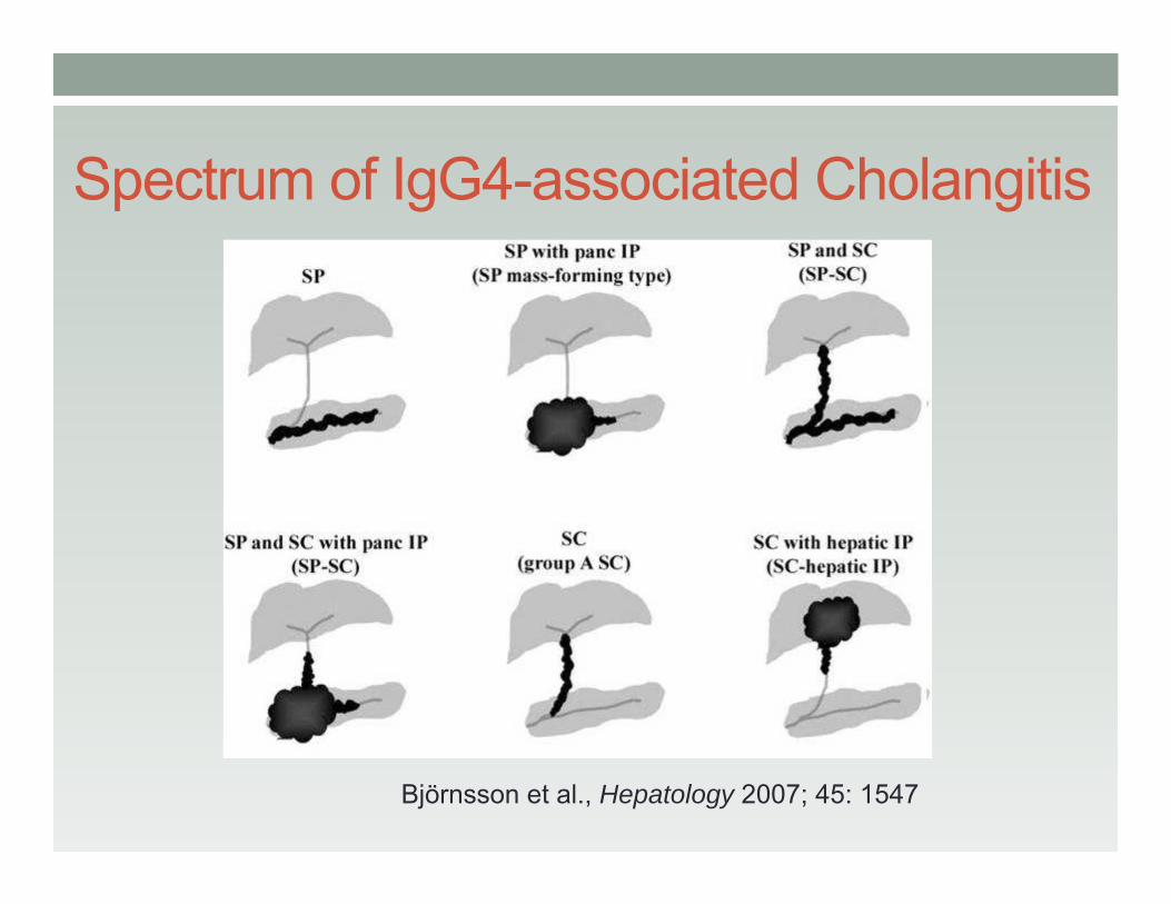
Spectrum of IgG4-associated Cholangitis
Björnsson et al., Hepatology 2007; 45: 1547
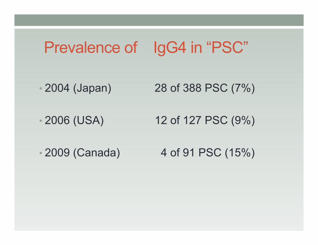
Prevalence of IgG4 in “PSC”
• 2004 (Japan) 28 of 388 PSC (7%)
• 2006 (USA) 12 of 127 PSC (9%)
• 2009 (Canada) 4 of 91 PSC (15%)
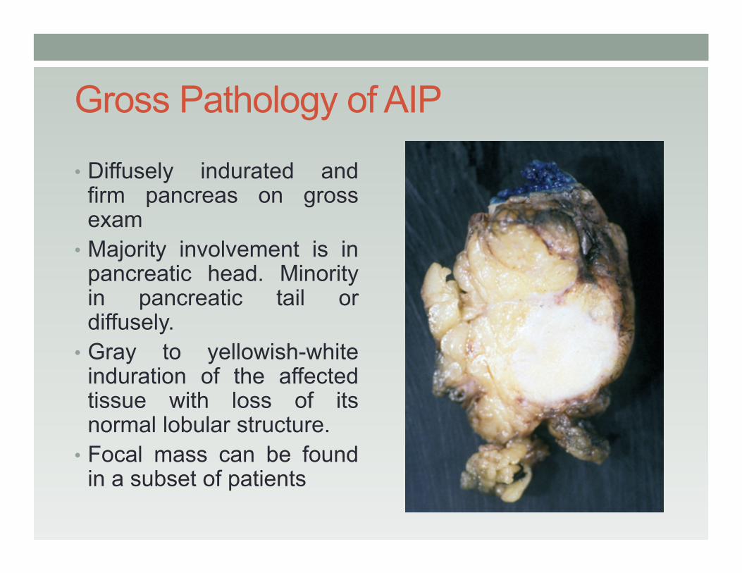
Gross Pathology of AIP
• Diffusely indurated andfirm pancreas on grossexam
• Majority involvement is inpancreatic head. Minorityin pancreatic tail ordiffusely.
• Gray to yellowish-whiteinduration of the affectedtissue with loss of itsnormal lobular structure.
• Focal mass can be foundin a subset of patients
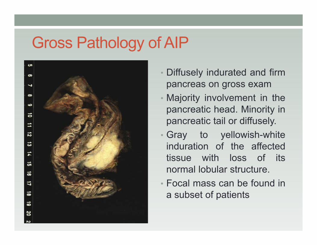
Gross Pathology of AIP
• Diffusely indurated and firmpancreas on gross exam
• Majority involvement in thepancreatic head. Minority inpancreatic tail or diffusely.
• Gray to yellowish-whiteinduration of the affectedtissue with loss of itsnormal lobular structure.
• Focal mass can be found ina subset of patients
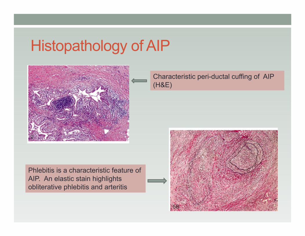
Histopathology of AIP
Characteristic peri-ductal cuffing of AIP (H&E)
Phlebitis is a characteristic feature of AIP. An elastic stain highlights obliterative phlebitis and arteritis
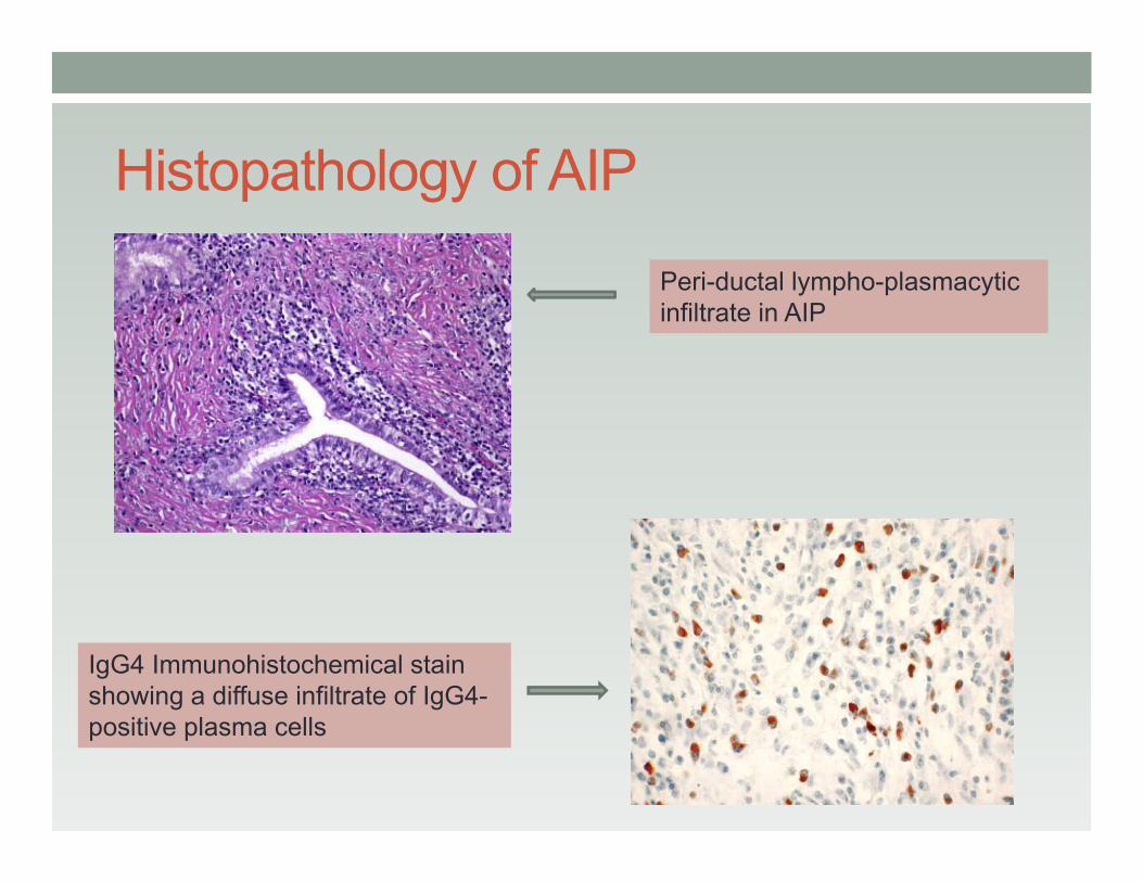
Histopathology of AIP
IgG4 Immunohistochemical stain showing a diffuse infiltrate of IgG4-positive plasma cells
Peri-ductal lympho-plasmacyticinfiltrate in AIP
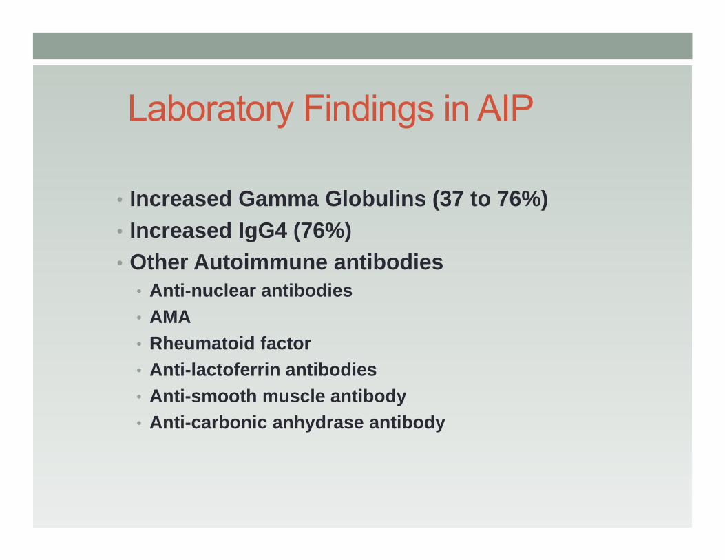
Laboratory Findings in AIP
• Increased Gamma Globulins (37 to 76%) • Increased IgG4 (76%)• Other Autoimmune antibodies
• Anti-nuclear antibodies• AMA• Rheumatoid factor• Anti-lactoferrin antibodies• Anti-smooth muscle antibody• Anti-carbonic anhydrase antibody
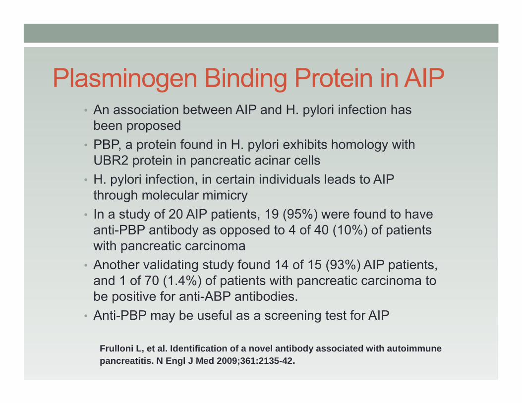
Plasminogen Binding Protein in AIP• An association between AIP and H. pylori infection has
been proposed• PBP, a protein found in H. pylori exhibits homology with
UBR2 protein in pancreatic acinar cells• H. pylori infection, in certain individuals leads to AIP
through molecular mimicry• In a study of 20 AIP patients, 19 (95%) were found to have
anti-PBP antibody as opposed to 4 of 40 (10%) of patients with pancreatic carcinoma
• Another validating study found 14 of 15 (93%) AIP patients, and 1 of 70 (1.4%) of patients with pancreatic carcinoma to be positive for anti-ABP antibodies.
• Anti-PBP may be useful as a screening test for AIP
Frulloni L, et al. Identification of a novel antibody associated with autoimmune pancreatitis. N Engl J Med 2009;361:2135-42.
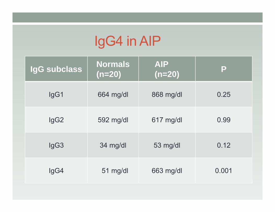
IgG4 in AIP
IgG subclass Normals(n=20)
AIP(n=20) P
IgG1 664 mg/dl 868 mg/dl 0.25
IgG2 592 mg/dl 617 mg/dl 0.99
IgG3 34 mg/dl 53 mg/dl 0.12
IgG4 51 mg/dl 663 mg/dl 0.001
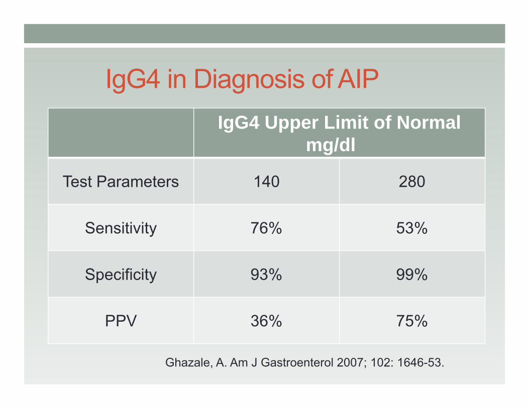
IgG4 in Diagnosis of AIPIgG4 Upper Limit of Normal
mg/dl
Test Parameters 140 280
Sensitivity 76% 53%
Specificity 93% 99%
PPV 36% 75%
Ghazale, A. Am J Gastroenterol 2007; 102: 1646-53.
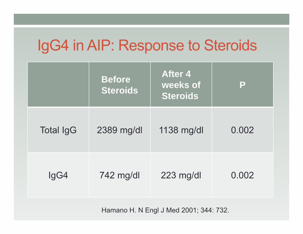
IgG4 in AIP: Response to Steroids
Before Steroids
After 4 weeks of Steroids
P
Total IgG 2389 mg/dl 1138 mg/dl 0.002
IgG4 742 mg/dl 223 mg/dl 0.002
Hamano H. N Engl J Med 2001; 344: 732.
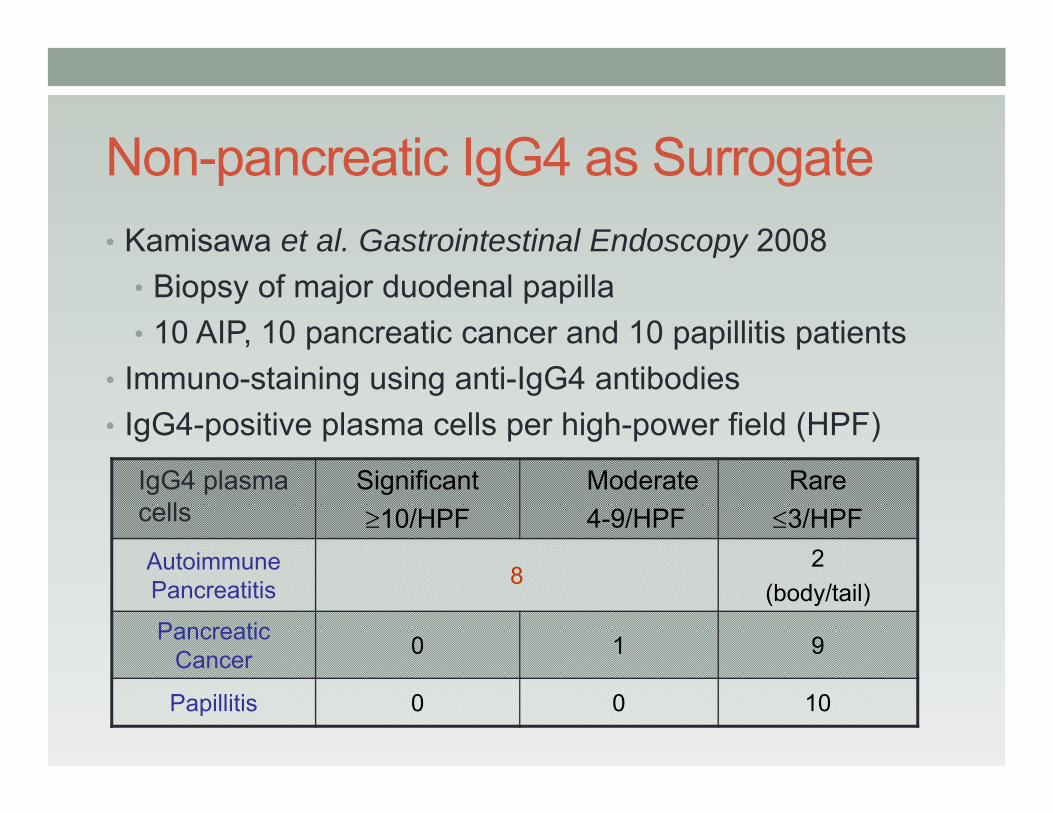
Non-pancreatic IgG4 as Surrogate• Kamisawa et al. Gastrointestinal Endoscopy 2008
• Biopsy of major duodenal papilla • 10 AIP, 10 pancreatic cancer and 10 papillitis patients
• Immuno-staining using anti-IgG4 antibodies• IgG4-positive plasma cells per high-power field (HPF)
IgG4 plasma cells
Significant≥10/HPF
Moderate4-9/HPF
Rare≤3/HPF
Autoimmune Pancreatitis 8
2(body/tail)
Pancreatic Cancer 0 1 9
Papillitis 0 0 10
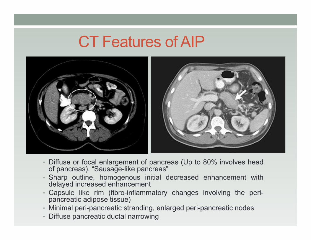
CT Features of AIP
• Diffuse or focal enlargement of pancreas (Up to 80% involves headof pancreas). “Sausage-like pancreas”
• Sharp outline, homogenous initial decreased enhancement withdelayed increased enhancement
• Capsule like rim (fibro-inflammatory changes involving the peri-pancreatic adipose tissue)
• Minimal peri-pancreatic stranding, enlarged peri-pancreatic nodes• Diffuse pancreatic ductal narrowing
PET and DWMR in AIP• FDG-PET will enhance both AIP and pancreatic cancer
• Extra-pancreatic involvement is highly suggestive of AIP
• MRCP will show delayed enhancement in AIP and long/ multiple PD strictures, but otherwise inferior to ERCP
• Diffusion-weighted MR (DWMR) will show high signal intensity areas both in AIP and pancreatic cancer• AIP: multiple and diffuse high signal intensity areas• Pancreatic cancer: solitary high signal intensity areas
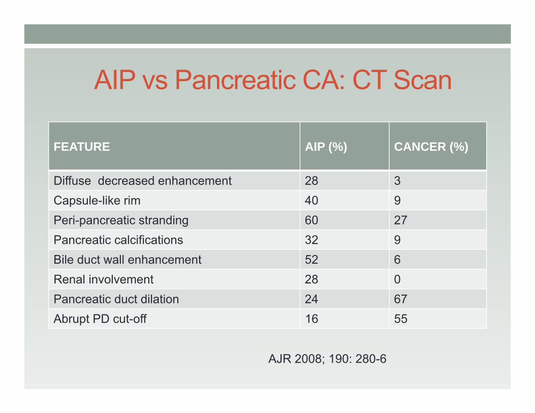
AIP vs Pancreatic CA: CT Scan
FEATURE AIP (%) CANCER (%)
Diffuse decreased enhancement 28 3Capsule-like rim 40 9Peri-pancreatic stranding 60 27Pancreatic calcifications 32 9Bile duct wall enhancement 52 6Renal involvement 28 0Pancreatic duct dilation 24 67Abrupt PD cut-off 16 55
AJR 2008; 190: 280-6
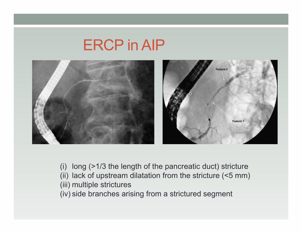
ERCP in AIP
(i) long (>1/3 the length of the pancreatic duct) stricture(ii) lack of upstream dilatation from the stricture (<5 mm) (iii) multiple strictures(iv) side branches arising from a strictured segment
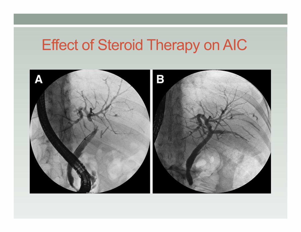
Effect of Steroid Therapy on AIC
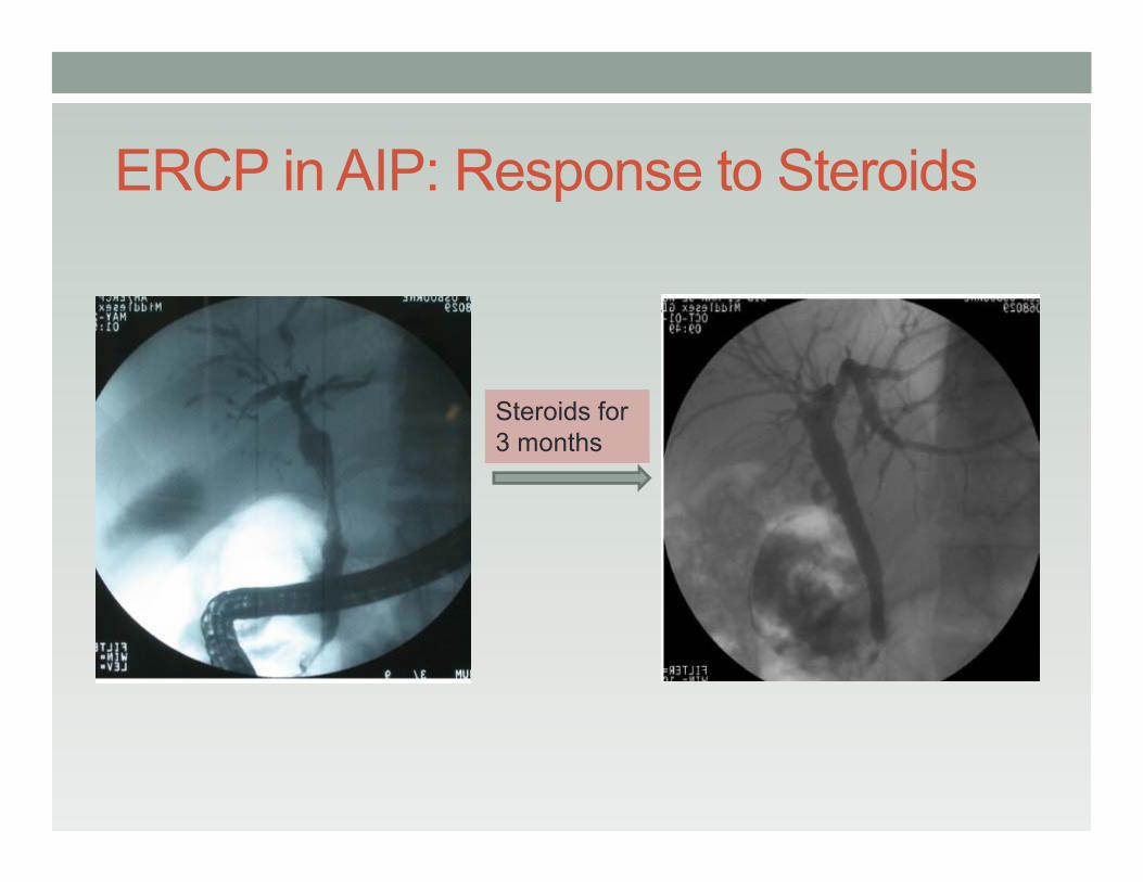
ERCP in AIP: Response to Steroids
Steroids for 3 months
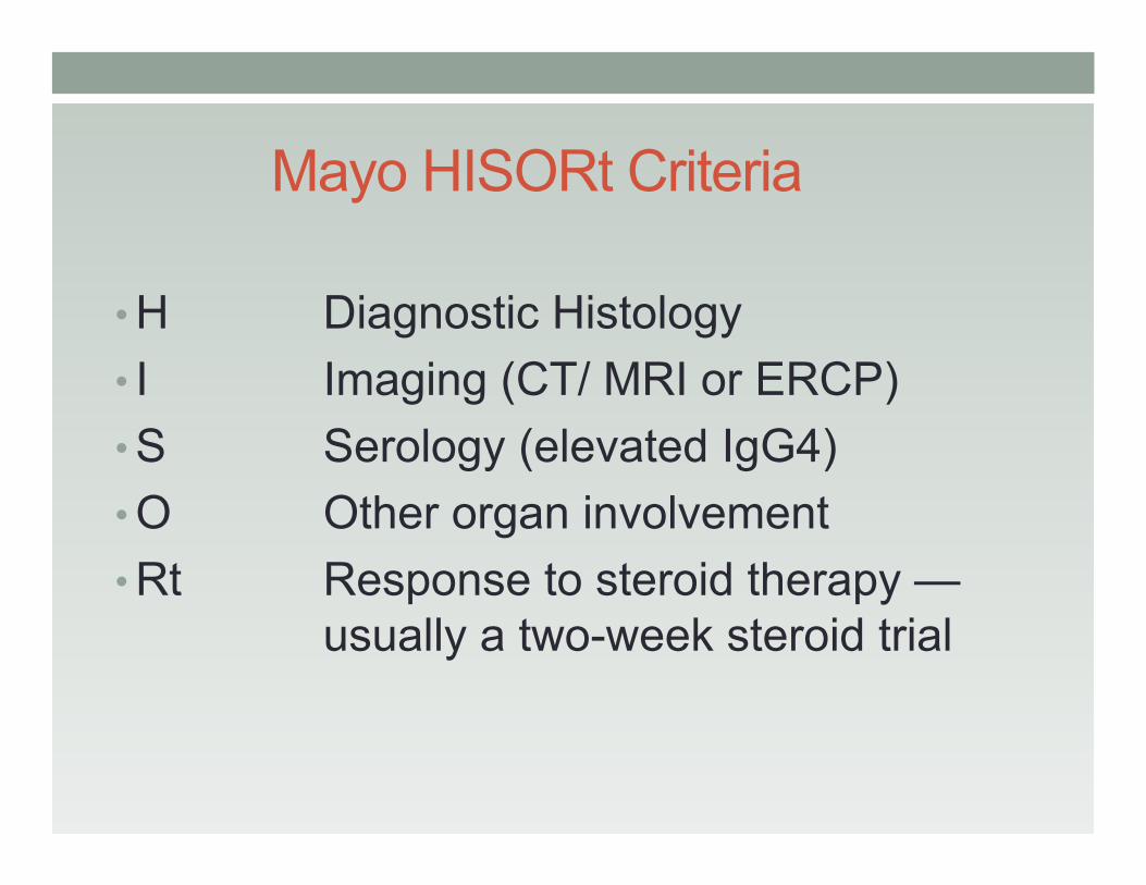
Mayo HISORt Criteria
• H Diagnostic Histology• I Imaging (CT/ MRI or ERCP)• S Serology (elevated IgG4)• O Other organ involvement• Rt Response to steroid therapy —
usually a two-week steroid trial
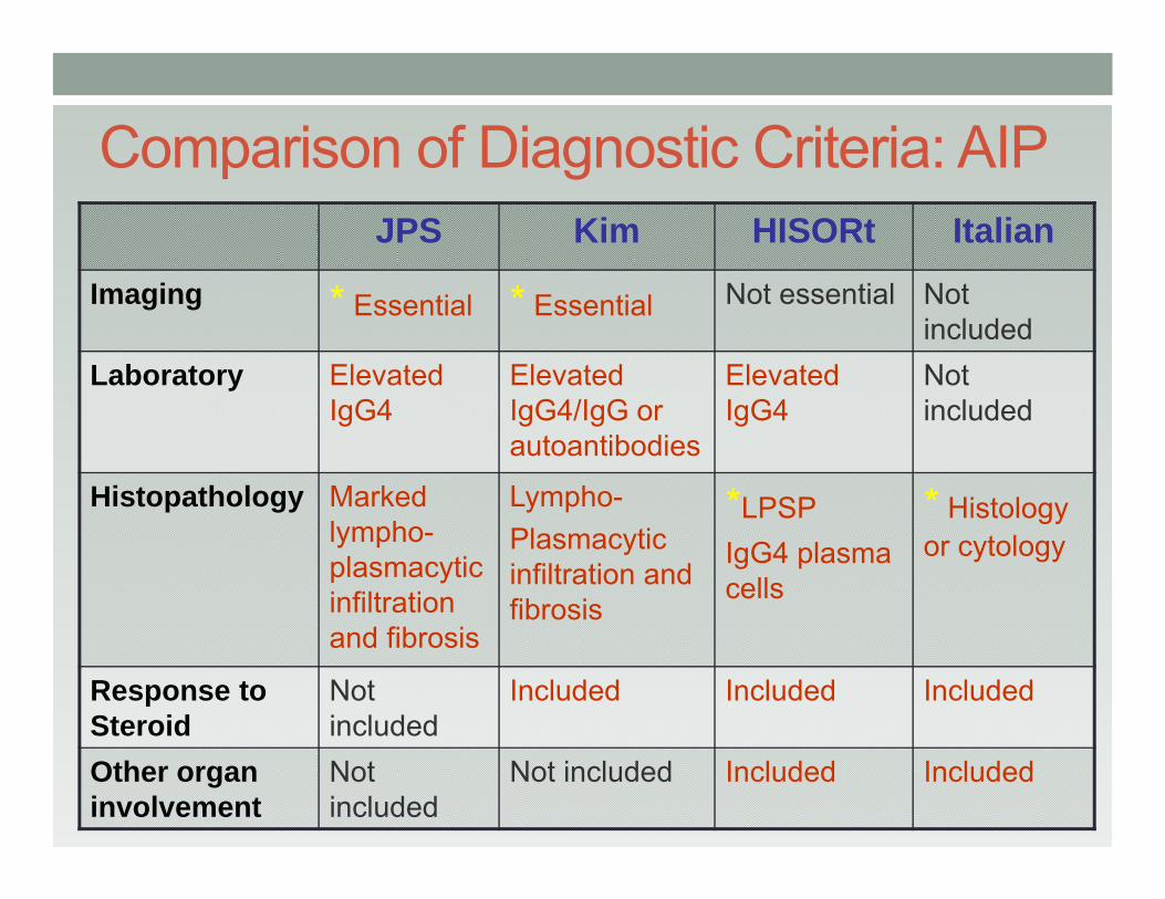
Comparison of Diagnostic Criteria: AIPJPS Kim HISORt Italian
Imaging * Essential * Essential Not essential Not included
Laboratory Elevated IgG4
Elevated IgG4/IgG or autoantibodies
Elevated IgG4
Not included
Histopathology Marked lympho-plasmacyticinfiltration and fibrosis
Lympho-Plasmacyticinfiltration and fibrosis
*LPSPIgG4 plasma cells
* Histology or cytology
Response to Steroid
Not included
Included Included Included
Other organ involvement
Not included
Not included Included Included
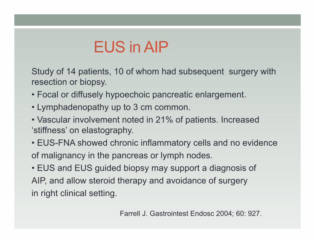
EUS in AIPStudy of 14 patients, 10 of whom had subsequent surgery with resection or biopsy.• Focal or diffusely hypoechoic pancreatic enlargement.• Lymphadenopathy up to 3 cm common.• Vascular involvement noted in 21% of patients. Increased ‘stiffness’ on elastography. • EUS-FNA showed chronic inflammatory cells and no evidenceof malignancy in the pancreas or lymph nodes.• EUS and EUS guided biopsy may support a diagnosis ofAIP, and allow steroid therapy and avoidance of surgeryin right clinical setting.
Farrell J. Gastrointest Endosc 2004; 60: 927.
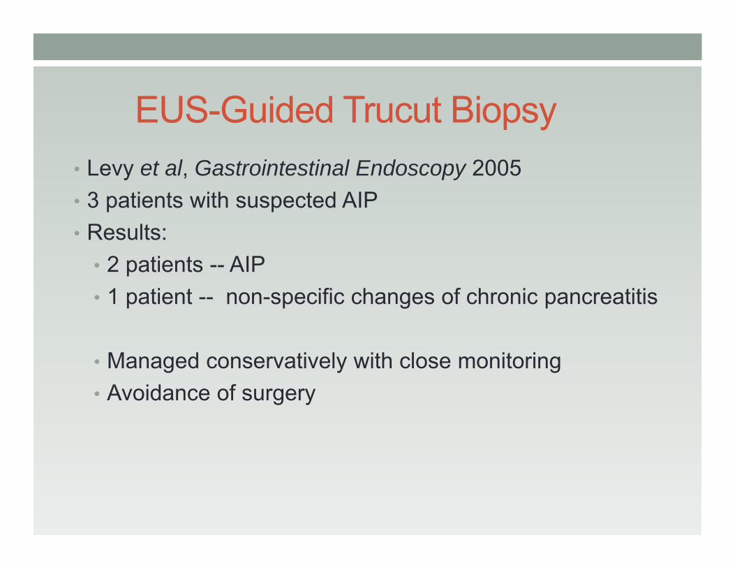
EUS-Guided Trucut Biopsy• Levy et al, Gastrointestinal Endoscopy 2005• 3 patients with suspected AIP• Results:
• 2 patients -- AIP• 1 patient -- non-specific changes of chronic pancreatitis
• Managed conservatively with close monitoring• Avoidance of surgery
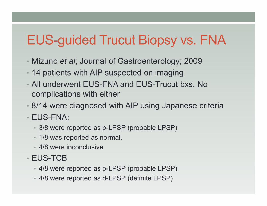
EUS-guided Trucut Biopsy vs. FNA• Mizuno et al; Journal of Gastroenterology; 2009• 14 patients with AIP suspected on imaging• All underwent EUS-FNA and EUS-Trucut bxs. No
complications with either• 8/14 were diagnosed with AIP using Japanese criteria• EUS-FNA:
• 3/8 were reported as p-LPSP (probable LPSP)• 1/8 was reported as normal,• 4/8 were inconclusive
• EUS-TCB• 4/8 were reported as p-LPSP (probable LPSP)• 4/8 were reported as d-LPSP (definite LPSP)
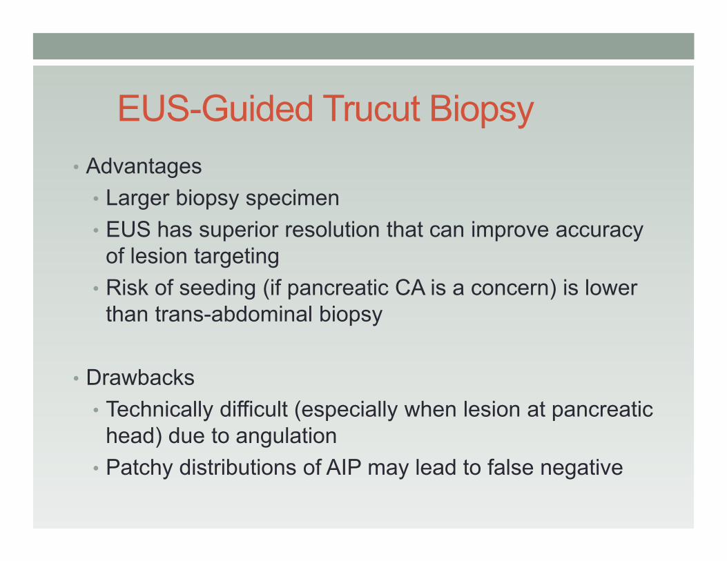
EUS-Guided Trucut Biopsy• Advantages
• Larger biopsy specimen• EUS has superior resolution that can improve accuracy
of lesion targeting• Risk of seeding (if pancreatic CA is a concern) is lower
than trans-abdominal biopsy
• Drawbacks• Technically difficult (especially when lesion at pancreatic
head) due to angulation• Patchy distributions of AIP may lead to false negative
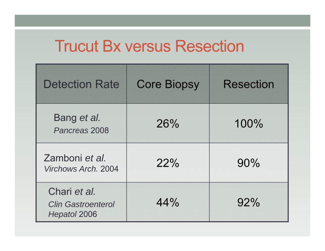
Trucut Bx versus Resection
Detection Rate Core Biopsy Resection
Bang et al. Pancreas 2008
26% 100%
Zamboni et al. Virchows Arch. 2004 22% 90%
Chari et al. Clin GastroenterolHepatol 2006
44% 92%
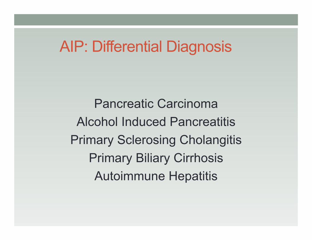
AIP: Differential Diagnosis
Pancreatic CarcinomaAlcohol Induced Pancreatitis
Primary Sclerosing CholangitisPrimary Biliary CirrhosisAutoimmune Hepatitis
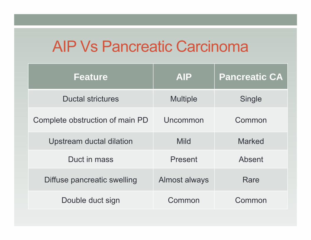
AIP Vs Pancreatic Carcinoma
Feature AIP Pancreatic CA
Ductal strictures Multiple Single
Complete obstruction of main PD Uncommon Common
Upstream ductal dilation Mild Marked
Duct in mass Present Absent
Diffuse pancreatic swelling Almost always Rare
Double duct sign Common Common
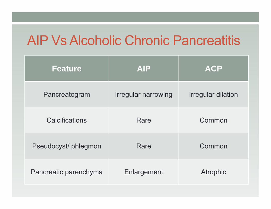
AIP Vs Alcoholic Chronic Pancreatitis
Feature AIP ACP
Pancreatogram Irregular narrowing Irregular dilation
Calcifications Rare Common
Pseudocyst/ phlegmon Rare Common
Pancreatic parenchyma Enlargement Atrophic
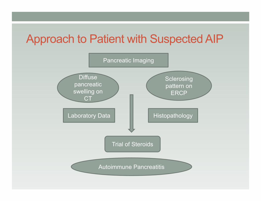
Approach to Patient with Suspected AIPPancreatic Imaging
Diffuse pancreatic swelling on
CT
Sclerosingpattern on
ERCP
Laboratory Data Histopathology
Trial of Steroids
Autoimmune Pancreatitis
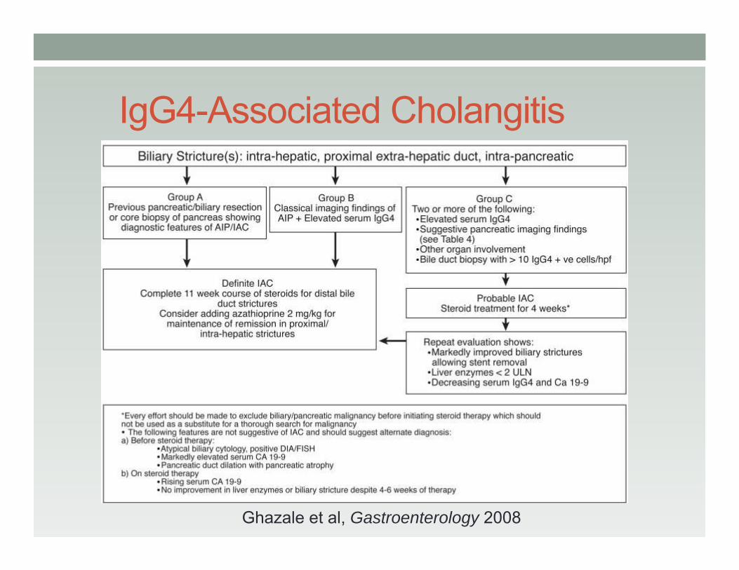
IgG4-Associated Cholangitis
Ghazale et al, Gastroenterology 2008
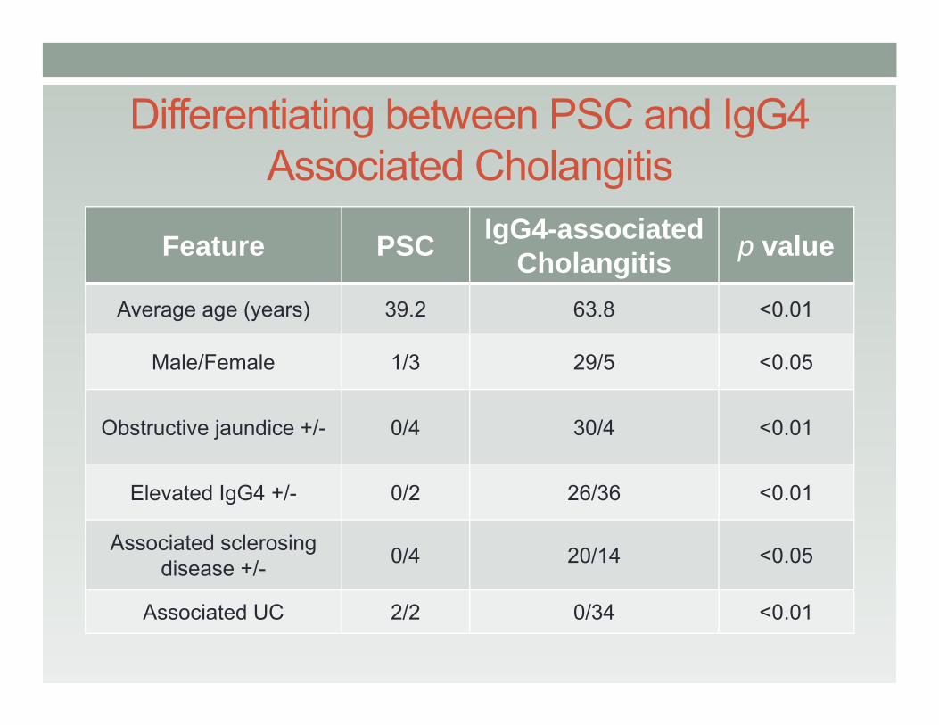
Differentiating between PSC and IgG4 Associated Cholangitis
Feature PSC IgG4-associatedCholangitis p value
Average age (years) 39.2 63.8 <0.01
Male/Female 1/3 29/5 <0.05
Obstructive jaundice +/- 0/4 30/4 <0.01
Elevated IgG4 +/- 0/2 26/36 <0.01
Associated sclerosingdisease +/- 0/4 20/14 <0.05
Associated UC 2/2 0/34 <0.01
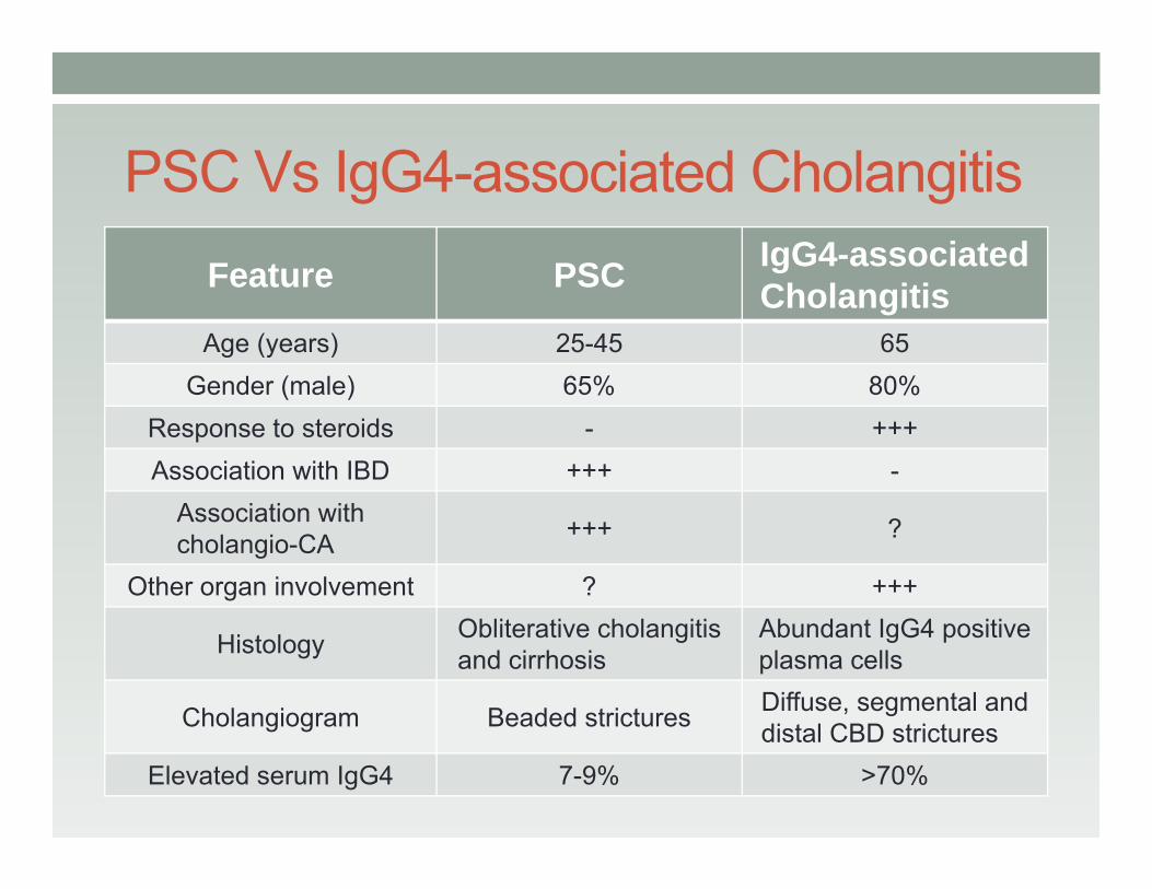
PSC Vs IgG4-associated CholangitisFeature PSC IgG4-associated
CholangitisAge (years) 25-45 65
Gender (male) 65% 80%Response to steroids - +++Association with IBD +++ -
Association with cholangio-CA +++ ?
Other organ involvement ? +++
Histology Obliterative cholangitis and cirrhosis
Abundant IgG4 positive plasma cells
Cholangiogram Beaded strictures Diffuse, segmental and distal CBD strictures
Elevated serum IgG4 7-9% >70%
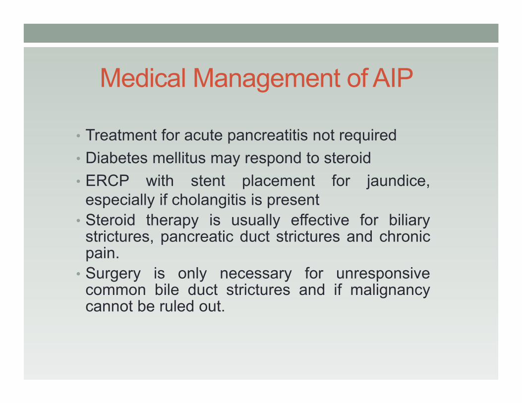
Medical Management of AIP
• Treatment for acute pancreatitis not required• Diabetes mellitus may respond to steroid• ERCP with stent placement for jaundice,
especially if cholangitis is present• Steroid therapy is usually effective for biliary
strictures, pancreatic duct strictures and chronicpain.
• Surgery is only necessary for unresponsivecommon bile duct strictures and if malignancycannot be ruled out.
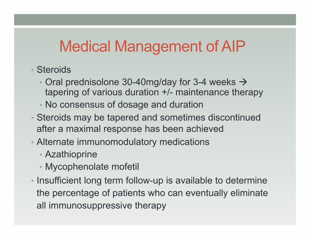
Medical Management of AIP• Steroids
• Oral prednisolone 30-40mg/day for 3-4 weeks tapering of various duration +/- maintenance therapy
• No consensus of dosage and duration• Steroids may be tapered and sometimes discontinued
after a maximal response has been achieved• Alternate immunomodulatory medications
• Azathioprine• Mycophenolate mofetil
• Insufficient long term follow-up is available to determine the percentage of patients who can eventually eliminate all immunosuppressive therapy.
Summary• Autoimmune pancreatitis is a recently described disorder presenting
with mild epigastric discomfort and jaundice• Patients may have associated autoimmune disorders and
autoantibodies• Imaging shows diffuse or focal pancreatic enlargement, and
pancreatic duct and bile duct stenoses.
• There is a low level of awareness of AIP. A high index of suspicion in the correct clinical setting is necessary to make a diagnosis.
• Steroid therapy is almost always effective
• AIP is clinically important since it is treatable, and may be mistaken for pancreatic carcinoma. It has been aptly called “The Great Masquerader”.
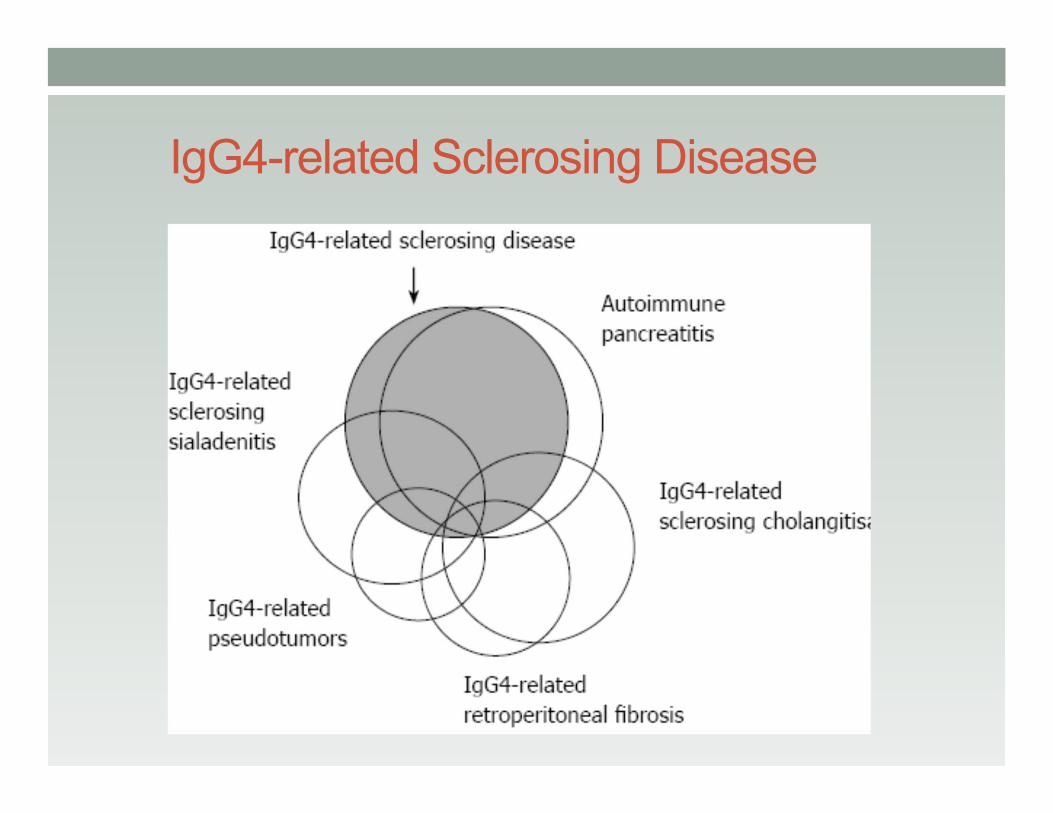
IgG4-related Sclerosing Disease