Embed Size (px)
Citation preview
CASE REPORT Open Access
Autoimmune enteropathy with a CD8+ CD7-
T-cell small bowel intraepithelial lymphocytosis:case report and literature reviewShrinivas Bishu1*, Violeta Arsenescu2, Eun Y Lee4, H David Vargas3, Willem JS de Villiers2 and Razvan Arsenescu2
Abstract
Background: Adult onset autoimmune enteropathy (AIE) is a rare condition characterized by diarrhea refractory todietary therapy diagnosed in patients with evidence of autoimmune conditions. Auto-antibodies to gut epithelialcells and other tissues are commonly demonstrated. Despite increasing awareness, the pathogenesis, histologic,immunologic and clinical features of AIE remain uncertain. There remains controversy regarding the diagnosticcriteria, the frequency and types of auto-antibodies and associated autoimmune conditions, and the extent andtypes of histologic and immunologic abnormalities. CD4+ T-cells are thought to at least responsible for thiscondition; whether other cell types, including B- and other T-cell subsets are involved, are uncertain. We present aunique case of AIE associated with a CD8+CD7- lymphocytosis and review the literature to characterize thehistologic and immunologic abnormalities, and the autoantibodies and autoimmune conditions associated withAIE.
Case Presentation: We present a case of immune mediated enteropathy distinguished by the CD8+CD7- intra-epithelial and lamina propria lymphocytosis. Twenty-nine cases of AIE have been reported. The majority of patientshad auto-antibodies (typically anti-enterocyte), preferential small bowel involvement, and predominately CD3+ CD4+ infiltrates. Common therapies included steroids or immuno-suppressive agents and clinical response withassociated with histologic improvement.
Conclusions: AIE is most often characterized (1) IgG subclass anti-epithelial cell antibodies, (2) preferential smallbowel involvement, and (3) CD3+ alphabeta TCR+ infiltrates; there is insufficient evidence to conclude CD4+ T-cellsare solely responsible in all cases of AIE.
BackgroundImmune enteropathies are heterogeneous group of rareconditions characterized by intractable diarrhea, damageto intestinal epithelia and constituent cells, and villousatrophy. These enteropathies may be associated withprimary immune deficiencies (PIDs) such as theimmune dysregulation, polyendocrinopathy, enteropathy,X-linked (IPEX) syndrome, common variable immunedeficiency and selective IgA deficiency, or may occur inpatients with auto-immune phenomena in the absenceof PIDs where it is termed auto-immune enteropathy(AIE) [1].
Typically, the diagnosis of immune mediated entero-pathy in the setting of PIDs is clear from the clinicalfeatures and a pre-existing diagnosis of PID. In contrast,AIE may be diagnosed in patients with no evidence ofPIDs, intractable diarrhea refractory to exclusion diets,and no evidence of celiac disease [1].Cumulatively, immune enteropathies, including AIE,
have an unclear pathogenesis. They are all thought to bemediated by immune phenomena, either through directdamage to gut epithelial cells by auto-reactive cells(IPEX) or auto-antibodies (AIE), or from a propensityfor bowel infections (IgA deficiency). However, the exactcell types causing damage and the mechanisms of dis-ease remain unclear in many of these conditions.Herein, we report a patient with an immune mediated
enteropathy distinguished from previously reported
* Correspondence: [email protected] of Internal Medicine, University of Kentucky Medical Center,800 Rose Street, Lexington, Kentucky, 40536, USAFull list of author information is available at the end of the article
Bishu et al. BMC Gastroenterology 2011, 11:131http://www.biomedcentral.com/1471-230X/11/131
© 2011 Bishu et al; licensee BioMed Central Ltd. This is an Open Access article distributed under the terms of the Creative CommonsAttribution License (http://creativecommons.org/licenses/by/2.0), which permits unrestricted use, distribution, and reproduction inany medium, provided the original work is properly cited.
cases by an unusual CD8+ CD7- IEL in the absence of aPID. We also review the literature to characterize thehistologic and immunophenotyic features of AIE, whichour patient’s enteropathy most closely resembles.
Case PresentationThe patient was a 28 year old Asian Indian female ofnon-consanguineous parents who presented with a 3year history of non-bloody diarrhea with abdominalcramping. Past medical history was significant for hepa-titis A. She denied alcohol or tobacco use, had nochronic medical conditions, was not on any medicationsincluding gut irritants such as aspirin/non-steroidialsand did not have a history of travel prior to onset ofsymptoms. Her last travel was to India one year prior topresentation, but she did not report any alleviation orexacerbation of her symptos with the travel/dietary/environmental change. She weighed 81 pounds at pre-sentation. Stool cultures were negative for pathogens.Basic labs, including the erythrocyte sedimentation rateand C-reactive protein were normal, but liver functiontests (LFTs) were elevated (Table 1). Abdominal com-puted tomography (CT) revealed hepatic hypodensitiesand mesenteric adenopathy. Serology was negative foranti-nuclear (ANA) and anti-smooth muscle antibodiesas well as antibodies for cytosolic anti-neutrophil cyto-plasm (cANCA), liver-kidney microsomes, liver cytosolicand soluble liver-pancreas antigen. However, pANCAwas positive (titer: > 1:20) and she had hyper-gamma-globulinemia (IgG: 2358 (630-1580 mg/dl), IgA: 578(100-400 mg/dl), IgM: 134 (37-247 mg/dl). Serum ceru-loplasmin was normal, and serology for hepatitis B andC were negative. Serum B12, folate and hematocrit werenormal.
Histologic examination of intestinal mucosa demon-strated chronic inflammation with duodenal intraepithe-lial cell lymphocytosis (IEL) (Table 1) (Figure 1). Celiacdisease and tropical sprue were therefore consideredbased on the clinical features and IEL. However, tissuetransglutaminase (TTG) IgA was normal (5.3, reference:< 7.0 AU), as were anti-enterocyte antibodies (only IgAtested) (both measured by enzyme linked immuno-absorbant assay of serum). Indirect immunofluorescenceon human intestinal tissue samples was not performed.The patient refused liver biopsy, but permitted CTguided biopsies of the enlarged retroperitoneal and peri-portal nodes; histologic analysis only demonstrated nor-mal stroma. She was diagnosed with auto-immunehepatitis (AIH) based on her revised original Interna-tional AIH group score of 12 (indicating probable AIHby criteria)[2].Over the next 21 months, she continued to have diar-
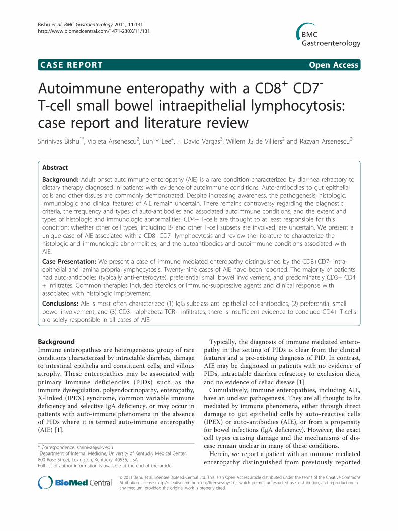
rhea despite dietary exclusion therapy, and elevatedLFTs (Table 1). Because serum folate and B12 remainedwithing normal limits for the duration of her care, shewas not treated with these agents. She did develop amild iron deficiency anemia, but never required bloodtransfusions. Endoscopy 15 months after presentationdemonstrated progression with grossly friable duodenalmucosa. There was marked IEL with a CD8+ T-cell sub-set without evidence of enteropathy type T-cell lym-phoma (Figure 2). Flow cytometry revealed a nearly100% T-cell preponderance, the majority of which werea polyclonal ab T-cell receptor (TCR)+ CD8+ CD7- sub-set (Table 1: 15 months). Clonality was assessed by poly-merase chain reaction of TCR chains. The minority ofIELs were CD4+ and CD8+ CD7+ subsets; there werenormal numbers of intra-epithelial gδ T-cells and rare
Table 1 Liver function test and histologic features pre- and post-prednisone
Pre-prednisone Post-prednisone
Presentation 15 months 21 months 26 months
Liver Function Tests
AST (U/L) 107 43 41 65
ALT (U/L) 95 46 97 82
ALP (U/L) 240 205 110 123
Albumin (g/dL)
2.2 1.5 2.1 3.5
Histology
Histology IELIntact villiChronic
inflammation
Intact villiDuodenum + jejunum+ illeum:
lymphoplastic infiltrateColon + cecum + rectum:lymphoplastic infiltrate
IELSubtotal villous atrophy
IELSubtotal villous atrophy Segmental areas normal
Immuno-phenotype
not obtained Jejunum + illeum: CD3+CD5+CD8+CD7-preponderance
Jejunum: Polyclonal abTCR+CD3+ preponderance
CD8+CD25-CD7- (70% of totalIEL)
Jejunum: Polyclonal abTCR+CD3+ (94% of total IEL)CD8+CD25-CD7- (41% of CD3+ fraction)
Bishu et al. BMC Gastroenterology 2011, 11:131http://www.biomedcentral.com/1471-230X/11/131
Page 2 of 10
B-cells (CD20+). Biopsies samples from the stomach andesophagus were normal, as was colonoscopy. There wasno evidence of reduced numbers or abnormal morphol-ogy of epithelial or goblet cells.Because of the marked IEL without laboratory or his-
tologic evidence of celiac disease, she was treated withCiprofloxicin® and Metronidazole® for potential bowelovergrowth and/or tropical sprue. However, six monthsof antibiotic therapy did not result in clinical or histolo-gic improvement (Table 1: 21 months). Although refrac-tory celiac disease is possible in this scenario, but thelack of elevated intra-epithelial gδ T-cells, normal TTGIgA, and her negative HLA-DQ2/DQ9 genotype makethis unlikely. A diagnosis of an immune mediatedenteropathy was therefore considered based on the lackof clinical or histologic response to dietary and antibio-tic therapy, and the lack of celiac disease serologic mar-kers. Prednisone (60 mg) daily was started.Steroid therapy resulted in marked improvement with
symptom resolution accompanied by a 20 lb weight
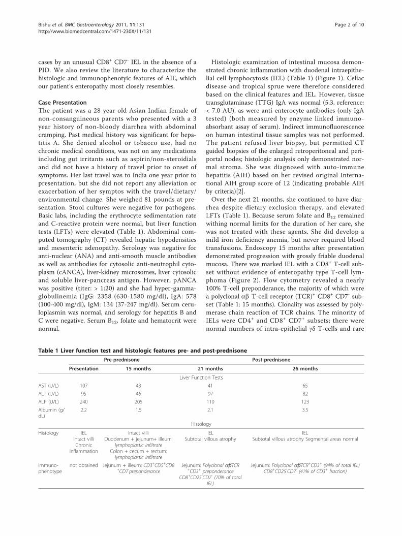
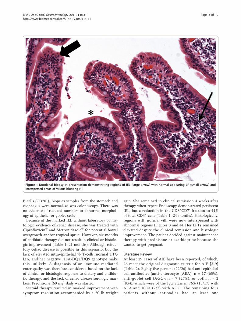
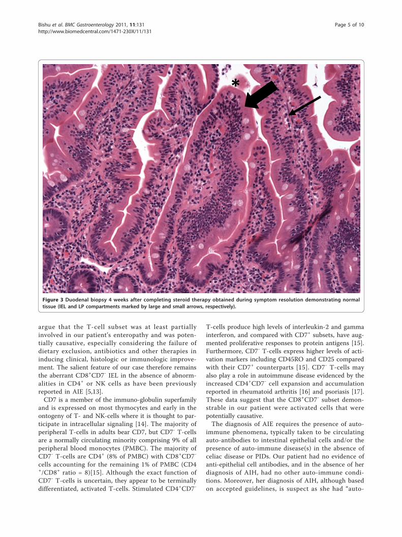
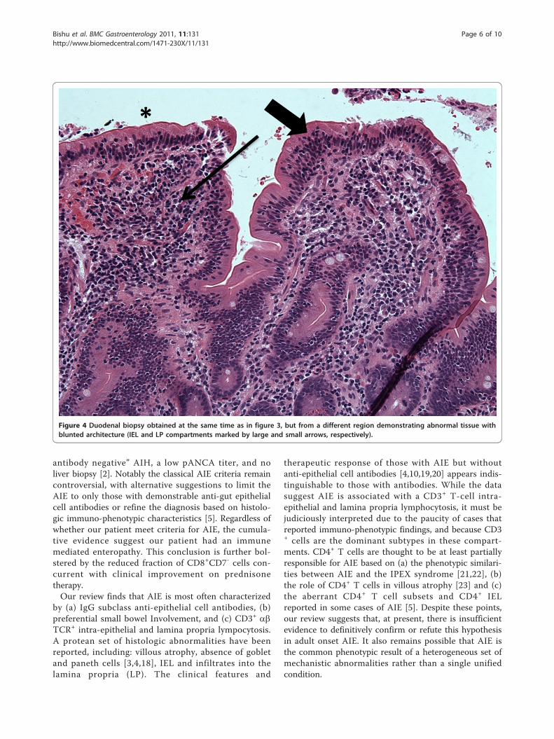
gain. She remained in clinical remission 4 weeks aftertherapy when repeat Endoscopy demonstrated persistentIEL, but a reduction in the CD8+CD7- fraction to 41%of total CD3+ cells (Table 1: 24 months). Histologically,regions with normal villi were now interspersed withabnormal regions (Figures 3 and 4). Her LFTs remainedelevated despite the clinical remission and histologicimprovement. The patient decided against maintenancetherapy with prednisone or azathioprine because shewanted to get pregnant.
Literature ReviewAt least 29 cases of AIE have been reported, of which,26 meet the original diagnostic criteria for AIE [3-9](Table 2). Eighty five percent (22/26) had anti-epithelialcell antibodies (anti-enterocyte (AEA): n = 17 (65%),anti-goblet cell (AGC): n = 7 (27%), or both: n = 2(8%)), which were of the IgG class in 76% (13/17) withAEA and 100% (7/7) with AGC. The remaining fourpatients without antibodies had at least one
*
Figure 1 Duodenal biopsy at presentation demonstrating regions of IEL (large arrow) with normal appearing LP (small arrow) andinterspersed areas of villous blunting (*).
Bishu et al. BMC Gastroenterology 2011, 11:131http://www.biomedcentral.com/1471-230X/11/131
Page 3 of 10

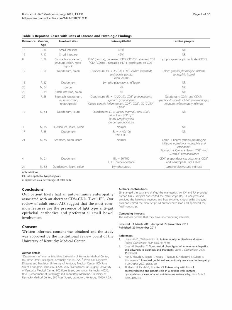
autoimmune condition (Table 2). Hypothyroidism wasmost frequently reported, but no disorder was stronglyassociated with AIE. The majority (69%) had other cir-culating tissue auto-antibodies, most commonly, ANA(Table 2).Fourteen studies detailed the sites of disease, and 9
provided some immuno-phenotypic analysis (Table 3).The small bowel was the most frequently involved (79%of cases), followed by the colon and stomach. Six (43%)patients had multiple sites and two had extensive disease(stomach, small intestinal and colonic disease) (’general-ized autoimmune gut disorder’)[5,10,11]. Nine patients(75%) had small bowel IEL at presentation while at leastone case reported concurrent colonic IEL. Complete orpartial clinical response with therapy was associatedwith improved IEL in all 8 cases that reported thesefindings [5-9,11,12]. The IEL was predominantly T-cells(CD3+) in at least three cases, and was furthermore CD8+ in at least one of these. T-cell (CD3+) lymphocytosiswas also reported in the lamina propria in four of five
cases that provided this data (Table 3). One casedemonstrated extensive abnormalities in CD4+ T-cells,including increased tumor necrosis factor (TNF)a pro-duction from activated rectal lymphocytes [5]. However,this finding must be cautiously extrapolated becausethat patient had active CMV colitis and it is thereforepossible that the TNFa response representepiphenomena.
DiscussionWe report a patient with an enteropathy associatedwith an aberrant CD8+CD7- T-cell IEL. Clinically, onhistology and by flow cytometry, our patient had char-acteristic features of an immune mediated enteropathy.The abberant T-cell subset in our patient was reprodu-cible, polyclonal, and correlated with the presence ofclinical symptoms. Moreover, the symptom resolutionand weight gain with steroid therapy correlated withimprovement in villous architecture and a decrease inthe CD8+CD7- T-cell subset. These facts strongly
Figure 2 Duodenal biopsy before steroid therapy demonstrating regions with persistent IEL (large arrow) with marked LPlymphocytosis (small arrow).
Bishu et al. BMC Gastroenterology 2011, 11:131http://www.biomedcentral.com/1471-230X/11/131
Page 4 of 10
argue that the T-cell subset was at least partiallyinvolved in our patient’s enteropathy and was poten-tially causative, especially considering the failure ofdietary exclusion, antibiotics and other therapies ininducing clinical, histologic or immunologic improve-ment. The salient feature of our case therefore remainsthe aberrant CD8+CD7- IEL in the absence of abnorm-alities in CD4+ or NK cells as have been previouslyreported in AIE [5,13].CD7 is a member of the immuno-globulin superfamily
and is expressed on most thymocytes and early in theontogeny of T- and NK-cells where it is thought to par-ticipate in intracellular signaling [14]. The majority ofperipheral T-cells in adults bear CD7, but CD7- T-cellsare a normally circulating minority comprising 9% of allperipheral blood monocytes (PMBC). The majority ofCD7- T-cells are CD4+ (8% of PMBC) with CD8+CD7-
cells accounting for the remaining 1% of PMBC (CD4+/CD8+ ratio = 8)[15]. Although the exact function ofCD7- T-cells is uncertain, they appear to be terminallydifferentiated, activated T-cells. Stimulated CD4+CD7-
T-cells produce high levels of interleukin-2 and gammainterferon, and compared with CD7+ subsets, have aug-mented proliferative responses to protein antigens [15].Furthermore, CD7- T-cells express higher levels of acti-vation markers including CD45RO and CD25 comparedwith their CD7+ counterparts [15]. CD7- T-cells mayalso play a role in autoimmune disease evidenced by theincreased CD4+CD7- cell expansion and accumulationreported in rheumatoid arthritis [16] and psoriasis [17].These data suggest that the CD8+CD7- subset demon-strable in our patient were activated cells that werepotentially causative.The diagnosis of AIE requires the presence of auto-
immune phenomena, typically taken to be circulatingauto-antibodies to intestinal epithelial cells and/or thepresence of auto-immune disease(s) in the absence ofceliac disease or PIDs. Our patient had no evidence ofanti-epithelial cell antibodies, and in the absence of herdiagnosis of AIH, had no other auto-immune condi-tions. Moreover, her diagnosis of AIH, although basedon accepted guidelines, is suspect as she had “auto-
*
Figure 3 Duodenal biopsy 4 weeks after completing steroid therapy obtained during symptom resolution demonstrating normaltissue (IEL and LP compartments marked by large and small arrows, respectively).
Bishu et al. BMC Gastroenterology 2011, 11:131http://www.biomedcentral.com/1471-230X/11/131
Page 5 of 10
antibody negative” AIH, a low pANCA titer, and noliver biopsy [2]. Notably the classical AIE criteria remaincontroversial, with alternative suggestions to limit theAIE to only those with demonstrable anti-gut epithelialcell antibodies or refine the diagnosis based on histolo-gic immuno-phenotypic characteristics [5]. Regardless ofwhether our patient meet criteria for AIE, the cumula-tive evidence suggest our patient had an immunemediated enteropathy. This conclusion is further bol-stered by the reduced fraction of CD8+CD7- cells con-current with clinical improvement on prednisonetherapy.Our review finds that AIE is most often characterized
by (a) IgG subclass anti-epithelial cell antibodies, (b)preferential small bowel Involvement, and (c) CD3+ abTCR+ intra-epithelial and lamina propria lympocytosis.A protean set of histologic abnormalities have beenreported, including: villous atrophy, absence of gobletand paneth cells [3,4,18], IEL and infiltrates into thelamina propria (LP). The clinical features and
therapeutic response of those with AIE but withoutanti-epithelial cell antibodies [4,10,19,20] appears indis-tinguishable to those with antibodies. While the datasuggest AIE is associated with a CD3+ T-cell intra-epithelial and lamina propria lymphocytosis, it must bejudiciously interpreted due to the paucity of cases thatreported immuno-phenotypic findings, and because CD3+ cells are the dominant subtypes in these compart-ments. CD4+ T cells are thought to be at least partiallyresponsible for AIE based on (a) the phenotypic similari-ties between AIE and the IPEX syndrome [21,22], (b)the role of CD4+ T cells in villous atrophy [23] and (c)the aberrant CD4+ T cell subsets and CD4+ IELreported in some cases of AIE [5]. Despite these points,our review suggests that, at present, there is insufficientevidence to definitively confirm or refute this hypothesisin adult onset AIE. It also remains possible that AIE isthe common phenotypic result of a heterogeneous set ofmechanistic abnormalities rather than a single unifiedcondition.
*
Figure 4 Duodenal biopsy obtained at the same time as in figure 3, but from a different region demonstrating abnormal tissue withblunted architecture (IEL and LP compartments marked by large and small arrows, respectively).
Bishu et al. BMC Gastroenterology 2011, 11:131http://www.biomedcentral.com/1471-230X/11/131
Page 6 of 10
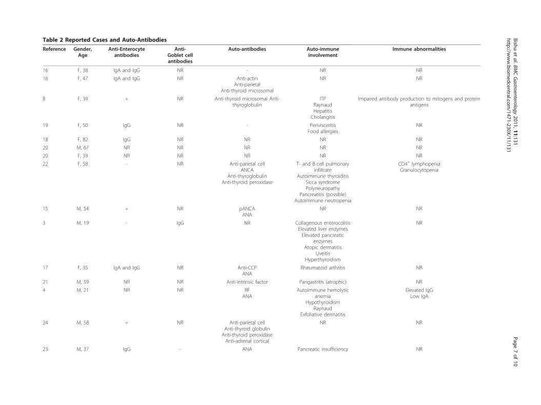
Table 2 Reported Cases and Auto-Antibodies
Reference Gender,Age
Anti-Enterocyteantibodies
Anti-Goblet cellantibodies
Auto-antibodies Auto-immuneinvolvement
Immune abnormalities
16 F, 38 IgA and IgG NR - NR NR
16 F, 47 IgA and IgG NR Anti-actinAnti-parietal
Anti-thyroid microsomal
NR NR
8 F, 39 + NR Anti-thyroid microsomal Anti-thyroglobulin
ITPRaynaudHepatitisCholangitis
Impaired antibody production to mitogens and proteinantigens
19 F, 50 IgG NR - PerivisceritisFood allergies
NR
18 F, 82 IgG NR NR NR NR
20 M, 67 NR NR NR NR NR
20 F, 39 NR NR NR NR NR
22 F, 58 - NR Anti-parietal cellANCA
Anti-thyroglobulinAnti-thyroid peroxidase
T- and B-cell pulmonaryinfiltrate
Autoimmune thyroiditisSicca syndromePolyneuropathy
Pancreatitis (possible)Autoimmune neutropenia
CD4+ lymphopeniaGranulocytopenia
15 M, 54 + NR pANCAANA
NR NR
3 M, 19 - IgG NR Collagenous enterocolitisElevated liver enzymesElevated pancreatic
enzymesAtopic dermatitis
UveitisHyperthyroidism
NR
17 F, 35 IgA and IgG NR Anti-CCPANA
Rheumatoid arthritis NR
21 M, 59 NR NR Anti-intrinsic factor Pangastritis (atrophic) NR
4 M, 21 NR NR RFANA
Autoimmune hemolyticanemia
HypothyroidismRaynaud
Exfoliative dermatitis
Elevated IgGLow IgA
24 M, 58 + NR Anti-parietal cellAnti-thyroid globulin
Anti-thyroid peroxidaseAnti-adrenal cortical
NR NR
23 M, 37 IgG - ANA Pancreatic insufficiency NR
Bishuet
al.BMCGastroenterology
2011,11:131http://w
ww.biom
edcentral.com/1471-230X/11/131
Page7of
10
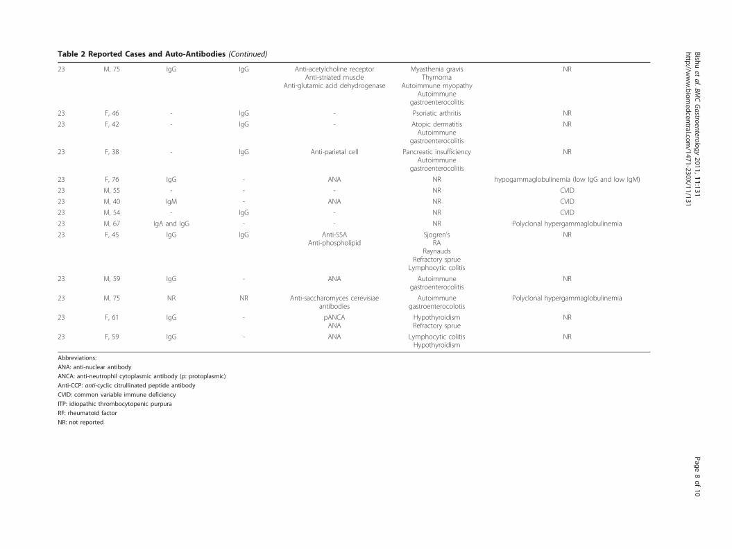
Table 2 Reported Cases and Auto-Antibodies (Continued)
23 M, 75 IgG IgG Anti-acetylcholine receptorAnti-striated muscle
Anti-glutamic acid dehydrogenase
Myasthenia gravisThymoma
Autoimmune myopathyAutoimmune
gastroenterocolitis
NR
23 F, 46 - IgG - Psoriatic arthritis NR
23 F, 42 - IgG - Atopic dermatitisAutoimmune
gastroenterocolitis
NR
23 F, 38 - IgG Anti-parietal cell Pancreatic insufficiencyAutoimmune
gastroenterocolitis
NR
23 F, 76 IgG - ANA NR hypogammaglobulinemia (low IgG and low IgM)
23 M, 55 - - - NR CVID
23 M, 40 IgM - ANA NR CVID
23 M, 54 - IgG - NR CVID
23 M, 67 IgA and IgG - - NR Polyclonal hypergammaglobulinemia
23 F, 45 IgG IgG Anti-SSAAnti-phospholipid
Sjogren’sRA
RaynaudsRefractory sprue
Lymphocytic colitis
NR
23 M, 59 IgG - ANA Autoimmunegastroenterocolitis
NR
23 M, 75 NR NR Anti-saccharomyces cerevisiaeantibodies
Autoimmunegastroenterocolotis
Polyclonal hypergammaglobulinemia
23 F, 61 IgG - pANCAANA
HypothyroidismRefractory sprue
NR
23 F, 59 IgG - ANA Lymphocytic colitisHypothyroidism
NR
Abbreviations:
ANA: anti-nuclear antibody
ANCA: anti-neutrophil cytoplasmic antibody (p: protoplasmic)
Anti-CCP: anti-cyclic citrullinated peptide antibody
CVID: common variable immune deficiency
ITP: idiopathic thrombocytopenic purpura
RF: rheumatoid factor
NR: not reported
Bishuet
al.BMCGastroenterology
2011,11:131http://w
ww.biom
edcentral.com/1471-230X/11/131
Page8of
10
ConclusionsOur patient likely had an auto-immune enteropathyassociated with an aberrant CD8+CD7- T-cell IEL. Ourreview of adult onset AIE suggest that the most com-mon features are the presence of IgG type anti-gutepithelial antibodies and preferential small bowelinvolvement.
ConsentWritten informed consent was obtained and the studywas approved by the institutional review board of theUniversity of Kentucky Medical Center.
Author details1Department of Internal Medicine, University of Kentucky Medical Center,800 Rose Street, Lexington, Kentucky, 40536, USA. 2Division of DigestiveDiseases and Nutrition, University of Kentucky Medical Center, 800 RoseStreet, Lexington, Kentucky, 40536, USA. 3Department of Surgery, Universityof Kentucky Medical Center, 800 Rose Street, Lexington, Kentucky, 40536,USA. 4Department of Pathology and Laboratory Medicine, University ofKentucky Medical Center, 800 Rose Street, Lexington, Kentucky, 40536, USA.
Authors’ contributionsSB analyzed the data and drafted the manuscript. VA, DV and RA providedhuman tissue samples and edited the manuscript (RA). EL analyzed andprovided the histologic sections and flow cytometric data. WdW analyzeddata and edited the manuscript. All authors have read and approved thefinal manuscript
Competing interestsThe authors declare that they have no competing interests.
Received: 11 March 2011 Accepted: 29 November 2011Published: 29 November 2011
References1. Unsworth DJ, Walker-Smith JA: Autoimmunity in diarrhoeal disease. J
Pediatr Gastroenterol Nutr 1985, 4:375-80.2. Czaja AJ, Bayraktar Y: Non-classical phenotypes of autoimmune hepatitis
and advances in diagnosis and treatment. World J Gastroenterol 2009,15:2314-28.
3. Hori K, Fukuda Y, Tomita T, Kosaka T, Tamura K, Nishigami T, Kubota A,Shimoyama T: Intestinal goblet cell autoantibody associated enteropathy.J Clin Pathol 2003, 56:629-30.
4. Al Khalidi H, Kandel G, Streutker CJ: Enteropathy with loss ofenteroendocrine and paneth cells in a patient with immunedysregulation: a case of adult autoimmune enteropathy. Hum Pathol2006, 37:373-6.
Table 3 Reported Cases with Sites of Disease and Histologic Findings
Reference Gender,Age
Involved sites Intra-epithelial Lamina propria
16 F, 38 Small intestine 46%a NR
16 F, 47 Small intestine 42%a NR
8 F, 39 Stomach, duodenum,jejunum, colon, recto-
sigmoid
12%a (normal), decreased CD3- CD103+, aberrant CD3+CD4+CD103-, increased HLA-II expression on CD3+
Lympho-plasmacytic infiltrate (CD3+)
19 F, 50 Duodenum, colon Duodenum: IEL = 48/100, CD3+ 50/mm (elevated),eosinophils (some);Colon: normal
Colon: lympho-plasmacytic infiltrate,eosinophils (some)
18 F, 82 Duodenum Lympho-plasmacytic infiltrate NR
20 M, 67 colon NR NR
20 F, 39 Small intestine, colon NR NR
22 F, 58 Stomach, duodenum,jejunum, colon,rectosigmoid
Duodenum: IEL = 10-20/100, CD8+ preponderanceJejunum: lymphocytosis
Colon: chronic inflammation, CD4+, CD8+, CD19+/20+,CD68+
Duodenum: CD3+ and CD43+lymphocytosis with CD68+ (macrophages)
Jejunum: Inflammatory infiltrate
15 M, 54 Duodenum, ileum Duodenum: IEL = 28/100 (normal), 50% CD8+,oligoclonal TCR ab+Ileum: lymphocytosisColon: lymphocytosis
NR
3 M, 19 Duodenum, ileum, colon Normal NR
17 F, 35 Duodenum IEL = > 40/10052% CD3+
NR
21 M, 59 Stomach, colon, ileum Normal Colon + Ileum: lympho-plasmacyticinfiltrate, occasional neutrophils and
eosinophilsStomach + Colon + Ileum: CD8+ and
CD45RO+ preponderance
4 M, 21 Duodenum IEL = 50/100CD8+ preponderance
CD4+ preponderance, occasional CD8+
and neutrophils, rare CD20+
24 M, 58 Duodenum, ileum, colon Lymphocytosis Lympho-plasmacytic infiltrate
Abbreviations:
IEL: Intra-epithelial lymphocytosis
a: expressed as a percentage of total cells
Bishu et al. BMC Gastroenterology 2011, 11:131http://www.biomedcentral.com/1471-230X/11/131
Page 9 of 10
5. Leon F, Olivencia P, Rodriguez-Pena R, Sanchez L, Redondo C, Alvarez I,Moreira V, Roy G: Clinical and immunological features of adult-onsetgeneralized autoimmune gut disorder. Am J Gastroenterol 2004,99:1563-71.
6. Daum S, Sahin E, Jansen A, Heine B, Riecken EO, Zeitz M, Schmidt W: Adultautoimmune enteropathy treated successfully with tacrolimus. Digestion2003, 68:86-90.
7. Corazza GR, Biagi F, Volta U, Andreani ML, De Franceschi L, Gasbarrini G:Autoimmune enteropathy and villous atrophy in adults. Lancet 1997,350:106-9.
8. Volta U, De Angelis GL, Granito A, Petrolini N, Fiorini E, Guidi M, Muratori P,Bianchi FB: Autoimmune enteropathy and rheumatoid arthritis: a newassociation in the field of autoimmunity. Dig Liver Dis 2006, 38:926-9.
9. Elwing JE, Clouse RE: Adult-onset autoimmune enteropathy in the settingof thymoma successfully treated with infliximab. Dig Dis Sci 2005,50:928-32.
10. Mitomi H, Tanabe S, Igarashi M, Katsumata T, Arai N, Kikuchi S, Kiyohashi A,Okayasu I: Autoimmune enteropathy with severe atrophic gastritis andcolitis in an adult: proposal of a generalized autoimmune disorder ofthe alimentary tract. Scand J Gastroenterol 1998, 33:716-20.
11. Carroccio A, Volta U, Di Prima L, Petrolini N, Florena AM, Averna MR,Montalto G, Notarbartolo A: Autoimmune enteropathy and colitis in anadult patient. Dig Dis Sci 2003, 48:1600-6.
12. Rolny P, Sigurjonsdottir HA, Remotti H, Nilsson LA, Ascher H, Tlaskalova-Hogenova H, Tuckova L: Role of immunosuppressive therapy in refractorysprue-like disease. Am J Gastroenterol 1999, 94:219-25.
13. Eiras P, Leon F, Camarero C, Lombardia M, Roldan E, Bootello A, Roy G:Intestinal intraepithelial lymphocytes contain a CD3- CD7+ subsetexpressing natural killer markers and a singular pattern of adhesionmolecules. Scand J Immunol 2000, 52:1-6.
14. Sempowski GD, Lee DM, Kaufman RE, Haynes BF: Structure and functionof the CD7 molecule. Crit Rev Immunol 1999, 19:331-48.
15. Reinhold U, Abken H, Kukel S, Moll M, Muller R, Oltermann I, Kreysel HW:CD7- T cells represent a subset of normal human blood lymphocytes. JImmunol 1993, 150:2081-9.
16. Schmidt D, Goronzy JJ, Weyand CM: CD4+ CD7- CD28- T cells areexpanded in rheumatoid arthritis and are characterized byautoreactivity. J Clin Invest 1996, 97:2027-37.
17. Moll M, Reinhold U, Kukel S, Abken H, Muller R, Oltermann I, Kreysel HW:CD7-negative helper T cells accumulate in inflammatory skin lesions. JInvest Dermatol 1994, 102:328-32.
18. Moore L, Xu X, Davidson G, Moore D, Carli M, Ferrante A: Autoimmuneenteropathy with anti-goblet cell antibodies. Hum Pathol 1995, 26:1162-8.
19. Casis B, Fernandez-Vazquez I, Barnardos E, Saiz A, Ballestin C, Morillas JD,Colina F, Solis-Herruzo JA: Autoimmune enteropathy in an adult withautoimmune multisystemic involvement. Scand J Gastroenterol 2002,37:1012-6.
20. Akram S, Murray JA, Pardi DS, Alexander GL, Schaffner JA, Russo PA,Abraham SC: Adult autoimmune enteropathy: Mayo Clinic Rochesterexperience. Clin Gastroenterol Hepatol 2007, 5:1282-90, quiz 1245.
21. Ochs HD, Oukka M, Torgerson TR: TH17 cells and regulatory T cells inprimary immunodeficiency diseases. J Allergy Clin Immunol 2009,123:977-83, quiz 984-5.
22. Baud O, Goulet O, Canioni D, Le Deist F, Radford I, Rieu D, Dupuis-Girod S,Cerf-Bensussan N, Cavazzana-Calvo M, Brousse N: Treatment of theimmune dysregulation, polyendocrinopathy, enteropathy, X-linkedsyndrome (IPEX) by allogeneic bone marrow transplantation. N Engl JMed 2001, 344:1758-62, others.
23. MacDonald TT, Spencer J: Evidence that activated mucosal T cells play arole in the pathogenesis of enteropathy in human small intestine. J ExpMed 1988, 167:1341-9.
Pre-publication historyThe pre-publication history for this paper can be accessed here:http://www.biomedcentral.com/1471-230X/11/131/prepub
doi:10.1186/1471-230X-11-131Cite this article as: Bishu et al.: Autoimmune enteropathy with a CD8+
CD7- T-cell small bowel intraepithelial lymphocytosis: case report andliterature review. BMC Gastroenterology 2011 11:131.
Submit your next manuscript to BioMed Centraland take full advantage of:
• Convenient online submission
• Thorough peer review
• No space constraints or color figure charges
• Immediate publication on acceptance
• Inclusion in PubMed, CAS, Scopus and Google Scholar
• Research which is freely available for redistribution
Submit your manuscript at www.biomedcentral.com/submit
Bishu et al. BMC Gastroenterology 2011, 11:131http://www.biomedcentral.com/1471-230X/11/131
Page 10 of 10