Embed Size (px)
Citation preview

This article appeared in a journal published by Elsevier. The attachedcopy is furnished to the author for internal non-commercial researchand education use, including for instruction at the authors institution
and sharing with colleagues.
Other uses, including reproduction and distribution, or selling orlicensing copies, or posting to personal, institutional or third party
websites are prohibited.
In most cases authors are permitted to post their version of thearticle (e.g. in Word or Tex form) to their personal website orinstitutional repository. Authors requiring further information
regarding Elsevier’s archiving and manuscript policies areencouraged to visit:
http://www.elsevier.com/copyright

Author's personal copy
Biosensors and Bioelectronics 26 (2011) 2201–2207
Contents lists available at ScienceDirect
Biosensors and Bioelectronics
journa l homepage: www.e lsev ier .com/ locate /b ios
Immobilization of metallothionein to carbon paste electrode surface viaanti-MT antibodies and its use for biosensing of silver
Libuse Trnkovaa, Sona Krizkovab, Vojtech Adamb, Jaromir Hubalekc, Rene Kizeka,b,∗
a Department of Chemistry, Masaryk University, Kotlarska 2, CZ-611 37 Brno, Czech Republicb Department of Chemistry and Biochemistry, Mendel University, Zemedelska 1, CZ-613 00 Brno, Czech Republicc Department of Microelectronics, Brno University of Technology, Udolni 53, CZ-602 00 Brno, Czech Republic
a r t i c l e i n f o
Article history:Received 2 August 2010Received in revised form15 September 2010Accepted 18 September 2010
Keywords:Heavy metalSilverMetallothioneinSquare wave voltammetryWaterEnvironmental analysis
a b s t r a c t
In this paper, heavy metal biosensor based on immobilization of metallothionein (MT) to the surface ofcarbon paste electrode (CPE) via anti-MT-antibodies is reported. First, the evaluation of MT electroactivitywas done. The attention was focused on the capturing of MT to the CPE surface. Antibodies incorporatedand mixed into carbon paste were stable; even after two weeks the observed changes in signal heightwere lower than 5%. Further, the interaction of MT with polyclonal chicken antibodies incorporated incarbon paste electrode was determined by square-wave voltammetry. In the voltammogram, two signals– labelled as cysMT and Wa – were observed. The cysMT corresponded to –SH moieties of MT and Wa corre-sponded to tryptophan residues of chicken antibodies. Time of interaction (300 s) and MT concentration(125 �g/ml) were optimized to suggest a silver(I) ions biosensor. Biosensor (CPE modified with anti-MT antibody) prepared under the optimized conditions was then used for silver(I) ions detection. Thedetection limit (3 S/N) for silver(I) ions was estimated as 0.5 nM. The proposed biosensor was tested bydetection spiking of silver(I) ions in various water samples (from very pure distilled water to rainwater).Recoveries varied from 74 to 104%.
© 2010 Elsevier B.V. All rights reserved.
1. Introduction
Toxic effect of silver(I) ions on water organisms has been repeat-edly reported (Bielmyer et al., 2008; Gorsuch and Klaine, 1998;Hogstrand et al., 1996; Wood et al., 1996). In water environment,silver(I) ions are stable in a wide range of pH. Under alkalic pH,AgOH and Ag(OH)2− are formed. In addition, in such environmentthere are many compounds which interact with silver(I) ions. Themost important ones are chloride anions, which form insoluble pre-cipitate with silver(I) ions (AgCl). The insoluble silver compoundsdo not represent any threat to aquatic organisms. Toxicity of silverin its soluble form is probably caused by its high affinity to proteinsand also to nucleic acids. The binding of silver ions into the activesites of enzymes leads to their distinctive inhibition.
The use of carbon electrode as working electrode for the deter-mination of silver has been previously reported by several authors(Guo and Khoo, 1999; Schildkraut et al., 1998; Svancara et al.,1996, 2001; Szymanski et al., 2010). Recently, our group intro-duced a heavy metal biosensor based on interaction of metal ionswith a low-molecular mass protein called metallothionein (MT)
∗ Corresponding author at: Department of Chemistry and Biochemistry, MendelUniversity, Zemedelska 1, CZ-613 00 Brno, Czech Republic. Tel.: +420 5 4513 3350;fax: +420 5 4521 2044.
E-mail address: [email protected] (R. Kizek).
(Adam et al., 2010; Eckschlager et al., 2009; Hamer, 1986) whichwas adsorbed on the surface of hanging mercury drop electrode(HMDE). This biosensor has been successfully used for the detec-tion of cadmium(II) and zinc(II) ions (Adam et al., 2005), cisplatin(Petrlova et al., 2006b), cisplatin-DNA adducts (Krizkova et al.,2007), and palladium(II) ions (Adam et al., 2007a). Hanging mercurydrop electrode has many advantages to be used for electroanaly-sis, but due to its physico-chemical properties it cannot be used inflow-automated instruments. Carbon electrodes represent a verypromising alternative for the detection of biomolecules and forsuggestion of biosensors (Cosnier, 1999; Li et al., 2007; Liu et al.,2008; Sivanesan and John, 2007). On the basis of the convincingresults with MT as a biological component, in this paper heavymetal biosensor based on immobilization of metallothionein tothe surface of carbon paste electrode (CPE) via anti-MT antibod-ies is suggested as capable tool for biosensing the silver. Schematicproposal of the suggested silver(I) ions biosensor with metalloth-ionein as a biological component and carbon paste electrode as atransducer is shown in Fig. 1.
2. Materials and methods
2.1. Chemicals and materials
Silver nitrate and all other reagents in ACS purity (chemi-cals meet the specifications of the American Chemical Society)
0956-5663/$ – see front matter © 2010 Elsevier B.V. All rights reserved.doi:10.1016/j.bios.2010.09.035

Author's personal copy
2202 L. Trnkova et al. / Biosensors and Bioelectronics 26 (2011) 2201–2207
Fig. 1. Suggestion of heavy metal biosensor based on immobilization of metallothionein on the surface of carbon paste electrode (CPE) via MT-antibodies. (I) Oxidationsignals of MT were measured at the surface of CPE; (II) carbon paste was mixed with polyclonal antibodies to MT; (III) CPE with the antibodies binds with MT; (IV) CPE withselectively bounded MT interacts with heavy metals.
were purchased from Sigma–Aldrich (USA), unless noted other-wise. Chicken antibody against metallothionein was obtained fromHena (Prague, Czech Republic) according to procedure publishedby Hodek et al. (2004). Stock standard solutions were preparedwith ACS water and stored in the dark at −20 ◦C. Working stan-dard solutions were freshly prepared on the day of experiment bydilution of the stock solutions. All solutions were filtered througha 0.45 �m nylon filter discs (MetaChem, Torrance, USA) prior toanalysis.
2.2. Dot immunobinding assay
For immunobinding assay the polyvinylidene fluoride (PVDF)membrane (Bio-Rad, USA) was used. Antigen (metallothioneinfrom rabbit liver, 1 �l) was applied with a micropipette and air-dried. Further the membrane was blocked in 2% bovine serumalbumin (BSA) in phosphate-buffered saline (PBS: 137 mM NaCl,2.7 mM KCl, 1.4 mM NaH2PO4, 4.3 mM Na2HPO4, pH 7.4) for 30 minwith constant shaking. Then, the membrane was rinsed in 0.05%(v/v) Tween-20 in PBS (PBS-T). The incubation with chicken pri-mary antibody (dilution 1:500 in 0.1% (w/v) BSA in PBS) wascarried out for 1 h at 37 ◦C under shaking. After the three timesrepeated washing in 0.05% PBS-T for 5 min the membrane wasincubated in the presence of anti-chicken antibody labelled withhorseradish peroxidase (dilution 1:1500 in 0.1% (w/v) BSA inPBS) for 1 h at 37 ◦C. Then the membrane was washed threetimes in 0.05% PBS-T for 5 min and incubated in chromogenicsubstrate (0.4 mg/ml AEC (3-aminoethyl-9-carbazole) in 0.01 Macetate buffer with 0.1% H2O2, pH 5.5). After the sufficient colouringthe reaction was stopped by rinsing in water. The dot intensity wasevaluated densitometrically by Biolight software (Vilber-Lourmat,France).
2.3. Electrochemical measurement
Square wave voltammetric (SWV) measurements were per-formed using an AUTOLAB analyser (EcoChemie, The Netherlands)connected to VA-Stand 663 (Metrohm, Switzerland), using astandard cell with three electrodes. Carbon paste electrode wasemployed as the working electrode. An Ag/AgCl/3 M KCl electrodeserved as the reference electrode. Glassy carbon electrode was usedas the auxiliary electrode. SWV parameters were as follows: initialpotential 0.0 V, end potential 1.6 V, modulation amplitude 25 mV,and step potential 0.5 mV. All experiments were carried out at roomtemperature. Borate buffer (0.2 M, pH 9.6) was used as the support-ing electrolyte. Savitzky and Golay filter included in the softwareGPES 4.9 supplied by EcoChemie was employed for background cor-rection (with following parameters: smoothing – level 2, baselinecorrection – peak width 0.03).
The carbon paste was made of 70% graphite powder and 30%mineral oil (free of DNase, RNase, and protease). Primary chickenantibody was diluted 1:500 with PBS buffer. Diluted solution withchicken antibody (100 �l) was mixed with 500 mg of carbon paste.The carbon paste was housed in a Teflon body of a 2.5 mm diameterof active disk surface. The electrode surface was polished beforeeach determination with a soft filter paper prior to measurement(Kizek et al., 2005; Masarik et al., 2003; Petrlova et al., 2007a,c).
2.4. Descriptive statistics
Results are expressed as mean ± S.D. unless noted otherwise.The detection limit (3 S/N) and quantification limit (10 S/N) werecalculated according to Long and Winefordner (1983), whereas Nwas expressed as standard deviation of noise determined in thesignal domain.
Author's personal copy
L. Trnkova et al. / Biosensors and Bioelectronics 26 (2011) 2201–2207 2203
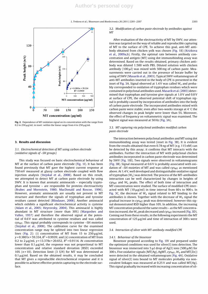
Fig. 2. Dependence of MT oxidation signal on its concentration with the range from0.2 to 250 �g/ml; in inset: within the linear range from 4 to 250 �g/ml.
3. Results and discussion
3.1. Electrochemical detection of MT using carbon electrode(oxidative signals of –SH groups)
This study was focused on basic electrochemical behaviour ofMT at the surface of carbon paste electrode (Fig. 1I). It has beenfound previously that MT gave the highest current response at750 mV measured at glassy carbon electrode coupled with flowinjection analysis (Stejskal et al., 2008). Based on this result,we attempted to detect MT at carbon paste electrode by usingSWV. It is known that aromatic aminoacids – especially trypto-phan and tyrosine – are responsible for proteins electroactivity(Brabec and Mornstein, 1980; MacDonald and Roscoe, 1996).However, aromatic aminoacids are usually not present in MTstructure and therefore the signals of tryptophan and tyrosineresidues cannot detected (Blindauer, 2008). Another aminoacidwhich exhibits a significant electrochemical activity is cysteine(Adam et al., 2005; Heyrovsky, 2004). This aminoacid is highlyabundant in MT structure (more than 30%) (Margoshes andVallee, 1957) and therefore the observed signal at the poten-tial of 0.6 V was attributed to cysteine residues and was calledcysMT. This signal probably corresponds to cysteine –SH moieties(Stejskal et al., 2008). The calibration curve for the examinedconcentration range may be splitted into two linear regressionlines (Fig. 2): (i) concentration of MT from 15 to 250 �g/ml,y = 0.3801x + 59.334, R2 = 0.9939; and (ii) concentration of MT from0.2 to 2 �g/ml, y = 13.378x + 20.652, R2 = 0.9114. At concentrationlower than 0.1 �g/ml, the response was not proportional to MTconcentration and relative standard deviation (RSD) exceeded10% (n = 10). Detection limit (3 S/N) for MT was determined as0.1 �g/ml. Based on the obtained results, it may be concludedthat MT gives a reproducible electrochemical response and it ispossible to achieve effective protein capturing to the electrode sur-face.
3.2. Modification of carbon paste electrode by antibodies againstMT
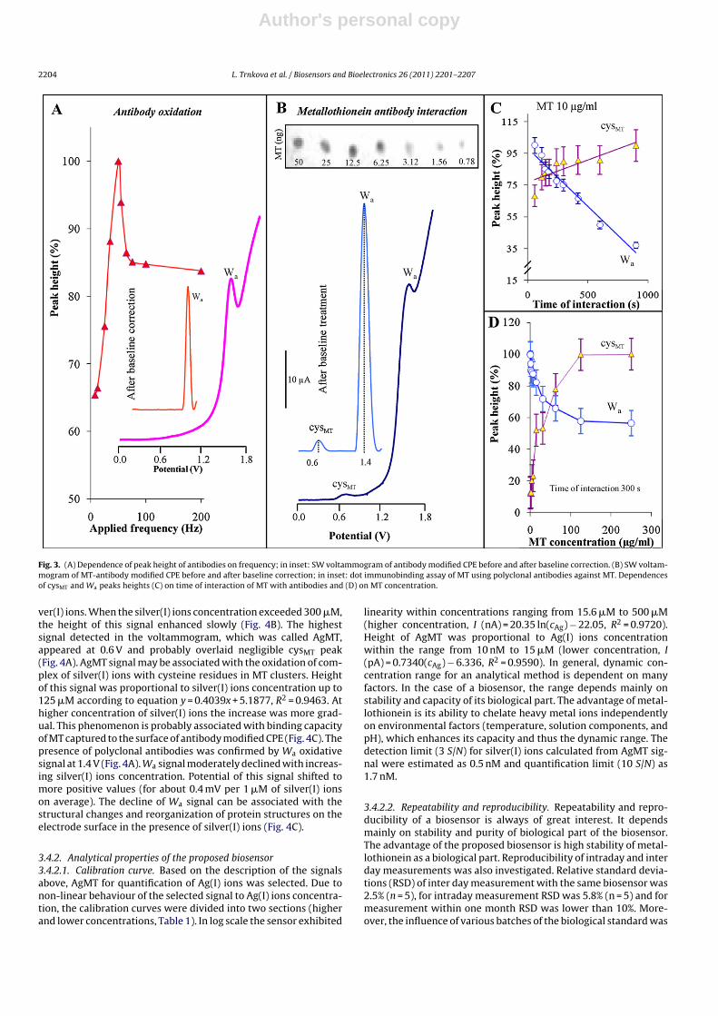
After evaluation of the electroactivity of MT by SWV, our atten-tion was targeted on the way of reliable and reproducible capturingof MT to the surface of CPE. To achieve this goal, anti-MT anti-body obtained from chicken yolk was chosen (Fig. 1II) (Krizkovaet al., 2009a,b). Firstly, the optimal rate between antibody con-centration and antigen (MT) using dot immunobinding assay wasdetermined. Based on the results obtained, primary chicken anti-body was diluted 1:500 with PBS. Diluted solution with chickenantibody (100 �l) was mixed with 500 mg of carbon paste. Mea-surements were carried out in the presence of borate buffer byusing of SWV (Masarik et al., 2003). Typical SWV voltammogram ofanti-MT antibodies inserted in the body of CPE is presented in theinset of Fig. 3A. Signal observed at 1.4 V was called Wa and proba-bly corresponded to oxidation of tryptophan residues which werecontained in polyclonal antibodies used. Masarik et al. (2003) deter-mined that tryptophan and tyrosine give signals at 1.0 V and 0.8 Vat surface of CPE, the observed potential shift of tryptophan sig-nal is probably caused by incorporation of antibodies into the bodyof carbon paste electrode. The incorporated antibodies mixed withcarbon paste were stable; even after two-weeks storage at 4 ◦C theobserved changes in peak height were lower than 5%. Moreover,the effect of frequency on voltammetric signal was examined. Thehighest signal was measured at 50 Hz (Fig. 3A).
3.3. MT capturing via polyclonal antibodies modified carbonpaste electrode
The interaction between polyclonal antibodies and MT using dotimmunobinding assay was tested (inset in Fig. 3B). It is evidentfrom the results obtained that even 0.78 ng of MT (e.g. 115 nM) canbe detected by this assay; it confirms that MT interacts with theantibodies. Further the interaction of MT with polyclonal chickenantibodies incorporated in carbon paste electrode was determinedby SWV (Fig. 1III). Two signals were observed in voltammograms(Fig. 3B). Signal measured at 0.6 V is probably associated with oxi-dation of –SH moieties of MT and was called cysMT as mentionedabove. At 1.4 V, well developed and distinguishable oxidative signalof tryptophan (Wa) was detected. The process of the MT-antibodiesinteraction can be well characterized by changes in heights ofcysMT and Wa peaks. The effect of both time of interaction andMT concentration were studied. The surface of modified CPE inter-acted with MT (10 �g/ml) in time interval from 60 s to 900 s. InFig. 3C, the decrease of Wa signal related to MT binding to theantibodies is shown. Together with the decrease of Wa signal thegradual increase in cysMT peak was determined; however this sig-nal demonstrated RSD higher than 10%. In addition, the increasingMT concentration produced the same results – as the MT concentra-tion increased, the Wa peak decreased and cysMT increased (Fig. 3D).Coming out from these results, in the following experiments the MTconcentration of 125 �g/ml and time of interaction of 300 s wereused.
3.4. Interaction of silver with MT-antibody-modified CPE
3.4.1. Behaviour of the biosensorBiosensor proposed according to Fig. 1IV and prepared under
the optimized conditions was used for silver(I) ions detection. Thebiosensor was immersed into 5 �l drop of Ag(I) ions (500 �M) for300 s. Five oxidative signals (MT(Ag), AgMT, cysMT, Wa and MT(Zn))were detected in the obtained voltammogram (Fig. 4A). Oxidativesignal of silver(I) ions bound to MT molecules probably via non-covalent linkages was measured at 0.25 V and was called MT(Ag).This signal gradually increased with increasing concentration of sil-
Author's personal copy
2204 L. Trnkova et al. / Biosensors and Bioelectronics 26 (2011) 2201–2207
Fig. 3. (A) Dependence of peak height of antibodies on frequency; in inset: SW voltammogram of antibody modified CPE before and after baseline correction. (B) SW voltam-mogram of MT-antibody modified CPE before and after baseline correction; in inset: dot immunobinding assay of MT using polyclonal antibodies against MT. Dependencesof cysMT and Wa peaks heights (C) on time of interaction of MT with antibodies and (D) on MT concentration.
ver(I) ions. When the silver(I) ions concentration exceeded 300 �M,the height of this signal enhanced slowly (Fig. 4B). The highestsignal detected in the voltammogram, which was called AgMT,appeared at 0.6 V and probably overlaid negligible cysMT peak(Fig. 4A). AgMT signal may be associated with the oxidation of com-plex of silver(I) ions with cysteine residues in MT clusters. Heightof this signal was proportional to silver(I) ions concentration up to125 �M according to equation y = 0.4039x + 5.1877, R2 = 0.9463. Athigher concentration of silver(I) ions the increase was more grad-ual. This phenomenon is probably associated with binding capacityof MT captured to the surface of antibody modified CPE (Fig. 4C). Thepresence of polyclonal antibodies was confirmed by Wa oxidativesignal at 1.4 V (Fig. 4A). Wa signal moderately declined with increas-ing silver(I) ions concentration. Potential of this signal shifted tomore positive values (for about 0.4 mV per 1 �M of silver(I) ionson average). The decline of Wa signal can be associated with thestructural changes and reorganization of protein structures on theelectrode surface in the presence of silver(I) ions (Fig. 4C).
3.4.2. Analytical properties of the proposed biosensor3.4.2.1. Calibration curve. Based on the description of the signalsabove, AgMT for quantification of Ag(I) ions was selected. Due tonon-linear behaviour of the selected signal to Ag(I) ions concentra-tion, the calibration curves were divided into two sections (higherand lower concentrations, Table 1). In log scale the sensor exhibited
linearity within concentrations ranging from 15.6 �M to 500 �M(higher concentration, I (nA) = 20.35 ln(cAg) − 22.05, R2 = 0.9720).Height of AgMT was proportional to Ag(I) ions concentrationwithin the range from 10 nM to 15 �M (lower concentration, I(pA) = 0.7340(cAg) − 6.336, R2 = 0.9590). In general, dynamic con-centration range for an analytical method is dependent on manyfactors. In the case of a biosensor, the range depends mainly onstability and capacity of its biological part. The advantage of metal-lothionein is its ability to chelate heavy metal ions independentlyon environmental factors (temperature, solution components, andpH), which enhances its capacity and thus the dynamic range. Thedetection limit (3 S/N) for silver(I) ions calculated from AgMT sig-nal were estimated as 0.5 nM and quantification limit (10 S/N) as1.7 nM.
3.4.2.2. Repeatability and reproducibility. Repeatability and repro-ducibility of a biosensor is always of great interest. It dependsmainly on stability and purity of biological part of the biosensor.The advantage of the proposed biosensor is high stability of metal-lothionein as a biological part. Reproducibility of intraday and interday measurements was also investigated. Relative standard devia-tions (RSD) of inter day measurement with the same biosensor was2.5% (n = 5), for intraday measurement RSD was 5.8% (n = 5) and formeasurement within one month RSD was lower than 10%. More-over, the influence of various batches of the biological standard was

Author's personal copy
L. Trnkova et al. / Biosensors and Bioelectronics 26 (2011) 2201–2207 2205
Fig. 4. (A) Typical SW voltammogram of MT-antibody biosensor after interaction with silver(I) ions. Dependences of (B) MT(Ag) and (C) AgMT and Wa peak height on silver(I)ions concentration. MT concentration: 125 �g/ml and time of interaction: 300 s. In inset in (B): the effect of 10 �M (white column) and 100 �M (black column) Cu(II), Hg(II),Pt(II), Cd(II), Zn(II), Fe(II) and Ni(II) ions on height of AgMT signal.
also tested. Data measured by biosensor prepared with standardobtained from different batches have relatively low RSD (less than10%). In addition, RSD between newly prepared biosensors wasexamined. RSD of inter day measurement was 7.5% (n = 5, numberof tested biosensors = 5). Besides repeatability and reproducibility,storage capacity was also investigated and was estimated 10 days.
3.4.2.3. Recovery. The proposed biosensor was tested by detectionof silver(I) ions spiked in various water samples (from very puredistilled water to rainwater) according to methodology publishedpreviously (Bugianesi et al., 2000; Causon, 1997). Changes in AgMTsignals were determined in raw sample. In tested water samplessilver(I) ions were not detected directly. The sample of water withspiked silver(I) ions interacted with biosensor for 300 s. The signalrecovery in water samples without impurities (Milli Q and distilledwater) was very good and varied between 101 and 104% (Table 1).In the case of tap water, rainwater and water Ponávka stream thesignal was influenced by sample matrix, which resulted in higherC.V. (from 7.1 to 14%) and lower recovery (from 74 to 93%).
3.4.2.4. Interferences. Due to the fact that biological part of biosen-sor was protein with the ability to bind almost all metal ions,the other metal ions (Cu(II), Hg(II), Pt(II), Cd(II), Zn(II), Fe(II), andNi(II)) were tested as interferences. The biosensor was immersedinto 5 �l drop of Ag(I) ions (10 �M) for 300 s and voltammogramwas measured. Then, the biosensor was immersed into 5 �l drop ofparticular metal ion (10 �M or 100 �M) for 300 s and voltammo-gram was measured again. The changes in height of AgMT signalwere measured. The effect of by the abovementioned metals on theheight of AgMT signal is shown in inset of Fig. 4B. Not only the same,but even 10 times higher concentration of other metal ions (but notthe Hg(II) ones) did not have considerable effect on AgMT signal.Ten times higher concentration of these ions caused more than 15%decrease in AgMT signal. This phenomenon may be associated withthe fact that Hg(II) ions have slightly higher affinity to MT comparedto Ag(I) ions accordingly to Hg(II) > Ag(I)–Cu(I) > Cd(II) > Zn(II). Nev-ertheless, the signals of single heavy metals present in MT structurecan be distinguished according to the peaks position which cor-responds to the formation of particular MT-heavy metal complex
Table 1Recovery of silver ions (AgNO3) measured in the presence of different types of waters (n = 5).
Compound of interest Sample matrix Filtrate (nA)a Spiking (nA)a,c Filtrate + spiking (nA)a Recovery (%)
Silver ions
Milli Q water
ndb 7.5 ± 0.2 (2.7)
7.8 ± 0.2 (2.6) 104Distilled water 7.6 ± 0.2 (2.6) 101Tap water 5.6 ± 0.8 (14) 74Ponávka stream 6.9 ± 0.5 (7.2) 92Rainwater 7.0 ± 0.5 (7.1) 93
a Silver ions current response; expressed as mean ± S.D. (C.V.%).b Not detected.c Silver ions current response (100 �mol/dm3); expressed as mean ± S.D. (C.V.%).

Author's personal copy
2206 L. Trnkova et al. / Biosensors and Bioelectronics 26 (2011) 2201–2207
(Adam et al., 2007b, 2005; Fabrik et al., 2009; Supalkova et al., 2008;Wu and Lin, 2004).
3.4.3. Comparison of the proposed biosensor with othertechniques
Electrochemistry approach seems to be very suitable for thedetermination of silver(I) ions. Various electrochemical meth-ods and electrode types can be used; the procedures differ inarrangement, electrodes construction, detection limits achieved,and influence by sample matrix (Table 1). The most often used areion-selective electrodes, with sensitivity comparable to biosensorproposed in this paper, but the interference with sample matrixis often problematic. Nevertheless, Gupta et al. (2009) used an ion-selective electrode for the determination of silver in blood samples.Using of carbon electrodes allows detection of a very low analyteconcentration and their application for real samples is more com-mon. Since the subnanomolar detection limits can be achieved, itcan be used for monitoring of trace amounts of silver in the envi-ronment (Javanbakht et al., 2009; Svancara et al., 1996). Enzymebiosensors exhibit a comparable detection limit in orders of tenthof micromoles per one litre, however the specificity of enzymesinhibition by heavy metals is of great concern (Verma and Singh,2005; Vopalensky et al., 2007).
Metallothionein (MT) can be used as a protein component ofbiosensors for heavy metals (Adam et al., 2007b; Bin et al., 2009; Fuet al., 2008; Varriale et al., 2007). Gonzalez-Bellavista et al. (2009)proposed a metallothionein-based silver biosensor where MT wasa part of ion-selective electrode with detection limit in orders of10−5 M in model solutions. Very good adsorption of MT to the sur-face of gold and hanging mercury drop (HMDE) electrodes has beenreported in numerous papers (Adam et al., 2005; Ju and Leech,2000; Petrlova et al., 2006a, 2007b; Trnkova et al., 2002). Thereforeadsorption of MT was used to introduce a metallothionein-basedsilver biosensor based on HMDE with 500 nM detection limit ofAg(I) ions (Krizkova et al., 2009c). In comparison to the bare car-bon paste electrode the detection limit of the sensor was ten timeslowered making it comparable to modified carbon paste electrodes(Krizkova et al., 2009d; Labuda and Vanickova, 1993; Mikelova etal., 2007; Wang et al., 2009; Ye and Khoo, 1997). In this study,1000-fold lowering of the detection limit compared to HMDE wasreached. This phenomenon may be associated with more effec-tive immune-based-capturing of MT onto the electrode surface ascompared to simple adsorption. Besides detection limit, the otheradvantage of the proposed biosensor is possibility of the miniatur-ization of carbon electrodes for in situ analysis.
4. Conclusions
Metallothioneins, low molecular mass proteins rich in cysteine,play an important role in the processes of heavy metals ionsmetabolism. Due to their unique physicochemical properties theyare able to bind heavy metals with high affinity (Zhang et al., 1997).This feature was used to suggest a simple biosensor based on immo-bilization of MT to the surface of carbon paste electrode via chickenanti-MT antibodies. The outlined biosensor was further successfullyemployed in detection of silver(I) ions. The main advantage of thisbiosensor is its easy miniaturization; carbon nanostructures withimmobilized MT might be used as working electrodes.
Acknowledgements
Financial support from the grants INCHEMBIOL MSM0021622412, MSM 0021630503, BIO-ANAL-MED LC06035, GACR526/07/0674, and GACR 102/08/1546 is highly acknowledged.
References
Adam, V., Fabrik, I., Eckschlager, T., Stiborova, M., Trnkova, L., Kizek, R., 2010. TRAC-Trends Anal. Chem. 29 (5), 409–418.
Adam, V., Hanustiak, P., Krizkova, S., Beklova, M., Zehnalek, J., Trnkova, L., Horna, A.,Sures, B., Kizek, R., 2007a. Electroanalysis 19 (18), 1909–1914.
Adam, V., Krizkova, S., Zitka, O., Trnkova, L., Petrlova, J., Beklova, M., Kizek, R., 2007b.Electroanalysis 19 (2–3), 339–347.
Adam, V., Petrlova, J., Potesil, D., Zehnalek, J., Sures, B., Trnkova, L., Jelen, F., Kizek, R.,2005. Electroanalysis 17 (18), 1649–1657.
Bielmyer, G.K., Brix, K.V., Grosell, A., 2008. Aquat. Toxicol. 87 (2), 81–87.Bin, Y.N., Gao, Y., Xiang, J., Ren, B., 2009. Surf. Interface Anal. 41 (10), 834–838.Blindauer, C.A., 2008. J. Inorg. Biochem. 102 (3), 507–521.Brabec, V., Mornstein, V., 1980. Biophys. Chem. 12 (2), 159–165.Bugianesi, R., Serafini, M., Simone, F., Wu, D.Y., Meydani, S., Ferro-Luzzi, A., Azzini,
E., Maiani, G., 2000. Anal. Biochem. 284 (2), 296–300.Causon, R., 1997. J. Chromatogr. B 689 (1), 175–180.Cosnier, S., 1999. Biosens. Bioelectron. 14 (5), 443–456.Eckschlager, T., Adam, V., Hrabeta, J., Figova, K., Kizek, R., 2009. Curr. Protein Pept.
Sci. 10 (4), 360–375.Fabrik, I., Kukacka, J., Baloun, J., Sotornik, I., Adam, V., Prusa, R., Vajtr, D., Babula, P.,
Kizek, R., 2009. Electroanalysis 21 (3–5), 650–656.Fu, Y., Xu, M.T., Li, X., Du, M., Wang, J.X., Zhou, F.M., 2008. Electroanalysis 20 (8),
888–893.Gonzalez-Bellavista, A., Atrian, S., Munoz, M., Capdevila, M., Fabregas, E., 2009.
Talanta 77 (4), 1528–1533.Gorsuch, J.W., Klaine, S.J., 1998. Environ. Toxicol. Chem. 17 (4), 537–538.Guo, S.X., Khoo, S.B., 1999. Electroanalysis 11 (12), 891–898.Gupta, V.K., Pal, M.K., Singh, A.K., 2009. Anal. Chim. Acta 631 (2), 161–169.Hamer, D.H., 1986. Annu. Rev. Biochem. 55, 913–951.Heyrovsky, M., 2004. Electroanalysis 16 (13–14), 1067–1073.Hodek, P., Koblas, T., Rydlova, H., Kubickova, B., Sulc, M., Hudecek, J., Stiborova, M.,
2004. Collect. Czech. Chem. Commun. 69 (3), 659–673.Hogstrand, C., Galvez, F., Wood, C.M., 1996. Environ. Toxicol. Chem. 15 (7),
1102–1108.Javanbakht, M., Divsar, F., Badiei, A., Fatollahi, F., Khaniani, Y., Ganjali, M.R.,
Norouzi, P., Chaloosi, M., Ziarani, G.M., 2009. Electrochim. Acta 54 (23), 5381–5386.
Ju, H.X., Leech, D., 2000. J. Electroanal. Chem. 484 (2), 150–156.Kizek, R., Masarik, M., Kramer, K.J., Potesil, D., Bailey, M., Howard, J.A., Klejdus, B.,
Mikelova, R., Adam, V., Trnkova, L., Jelen, F., 2005. Anal. Bioanal. Chem. 381 (6),1167–1178.
Krizkova, S., Adam, V., Eckschlager, T., Kizek, R., 2009a. Electrophoresis 30 (21),3726–3735.
Krizkova, S., Adam, V., Petrlova, J., Zitka, O., Stejskal, K., Zehnalek, J., Sures, B., Trnkova,L., Beklova, M., Kizek, R., 2007. Electroanalysis 19 (2–3), 331–338.
Krizkova, S., Blahova, P., Nakielna, J., Fabrik, I., Adam, V., Eckschlager, T., Beklova,M., Svobodova, Z., Horak, V., Kizek, R., 2009b. Electroanalysis 21 (23),2575–2583.
Krizkova, S., Huska, D., Beklova, M., Hubalek, J., Adam, V., Trnkova, L., Kizek, R., 2009c.Environ. Toxicol. Chem. 29 (3), 492–496.
Krizkova, S., Krystofova, O., Trnkova, L., Hubalek, J., Adam, V., Beklova, M., Horna, A.,Havel, L., Kizek, R., 2009d. Sensors 9 (9), 6934–6950.
Labuda, J., Vanickova, M., 1993. Electroanalysis 5 (2), 141–144.Li, Y.X., Wang, P., Wang, L., Lin, X.Q., 2007. Biosens. Bioelectron. 22 (12), 3120–
3125.Liu, A.L., Zhang, S.B., Chen, W., Lin, X.H., Xia, X.H., 2008. Biosens. Bioelectron. 23 (10),
1488–1495.Long, G.L., Winefordner, J.D., 1983. Anal. Chem. 55 (7), A712–A724.MacDonald, S.M., Roscoe, S.G., 1996. J. Colloid Interface Sci. 184 (2), 449–455.Margoshes, M., Vallee, B.L., 1957. J. Am. Chem. Soc. 79 (17), 4813–4814.Masarik, M., Kizek, R., Kramer, K.J., Billova, S., Brazdova, M., Vacek, J., Bailey, M., Jelen,
F., Howard, J.A., 2003. Anal. Chem. 75 (11), 2663–2669.Mikelova, R., Baloun, J., Petrlova, J., Adam, V., Havel, L., Petrek, H., Horna, A., Kizek,
R., 2007. Bioelectrochemistry 70 (2), 508–518.Petrlova, J., Krizkova, S., Supalkova, V., Masarik, M., Adam, V., Havel, L., Kramer, K.J.,
Kizek, R., 2007a. Plant Soil Environ. 53 (8), 345–349.Petrlova, J., Krizkova, S., Zitka, O., Hubalek, J., Prusa, R., Adam, V., Wang, J., Beklova,
M., Sures, B., Kizek, R., 2007b. Sens. Actuator B-Chem. 127 (1), 112–119.Petrlova, J., Masarik, M., Potesil, D., Adam, V., Trnkova, L., Kizek, R., 2007c. Electro-
analysis 19 (11), 1177–1182.Petrlova, J., Potesil, D., Mikelova, R., Blastik, O., Adam, V., Trnkova, L., Jelen, F., Prusa,
R., Kukacka, J., Kizek, R., 2006a. Electrochim. Acta 51 (24), 5112–5119.Petrlova, J., Potesil, D., Zehnalek, J., Sures, B., Adam, V., Trnkova, L., Kizek, R., 2006b.
Electrochim. Acta 51 (24), 5169–5173.Schildkraut, D.E., Dao, P.T., Twist, J.P., Davis, A.T., Robillard, K.A., 1998. Environ.
Toxicol. Chem. 17 (4), 642–649.Sivanesan, A., John, S.A., 2007. Biosens. Bioelectron. 23 (5), 708–713.Stejskal, K., Krizkova, S., Adam, V., Sures, B., Trnkova, L., Zehnalek, J., Hubalek, J.,
Beklova, M., Hanustiak, P., Svobodova, Z., Horna, A., Kizek, R., 2008. IEEE Sens. J.8 (9), 1578–1585.
Supalkova, V., Beklova, M., Baloun, J., Singer, C., Sures, B., Adam, V., Huska, D., Pikula,J., Rauscherova, L., Havel, L., Zehnalek, J., Kizek, R., 2008. Bioelectrochemistry 72(1), 59–65.
Svancara, I., Kalcher, K., Diewald, W., Vytras, K., 1996. Electroanalysis 8 (4),336–342.

Author's personal copy
L. Trnkova et al. / Biosensors and Bioelectronics 26 (2011) 2201–2207 2207
Svancara, I., Vytras, K., Barek, J., Zima, J., 2001. Crit. Rev. Anal. Chem. 31 (4), 311–345.Szymanski, M., Turner, A.P.F., Porter, R., 2010. Electroanalysis 22 (2), 191–198.Trnkova, L., Kizek, R., Vacek, J., 2002. Bioelectrochemistry 56 (1–2), 57–61.Varriale, A., Staiano, M., Rossi, M., D’Auria, S., 2007. Anal. Chem. 79 (15), 5760–5762.Verma, N., Singh, M., 2005. Biometals 18 (2), 121–129.Vopalensky, P., Ruml, T., Kotrba, P., 2007. Chem. Listy 101 (6), 468–479.
Wang, F., Liu, Q.Y., Wu, Y.J., Ye, B.X., 2009. J. Electroanal. Chem. 630 (1–2), 49–54.Wood, C.M., Hogstrand, C., Galvez, F., Munger, R.S., 1996. Aquat. Toxicol. 35 (2),
93–109.Wu, C.M., Lin, L.Y., 2004. Biosens. Bioelectron. 20 (4), 864–871.Ye, R.D., Khoo, S.B., 1997. Electroanalysis 9 (6), 481–489.Zhang, B.L., Sun, W.Y., Tang, W.X., 1997. J. Inorg. Biochem. 65 (4), 295–298.