Embed Size (px)
Citation preview

This article appeared in a journal published by Elsevier. The attachedcopy is furnished to the author for internal non-commercial researchand education use, including for instruction at the authors institution
and sharing with colleagues.
Other uses, including reproduction and distribution, or selling orlicensing copies, or posting to personal, institutional or third party
websites are prohibited.
In most cases authors are permitted to post their version of thearticle (e.g. in Word or Tex form) to their personal website orinstitutional repository. Authors requiring further information
regarding Elsevier’s archiving and manuscript policies areencouraged to visit:
http://www.elsevier.com/authorsrights

Author's personal copy
Complementary cross-section based protocol of investigation ofpolychrome samples of a 16th century Moravian Sculpture by optical,vibrational and mass spectrometric techniques
Stepanka Kuckova a,b,⁎, Irina Crina Anca Sandu c,1, Michaela Crhova a, Radovan Hynek a,Igor Fogas d,2, Vania Solange Muralha e, Andrei Victor Sandu f,g
a Department of Biochemistry and Microbiology, Institute of Chemical Technology, Technická 3, 166 28 Prague 6, Czech Republicb Department of Chemistry and Chemical Education, Charles University, M.D. Rettigové 4, 116 39 Prague 1, Czech Republicc REQUIMTE and Department of Conservation and Restoration, Faculty of Sciences and Technology (FCT), Nova University of Lisbon (UNL), 2829-516 Caparica, Portugald Moravian Gallery Brno, Husova 18, 662 26 Brno, Czech Republice Research Unit VICARTE: Vidro e Cerâmica para as Artes, Faculty of Sciences and Technology, NOVA University of Lisbon, Portugalf Gheorghe Asachi Technical University of Iasi, Faculty of Materials Science and Engineering, Iasi, Romaniag Romanian Inventors Forum (FIR), Str. Sf. P. Movila 3, L11, III/3, RO 700089 Iasi, Romania
a b s t r a c ta r t i c l e i n f o
Article history:Received 10 June 2013Received in revised form 1 July 2013Accepted 1 July 2013Available online 10 July 2013
Keywords:Cross-sectionBinders and pigmentsMALDI-TOF mass spectrometryNano-LC-ESI-Q-TOF mass spectrometryMedieval polychrome sculptureCoccoliths
On the occasion of its restoration treatments, the rare medieval polychrome sculpture entitled “The Mourn-ing of Jesus Christ” (16th century, belonging to the Moravian Gallery in Brno, Czech Republic) was subject to adetailed analytical investigation in order to identify the organic and inorganic materials present in the poly-chrome layers and to map their distribution in the paint structure. This knowledge is extremely useful espe-cially for art objects that suffered reconstructions and/or over paintings or other interventions and can be ofhelp to the restorers in deciding the best conditions and materials for the treatment. In this case, the identi-fication of all components was very important in terms of the assessment of the original painting techniquesand discrimination of the presence of over painting layers, being complemented by the study of the construc-tion process and of the history of the artwork.Until recently, the identification of proteinaceous binders contained in small samples taken from poly-chrome or painted works of art was very complicated and nearly impossible in the case of cross-sectionanalyses. Therefore, the novelty of this paper is the complementary applications of several analyticaltechniques, such as optical microscopy (OM), nano-LC-ESI-Q-TOF and MALDI-TOF mass spectrometry,microRaman spectroscopy and scanning electronmicroscopy coupled with energy dispersive X-ray analysis(SEM-EDX) directly on cross-sections. This approach is especially useful for this case as only few sampleswere available for binder and pigment identification and it allowed the preservation of the samples ascross-sections for further analyses.
© 2013 Elsevier B.V. All rights reserved.
1. Research aims
The research results presented in this paper are part of a largerstudy of materials and techniques used in a Czech Medievalpolychrome sculpture illustrating “The Mourning of Jesus Christ”dated around 1500 (Moravian Gallery in Brno, Czech Republic). This
contribution is the second part of a paper already published byKuckova, Sandu et al. [1], where the simultaneous identification andlocalization of proteinaceous binders on cross-section by MALDI-TOF MS (Matrix-Assisted Laser Desorption/Ionisation-Time of FlightMass Spectrometry) was described.
The two objectives of the research presented here were:
– to compare two mass spectrometric techniques (MALDI-TOF andnano-LC-ESI-Q-TOF) applied for proteinaceous binder's identifica-tion on cross-sections;
– to identify and map the organic and inorganic components of thepolychrome layers in order to characterize the polychrome mate-rials and techniques of the sculpture and to confirm the art workprovenience.
Microchemical Journal 110 (2013) 538–544
⁎ Corresponding author at: Department of Biochemistry and Microbiology, Instituteof Chemical Technology, Technická 3, 166 28 Prague 6, Czech Republic. Tel.: +420 22044 4339.
E-mail addresses: [email protected] (S. Kuckova), [email protected] (I.C.A. Sandu),[email protected] (M. Crhova), [email protected] (R. Hynek), [email protected](I. Fogas), [email protected] (V.S. Muralha), [email protected] (A.V. Sandu).
1 Tel.: +351 212948322/11305.2 Tel.: +420 532169161.
0026-265X/$ – see front matter © 2013 Elsevier B.V. All rights reserved.http://dx.doi.org/10.1016/j.microc.2013.07.002
Contents lists available at ScienceDirect
Microchemical Journal
j ourna l homepage: www.e lsev ie r .com/ locate /mic roc
Author's personal copy
2. Introduction
Currently, most of advanced analytical methods (e.g. GC-MS, LC-MS,opticalmethods), which are used for the identification of proteinaceousbinders in works of art, are able to identify and distinguish at least egg,animal glue and milk proteins [4–8]. Also, though only in rare cases,some immunochemical methods (ELISA, IFM, nano-SERS antibodies,chemiluminescence) staining tests and fluorescence microscopy[9,10], and mass spectrometric methods (MALDI-TOF) [8,11–15]were reported in studies for the localization and simultaneousmappingof various protein-based materials in individual layers of samples thathave been prepared to form polished cross-sections [14,15].
Both mass spectrometric methods MALDI-TOF and nano-LC-ESI-Q-TOF are working with specific peptides that are obtained afterenzymatic cleavage of proteins contained in artwork's samples usingtrypsin. Trypsin cleaves peptide bonds only behind two basic aminoacids — lysine and arginine. In the case of MALDI-TOF MS the uniquepeptidemixture (fingerprint of binder) is analysed by amass spectrom-eter and then compared and assigned to a binder from a reference data-base of protein binders [16]. Nano-LC-ESI-Q-TOF mass spectrometrydetermines the order of amino acids in the peptides and the sequencescompared to the publicly available databases of proteins. Nowadays thehighly impacted proteomic journals usually demand two peptides for areliable determination of individual proteins.
As far as the identification of inorganic pigments is concerned,there are many dedicated analytical techniques as the pigments andinert materials used in paint or polychrome layers represent theirmajor component and their identification could serve as a tool for at-tribution or dating of the artwork [17,18]. The inorganic constituentsare identified mainly using scanning electron microscopy coupledwith energy dispersive X-ray spectrometry (SEM-EDX) [19,20],Raman spectroscopy [19], classical Fourier Transform Infrared spec-troscopy (FTIR) [19–21] and infrared microspectroscopy (microFTIR)using a synchrotron source which allows the collection of high signal-to-noise ratio infrared spectra with diffraction-limited spatial res-olution across the entire spectral range [22]. X-ray spectroscopy(XRF) [23] has also become established as one of the most impor-tant tools for non-destructively identifying specific elements forpigment and inert material identification. The metals could bealso determined using graphite furnace atomic absorption spec-troscopy (GFAAS) and inductively coupled plasma mass spec-trometry (ICP-MS), where the previous extraction of pigments isnecessary [24]. X-ray diffraction [21], especially X-ray micro-diffraction [25], is useful in material research of painting layers withcomplex stratigraphy and composition and it could distinguish inorganicphases of different natural provenances and reveal their degradationproducts.
In this work the inorganic and protein-based constituents of thepaint layers of a 16th century polychrome sculpture are analysedusing a complementary protocol of investigation, based on the use ofoptical microscopy (OM), nano-LC-ESI-Q-TOF and MALDI-TOF massspectrometry,microRaman spectroscopy and scanning electronmicros-copy coupledwith energy dispersive X-ray analysis (SEM-EDX) directlyon cross-sections.
3. Experimental
3.1. Sampling
The sculpture (Fig. 1), owned by the Convent of Minorits in Brno(Czech Republic), is a complex work of figurative woodcarving(illustrating the theme of “TheMourning of Jesus Christ”, comprising11 male and female characters standing around the laid body of Christafter the Descent from the Cross) shaped in high relief (97 cm ×136 cm × 29 cm), made of four massive lime wood blocks togetherwith several supplements dating around the year 1520. In Fig. 1 the
image of the entire sculpture is given and for each statue a letter wasused to label the represented character: A: Jesus Christ; B: Saint-Anna;C: Saint-John the Baptist; D: Mary Magdalene; E: Mary of Kleofas;F: Saint-Mary; G: Mary Salome; H: Joseph from Arimathea; I: Nikodem;J: member of Synedrium; K: Pharisee (Simon of Cyrene); L: member ofSynedrium.
A suspicion of a possible relationship between the sculpture (Fig. 1)and a 1500 printed engraving of a woodcarving work (Fig. 2) made byAlbrecht Dürer arose due to the stylistic and compositional characteris-tics of both [2,3].
The polychrome's stratigraphy is made of many paint layers (fromthe Gothic, Renaissance, Baroque and 19th and 20th centuries) appliedover awhite ground layer. Unfortunately, the sculpturewas damaged inlarge areas, because of over-paintings probably made during the 19thand 20th centuries and other interventions leading to missing partssuch as a finger or incarnate areas, extensive abrasions and cracks,which were appropriately used for sampling. As shown in Fig. 1, atotal of 12 samples was taken from the sculpture (M1/M1a to M11)and embedded as cross-sections [1].
3.2. Materials and analytical protocol
3.2.1. Materials and reagentsThe NH4HCO3 and acetonitrile were bought from Lachema Brno
(Brno, CzechRepublic). Trypsin (TPCK) comes fromPromega Corporation(Madison,WI, USA). The reverse phase ZipTipwas bought fromMilliporeCorporation (Bedford, MA, USA), and 2,5-dihydroxybenzoic acid (DHB)and trifluoracetic acid were bought from Sigma (Saint-Louis, MO, USA).Mecaprex 2S polyester embedding resin and peroxide of methyl ethylketone hardener come from Presi (Grenoble, France).
3.2.2. Cross-sections and OM observationAfter curing of the polyester resin, the blocks were cut and
polished to reveal the paint/ground composite in cross-section. Thecross-sections were dry polished with successively finer grades ofmicromesh abrasive cloths (600, 800, 1200 and 4000 mesh). Feltwas used for the final polishing. Water or other aqueous-basedliquids are not used during polishing since they could dissolve theproteinaceous component in the samples. The cross-sections wereobserved at different magnifications (from 50× to 500×) using anAxioplan Zeiss 2 imaging binocular microscope and the images wereacquired using a Nikon DXM1200F digital camera, coupled to the mi-croscope (provided with a mercury lamp HBO100 and a halogen lampHAL100). Visual light observations (illumination position for darkfield observation, abbreviated as f2) were performed in reflectiongeometry. Images were captured and treated using Nikon ACT-1software.
3.2.3. Protein digestion and purificationOn the surface of the cross-sections,10 μL of 50 mM NH4HCO3
containing approximately 10 μg/mL of trypsin was applied and left toreact at room temperature for two hours. After the trypsin digestion,the solutions were taken from the surfaces and purified on a reversephase ZipTip. After equilibrating, binding and washing steps, targetcompounds were desorbed from the stationary phase [26]. Thesolutions were consequently used for analyses by MALDI-TOF MS andnano-LC-ESI-Q-TOF (3.2.4 and 3.2.5).
3.2.4. Matrix-assisted laser desorption/ionisation-time of flight (MALDI-TOF)
An aliquot of the elution solution containing peptides (2 μL) wasmixed with 4 μL of 2,5-dihydroxybenzoic acid (DHB) solution — 18 mgof DHB in 1 mL of mixture of acetonitrile/0.1% trifluoracetic acid inwater (1/2 [v/v]). The part of the resulting mixture (2.8 μL) was fortwo times spotted on the stainless steel MALDI target and dried in air.Mass spectra were acquired by a Bruker-Daltonics Biflex IV MALDI-
539S. Kuckova et al. / Microchemical Journal 110 (2013) 538–544

Author's personal copy
TOF mass spectrometer equipped with a standard nitrogen laser(337 nm) in positive reflector mode with a mass accuracy of 0.2 Da;at least 200 laser shots were collected for each spectrum. The spectra
were analysed using XMASS (Bruker), mMass software [27] and ahomemade database of reference proteinaceous binders [28].
3.2.5. Mass spectrometry nano-LC-ESI-Q-TOFMeasurement was carried out using UHPLC Dionex Ultimate 3000
RSLC nano (Dionex, Germany) connected with a mass spectrometerESI-Q-TOF Maxis Impact (Bruker, Germany). 10 μL of a peptide solu-tion was previously dried and then dissolved in a 97:3:0.1% mixtureof water:acetonitrile:formic acid. Consequently they were loaded ona trap column Acclaim PepMap 100 C18 (100 μm × 2 cm, size ofreverse phase particles = 5 μm, Dionex, Germany) with a flow rateof mobile phase A = 5 μL/min for 5 min. The peptides were elutedfrom the trap column to an analytical column Acclaim PepMap RSLCC18 (75 μm × 250 mm, size of reverse phase particles = 2 μm)using the following gradient: 0 min 3% B, 5 min 3% B, 85 min 50% B,86 min 90% B, 95 min 90% B, 96 min 3% B, 110 min 3% B. Mobilephase A was 0,1% formic acid in water and mobile phase B was 0,1%formic acid in acetonitrile. The flow rate during gradient separationwas set to 0.3 μL/min. Peptides were eluted directly to the ESI source— Captive spray (Bruker Daltonics, Germany). Measurement wascarried out in positive ion mode with precursor selection in therange of 400–2200 Da; from each MS spectrum, up to ten precursorswere selected for fragmentation.
Peak lists were extracted from raw data by Data Analysis (BrukerDaltonics, Germany). Proteins were identified using Mascot version2.2.04 (Matrix Science, UK) by searching protein database Uniprotversion 20110-12. Parameters for database search were set as follows:Oxidation of methionine and hydroxylation of proline as variablemodifications, tolerance of 50 ppm in MS mode and 0.05 Da in MS/MS mode.
3.2.6. microRaman on cross-sectionsThe equipment used was a Labram 300 Jobin Yvon spectrometer,
equipped with a He–Ne laser of 17 mW power operating at 632.8 nmand a solid state laser operating at 532 nm. The laser beamwas focusedwith a 50× or 100× Olympus objective lens. The laser energy wasfiltered up to 10% using a neutral density filter for all analyses. The
Fig. 1. Entire view of the polychrome sculpture with the sampling areas for the 12 samples.
Fig. 2. The printed engraving by Albrecht Dürer (1500) illustrating the Mourning ofChrist scene.
540 S. Kuckova et al. / Microchemical Journal 110 (2013) 538–544

Author's personal copy
attribution of the Raman spectrawasmade using databases of referencematerials reported in the literature [29,30].
3.2.7. SEM-EDX on cross-sectionsA VEGA II LSH scanning electron microscope (TESCAN — Czech
Republic), coupled with an EDX-QUANTAX QX2 (ROENTEC Bruker —Germany) spectrometer was used. The EDX Quantax QX2 uses adetector of third generation Xflash, that does not need cooling withnitrogen and is 10 times faster than a traditional detector based onSi(Li). The SEM images and EDX spectra have been acquired in thefollowing conditions: 20 and 30 kV voltage; 1 × 10⁻³ Pa; workingdistance of 11–20 mm (16.6 mm for EDX) and magnification: 78× to1000×. The cross-sections were covered with a fine layer of graphiteusing a specific “sputter coater”.
4. Results and discussions
The results obtained from SEM-EDX and Raman spectroscopyperformed on embedded fragments of polychrome layers are givenin Table 1.
The mainly identified pigments were those commonly used inmedieval times: lead white, lead red, vermillion, red ochre (hematite),massicot, ultramarine blue, and indigo. The SEM-EDX also identifiedthe presence of Co in the M11 sample (probably attributed to a Co-based pigment, maybe Co blue as Al was also identified in the samearea as the cobalt — Fig. 3c) and of Ba in several samples (M1, M8, M9and M11) suggesting the use of barite (Ba sulphate) probably duringthe intervention of over-painting or puttying (this type of materialbeing in use in modern times as a filler or addition to white pigments).The ground layers aremade of calciumcarbonate (chalk),which containsfragments of shells (coccoliths) indicating a marine sedimentary fossilorigin of the calcite. The presence of coccoliths was already reported byP. Dietemann as part of the ground layers in polychrome sculptures inmedieval times in Germany [31].
Fig. 3 gives a selection of 3 cross-sections on which OM, SEM-EDXand microRaman were performed. The first fragment (M1a — Fig. 3a)was taken from Jesus Christ's finger incarnate and the OM images inVis and UV light show a series of overlapped layers over the ground(the most superficial ones could be attributed to an over-painting).The second case (Fig. 3b) shows a green area of over-painting fromthe knee drapery of S. John, with the optical microscopy unveilingnot more than 5 overlapped layers, each of them of different colour,granulometry and fluorescence pattern. The composition and struc-ture of the layers appear heterogeneous (big grains of differentshapes, inside a more or less darker green matrix) and correlatingthe microRaman with the SEM-EDX results (Table 1) it seems thatthey contain a mixture of pigments (massicot, lead white, red ochre,a Cu-based pigment) with some clay mineral constituents (theSEM-EDX identified Fe, Al, Si). Sample M11 (Fig. 3c) is another caseof an over-painted area fragment, displaying not less than sevenlayers of different colours, with a heterogeneous composition. Besides
the ultramarine, a CO-blue pigment is also responsible for the bluecolour of several layers, mixed with other pigments (red ochre, redlead) to give different tones of green, ochre and blue.
Fig. 4 shows the example of sample M5, which was taken from thewelt of John's cloak. The distribution of chemical elements in the EDXmapping together with the observation of the sequence and patternsof layers under OM and the pigment identification by microRamancharacterizes a complex and multi-layer structure with coccoliths inthe calcitic ground and remains of gold leaf between the red upperlayers of paint.
The remains of former gildings, which were used on most of therepresented characters, were found in samples M5 and M10 (Fig. 5).If in the case of sample M5 an ochre bole layer is visible under thethin leaf (Fig. 5a), in the sample M10 the leaf seems to be applieddirectly over the white ground (Fig. 5b). The MALDI-TOF-MS detectedanimal glue and traces of egg proteins in both samples [1]. Unfortunately,it is nearly impossible to assign the individual binders to the groundlayer or to the adhesive under-layer of the gildings, because the groundlayers contain oil that could come from egg yolk or it could be added asvegetal oil and the gilding under-layer is too thin for the analyses.
The dyes Sypro Ruby and Oil red O were previously used on thepolished cross-sections to detect layers containing proteins and oils[1]. The enzymatic cleavage was performed directly on the cross-sections and after the desalting the solutions containing peptideswere analysed by both mass spectrometric methods MALDI-TOF andnano-LC-ESI-Q-TOF. MALDI-TOF mass spectrometry in most of thesamples found mainly collagen binders and to a lesser extent, eggproteins [1]. The mass spectrum obtained from sample M10 isshown in Fig. 6.
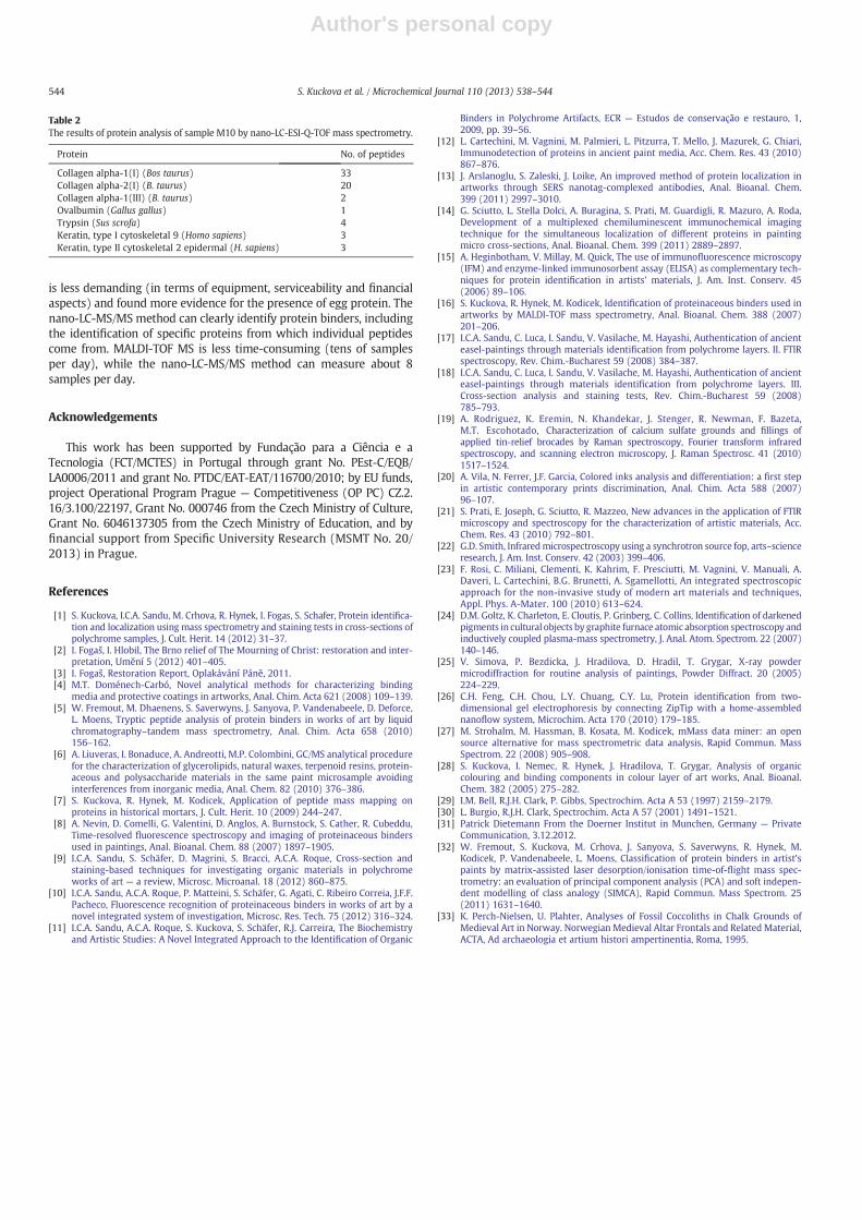
Nano-LC-ESI-Q-TOF confirms the previous results obtained byMALDI-TOF MS and identified mainly collagens, keratin contamina-tion (proteins derived from dermal cells) and traces of egg protein(only by one peptide) in the same samples (e.g. sample M10 —
Fig. 6, Table 2). Although this technique can uniquely identify pro-teins and all found collagens were assigned to the Bos taurus, mainlyin the case of collagens it could hardly identify their species specificity[32], because the collagens are very old evolutionary proteins with ahighly conserved amino acid composition. The identified keratinsmost probably originate from surface contamination of polishedsections, which may have occurred during their preparation ormanipulation.
5. Conclusions
The results obtained by visible and fluorescent light OM, SEM-EDXand micro-Raman spectroscopy revealed the classical structure of amulti-layer polychromy and the composition of the materials presentin this medieval polychrome statue. The ground layer is made from“marine” chalk, containing the remains of calcareous nano-fossils ofeukaryote phytoplankton, known as coccoliths. The presence ofthese nano-fossils in the ground layer cannot add information on
Table 1The inorganic constituents of 11 samples from “The Mourning of Jesus Christ”, identified by Raman spectroscopy and SEM-EDX.
Sample Vermillion Cu-based pigment Ultramarine Indigo Hematite (red iron oxide) Lead red Lead white Massicot Calcite Ba sulphate
M1 √ √ √ √ √M1a √ √ √M2 √ √ √ √M4 √ √ √ √M5 √ √ √M6 √ √M7 √ √M8 √ √ √M9 √ √ √ √M10 √M11 √ √ √ √ √
541S. Kuckova et al. / Microchemical Journal 110 (2013) 538–544

Author's personal copy
the creation of the figurative woodcarving, because they are the com-mon constituents of the calcium carbonate sediments and such mate-rial has been used all around medieval Europe [33]. Other inorganicpigments identified in the paint layers have also been commonlyused since the Medieval Ages, therefore the provenience of the artwork could not be securely determined based on these data. Thepresence of over-painting layers/materials was assessed in several
samples taken from damaged areas and few pigments/fillers belongingto modern times (Co-based pigment, barite) confirmed the presence oflayers of posterior intervention.
Identification of binders together with pigments and fillers in dif-ferent layers of the analyzed polychrome samples allowed restorersto compositionally and stratigraphically characterize the decorationof the sculpture and to assess the presence of historically known
Fig. 3. Cross-section observation and related sampling areas for 3 samples: a) sample M1a; b) sample M4; c) sample M11.
Fig. 4. Characterization of sample M5 using OM and SEM-EDX.
542 S. Kuckova et al. / Microchemical Journal 110 (2013) 538–544

Author's personal copy
techniques (paint and gilding techniques) useful for further selectionof the most appropriate treatment of restoration.
Based on the results of this detailed analytical protocol the wrinkledpaint layers added during the 20th century, applied in very thick layersto correct defects on the heavily damaged polychromy, were removed.The early 19th century over-paints (in Renaissance, Rococo andBaroque style), were also removed. It was found that the first Gothic
re-paints with artistic high quality are enough to maintain an authenticexpression of the work in its entirety. The substantial parts of the olderpolychrome were found and many details of carvingmastery of anony-mous Late Gothic artists also excel.
Both mass spectrometric methods MALDI-TOF and nano-LC-MS/MSare able to identify protein binders in their mixtures, even in samplesembedded as polished cross-sections. MALDI-TOF mass spectrometry
Fig. 5. Two samples with gold leaf, details of the OM Vis images and EDX spectrum for sample M5.
Fig. 6. Mass spectrum of a fragment from sample M10. The red circles labelled egg proteins and the green arrows animal glue.
543S. Kuckova et al. / Microchemical Journal 110 (2013) 538–544
Author's personal copy
is less demanding (in terms of equipment, serviceability and financialaspects) and found more evidence for the presence of egg protein. Thenano-LC-MS/MS method can clearly identify protein binders, includingthe identification of specific proteins from which individual peptidescome from. MALDI-TOF MS is less time-consuming (tens of samplesper day), while the nano-LC-MS/MS method can measure about 8samples per day.
Acknowledgements
This work has been supported by Fundação para a Ciência e aTecnologia (FCT/MCTES) in Portugal through grant No. PEst-C/EQB/LA0006/2011 and grant No. PTDC/EAT-EAT/116700/2010; by EU funds,project Operational Program Prague — Competitiveness (OP PC) CZ.2.16/3.100/22197, Grant No. 000746 from the Czech Ministry of Culture,Grant No. 6046137305 from the Czech Ministry of Education, and byfinancial support from Specific University Research (MSMT No. 20/2013) in Prague.
References
[1] S. Kuckova, I.C.A. Sandu, M. Crhova, R. Hynek, I. Fogas, S. Schafer, Protein identifica-tion and localization using mass spectrometry and staining tests in cross-sections ofpolychrome samples, J. Cult. Herit. 14 (2012) 31–37.
[2] I. Fogaš, I. Hlobil, The Brno relief of The Mourning of Christ: restoration and inter-pretation, Umění 5 (2012) 401–405.
[3] I. Fogaš, Restoration Report, Oplakávání Páně, 2011.[4] M.T. Doménech-Carbó, Novel analytical methods for characterizing binding
media and protective coatings in artworks, Anal. Chim. Acta 621 (2008) 109–139.[5] W. Fremout, M. Dhaenens, S. Saverwyns, J. Sanyova, P. Vandenabeele, D. Deforce,
L. Moens, Tryptic peptide analysis of protein binders in works of art by liquidchromatography–tandem mass spectrometry, Anal. Chim. Acta 658 (2010)156–162.
[6] A. Liuveras, I. Bonaduce, A. Andreotti, M.P. Colombini, GC/MS analytical procedurefor the characterization of glycerolipids, natural waxes, terpenoid resins, protein-aceous and polysaccharide materials in the same paint microsample avoidinginterferences from inorganic media, Anal. Chem. 82 (2010) 376–386.
[7] S. Kuckova, R. Hynek, M. Kodicek, Application of peptide mass mapping onproteins in historical mortars, J. Cult. Herit. 10 (2009) 244–247.
[8] A. Nevin, D. Comelli, G. Valentini, D. Anglos, A. Burnstock, S. Cather, R. Cubeddu,Time-resolved fluorescence spectroscopy and imaging of proteinaceous bindersused in paintings, Anal. Bioanal. Chem. 88 (2007) 1897–1905.
[9] I.C.A. Sandu, S. Schäfer, D. Magrini, S. Bracci, A.C.A. Roque, Cross-section andstaining-based techniques for investigating organic materials in polychromeworks of art — a review, Microsc. Microanal. 18 (2012) 860–875.
[10] I.C.A. Sandu, A.C.A. Roque, P. Matteini, S. Schäfer, G. Agati, C. Ribeiro Correia, J.F.F.Pacheco, Fluorescence recognition of proteinaceous binders in works of art by anovel integrated system of investigation, Microsc. Res. Tech. 75 (2012) 316–324.
[11] I.C.A. Sandu, A.C.A. Roque, S. Kuckova, S. Schäfer, R.J. Carreira, The Biochemistryand Artistic Studies: A Novel Integrated Approach to the Identification of Organic
Binders in Polychrome Artifacts, ECR — Estudos de conservação e restauro, 1,2009, pp. 39–56.
[12] L. Cartechini, M. Vagnini, M. Palmieri, L. Pitzurra, T. Mello, J. Mazurek, G. Chiari,Immunodetection of proteins in ancient paint media, Acc. Chem. Res. 43 (2010)867–876.
[13] J. Arslanoglu, S. Zaleski, J. Loike, An improved method of protein localization inartworks through SERS nanotag-complexed antibodies, Anal. Bioanal. Chem.399 (2011) 2997–3010.
[14] G. Sciutto, L. Stella Dolci, A. Buragina, S. Prati, M. Guardigli, R. Mazuro, A. Roda,Development of a multiplexed chemiluminescent immunochemical imagingtechnique for the simultaneous localization of different proteins in paintingmicro cross-sections, Anal. Bioanal. Chem. 399 (2011) 2889–2897.
[15] A. Heginbotham, V. Millay, M. Quick, The use of immunofluorescence microscopy(IFM) and enzyme-linked immunosorbent assay (ELISA) as complementary tech-niques for protein identification in artists' materials, J. Am. Inst. Conserv. 45(2006) 89–106.
[16] S. Kuckova, R. Hynek, M. Kodicek, Identification of proteinaceous binders used inartworks by MALDI-TOF mass spectrometry, Anal. Bioanal. Chem. 388 (2007)201–206.
[17] I.C.A. Sandu, C. Luca, I. Sandu, V. Vasilache, M. Hayashi, Authentication of ancienteasel-paintings through materials identification from polychrome layers. II. FTIRspectroscopy, Rev. Chim.-Bucharest 59 (2008) 384–387.
[18] I.C.A. Sandu, C. Luca, I. Sandu, V. Vasilache, M. Hayashi, Authentication of ancienteasel-paintings through materials identification from polychrome layers. III.Cross-section analysis and staining tests, Rev. Chim.-Bucharest 59 (2008)785–793.
[19] A. Rodriguez, K. Eremin, N. Khandekar, J. Stenger, R. Newman, F. Bazeta,M.T. Escohotado, Characterization of calcium sulfate grounds and fillings ofapplied tin-relief brocades by Raman spectroscopy, Fourier transform infraredspectroscopy, and scanning electron microscopy, J. Raman Spectrosc. 41 (2010)1517–1524.
[20] A. Vila, N. Ferrer, J.F. Garcia, Colored inks analysis and differentiation: a first stepin artistic contemporary prints discrimination, Anal. Chim. Acta 588 (2007)96–107.
[21] S. Prati, E. Joseph, G. Sciutto, R. Mazzeo, New advances in the application of FTIRmicroscopy and spectroscopy for the characterization of artistic materials, Acc.Chem. Res. 43 (2010) 792–801.
[22] G.D. Smith, Infraredmicrospectroscopy using a synchrotron source fop, arts–scienceresearch, J. Am. Inst. Conserv. 42 (2003) 399–406.
[23] F. Rosi, C. Miliani, Clementi, K. Kahrim, F. Presciutti, M. Vagnini, V. Manuali, A.Daveri, L. Cartechini, B.G. Brunetti, A. Sgamellotti, An integrated spectroscopicapproach for the non-invasive study of modern art materials and techniques,Appl. Phys. A-Mater. 100 (2010) 613–624.
[24] D.M. Goltz, K. Charleton, E. Cloutis, P. Grinberg, C. Collins, Identification of darkenedpigments in cultural objects by graphite furnace atomic absorption spectroscopy andinductively coupled plasma-mass spectrometry, J. Anal. Atom. Spectrom. 22 (2007)140–146.
[25] V. Simova, P. Bezdicka, J. Hradilova, D. Hradil, T. Grygar, X-ray powdermicrodiffraction for routine analysis of paintings, Powder Diffract. 20 (2005)224–229.
[26] C.H. Feng, C.H. Chou, L.Y. Chuang, C.Y. Lu, Protein identification from two-dimensional gel electrophoresis by connecting ZipTip with a home-assemblednanoflow system, Microchim. Acta 170 (2010) 179–185.
[27] M. Strohalm, M. Hassman, B. Kosata, M. Kodicek, mMass data miner: an opensource alternative for mass spectrometric data analysis, Rapid Commun. MassSpectrom. 22 (2008) 905–908.
[28] S. Kuckova, I. Nemec, R. Hynek, J. Hradilova, T. Grygar, Analysis of organiccolouring and binding components in colour layer of art works, Anal. Bioanal.Chem. 382 (2005) 275–282.
[29] I.M. Bell, R.J.H. Clark, P. Gibbs, Spectrochim. Acta A 53 (1997) 2159–2179.[30] L. Burgio, R.J.H. Clark, Spectrochim. Acta A 57 (2001) 1491–1521.[31] Patrick Dietemann From the Doerner Institut in Munchen, Germany — Private
Communication, 3.12.2012.[32] W. Fremout, S. Kuckova, M. Crhova, J. Sanyova, S. Saverwyns, R. Hynek, M.
Kodicek, P. Vandenabeele, L. Moens, Classification of protein binders in artist'spaints by matrix-assisted laser desorption/ionisation time-of-flight mass spec-trometry: an evaluation of principal component analysis (PCA) and soft indepen-dent modelling of class analogy (SIMCA), Rapid Commun. Mass Spectrom. 25(2011) 1631–1640.
[33] K. Perch-Nielsen, U. Plahter, Analyses of Fossil Coccoliths in Chalk Grounds ofMedieval Art in Norway. Norwegian Medieval Altar Frontals and Related Material,ACTA, Ad archaeologia et artium histori ampertinentia, Roma, 1995.
Table 2The results of protein analysis of sample M10 by nano-LC-ESI-Q-TOF mass spectrometry.
Protein No. of peptides
Collagen alpha-1(I) (Bos taurus) 33Collagen alpha-2(I) (B. taurus) 20Collagen alpha-1(III) (B. taurus) 2Ovalbumin (Gallus gallus) 1Trypsin (Sus scrofa) 4Keratin, type I cytoskeletal 9 (Homo sapiens) 3Keratin, type II cytoskeletal 2 epidermal (H. sapiens) 3
544 S. Kuckova et al. / Microchemical Journal 110 (2013) 538–544