Embed Size (px)
Citation preview
Authority on Radiation Protection Radiation Machine Registration Permit Manual
BASED ON THE
“DELAWARE RADIATION CONTROL REGULATIONS”
Adopted July 22, 1969
AMENDED May 1, 2014
BY THE
AUTHORITY ON RADIATION PROTECTION
In conformance with 16 Del. C. § 7406 (c)
STATE OF DELAWARE Radiation Machine Registration Permit Manual
TABLE OF CONTENTS
MESSAGE TO DELAWARE RADIATION MACHINE SOURCE FACILITIES 1 SECTION A Radiation Machine Source Permit 2 SECTION B Application Requirements 3
1) Application Form 3 2) Application Requirements – Shielding Plan 4
SECTION C Renewal of Existing Permits 4
SECTION D Request to Terminate 5
1) Application Form 5 2) Required Documentation 5
SECTION E Annual Fees 6
1) Mailing Invoice 6 2) Failure to Pay Fee (30 days) 6 3) Failure to Pay Fee (60 days) 6
SECTION F Inspections 6
1) Routine Inspections 6 2) Non-Routine Inspections 7
• Follow-up • Complaint • Investigation
3) Inspection Report (ORC R-11 Form) 7 4) Correction of Violations 7
SECTION G Administrative Penalties 8 SECTION H CONTACT INFORMATION FOR OFFICE OF RADIATION CONTROL 8 APPENDIX A Annual Fee Schedule 9 APPENDIX B RSO Responsibilities 10 APPENDIX C Education & Training 11 APPENDIX D Inspection Report (ORC-R11) 12 APPENDIX E Fundamentals of X-Ray 14 APPENDIX F DEFINITIONS 18
PREFACE

1
Message to Delaware Radiation Machine Source Facilities
Radiation safety is the business--and the responsibility--of every person taking
part in the use of radiation for diagnostic, therapeutic, industrial, or research purposes.
Using ionizing radiation in a way that is safe for patients, staff, and the general
public is the personal responsibility of every authorized user of radiation. To exercise
that responsibility, each person who uses ionizing radiation sources in Delaware must
have a basic understanding of how the equipment works, of necessary safety principles
and practices, and of the Delaware regulations for control of ionizing radiation. The
owners and operators of registered radiation machine source facilities permitted to
operate in Delaware are obligated to ensure that facilities are designed and constructed
properly, that radiation service providers used are state-registered, and that radiation
machine sources are used in compliance with established safety and health standards
and regulations.
To assure that proper safety standards are met, Delaware's Authority on
Radiation Protection requires that all Radiation Machine Source Facilities hold a
registration permit in good standing, and be subject to periodic safety inspections. This
manual contains instructions and other necessary information to assist radiation
machine source facilities to obtain registration, and achieve compliance with regulations
established by the State of Delaware.
F. Esposito, M.D., Chair, 2014 F. Fisher-Tyler, CIH, Administrator Authority on Radiation Protection Office of Radiation Control State of Delaware Delaware Division of Public Health

2
RADIATION MACHINE REGISTRATION A. The Radiation Machine Registration The Delaware Radiation Machine Registration is a permit to operate radiation
machines in Delaware, as required by Delaware Radiation Control Regulations. It is issued to qualified facility owner/operators who meet the requirements of the Delaware Radiation Control Regulations, as amended, as carried out by the Office of Radiation Control (ORC) in the Division of Public Health, the Administrative Agent for the Authority on Radiation Protection, a Governor – appointed public board.
1) The radiation machine registration permit is valid for the period specified
on the permit. 2) Under a provision of the Delaware Radiation Control Regulations, all
facility radiation machine registration permits, and radiation technologist/technician certificates in medical/dental facilities, must be posted in a conspicuous location within the registered facility.
3) The registration permit is non-transferable. If a facility changes
ownership, the existing permit will be terminated, and a new permit issued to the company acquiring the permitted facility, upon submittal of an official application form.
4) The facility registration permit is kept in good standing by payment of an
annual fee in compliance with the Delaware Radiation Control Regulations, as amended.
5) The permit-holder is subject to the annual permit fee regardless of
radiation machine inventory status (eg. installed, activated, stored), for as long as the x-ray equipment remains on the property and until such time as all equipment is disposed of, and the registration permit is officially terminated.
B. Application Requirements for Obtaining a New Radiation Machine Source Permit 1. Application to register an existing radiation machine source facility:
The Owner/Operator of the radiation machine source facility must submit a completed, signed, official application form (ORC-R1) to:
Delaware Office of Radiation Control 417 Federal Street
Dover, Delaware 19901

3
The application must consist of:
A request that a registration permit be issued by filing the appropriate, official application form (ORC-R1).
Complete information, including: applicant's full company name,
owner/operator name (with IRS Employer Identification or Social Security number), individual responsible for radiation protection (Radiation Safety Officer), name of licensed practitioner (for healing arts radiation use only), radiation service companies used, radiation procedures performed at the facility, radiation machine registration fee category and modalities used, radiation machine equipment inventory and status, signature of owner/operator.
Applicants are instructed to mail a copy of the completed official
application form to the address specified on the application form, and retain a copy of the application for their records.
2. Application to open a new radiation machine facility (renovation or new construction) must include:
A request that a registration permit be issued by filing the appropriate, official application form (ORC-R1).
Prior to construction, the floor plans, shielding specifications and equipment arrangement of all new installations, or modifications of existing installations utilizing ionizing radiation sources with maximum energy greater than 70 kVP (kilovolts peak) shall be submitted to the Division of Public Health Office of Engineering for review, see contact information below:
Division of Public Health Office of Engineering Edgehill Shopping Center 43 South DuPont Hwy. Dover, Delaware 19901 Phone: (302) 741-8640 Fax: (302) 741-8641
Submission of a Shielding Plan to the Division of Public Health
Office of Engineering for all facilities planning to use radiation machines for modalities below, which may have maximum energy exceeding 70 kVP, by filing the appropriate, official Minimum Shielding Requirements Report form (ORC-R15A), completed by a registered Radiation Service Provider. Radiation Machine Facilities or rooms that require a shielding plan include:
• Dental panoramic or cephalometric x-ray • Dental Cone Beam Computed Tomography (CT) • Stationary radiographic or fluoroscopic x-ray • Mobile x-ray machine used routinely in one location • Computed Tomography (CT) scanner

4
• Mammography • Linear Accelerator or other therapy machine
Submission of a floor plan, with radiation machine and remote
operational switch location specified for facilities planning to use radiation machines with maximum energy below or at 70 kVP, including:
• Dental intraoral (e.g. bitewing, periapicals) • Bone densitometry • Podiatry • Mini C-Arm (e.g. Orthopedic)
The Division of Public Health, Office of Engineering issues a
Certificate of Approval to Construct directly to the applicant submitting the radiation shielding plan for review.
Upon receipt of a complete application package and conclusion of
pre-operational inspection (if required), the Office of Radiation Control will issue a Certificate of Approval to Operate and a new radiation machine facility registration permit.
3. See Delaware Radiation Control Regulations, 4465 Part B, Section 5.0
Shielding Plan Review, for additional information.
C. Renewal of Existing Radiation Machine Source Permit
1. Radiation Machine Source permits are renewed prior to the expiration date specified on the permit and must be posted in a public location to remain in full compliance with the regulations.
2. Radiation Machine Source facilities will be mailed a renewed registration
permit prior to the expiration date on the current permit. In addition, a datasheet populated with information from the most recent application on file will be enclosed with the renewed permit. Facilities are instructed to review the information on the datasheet for accuracy, make changes if needed, sign, date and mail or fax the marked-up form back to the Office of Radiation Control. If no changes are needed, the datasheet should be retained by the facility.
Note: Renewal of registration permit is NOT related to the annual
permit fee billing cycle. Do not submit payment at renewal. Permit Fees are payable only when invoice is received in the fall of each year.
5
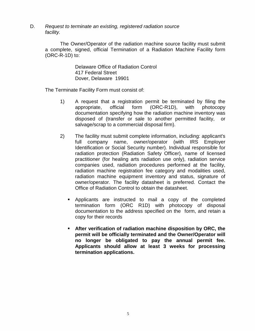
D. Request to terminate an existing, registered radiation source facility.
The Owner/Operator of the radiation machine source facility must submit a complete, signed, official Termination of a Radiation Machine Facility form (ORC-R-1D) to:
Delaware Office of Radiation Control 417 Federal Street
Dover, Delaware 19901 The Terminate Facility Form must consist of:
1) A request that a registration permit be terminated by filing the appropriate, official form (ORC-R1D), with photocopy documentation specifying how the radiation machine inventory was disposed of (transfer or sale to another permitted facility, or salvage/scrap to a commercial disposal firm).
2) The facility must submit complete information, including: applicant's
full company name, owner/operator (with IRS Employer Identification or Social Security number). Individual responsible for radiation protection (Radiation Safety Officer), name of licensed practitioner (for healing arts radiation use only), radiation service companies used, radiation procedures performed at the facility, radiation machine registration fee category and modalities used, radiation machine equipment inventory and status, signature of owner/operator. The facility datasheet is preferred. Contact the Office of Radiation Control to obtain the datasheet.
Applicants are instructed to mail a copy of the completed
termination form (ORC R1D) with photocopy of disposal documentation to the address specified on the form, and retain a copy for their records
After verification of radiation machine disposition by ORC, the
permit will be officially terminated and the Owner/Operator will no longer be obligated to pay the annual permit fee. Applicants should allow at least 3 weeks for processing termination applications.

6
E. Annual Permit Fees
Each radiation machine source permit-holder shall receive an invoice and pay an annual fee based on the fee schedule signed into law in June 2008, regardless of source status (installed, activated, or stored). See Appendix A for Fee Schedule. 1) Mailing Invoice:
Radiation machine source permit-holders will be mailed an invoice for their annual fee, in the fall of each calendar year, with bill payable within 30 days of invoice date. Owner/Operators are instructed to make check payable to “Delaware Office of Radiation Control,” and mail the check AND copy of invoice to the attention of the Office of Radiation Control, to the address specified on the invoice (417 Federal Street, Dover, DE 19901).
2) Failure to Pay Fee (30 days):
Radiation machine source permit-holders who do not pay their permit fee within 30 days following the invoice due date will receive a reminder letter via certified mail, with instruction to pay their fee immediately, or risk issuance of an administrative penalty in the amount of $500.
3) Failure to Pay Fee (60 days):
Radiation machine source permit-holders who do not pay their permit fee within 60 days following the invoice due date will be issued an administrative penalty in the amount of $500 by the Delaware Authority on Radiation Protection, and shall be ordered to pay their fee.
Radiation machine source permit-holders who do not pay their permit fee AND the administrative penalty amount within 10 days of the date on the administrative penalty letter will be forwarded to the Department of Justice for enforcement action through the Office of the Attorney General.
F. Inspections
1) Routine Inspections:
Periodic, routine inspections of registered machine source facilities shall be performed at frequencies established by the Authority on Radiation Protection. An inspection of a registered facility shall be performed at least every two (2) years for medical facilities utilizing angiography, radiography, fluoroscopy, computed tomography (CT), and radiation therapy modalities, and at least every four (4) years for other registered facilities, including dental, bone densitometry, podiatry, veterinary, academic and industrial.
7
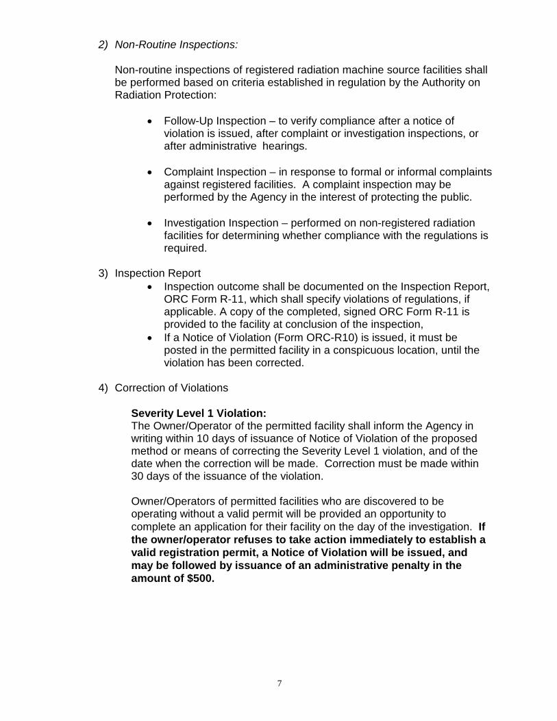
2) Non-Routine Inspections:
Non-routine inspections of registered radiation machine source facilities shall be performed based on criteria established in regulation by the Authority on Radiation Protection:
• Follow-Up Inspection – to verify compliance after a notice of violation is issued, after complaint or investigation inspections, or after administrative hearings.
• Complaint Inspection – in response to formal or informal complaints
against registered facilities. A complaint inspection may be performed by the Agency in the interest of protecting the public.
• Investigation Inspection – performed on non-registered radiation
facilities for determining whether compliance with the regulations is required.
3) Inspection Report • Inspection outcome shall be documented on the Inspection Report,
ORC Form R-11, which shall specify violations of regulations, if applicable. A copy of the completed, signed ORC Form R-11 is provided to the facility at conclusion of the inspection,
• If a Notice of Violation (Form ORC-R10) is issued, it must be posted in the permitted facility in a conspicuous location, until the violation has been corrected.
4) Correction of Violations
Severity Level 1 Violation: The Owner/Operator of the permitted facility shall inform the Agency in writing within 10 days of issuance of Notice of Violation of the proposed method or means of correcting the Severity Level 1 violation, and of the date when the correction will be made. Correction must be made within 30 days of the issuance of the violation.
Owner/Operators of permitted facilities who are discovered to be operating without a valid permit will be provided an opportunity to complete an application for their facility on the day of the investigation. If the owner/operator refuses to take action immediately to establish a valid registration permit, a Notice of Violation will be issued, and may be followed by issuance of an administrative penalty in the amount of $500.

8
Severity Level 2 Violation: The Owner/Operator of the permitted facility shall correct the violation as soon as possible, and is allowed up to 60 days to make the correction. G. Administrative Penalties
Whomever violates any rules, regulations or orders of the Authority may be assessed an administrative penalty in an amount not to exceed $500 for a first offense, or an amount not to exceed $750 for any subsequent offense. Each violation shall be considered a separate offense.
H. Contact Information The Office of Radiation Control can be contacted at:
Phone Number: (302) 744-4546
Fax Number: (302) 739-3839
Mailing Address:
Delaware Office of Radiation Control 417 Federal Street Dover, Delaware 19901
See Office of Radiation Control (ORC) webpage for additional information, manuals and application forms.
http://www.dhss.delaware.gov/dph/hsp/orc.html

9
Appendix A ANNUAL FEE SCHEDULE
For purposes of the fee schedule set out above, the following definitions apply: “Medical Modalities” means radiography, fluoroscopy, computed tomography, angiography, stereotactic breast biopsy systems, and radiation therapy utilized in humans. “Non-medical Modalities” means radiography, fluoroscopy, analytical equipment (including electron microscopes, fluorescence analysis and x-ray diffraction equipment), computed tomography, and particle accelerators not utilized on humans. Owner/Operators must pay radiation machine source fees for all fee categories which apply to their facilities. For example, a facility performing medical modalities and dental modalities in the same facility would be obligated to pay the appropriate medical category fee, and the dental category fee. Note:
Radiation machine source permit-holders will be mailed an invoice for their annual fee, in the fall of each calendar year, with bill payable within 30 days of invoice date. Owner/Operators are instructed to make check payable to “Delaware Office of Radiation Control,” and mail the check AND copy of invoice to the attention of the Office of Radiation Control, to the address specified on the invoice (417 Federal Street, Dover, DE 19901).
Category Fee ($) Description I 1,370 Facilities with a total of five or more of the medical
modalities or non-medical modalities listed below. II 1,030 Facilities with a total of three or four of the medical
modalities or non-medical modalities listed below. III 690 Facilities with two of the medical modalities listed below. IV 275 Facilities with one of the medical modalities listed below,
and an annual patient workload of 750 examinations or more.
V 140 Facilities with one of the medical modalities listed below, and an annual patient workload of less than 750 examinations, or all other radiation installations with one or two of the non-medical modalities listed below except as listed under Category VI.
VI 75 Dental, podiatric, bone densitometry or veterinary installations.

10
Appendix B RSO RESPONSIBILITIES
Radiation Machine Facility Radiation Safety Officer (RSO) Responsibilities
The applicant or registration permit-holder, shall require each individual assigned to fulfill responsibilities and duties as Radiation Safety Officer (RSO) to be an individual who has training and experience in the safe and effective use of radiation machines and the potential radiation hazards and emergency precautions applicable to the type(s) of activity or facility for which the individual is seeking to perform RSO duties, to include:
1. Establishing and overseeing operating and safety procedures that maintain radiation exposures as low as reasonably achievable (ALARA), and to review them periodically to ensure that the procedures are current and conform with these regulations;
2. Ensuring that individual monitoring devices are properly used by
occupationally exposed personnel as required by the regulations, that records are kept of the monitoring results, and that timely notifications are made as required by 4465 Part D;
3. Investigating and reporting to the agency each known or suspected
case of radiation exposure to an individual or radiation level detected in excess of limits established by these regulations and each theft or loss of source(s) of radiation, determining the cause, and taking steps to prevent its recurrence;
4. Having a thorough knowledge of management policies, administrative
procedures and records of the registration permit-holder and keeping management informed on a periodic basis of the performance of the registrant’s radiation protection program, if applicable;
5. Assuming control and having the authority to institute corrective
actions including shutdown of operations when necessary in emergency situations or unsafe conditions;
6. Maintaining records as required by these regulations; and
7. Ensuring that personnel are adequately trained and complying with
these regulations, the conditions of the certificate of registration, and the operating and safety procedures of the registered permit-holder.

11
Appendix C EDUCATION & TRAINING EDUCATION AND TRAINING FOR PERSONS PERFORMING RADIATION MACHINE
ASSEMBLY, INSTALLATION OR REPAIR All persons performing radiation machine assembly, installation or repair shall meet the general requirements in subparagraph 1. of this paragraph, and one or more of the specialized requirements in subparagraph 2. of this paragraph. 1. General requirements include: (a) Experience or education providing familiarity with the type(s) of equipment
to be serviced, to include radiation safety; (b) Knowledge of protective measures to reduce potentially hazardous conditions; and (c) Six months of supervised assembly and repair of the type(s) of equipment
to be serviced. 2. Specialized requirements include: (a) One year of formal training (may be satisfied by factory school, military
technical training school, or other courses in radiation machine assembly, installation or repair techniques) or an associate's degree in biomedical equipment repair;
(b) A bachelor's degree in electrical engineering with specialized training in
radiation producing devices; or (c) A combination of training and experience equal to clause (a) of this
subparagraph.

12
Appendix D
INSPECTION REPORT (ORC-R11) FOR OFFICIAL USE ONLY
DELAWARE HEALTH AND SOCIAL SERVICES Division of Public Health
Office of Radiation Control (302) 744-4546
Page 1 of 2 Registration # Date of Inspection: Exp. Date / / Facility Name Specify Facility Type Address Hospital Medical City, State, ZIP Dental Veterinary Phone Podiatric Chiropractic Total # of x-ray tubes at this location #Tubes Inspected Industrial Other Number of Violations (see comments) Violation Severity Level I II (circle)
GENERAL ADMINISTRATIVE ITEMS A. Total Number Users B. Total Number Occupationally Exposed C. # Certified Radiation Techs: Lic. Prac. Dental Hyg Others D. Employees Monitored: Y N E. Total Number Monitored F. Monitoring Records Satisfactory: Y N G. Monitoring System Adequate: Y N N/A H. Number over : WB 1250 mR/Quarter: WB 5000 mR/Year: WB Ext 3750 mR/Quarter: Ext 15000 mR/year: Ext I. Number Patients Radiographed (Avg./week) J. Patient Work load (#/year) :_______ (avg./week X 4weeks X 12months) K. Number Patients Fluoroscoped (Avg./week) L. Number Therapy Treatments (Avg./week) M. Number Nuclear Medicine Administrations (Avg./Week) N. Brand/Type X-ray Film Used O. Type of Intensifying Screen P. Digital Y N ___________________________________________________________
Q. Radioactive Material Present: Y N R. Registered By: DE Licensed By: NRC S. Current State Registration Posted: Y N T. Notice to Employees Posted: Y N U. Written Safety Procedures: Y N V. Records/Corr./Copy of DRCR: Y N W. ORC-R1 data current: Y N
X. Reason for Inspection: Regular Follow-Up Investigation Complaint Other ORC-R11/Facility (05/14)
13
Page 2 of 2
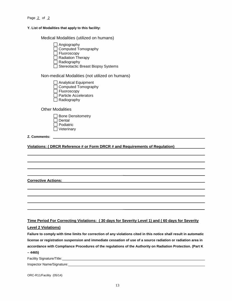
Y. List of Modalities that apply to this facility:
Medical Modalities (utilized on humans)
Angiography Computed Tomography Fluoroscopy Radiation Therapy Radiography Stereotactic Breast Biopsy Systems
Non-medical Modalities (not utilized on humans)
Analytical Equipment Computed Tomography Fluoroscopy Particle Accelerators Radiography
Other Modalities
Bone Densitometry Dental Podiatric Veterinary
Z. Comments:
Violations: ( DRCR Reference # or Form DRCR # and Requirements of Regulation)
_______________________________________
Corrective Actions:
_______________________________________
Time Period For Correcting Violations: ( 30 days for Severity Level 1) and ( 60 days for Severity
Level 2 Violations) Failure to comply with time limits for correction of any violations cited in this notice shall result in automatic
license or registration suspension and immediate cessation of use of a source radiation or radiation area in
accordance with Compliance Procedures of the regulations of the Authority on Radiation Protection. (Part K
– 4465)
Facility Signature/Title:
Inspector Name/Signature:
ORC-R11/Facility (05/14)

14
Appendix E
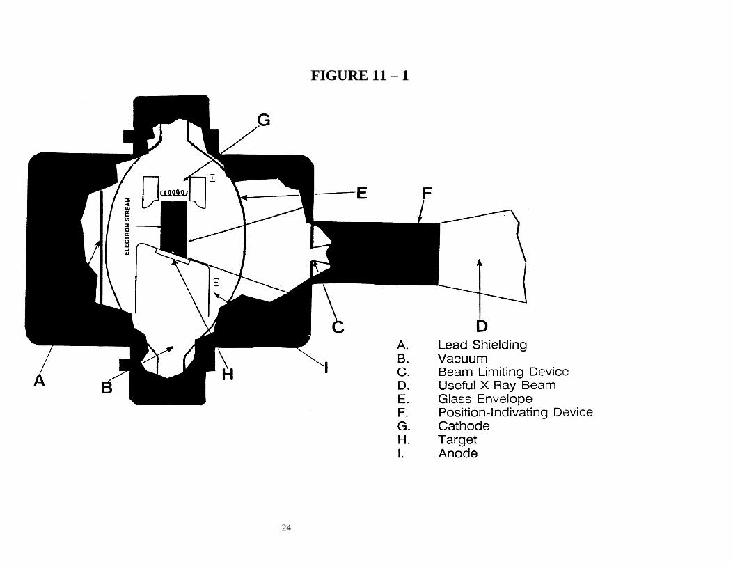
FUNDAMENTALS OF X-RAYS Radiation Physics X-rays are electromagnetic waves, like visible light, microwaves, and radio waves. The difference between these different types of electromagnetic radiation is in their wavelengths, the distance between adjacent peaks of the waves. Our eyes are equipped to see radiation in a certain range of these wavelengths which we call visible light. Radiation at other wavelengths, invisible to the human eye, can only be detected by means of special sensors. Electromagnetic radiation of most wavelengths passes through some materials, for example through air, without either being absorbed or reflected. When visible light passes through such a material we call it transparent. Other materials pass only a fraction of the radiation incident on them and either absorb or reflect the rest (we call such materials translucent or attenuating) and still others absorb or reflect most the radiation incident on them (we call these opaque). The shorter the wavelength of the radiation is the more energy it carries and the greater is the chance of its passing through various materials without being absorbed or reflected. X-rays have very short wavelengths. They thus carry considerable more energy and are able to pass through many materials without a large fraction of them being absorbed. In fact, the fraction of incident x-rays that is absorbed by a given material depends mostly on the material’s density. Thus, dense materials such as lead and gold absorb a much greater fraction of incident x-rays, at a given thickness of the material, than do “light” materials, such as water. Similarly, x-rays pass mostly unabsorbed through most biological tissues such as muscle and various organs, but they are more strongly absorbed by bone, which has a higher density. X-ray imaging takes advantage of this difference in the x-ray attenuating properties of various tissues in the body. What we finally see as a finished radiograph is the result of variations in the intensity of the x-ray beam due to differences in attenuation by different tissues. After passing through the body, the x-rays, which have passed through without being attenuated, impinge on fluorescent screens between which a photographic film is sandwiched. The fluorescent screens are made of a dense material designed to absorb most of the incident beam and, on absorbing the x-rays, emitting visible light to which the photographic film is sensitive. They thus expose the film, which is then developed to display the radiograph. How X-rays Are Made An x-ray tube is a special case of electronic vacuum tube. It always has a glass envelope with several electrical leads sealed into it, enclosing several electronic elements, with the air removed from it as completely as possible. A schematic diagram of a tube head, which includes the tube itself (glass envelope + contents), is shown as Figure II-1; a typical control panel is shown as Figure II-2.

15
This section describes how x-rays are generated. Though somewhat oversimplified, the concept is accurate. These steps always occur, though in some modern equipment some of them may happen in a rapid and automatic sequence. 1. The x-ray machine's main power switch is turned ON and the "X-ray
Ready" light comes ON. Ordinarily, this is done at the beginning of the workday.
2. The current is adjusted to the value called for by the technique chart,
using the current (mA) adjustment control knob. This current heats the filament of the negatively charged cathode, which begins to give off electrons when it gets hot enough. Once the filament gets hot enough to begin emitting electrons (its threshold temperature), it emits them more rapidly, the hotter it gets. A higher current produces a hotter filament which in turn produces a higher rate of electron emission.
At this stage, the electrons have nowhere in particular to go. Generally,
they fly a short distance out into the vacuum, stay there awhile, and finally fall back into the surface of the cathode. This makes a sort of "cloud" of free electrons around the hot cathode, just waiting for something to happen.
3. The high-voltage supply is adjusted to the value prescribed by the
technique chart (or by the Licensed Practitioner, in case of special radiography), using the kVp control knob. A typical value is 70,000 peak volts (70 kVp), nearly 1000 times the voltage of ordinary "house" voltage. (This voltage is lethally high; the leads are totally enclosed to protect anyone from touching them.
4. When the operator positions the patient (the object) for the radiograph, he
sets the time knob to the appropriate time interval. 5. At this point, the x-ray machine is ready to be used. The electron cloud
around the cathode is stabilized according to the mA setting; the high voltage has been set to the desired kVp setting; the time interval is set. NO X-RAYS ARE GENERATED YET.
6. When the operator (having taken a safe position behind a protection
barrier or 12 feet from the x-ray tube presses the Remote Activator switch (Deadman Switch/ exposure button), (a) the high voltage is imposed between cathode and anode, (b) the automatic timer starts, and (c) the "X-ray ON" light comes ON.
The anode, now bearing a strong positive charge, attracts the free electrons in the cloud around the cathode. The electrons fly through the vacuum to strike (and stick to) the anode, striking the target with so much energy that it emits x-rays and gets quite hot. The higher the kVp, the faster the electrons move to the anode, the harder they strike the target, and the more energetic the x-rays are--that is, the more penetrating capability they have.
16
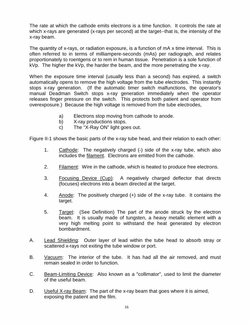
The rate at which the cathode emits electrons is a time function. It controls the rate at which x-rays are generated (x-rays per second) at the target--that is, the intensity of the x-ray beam. The quantity of x-rays, or radiation exposure, is a function of mA x time interval. This is often referred to in terms of milliampere-seconds (mAs) per radiograph, and relates proportionately to roentgens or to rem in human tissue. Penetration is a sole function of kVp. The higher the kVp, the harder the beam, and the more penetrating the x-ray. When the exposure time interval (usually less than a second) has expired, a switch automatically opens to remove the high voltage from the tube electrodes. This instantly stops x-ray generation. (If the automatic timer switch malfunctions, the operator's manual Deadman Switch stops x-ray generation immediately when the operator releases finger pressure on the switch. This protects both patient and operator from overexposure.) Because the high voltage is removed from the tube electrodes,
a) Electrons stop moving from cathode to anode. b) X-ray productions stops. c) The “X-Ray ON” light goes out. Figure II-1 shows the basic parts of the x-ray tube head, and their relation to each other: 1. Cathode: The negatively charged (-) side of the x-ray tube, which also
includes the filament. Electrons are emitted from the cathode. 2. Filament: Wire in the cathode, which is heated to produce free electrons. 3. Focusing Device (Cup): A negatively charged deflector that directs
(focuses) electrons into a beam directed at the target. 4. Anode: The positively charged (+) side of the x-ray tube. It contains the
target. 5. Target: (See Definition) The part of the anode struck by the electron
beam. It is usually made of tungsten, a heavy metallic element with a very high melting point to withstand the heat generated by electron bombardment.
A. Lead Shielding: Outer layer of lead within the tube head to absorb stray or
scattered x-rays not exiting the tube window or port. B. Vacuum: The interior of the tube. It has had all the air removed, and must
remain sealed in order to function. C. Beam-Limiting Device: Also known as a "collimator", used to limit the diameter
of the useful beam. D. Useful X-ray Beam: The part of the x-ray beam that goes where it is aimed,
exposing the patient and the film.

17
E. Glass Envelope: The shell of the x-ray tube, vacuum-tight. F. Position-Indicating Device: A device used to aim the primary x-ray beam. Figure II-2 shows controls and meters. The components of this figure perform the following functions: A. Remote Activator: Remote switch that activates the x-ray machine. This is
preferably a Deadman Switch, i.e., a switch which is made so that it is activated only by the operator's continuous pressure, also known as the exposure button.
B. kVp Meter: Indicates the peak voltage (kilovolts) between cathode and anode. C. mA Meter: Indicates the current (milliamperes) flowing between cathode and
anode. D. X-ray Ready Light: Indicates that the machine is warmed up and ready to
operate. E. X-ray ON Light: Lights only during the brief period when the x-ray machine
operates. F. Timer: Sets the time interval during which the machine generates the x-ray
beam. The timer is connected to the TIMER SWITCH. The operator turns the machine ON manually, using the Remote Activator, and then the timer switch turns OFF automatically after the preset time interval has elapsed.
G. mA Adjustment Knob: Allows you to alter the tube current (mA), by controlling
the input. The procedures for individual x-ray machines prescribe the normal range of current values.
H. kVp Adjustment Knob: Allows the operator to select the (operating) voltage
across the x-ray tube needed to penetrate the part being x-rayed. I. Main Power ON/OFF Switch: Connects/disconnects electrical power to the x-
ray machine.

18
Appendix F
DEFINITIONS AGENCY The administrative agent of the Authority on Radiation
Protection; i.e., the Office of Radiation Control, Division of Public Health, Delaware Department of Health and Social Services.
AMPERE Unit of electric current. One ampere is produced by
one volt acting through resistance of 1 ohm. ANODE The positively-charged side of the x-ray tube. It
contains the target. AUTHORITY Delaware's Authority on Radiation Protection as
specified by 16 Del. Code 7404. BARRIER
A radiation-absorbing material such as lead, concrete, or plaster, used to protect an individual or an area by reducing exposure.
BITEWING RADIOGRAPHS
Intra-oral films that show the crown portions of opposing teeth in the biting position.
CASSETTES A holder for x-ray film that protects the film from
exposure to visible light but permits penetration of x-rays. Cassettes may be plastic, cardboard or metal.
CATHODE The negatively-charged side of the x-ray tube. It
contains the filament and the focusing device. CENTRAL RAY The x-ray that is located in the center of the x-ray beam
as it leaves the tube head. CERTIFICATE A document issued by the Agency recognizing the
successful completion of an Authority-approved Certification Examination. Unless otherwise specified, a “certificate” allows practice of Radiation Technology to the level of examination passed. A “temporary certificate” may be issued under certain circumstances.
19
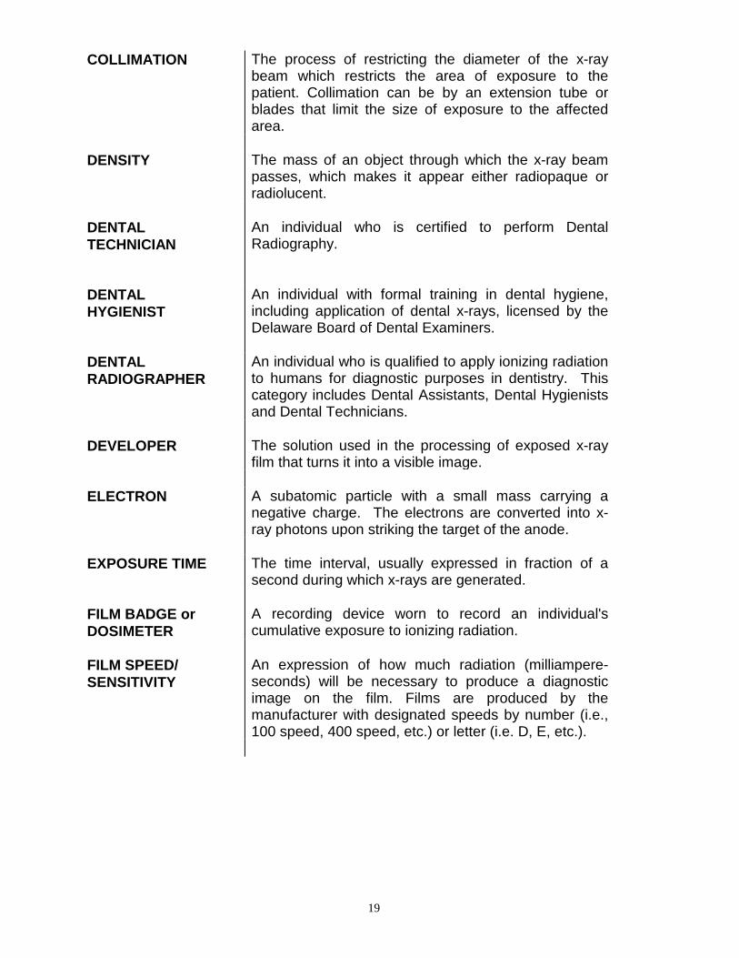
COLLIMATION The process of restricting the diameter of the x-ray beam which restricts the area of exposure to the patient. Collimation can be by an extension tube or blades that limit the size of exposure to the affected area.
DENSITY The mass of an object through which the x-ray beam
passes, which makes it appear either radiopaque or radiolucent.
DENTAL TECHNICIAN
An individual who is certified to perform Dental Radiography.
DENTAL HYGIENIST
An individual with formal training in dental hygiene, including application of dental x-rays, licensed by the Delaware Board of Dental Examiners.
DENTAL RADIOGRAPHER
An individual who is qualified to apply ionizing radiation to humans for diagnostic purposes in dentistry. This category includes Dental Assistants, Dental Hygienists and Dental Technicians.
DEVELOPER The solution used in the processing of exposed x-ray
film that turns it into a visible image. ELECTRON A subatomic particle with a small mass carrying a
negative charge. The electrons are converted into x-ray photons upon striking the target of the anode.
EXPOSURE TIME The time interval, usually expressed in fraction of a
second during which x-rays are generated. FILM BADGE or DOSIMETER
A recording device worn to record an individual's cumulative exposure to ionizing radiation.
FILM SPEED/ SENSITIVITY
An expression of how much radiation (milliampere-seconds) will be necessary to produce a diagnostic image on the film. Films are produced by the manufacturer with designated speeds by number (i.e., 100 speed, 400 speed, etc.) or letter (i.e. D, E, etc.).

20
FIXER The chemical solution used in the processing of exposed x-ray film that preserves the developed image by removing the unexposed silver halide crystals. The proper “fixing” of a film allows for extended archival quality.
FOCAL-FILM DISTANCE (FFD) or SOURCE IMAGE DISTANCE (SID)
The distance from the focal spot (target) at the anode of the x-ray tube to the film. It is usually expressed in inches, for example 8-inch FFD. More recently called the source image distance or SID.
FULL MOUTH SURVEY
A series of intra-oral radiographs that gives diagnostic information for all teeth and desired bony areas. It is usually composed of peri-apical and bite-wing films.
IMAGE Any likeness of an object reproduced on photographic
film or other viewing device. The image is the entire radiograph.
ION An electrically charged (+ or -) particle. IN-UTERO EXPOSURE
Radiation exposure to the unborn fetus, resulting from irradiation of the pregnant mother.
IONIZATION Process whereby electrically neutral atoms or
molecules are converted to positively or negatively charged fragments on exposure to x-rays.
IONIZING RADIATION
The kind of radiation that produces ions when interacting with matter. Dental equipment and medical equipment produce this type of radiation.
KILOVOLT (kVp) One thousand (1000) volts. Used in radiology to
describe the kilovoltage setting used to expose a particular body part. The thicker (denser) the part, the higher the kVp setting required to penetrate the part to produce a diagnostic image. kVp determines the quality of the x-ray beam.
LICENSED PRACTITIONER
An individual licensed to practice medicine, dentistry, podiatry, chiropractic, or osteopathy in Delaware. In other words, any individual licensed to prescribe therapeutic or diagnostic radiation for human patients. In addition, this category includes dental hygienists who cannot prescribe radiation.
21
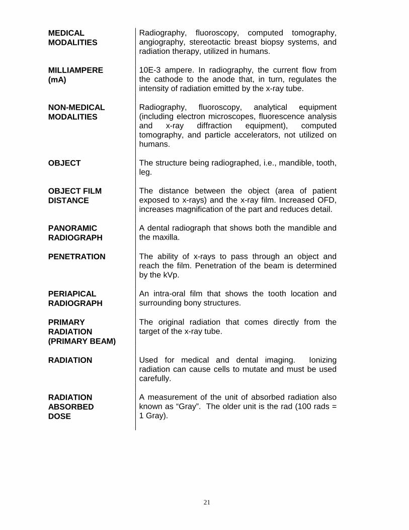
MEDICAL MODALITIES
Radiography, fluoroscopy, computed tomography, angiography, stereotactic breast biopsy systems, and radiation therapy, utilized in humans.
MILLIAMPERE (mA)
10E-3 ampere. In radiography, the current flow from the cathode to the anode that, in turn, regulates the intensity of radiation emitted by the x-ray tube.
NON-MEDICAL MODALITIES
Radiography, fluoroscopy, analytical equipment (including electron microscopes, fluorescence analysis and x-ray diffraction equipment), computed tomography, and particle accelerators, not utilized on humans.
OBJECT The structure being radiographed, i.e., mandible, tooth,
leg. OBJECT FILM DISTANCE
The distance between the object (area of patient exposed to x-rays) and the x-ray film. Increased OFD, increases magnification of the part and reduces detail.
PANORAMIC RADIOGRAPH
A dental radiograph that shows both the mandible and the maxilla.
PENETRATION The ability of x-rays to pass through an object and
reach the film. Penetration of the beam is determined by the kVp.
PERIAPICAL RADIOGRAPH
An intra-oral film that shows the tooth location and surrounding bony structures.
PRIMARY RADIATION (PRIMARY BEAM)
The original radiation that comes directly from the target of the x-ray tube.
RADIATION Used for medical and dental imaging. Ionizing
radiation can cause cells to mutate and must be used carefully.
RADIATION ABSORBED DOSE
A measurement of the unit of absorbed radiation also known as “Gray”. The older unit is the rad (100 rads = 1 Gray).

22
RADIATION EXPOSURE
The process of being struck by radiation, either primary or secondary.
RADIATION SERVICE PROVIDER
A company or individual who provides the following services for compensation: a. installation and/or servicing of radiation machines and associated machine components; b. calibration of radiation machines or radiation measurement instruments or devices; c. radiation protection or health physics consultations or surveys; d. personnel dosimeter services; e. radiation shielding per NCRP Report # 49 and # 147; f. radiation therapy physicist (operator of therapeutic radiation machine).
RADIATION TECHNICIAN
Section IV of the Radiation Technologist/Technician Certification Regulation – means any individual who has not graduated from a JRCERT-approved or CODA program in radiation technology, but has passed a State of Delaware-approved examination.
RADIATION TECHNOLOGIST
Any individual who is a dental hygienist, a medical radiologic technologist, a nuclear medicine technologist, or a radiation therapist who has completed an approved program and holds a national credential acceptable to the Authority.
RADIOGRAPH The finished visual image of the part produced by
exposing an object to radiation and recording that exposure on x-ray film and then chemically processing the film.
ROENTGEN The special unit of exposure for photons in air in the
system of units using centimeters, grams and seconds (cgs system); equal to 2.58 x 10E-4 C/kg (exactly).
SCATTERED RADIATION
Radiation that changes direction during its passage through matter. It may also be changed in its energy, by attenuation, i.e., become "softer." It is one form of secondary radiation. Scattered radiation can present a serious danger to the operator if appropriate protective measures (time, distance, shielding) are not used.

23
SECONDARY RADIATION
Radiation that comes from any matter being exposed to primary radiation. Secondary x-rays are less penetrating ("softer") than primary x-rays.
SHIELDING Preventing or hindering the passage of radiation, by
use of one or more barriers that attenuate the x-rays. Lead aprons, leaded walls, collimation are all forms of shielding. Patients should have gonadal shielding applied before any radiation that may expose the gonadal region.
SHIELDING PLAN REVIEW
Report containing facility design and construction information relevant to controlling radiation levels within dose limits established for: members of the general public, minors, or in-utero exposure.
TARGET That part of the anode that the high-speed electrons
strike, and that produces x-rays and heat. It is usually made of tungsten.
TECHNIQUE Term used to define the exposure to the patient based
on mA, time, and kVp used to make the radiograph. TISSUE SENSITIVITY
A measure of the tendency of a given tissue type to mutate when exposed to ionizing radiation. Some tissues (for example, epithelium) are very radiosensitive, while others (for example, bone) are relatively radio-resistant.
TOTAL BODY EXPOSURE
The radiation dosage that describes the effect of an exposure on the entire body of the person.
TUBE X-ray tube containing the cathode and anode where x-
rays are produced. USEFUL BEAM The part of the primary radiation that goes where it is
aimed and exposes the patient. WORKLOAD The degree of use of an x-ray source. The work load of
a medical imaging x-ray tube is the time integral of the x-ray tube current and is given in units of milliampere-minutes (mAmin). The total workload per week (W tot) is the total workload over a specific period expressed in mA min/week._.
X-RAYS Electromagnetic radiation typically produced by high-energy electrons impinging on a metal target.
24
FIGURE 11 – 1

25
FIGURE 11

25


![An In Vitro Study for the Detection of Breast Cancer by ...as Computed Tomography (CT) contrast agents [14-16]. Computed Tomography is one of the most common imaging modalities in](https://img.dokumen.tips/doc/110x75/60016d073c43147177729a83/an-in-vitro-study-for-the-detection-of-breast-cancer-by-as-computed-tomography.jpg)