Embed Size (px)
Citation preview

Megalocytivirus infections of finfish
Australian and New Zealand Standard Diagnostic Procedures (ANZSDP) for Megalocytivirus infections of finfish
Version 2019
MStJ Crane AAHL Fish Diseases LaboratoryCSIRO Australian Animal Health LaboratoryPrivate Bag 24Geelong VIC [email protected]
NJG Moody AAHL Fish Diseases LaboratoryCSIRO Australian Animal Health LaboratoryPrivate Bag 24Geelong VIC [email protected]
i

Megalocytivirus infections of finfish
SummaryThe purpose of this ANZSDP is to describe methods of detection of Megalocytivirus infections in finfish for confirmation of the cause of disease in clinically-affected finfish or for surveillance purposes (detection of sub-clinical infections).
Megalocytivirus is one of the five genera of the family Iridoviridae. Megalocytiviruses are the aetiological agents of severe disease associated with high mortality in a range of marine and freshwater finfish species. While infectious spleen and kidney necrosis virus (ISKNV) is the type species causing mass mortalities in China since 1994, megalocytiviral disease, was first reported in 1990 in farmed red sea bream in Japan. Electron microscopic analysis of enlarged cells in the spleen, kidney and other organs demonstrated virions characteristic of iridoviruses and the virus was named red sea bream iridovirus (RSIV). Since then other Megalocytiviruses—including ISKNV, dwarf gourami iridovirus (DGIV), turbot reddish body iridovirus (TRBIV), Taiwan grouper iridovirus (TGIV) and rock bream iridovirus (RBIV)—have caused mass mortalities in cultured juvenile fish in western Japan and several other Asian countries and continue to threaten both marine and freshwater wild and aquaculture species.
RSIV is the most characterised of the Megalocytiviruses and has been shown to have a very broad host range, reported in fish species from at least thirteen fish families within the order Perciformes, including Sparidae (porgies), Moronidae (temperate basses), Scombridae (mackerels and tuna), Oplegnanthidae (knifejaws), Serranidae (sea basses), Centropomidae (snooks) and Carangidae (jacks and pompanos) and families, from other orders, including Pleuronectidae (righteye flounders), Tetraodontidae (puffers). Similarly, ISKNV also has a very broad host range; as well as marine and freshwater food and game fish, a number of ornamental fish such as freshwater angelfish (Pterophyllum scalare), other Cichlidae, swordtails, sailfin mollies, and other common live-bearers (Poeciliidae) and a number of species of gourami (Osphronemidae and Helostomatidae), are also susceptible to infection and disease.
Clinical signs are non-specific and include lethargy, severe anaemia, and petechial haemorrhages of the gills. Characteristic histopathology, enlarged cells (hence the name) that stain intensely with Giemsa, can be found in the spleen, heart, kidney, intestine and gills of infected fish. Interestingly, RSIV, but not ISKNV, can be isolated using cultures of the grunt fin (GF) cell line. A number of diagnostic PCR tests, both conventional and real-time, are available for confirmatory testing. Sequence analysis of PCR amplicons provides further phylogenetic information.
ii
Megalocytivirus infections of finfish
Contents
Summary.............................................................................................................................ii
1. Diagnostic overview.....................................................................................................1
Aetiology.........................................................................................................................................1
Clinical signs....................................................................................................................................1
Epidemiology..................................................................................................................................1
Occurrrence and distribution..........................................................................................................2
Pathology........................................................................................................................................7
Diagnostic tests...............................................................................................................................7
Guidance on safety and biosecurity requirements.........................................................................8
2. Test methods................................................................................................................9
Storage of tissue specimens...........................................................................................................9
Microscopic methods.....................................................................................................................9
Real-time PCR (qPCR)......................................................................................................................9
Conventional PCR tests.................................................................................................................12
One-step PCR................................................................................................................................12
Nested PCR (nPCR)........................................................................................................................14
Virus isolation (RSIV only).............................................................................................................16
References........................................................................................................................20
Appendix A: Reagents and kits..........................................................................................26
RSIV-infected GF cell culture supernatant for PCR positive controls............................................26
Suppliers of reagents and kits.......................................................................................................26
Tables
Table 1 Host range for RSIV-like megalocytiviruses................................................................................2
Table 2 Host range for ISKNV-like megalocytiviruses.............................................................................5
Table 3 Host range for TRBIV-like megalocytiviruses.............................................................................6
Table 4 Methods currently used for surveillance and diagnostic testing for Megalocytivirus................8
Table 5 Primer sequences used in Megalocytivirus qPCR test..............................................................10
Table 6 Probe sequence used in Megalocytivirus qPCR test................................................................10
Table 7 Tissue samples required for testing.........................................................................................11
iii

Megalocytivirus infections of finfish
Table 8 Primers used in the Megalocytivirus nPCR test........................................................................14
Table 9 Tissue samples required for testing.........................................................................................15
Table 10 Tissue samples required for testing.......................................................................................17
Figures
Figure 1 Example of Megalocytivirus conventional PCR test results....................................................13
Figure 2 Example of positive nPCR results on serial 10-fold dilutions of RSIV......................................16
Figure 3 Examples of RSIV-infected and uninfected GF cell cultures....................................................19
iv

Megalocytivirus infections of finfish
1. Diagnostic overview
Aetiology
Megalocytiviruses, reported from a range of marine and freshwater fish species, are a group of closely related species/strains/variants within the genus Megalocytivirus, including red sea bream iridovirus (RSIV), infectious spleen and kidney necrosis virus (ISKNV), dwarf gourami iridovirus (DGIV), turbot reddish body iridovirus (TRBIV), Taiwan grouper iridovirus (TGIV) and rock bream iridovirus (RBIV).
Due to virus designations being based on their host species rather than following any systematic classification, the taxonomy of viruses within Megalocytivirus is confusing. Nevertheless, based on the major capsid protein (MCP) and ATPase gene phylogenetic analyses, the genus can be divided into three main clusters represented by RSIV, ISKNV and TRBIV (Song et al., 2008; Huang et al., 2011; Kurita and Nakajima, 2012). In addition, a fourth Megalocytivirus has been detected in the threespine stickleback, Gasterosteus aculeatus (Marcos-Lopez et al., 2011), in North America that appears to be quite distinct from the other three genotypes (Waltzek et al., 2012).
More recently an iridovirus, named scale drop disease virus (SDDV) was isolated and characterized from Lates calcarifer suffering scale drop disease (de Groof et al., 2015). The authors describe SDDV as a novel member of the Megalocytivirus, however based on low nucleotide identity, low G+C percentage and different pathology, SDDV may actually belong in a new genus in the Iridoviridae and not in the Megalocytivirus genus.
Clinical signs
Clinical signs are non-specific, can vary depending on the host species, virus genotype and environmental conditions (Sano et al., 2002; Sudthongkong et al., 2002) and include exterior discolouration, lethargy, severe anaemia indicated by pale gills, petechial haemorrhages of the gills and abnormal respiratory movements, caused by the anaemia (Inouye et al., 1992; Jung et al., 1997; Nakajima & Maeno, 1998).
Epidemiology
Megalocytiviruses are the aetiological agents of significant disease and associated mortality in a range of cultured marine and freshwater finfish species (RSIV-like, Table 1; ISKNV-like, Table 2; TRBIV-like, Table 3). Mortality rates range between 0% and 100% with susceptibility generally decreasing with age (Choi et al., 2006). Other factors influencing severity of disease include host species, water temperature and farming practices (Jun et al., 2009). Disease outbreaks occur mostly in the summer at temperatures greater than 25°C (He et al., 2002). Transmission is horizontal via the water (He et al., 2002); it is unknown whether or not vertical transmission of the virus occurs (OIE, 2015). These viruses have had such a devastating effect on mariculture in Japan that research on development of vaccines has progressed steadily and, currently, there are various megalocytivirus vaccines available for several species of fish (Nakajima et al., 1999; Caipang et al., 2006; Shimmoto et al., 2010; Fu et al., 2012; Dong et al., 2013; Oh et al., 2014). It is clear that sub-clinical infections occur (Choi et al., 2006; Jeong et al., 2008; Jun et al., 2009; Rimmer et al., 2012; Subramaniam et al., 2014; Mohr et al., 2015) from which overt disease can progress under sub-optimal environmental conditions.
1

Megalocytivirus infections of finfish
Occurrrence and distribution
The diseases caused by megalocytiviruses are found widely in:
North America (Marcos-Lopez et al., 2011; Waltzek et al., 2012)
East and South-East Asia
Taiwan (Chou et al., 1998) Thailand (Danayadol et al., 1997; Miyata et al., 1997) China (He et al., 2002; Weng et al., 2002; Dong et al., 2010) Singapore (Gibson-Kueh et al., 2003; 2004) South Korea (Jung & Oh, 2000; Kim et al., 2002; Jeong et al., 2003; 2008) Japan (Matsuoka et al., 1996; Kawakami & Nakajima, 2002) Malaysia (Oseko et al., 2004; Razak et al., 2014; Subramaniam et al., 2014) Indonesia (Mahardika et al., 2004; Kim et al., 2010) Taiwan (Murali et al., 2002; Wang et al., 2009)
ornamental fish species imported into many countries around the world (Rodger et al., 1997; Sudthongkong et al., 2002; Whittington & Chong, 2007; Kim et al., 2010; Sriwanayos et al., 2013; Subramaniam et al., 2014; Nolan et al., 2015).
Table 1 Host range for RSIV-like megalocytiviruses
Host common name Host scientific name Natural/experimental infection (route of infection
References
Order: Perciformes
Family: Serranidae
Tiger grouper Epinephelus fuscoguttatus
Natural Gibson-Kueh et al., 2004
Banded grouper/Yellow grouper
E. awoara (Temminck & Schlegel)
Natural Matsuoka et al., 1996; Kawakami & Nakajima, 2002
Red spotted grouper/Hong Kong grouper
E. akaara Natural Matsuoka et al., 1996; Kawakami & Nakajima, 2002
Sevenbanded grouper/Convict grouper
E. septemfasciatus Natural Matsuoka et al., 1996; Kawakami & Nakajima, 2002
Brown-spotted grouper/Malabar grouper
E. malabaricus Natural and experimental (inoculation)
Danayodol et al 1997; Kawakami & Nakajima, 2002; Sano et al., 2002
Kelp grouper E. moara Natural Kawakami & Nakajima, 2002
Orange-spotted grouper E. coioides Natural Kawakami & Nakajima, 2002
Giant grouper/King grouper
E. lanceolatus Natural Wang et al., 2009; Huang et al., 2011
Longtooth grouper E. bruneus Natural OIE, 2015
Humpback grouper/Barramundi cod
Cromileptes altivelis Experimental (inoculation)
Mahardika et al., 2004
Family: Carangidae
2

Megalocytivirus infections of finfish
Host common name Host scientific name Natural/experimental infection (route of infection
References
Japanese amberjack Seriola quinqueradiata
Natural and experimental (inoculation)
Matsuoka et al., 1996; Kawakami & Nakajima, 2002; Jeong et al., 2003; Ito et al., 2013
Yellowtail amberjack S. lalandi Natural OIE 2015
Amberjack/Greater amberjack
S. dumerili Natural Matsuoka et al., 1996; Kawakami & Nakajima, 2002
Goldstriped amberjack S. aureovittata Natural Matsuoka et al., 1996; Kawakami & Nakajima, 2002
hybrid of yellowtail amberjack and Japanese amberjack
S. lalandi × S. quinqueradiata
Natural OIE 2015
Striped jack Pseudocaranx dentex Natural Matsuoka et al., 1996; Kawakami & Nakajima, 2002
Buri-hira hybrid S. quinqueradiata x S. aureovittata
Natural Kawakami & Nakajima, 2002
Snubnose dart/ Snubnose pompano
Trachinotus blochii Natural Matsuoka et al., 1996; Kawakami & Nakajima, 2002
Horse mackerel/ Japanese jack mackerel
Trachurus japonicus Natural Matsuoka et al., 1996; Kawakami & Nakajima, 2002
Family: Centropomidae
Barramundi Lates calcarifer Natural Oseko et al., 2004; Wang et al., 2009
Mandarinfish Siniperca chuatsi Natural and experimental (inoculation)
Fu et al., 2011, Dong et al., 2010; 2013
Family: Sparidae
Red sea bream Pagrus major Natural Matsuoka et al., 1996; Kawakami & Nakajima, 2002; Do et al., 2005
Black sea bream/ Black porgy
Acanthopagrus schlegeli
Natural Matsuoka et al., 1996; Kawakami & Nakajima, 2002; Jeong et al., 2003
Yellowfin sea bream Acanthopagrus latus Natural OIE 2015
Crimson sea bream Evynnis japonica Natural Matsuoka et al., 1996; Kawakami & Nakajima, 2002
Silver sea bream Rhabdosargus sarba Natural Huang et al., 2011
Family: Oplegnathidae
Striped beakperch/Japanese parrotfish/Rock bream
Oplegnathus fasciatus Kroyer
Natural Matsuoka et al., 1996; Jung & Oh, 2000; Kim et al., 2002; Kawakami & Nakajima, 2002; Jeong et al., 2003; Do et al., 2005; Song et al., 2008
3

Megalocytivirus infections of finfish
Host common name Host scientific name Natural/experimental infection (route of infection
References
Spotted parrotfish/Spotted knifejaw
Oplegnathus punctatus
Natural Matsuoka et al., 1996; Kawakami & Nakajima, 2002; Dong et al., 2010
Family: Moronidae
Sea perch Lateolabrax sp. Natural Jeong et al., 2003
Sea bass/Japanese sea perch
Lateolabrax japonicas
Natural Matsuoka et al., 1996; Do et al., 2005
Family: Scombridae
Northern bluefin tuna Thunnus thynnus Natural Matsuoka et al., 1996; Kawakami & Nakajima, 2002
Japanese Spanish mackerel
Scomberomorus niphonius
Natural Kawakami & Nakajima, 2002
Chub mackerel Scomber japonicus Natural Kawakami & Nakajima, 2002
Family: Sciaenidae
Croceine croaker Larimichthys crocea Natural Chen et al., 2003
Family: Rachycentridae
Cobia Rachycentron canadum
Natural Kawakami & Nakajima, 2002
Family: Haemulidae
Threeline grunt/Chicken grunt
Parapristipoma trilineatum
Natural Matsuoka et al., 1996; Kawakami & Nakajima, 2002
Threeband sweetlips/Crescent sweetlips
Plectorhynchus cinctus
Natural Kawakami & Nakajima, 2002
Family: Lethrinidae
AdjutantChinese emperor
Lethrinus haematopterus
Natural Kawakami & Nakajima, 2002
Family: Leiognathidae
Common ponyfish Leiognathus equulus Natural Wang et al., 2009
Family: Kyphosidae
Largescale blackfish Girella punctata Natural Matsuoka et al., 1996; Kawakami & Nakajima, 2002
Order: Mugiliformes
Family: Mugilidae
Mullet Mugil cephalus L. Natural Gibson-Kueh et al., 2004
Order: Scorpaeniformes
Family: Scorpaenidae
Rockfish Sebastes schlegeli Natural Kim et al., 2002; Do et al., 2005
Order: Tetraodontiformes
4

Megalocytivirus infections of finfish
Host common name Host scientific name Natural/experimental infection (route of infection
References
Family: Tetradontidae
Tiger puffer/Torafugu
Takifugu rubripes Natural Matsuoka et al., 1996; Kawakami & Nakajima, 2002
Table 2 Host range for ISKNV-like megalocytiviruses
Host Common Name Host Scientific Name Natural/experimental infection (route of infection
References
Order: Perciformes
Family: Percichthyidae
Mandarin fish/Chinese perch
Siniperca chuatsi (Basilewsky)
Natural and experimental (natural)
He et al., 2000; 2002; Tanaka et al., 2014
Murray cod Macullochella peelii peelii
Natural and experimental (co-habitation)
Go and Whittington, 2006
Family: Centrarchidae
Largemouth bass Micropterus salmoides Experimental (inoculation)
He et al., 2002
Family: Eleotridae
Marble goby Oxyeleotris marmoratus Natural Wang et al., 2011
Family: Osphronemidae
Dwarf gourami Colisa lalia Natural Sudthongkong et al., 2002; 2002a; Go & Whittington, 2006
Pearl gourami Trichogaster leeri Experimental (inoculation)
Jeong et al 2008
Silver gourami T. microlepis Experimental (inoculation)
Jeong et al 2008
Family: Cichlidae
Red tiger oscar Astronotus ocellatus Natural Nolan et al 2015
Family: Centropomidae
Barramundi Lates calcarifer Natural Oseko et al., 2004; Huang et al., 2011
Family: Ephippidae
Batfish Platax orbicularis Natural Sriwanayos et al., 2013
Family: Sciaenidae
Red drum Sciaenops ocellatus Natural Oseko et al., 2004
Family: Serranidae
Orange-spotted grouper E. coioides Natural Huang et al., 2011; Razak et al., 2014
5

Megalocytivirus infections of finfish
Host Common Name Host Scientific Name Natural/experimental infection (route of infection
References
Brown-marbled grouper E. fuscoguttatus Natural Razak et al., 2014
Humpback grouper Cromileptes altivelis Natural Razak et al., 2014
Giant grouper E. lanceolatus Natural Razak et al., 2014
Hybrid grouper E. fuscoguttatus x E. lanceolatus
Natural Razak et al., 2014
Family: Apogonidae
Banggai cardinalfish Pterapogon kauderni Natural Weber et al 2009
Order: Cyprinodontiformes
Family: Aplocheilidae
African lampeye Aplocheilichthys normani
Natural Sudthongkong et al., 2002; 2002a
Family: Nothobranchiidae
Blue lyretail/Steel blue killifish
Fundulopanchax gardneri
Natural Nolan et al 2015
Family: Poeciliidae
Green swordtail Xiphophorus hellerii Natural Nolan et al 2015
Common platy Xiphophorus maculatus Natural Nolan et al 2015
Sailfin molly Poecilia latipinna Natural Nolan et al 2015
Order: Cypriniformes
Family: Cyprinidae
Zebrafish Danio rerio Exp (inoculation) Xu et al., 2008
Table 3 Host range for TRBIV-like megalocytiviruses
Host Common Name Host Scientific Name Natural/experimental infection (route of infection
References
Order: Perciformes
Family: Oplegnathidae
Striped beakperchJapanese parrotfishRock bream
Oplegnathus fasciatus Kroyer
Natural Song et al., 2008; Huang et al., 2011
Order: Pleuronectiformes
Family: Pleuronectidae
Spotted halibut Verasper variegatus Natural Kawakami & Nakajima, 2002
Family: Scophthalmidae
Turbot Scophthalmus maximus Natural Kawakami & Nakajima, 2002
6

Megalocytivirus infections of finfish
Host Common Name Host Scientific Name Natural/experimental infection (route of infection
References
Family: Paralichthyidae
Korean flounderJapanese flounderBastard halibut
Paralichthys olivaceus Natural Matsuoka et al., 1996; Kawakami & Nakajima, 2002
Sub-clinical infection with ISKNV of various ornamental species has been detected by PCR testing (Jeong et al., 2008; Kim et al., 2010; Rimmer et al., 2012; Becker et al., 2013; Subramaniam et al., 2014; Mohr et al., 2015) and include pearl gourami Trichogaster leeri, silver/moonlight gourami T. microlepis, blue/gold/three-spotted gourami T. trichopterus, dwarf gourami Colisa lalia, kissing gourami Helostoma temminkii, honey gourami C. chuna, platy Xiphophorus maculatus, molli Poecilia sphenopsx, guppy Lebistes reticulatus, oscar Astronotus ocellatus, neontetra Hyphessobrycon innesi, swordtail Xiphophorus helleri, paradise fish Macropodus opercularis, freshwater angelfish Pterophyllum scalare and P. eimekei, zebrafish Danio rerio, ram cichlid Mikrogeophagus ramirezi, and thick-lipped gourami C. labiosa.
Pathology
Pathology can vary and is dependent on various factors such as the species and age of infected fish, the virus genotype and environmental conditions, e.g., water temperature. Gross lesions include petechial haemorrhages in the gill, pale gills (anaemia), exterior discolouration and splenomegaly. The disease is characterised by the presence of enlarged cells that stain intensely with haematoxylin-eosin in histological sections of spleen, heart, kidney, intestine and gill of infected fish (Inouye et al., 1992; Jung et al., 1997; Gibson-Kueh et al., 2003; Chao et al., 2004; Lee et al., 2009). These enlarged cells can also be observed in Giemsa-stained tissue imprints of the spleen and other tissues.
Electron microscopic examination demonstrates that the darkly staining component of these enlarged cells is an inclusion body containing virions with morphology typical of iridoviruses. Virion diameter ranges from 175 to 196 nm (Jung et al., 1997).
Diagnostic tests
Clinical signs are non-specific and definitive diagnosis requires laboratory investigation. Diagnosis can be achieved using a combination of techniques including detection of enlarged cells in Giemsa-stained tissue imprints and H&E-stained histological sections of the spleen, detection of the viral genome by molecular tests and, for RSIV, virus isolation in cell culture followed by identification of Megalocytivirus by molecular methods.
Tests on fixed/preserved materialIn suspect cases, enlarged cells in tissue imprints from spleen, heart and/or kidney may be observed as well as in histological sections. Presence of virus can be confirmed in the tissue sections by transmission electron microscopy. Real-time polymerase chain reaction (qPCR) is a rapid and highly sensitive method for the detection of viral DNA in tissue preserved in 80–95% analytical grade ethanol. Confirmation of qPCR results by conventional PCR/nPCR and sequencing of amplicons should be undertaken to confirm the positive qPCR result.
7

Megalocytivirus infections of finfish
Tests on unfixed materialDetection of Megalocytivirus in unfixed (fresh) tissue is undertaken by qPCR/PCR/nPCR, or virus isolation of RSIV in cultures of susceptible cell lines (such as BF-2 or GF cell line). Virus isolation can be undertaken in cultures of either cell line with confirmation of RSIV isolation in cell culture undertaken by qPCR/PCR/nPCR.
Summary for recommended tests for surveillance and diagnostic testingThe methods currently available for targeted surveillance and diagnosis of Megalocytivirus are listed in Table 4. The assigned designations are based on those of the OIE Manual (OIE, 2015), published research and authors experience.
Table 4 Methods currently used for surveillance and diagnostic testing for Megalocytivirus
Method Targeted surveillance
Presumptive surveillance
Confirmatory diagnosis
Histology d b b
Transmission EM d c d
Virus isolation (RSIV only) b b b
Virus isolation followed by PCR and sequencing (RSIV only)
a a a
nPCR c a b
qPCR a a b
PCR followed by sequencing c a a
a Recommended method for reasons of availability, utility and diagnostic specificity and sensitivity. b Standard method with good diagnostic sensitivity and specificity, but cost and availability limit its application. c The method has application in some situations, but other factors severely limit its application. d The method is not recommendd for this purpose.Source: The OIE Manual (OIE, 2015)
Determining the suitability of these designations will involve examining issues of reliability, sensitivity, specificity and utility. Although not all the tests listed as category ‘a’ or ‘b’ have undergone formal standardisation and validation, their routine nature and the fact they have been used widely within dubious results, makes them acceptable (OIE, 2015).
Guidance on safety and biosecurity requirements
Megalocytivirus is not known or suspected to cause human infection. Further, these viruses are considered exotic to Australia and New Zealand. The virus is known to have entered Australia in imported ornamental fish and stronger imports controls have been developed to manage this risk. Appropriate laboratory containment is required to minimise biosecurity risk of spreading outside of the laboratory environment, and other relevant regulatory requirements involving handling of infectious materials or amplification of infectious agents must be met.
8

Megalocytivirus infections of finfish
2. Test methodsThe target audience of this ANZSDP are staff working in laboratories performing routine diagnostic testing, preferably within an accredited quality system (ISO/IEC 17025:2005). As such, competencies relating to health and safety requirements for reagents and equipment used, and technical skills required to perform the assays and interpret the results have been assumed.
Storage of tissue specimens
Samples for histology or electron microscopic analysis should be placed in the appropriate fixative (at least 10-fold volume excess) immediately after euthanasia of the fish and processed using standard procedures.
Tissue preserved in 80–95% analytical grade ethanol (at least 10-fold volume excess) for PCR tests can be transported at ambient temperature. For longer term storage, samples should be held at temperatures less than 10°C.
Unfixed samples should be held at temperatures less than 10°C at all times and transported with wet ice to the laboratory within 24 hours. If samples cannot be transported within 24 hours they should be frozen at – 70°C or lower, until transport to the laboratory with dry ice. Samples can be stored at – 70°C or lower for at least two years without loss of integrity.
Microscopic methods
For diseased fish, standard processing of formalin-fixed tissues (spleen, heart, kidney, liver, intestine or gill) for histological examination should be carried out. The presence of abnormally enlarged cells would suggest infection with Megalocytivirus (Inouye et al., 1992; Jung et al., 1997; Gibson-Kueh et al., 2003; Chao et al., 2004; Lee et al., 2009; OIE, 2015).
If available, processing of glutaraldehyde-fixed tissues for electron microscopic examination can be undertaken and may reveal the presence of virions with morphology typical of iridoviruses. Virion diameter ranges from 175 to 196 nm (Jung et al., 1997).
Real-time PCR (qPCR)
Principle of the testqPCR is used to amplify a specific sequence from a DNA target. Incorporation of a sequence-specific probe with a fluorescent dye at the 5’ end and a quencher at the 3’ end increases the specificity of the assay, as the probe must also bind to a specific target sequence with the specific primers during the annealing stage. As the PCR amplifies the specific sequence from extracted DNA to produce multiple copies, the 5’ to 3’ exonuclease activity of the Taq polymerase releases the fluorophore from the bound probe. As the effect of the quencher has been eliminated, the fluorescence emission is detected in real time. Thus as the amount of target sequence is increased by the PCR, the amount of fluorescence increases as the PCR continues. Several qPCR methods for Megalocytiviruses are available, for example, in New Zealand the test used (Gias et al., 2011) is an IANZ-accredited test (Jones, pers. comm.) while, in Australia, there are two qPCR tests in use (Rimmer et al., 2012; Mohr et al., 2015). The TaqMan qPCR described here (Mohr et al., 2015) was developed at the Commonwealth Scientific Industrial Research Organisation (CSIRO) Australian Animal Health Laboratory (AAHL) Fish Diseases Laboratory (AFDL) and has been adopted by the Australian
9

Megalocytivirus infections of finfish
Laboratories for Emergency Animal Disease Diagnosis and Response (LEADDR). The primers, probe and cycling conditions used in these procedures are fully described.
Due to the highly sensitive nature of PCR tests, highly developed technical skills as well as quality control procedures and separate work areas for the different components of the PCR test are required to avoid cross-contamination and production of false-positive results.
Reagents and materials Transportation medium (TM). Hank’s balanced salt solution (Life Technologies) containing
200 IU/mL penicillin, 200 µg/mL streptomycin sulphate and 2% (v/v) foetal bovine serum. Any transportation medium can be used after validation for use.
Preservatives 80% (v/v) ethanol (analytical-grade absolute ethanol).
QIAamp Viral RNA Mini Kit (Qiagen) or MagMAX™-96 Viral RNA Isolation Kit (Life Technologies). Both kits have been validated for use by AFDL for RNA and DNA extractions. Alternative kits should be validated before use.
Primers(Table 5) and probe (
Error: Reference source not found).
Molecular grade water.
Real-time PCR instrument (Life Technologies 7500 Fast). Any real-time PCR instrument can be used after validation.
Table 5 Primer sequences used in Megalocytivirus qPCR test
Primers Sequence
RSIV RT F 5’- TGA CCA GCG AGT TCC TTG ACT T -3’
RSIV RT R 5’- CAT AGT CTG ACC GTT GGT GAT ACC -3’
18S F 5'-CGG CTA CCA CAT CCA AGG AA-3'
18S R 5'-GCT GGA ATT ACC GCG GCT -3'
Source: Mohr et al 2015
Table 6 Probe sequence used in Megalocytivirus qPCR test
Probe Sequence
RSIV Probe 5’- 6FAM AAC GCC TGC ATG ATG CCT GGC TAMRA -3’
18S Probe 5'-VIC TGC TGG CAC CAG ACT TGC CCT C TAMRA-3'
Source: Mohr et al 2015
Test procedureWhole fry or tissue samples are homogenised in TM. The tissue sample required is dependent on the size of the fish (Table 7) with a 1:10 (w/v) tissue: TM ratio required for subsequent analysis. Homogenisation can be achieved using a mortar and pestle, bead beater, or stomacher bag and 2 lb hammer.
Table 7 Tissue samples required for testing
Fish size (length) Tissue sample
<1 cm Whole fish
10

Megalocytivirus infections of finfish
Fish size (length) Tissue sample
1–5 cm Whole viscera
5–8 cm Spleen, hear, kidney, liver
>8 cm Spleen
For smaller sample volumes, tissue is homogenised with a mortar and pestle. The homogenate is clarified by centrifugation at 3000 x g for 15 minutes at 5°C. An aliquot of the clarified supernatant is used for DNA extraction using the QIAamp Viral RNA Mini Kit (Qiagen, Cat. No. 51306 for 250 extractions), according to the manufacturer’s instructions.
Cell-free supernatants from cell cultures can be used directly for DNA extraction. DNA is extracted from clarified supernatant samples using the QIAamp Viral RNA Mini Kit (Qiagen, Cat. No. 52904 for 50 extractions), according to the manufacturer’s instructions.
For robotic extraction of DNA, nucleic acid is extracted using the MagMAX-96 Viral RNA Isolation Kit (Life Technologies, Cat. No. AMB1836) and AB MagMAX Express-96 Magnetic Particle Processor, according to the manufacturer’s instructions.
The Megalocytivirus TaqMan qPCR is multiplexed with primers and probe specific for 18S ribosomal RNA (Applied Biosystems) to validate the nucleic acid extraction procedure. Samples are tested in duplicate and each 25 µl qPCR mixture is made up of 2 µl extracted nucleic acid template, 12.5 µl TaqMan Universal PCR Master Mix (Life Technologies), a final concentration of 900 nM for each Megalocytivirus primer, 250 nM for the probe and 100 nM for each 18S primer (Table 6) and probe (Table 7) and molecular-grade water. At AAHL, the qPCR assays are performed in a 7500 Fast Real-Time PCR System (Life Technologies) and analysed with the 7500 software. PCR amplifications are conducted in a thermal cycler programmed as follows:
1) 1 cycle of 50°C for 2 minutes
2) 1 cycle of 95°C for 10 minutes
3) 45 cycles of 95°C for 15 seconds
4) 60°C for 60 seconds
To be classified as positive samples must generate typical amplification curves. A negative extraction control, no template control and RSIV positive control are included in each assay plate.
For the assay and test results to be accepted the following criteria must be fulfilled: All negative controls must have no evidence of typical amplification curves. Each positive control must yield a typical amplification curve and mean CT values within the acceptable range according to quality control data accumulated by the National Association of Testing Authorities Australia (NATA)-accredited diagnostic laboratory. Test samples with typical amplification curves are considered positive. At AFDL any sample producing a typical amplification curve is considered positive and confirmatory testing, such as conventional PCR/nPCR and sequencing of amplicons, is undertaken. This is an acceptable alternative to using a CT cut-off to determine positive or negative status of a test sample. Each laboratory will need to determine their own appropriate cut-off values according to results of testing during implementation of the assays.
Interpretation of resultsA positive qPCR result is indicative of the presence of Megalocytivirus DNA in the sample. However, a positive qPCR does not indicate whether or not the sample contains infectious virus. Follow-up testing with conventional PCR/nPCR and sequencing of amplicons should be undertaken where
11

Megalocytivirus infections of finfish
samples are test-positive from facilities, species, or geographical locations in which Megalocytivirus infections have not been reported. This is especially important when no other diagnostic test has been used. A negative result from finfish tissue is indicative of the absence of Megalocytivirus DNA in the sample.
Conventional PCR tests
The conventional OIE RSIV/ISKNV and the OIE RSIV PCR can be used to confirm positive qPCR samples. Details are published in the OIE Manual of Diagnostic Tests for Aquatic Animals (OIE, 2015). Alternatively, there are more recent conventional PCR assays designed to detect and differentiate viruses of the Megalocytivirus genus (Kurita & Nakajima, 2012) and a nested PCR designed to detect all megalocytiviruses (Rimmer et al. 2012).
One-step PCR
Principle of the testOne-step PCR is used to amplify a specific sequence from a DNA target to produce sufficient multiple copies which, after electrophoresis through a 1.5% to 2% agarose gel (using standard procedures) are visualised with SYBRSafe gel stain according to the manufacturer’s instructions. The Megalocytivirus one-step PCR test is based on the major capsid protein primers MCP-uni332-F3 (5’- AGG TGT CGG TGT CAT TTA ACG ACC TG -3’) and MCP-uni1108-R8 (5’-TCT CAG GCA TGC TGG GCG CAA AG-3’) (Kurita & Nakajima, 2012) and PCR cycling conditions (95°C for 15 minutes, then 35 cycles of 94°C for 30 seconds, 58°C for 60 seconds, 72°C for 60 seconds followed by a final incubation of 72°C for 7 minutes) to amplify a 777bp sequence (Mohr et al. 2015).
Due to the highly sensitive nature of the PCR, highly developed technical skills as well as quality control procedures and separate work areas for the different components of the PCR test are essential to avoid contamination and production of false positive results.
Reagents and materials Extracted nucleic acid from submitted samples.
Primers (Table 5).
Molecular grade water.
HotStarTaq™ Master Mix Kit (QIAGEN). Any Taq could be used; however, not all enzymes perform to the same standard, and comparative testing should be undertaken if reagents are changed. The HotStarTaq™ Master Mix Kit is specifically mentioned as this reagent was used for the optimisation and validation work for the Megalocytivirus PCR and the hot start format reduces the risk of reagent degradation due to temperature fluctuations, and of contamination due to a reduction in the number of components the operator must add.
DNA ladder and loading dye. Any commercially available DNA ladder that contains bands which enable easy confirmation of the size of the amplicons (~430bp and 167bp) can be used.
Agarose.
Dye (such as SYBRSafe gel stain).
Thermal Cycler (Eppendorf MasterCycler).
Gel electrophoresis system.
Gel documentation system.
12

Megalocytivirus infections of finfish
Test procedurePCR is performed in a 25 µL reaction mixture, containing 2 µL DNA sample, 2X Reaction Mix, 0.18 µM of each primer, 1 µL HotStarTaq™ Master Mix Kit and molecular-grade water. Reactions are conducted in a thermal cycler programmed as follows: 95°C for 15 minutes, then 35 cycles of 94°C for 30 seconds, 58°C for 60 seconds and 72°C for 60 seconds with a final extension at 72°C for 7 minutes. A negative extraction control, no template control and RSIV positive control are included in each assay run.
Reaction products are analysed after electrophoresis through a 1.5% to 2% agarose gel (using standard procedures) and amplicons are visualised with SYBRSafe gel stain according to the manufacturer’s instructions.
The negative controls must have no evidence of specific amplicons. A positive reaction for the primary PCR results in the production of a discrete 777bp amplicon (Error: Reference source not found). Amplicons should be sequenced for confirmation of the presence of Megalocytivirus.
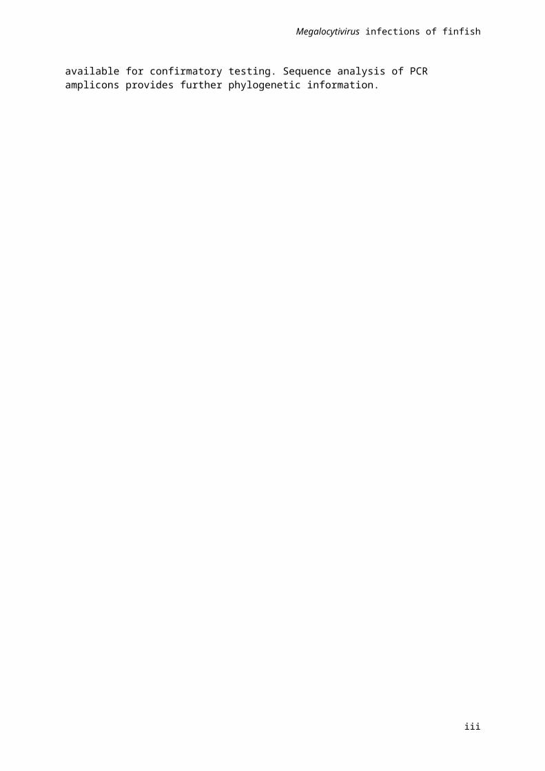
Figure 1 Example of Megalocytivirus conventional PCR test results
Note: Test uses the primers described by Kurita & Nakajima (2012) to generate a 777bp amplicon. Lanes 1 to 12: test samples, M: 100bp molecular weight marker, N1: negative extraction control, N2: no template control, P: positive control.
Sequence analysis of PCR ampliconsPCR amplicons are purified from the agarose using the QIAquick Gel Extraction Kit (Qiagen, Cat. No. 28704 for 50 extractions) and sequenced by conventional Sanger sequencing, with both the forward and reverse primers. Primer trimmed consensus sequences are then compared with Megalocytivirus sequences using software (such as Geneious Pro or Biomatters) or BLAST on the NCBI website.
Interpretation of resultsPositive PCR results from finfish tissue are indicative of the presence of Megalocytivirus DNA in the sample. However, a positive PCR does not indicate whether or not the sample contains infectious virus. Amplicons from positive PCR results from facilities, species or geographical locations where Megalocytivirus infections have not been reported should be sequenced and the sequence compared with known Megalocytivirus sequences to confirm the result. This is especially important when no other diagnostic test has been used. A negative result from finfish tissue is indicative of the absence of Megalocytivirus DNA in the sample.
Nested PCR (nPCR)
Principle of the testnPCR is used to amplify a specific sequence from a DNA target. The primary (first-step) PCR amplifies the specific sequence to produce multiple copies. To achieve even greater sensitivity a second, or
13

Megalocytivirus infections of finfish
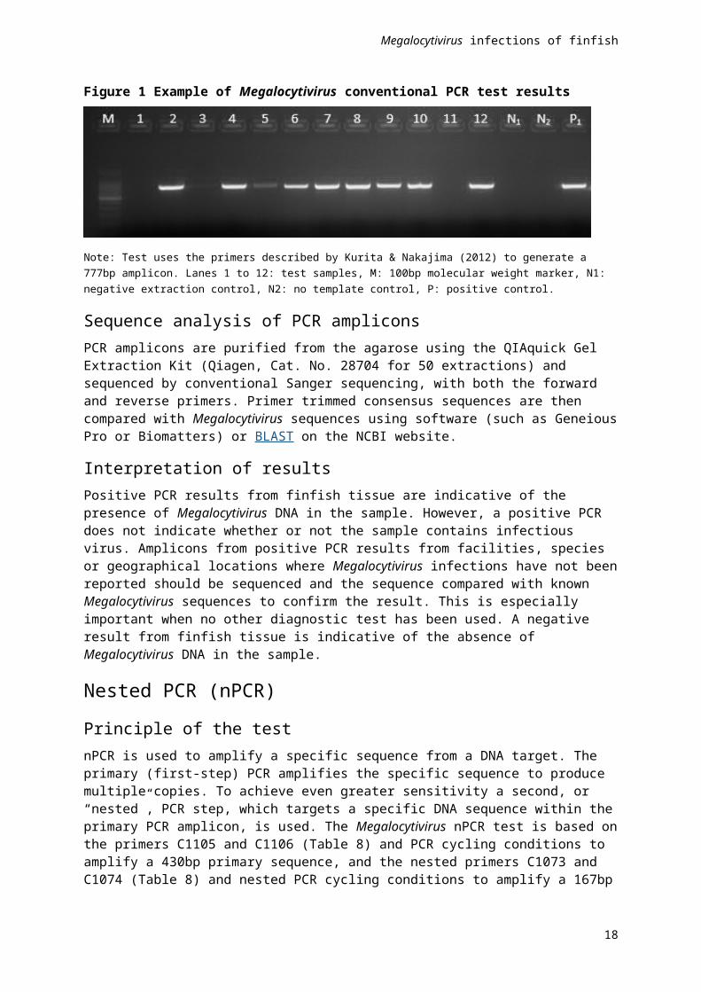
“nested”, PCR step, which targets a specific DNA sequence within the primary PCR amplicon, is used. The Megalocytivirus nPCR test is based on the primers C1105 and C1106 (Table 8) and PCR cycling conditions to amplify a 430bp primary sequence, and the nested primers C1073 and C1074 (Table 8) and nested PCR cycling conditions to amplify a 167bp secondary sequence from the Megalocytivirus major capsid protein gene (Rimmer et al, 2012).
Table 8 Primers used in the Megalocytivirus nPCR test
Primer Sequence
C1105 5’- GGG TTC ATC GAC ATC TCC GCG -3’
C1106 5’- AGG TCG CTG CGC ATG CCA ATC -3’
C1073 5’- AAT GCC GTG ACC TAC TTT GC -3’
C1074 5’- GAT CTT AAC ACG CAG CCA CA -3’
Due to the highly sensitive nature of the PCR, highly developed technical skills as well as quality control procedures and separate work areas for the different components of the PCR test are essential to avoid contamination and production of false positive results.
Reagents and materials Transport Medium (TM). Hank’s balanced salt solution (Life Technologies) containing 200 IU/mL
penicillin, 200 µg/mL streptomycin sulphate and 2% (v/v) foetal bovine serum.
Fixatives.
QIAamp Viral RNA Mini Kit (Qiagen, Cat. No. 52904 for 50 extractions) or MagMAX™-96 Viral RNA Isolation Kit (Life Technologies, Cat. No. AMB1836). Both kits have been validated for use by AFDL for RNA and DNA extractions. Alternative kits should be validated for use.
Primers (Table 8).
Molecular grade water.
HotStarTaq™ Master Mix Kit (QIAGEN). Any Taq could be used; however, not all enzymes perform to the same standard, and comparative testing should be undertaken if reagents are changed. The HotStarTaq™ Master Mix Kit is specifically mentioned as this reagent was used for the optimisation and validation work for the Megalocytivirus PCR and the hot start format reduces the risk of reagent degradation due to temperature fluctuations, and of contamination due to a reduction in the number of components the operator must add.
DNA ladder and loading dye. Any commercially available DNA ladder that contains bands which enable easy confirmation of the size of the amplicons (approximately 430bp and 167bp) can be used.
Agarose.
Dye (such as SYBRSafe gel stain).
Thermal Cycler (Eppendorf MasterCycler).
Gel electrophoresis system.
Gel documentation system.
14

Megalocytivirus infections of finfish
Test procedureWhole larvae/fry or organ samples are homogenised in TM. The tissue sample required is dependent on the size of the fish (Table 9) with a 1:10 (w/v) tissue:TM ratio required for subsequent analysis. Homogenisation can be achieved using a mortar and pestle, bead beater, or stomacher bag and 2 lb hammer.
Table 9 Tissue samples required for testing
Fish size (length) Tissue sample
<1 cm Whole fish
1 – 5 cm Whole viscera
5 – 8 cm Spleen, heart, kidney, liver
>8 cm Spleen
For smaller sample volumes, tissue is homogenised using a mortar and pestle. The homogenate is clarified by centrifugation at 3000 x g for 15 minutes at 4°C. An aliquot of the clarified supernatant is used for DNA extraction.
DNA is extracted according to the manufacturer’s instructions.
PCR is performed in a 25 µL reaction mixture, containing 2 µL DNA sample, 2x Reaction Mix, 0.36 µM of each primer (C1105 and C1106), 12.5 µL HotStarTaq™ Master Mix Kit and molecular grade water. Reactions are conducted in a thermal cycler programmed as follows: 95°C for 15 minutes, then 30 cycles of 94°C for 30 seconds, 55°C for 30 seconds and 72°C for 60 seconds with a final extension at 72°C for 7 minutes.
Nested PCR amplification is carried out in a 25 µl reaction mix, containing 2 µl of the primary PCR reaction, 0.36 µM of each primer (C1073 and C1074), 12.5 µL HotStarTaq™ Master Mix and molecular grade water. Cycling conditions are as for the primary PCR.
Reaction products are analysed after electrophoresis through a 1.5% to 2% agarose gel (using standard procedures) and amplicons are visualised with SYBRSafe gel stain according to the manufacturer’s instructions.
Negative controls must have no evidence of specific amplicons. A positive reaction for the primary PCR results in the production of a discrete 430 bp amplicon and, for the nested PCR, the production of a discrete 167 bp amplicon (Error: Reference source not found). Amplicons should be sequenced for Megalocytivirus identification.
Interpretation of resultsPositive nPCR results from finfish tissue are indicative of the presence of Megalocytivirus DNA in the sample. However, a positive nPCR does not indicate whether or not the sample contains infectious virus. Amplicons from positive nPCR results from facilities, species or geographical locations where Megalocytivirus infections have not been reported should be sequenced and the sequence compared with known Megalocytivirus sequences to confirm the result. This is especially important when no other diagnostic test has been used. A negative result from finfish tissue is indicative of the absence of Megalocytivirus DNA in the sample.
15

Megalocytivirus infections of finfish
Figure 2 Example of positive nPCR results on serial 10-fold dilutions of RSIV
Note: Test uses the primers described by Rimmer et al, (2012). Upper gel: Primary PCR–positive amplicon of 430bp). Lower gel: nPCR–positive amplicon of 167bp. M: molecular weight marker, N1: negative extraction control, N2: no template control, P: positive control, N3: nested PCR no template control.
Virus isolation (RSIV only)
Principle of the testThe grunt fin (GF) cell line (American Type Culture Collection Catalogue number CCL-58) is the preferred cell line for isolation of RSIV and the following method is specific for cell cultures of the GF cell line. Refer to the OIE Manual (2015) for the method using the BF-2 cell line.
Virus isolation in monolayer cultures of susceptible cell lines is considered a sensitive test for determining the presence of infectious virus (OIE, 2015). For tissue samples submitted in appropriate condition (unfixed, stored under correct temperature and transportation conditions), processed correctly for virus isolation and inoculated onto cultures of susceptible cell lines in a PC3 facility, then incubated under appropriate conditions, the development of viral cytopathic effect (CPE) indicates the presence of infectious virus.
Reagents and materialsGrunt fin (GF) cell line (American Type Culture Collection Catalogue number CCL-58).
Growth medium: Earle's minimum essential medium with non-essential amino acids (EMEM) supplemented with 2mM glutamine (unless included in commercial medium), 100IU penicillin/100µg streptomycin/mL (or 50µg/mL gentamycin), 20% (v/v) foetal bovine serum (FBS), 3.5g/L sodium chloride.
16

Megalocytivirus infections of finfish
Maintenance medium: Earle's minimum essential medium with non-essential amino acids (EMEM) supplemented with 2mM glutamine (unless included in commercial medium), 100 IU penicillin/100 µg streptomycin /mL (or 50 µg/mL gentamycin), 5% (v/v) FBS, 3.5 g/L sodium chloride.
Equipment required for the establishment and maintenance of a fish cell culture laboratory has been discussed previously (Crane and Williams, 2008).
Test procedureWhole fry or organ samples are homogenised in TM and should be kept chilled (4°C to 10°C) during processing. Sample preparation and virus isolation are ideally undertaken on the day of sample receipt. If this is not possible, samples should be stored at –80°C. All manipulations are undertaken in a Class II Biological Safety Cabinet using aseptic technique and sterile equipment and reagents. The sample/tissue required is dependent on the size of the fish (Table 10).
Table 10 Tissue samples required for testing
Fish size (length) Tissue sample
<1 cm Whole fish
1–5 cm Whole viscera
5–8 cm Spleen, heart, kidney, liver
>8 cm Spleen
Prepare two sets of sterile centrifuge tubes labelled with the submission identification (ID) number, sample number and dilution. One set is used for the 1/10 sample dilution and the other set for the 1/100 sample dilution. Add 4.5 mL TM to each 1/100 tube. A clarified 1:10 (w/v) tissue suspension in TM is required after homogenisation. Therefore, weigh a sample container with a tissue sample and subtract the weight of an empty sample container to obtain an estimate of the weight of the tissue sample. Operating within a Class II Biological Safety Cabinet and using 10 mL sterile pipettes, dispense 4.5 mL TM into each 1/100 tube. Place all tubes in a test tube rack sitting in an ice slurry. Homogenise the tissue sample and resuspend in extra TM to achieve a 1:10 (w/v) tissue homogenate. Transfer to the tubes labelled 1/10 and clarify the homogenate by centrifugation at 3000 × g for 15 minutes at 4°C. Pipette 0.5 mL of the supernatant from each 1/10 tissue sample dilution into the corresponding 1/100 test tubes containing 4.5 mL TM.
The procedure described is for virus isolation in cultures of the GF cell line established in 24-well tissue culture plates. If different culture vessels are used (such as 96-well plates or 25 cm² tissue culture flasks), volumes are adjusted proportionally. Cells are seeded at a density of 3.5 x 10⁵ cells/mL with 1.5 mL of cell suspension (in growth medium) and incubated at 25°C overnight. Cultures should be less than 24 hours old when inoculated with the diagnostic sample. On the day of sample inoculation, examine cell cultures to be used by inverted light microscopy. Ensure that they are approximately 70% to 80% confluent, free from overt microbial contamination, and that mitotic figures are visible (that is, the cultures contain actively dividing cells). Any problems should be noted and, if necessary, fresh cultures prepared for use on the next day. Discard the cell culture medium from the cultures. Inoculate duplicate cultures with 150 µL of each sample dilution (1/10 and 1/100). One set (=column) of four well-cultures on each 24-well tissue culture plate should be used as negative controls, which are inoculated with 150 µL TM only. Incubate the cultures at 25°C for 1 hour to allow adsorption of any virus present. Following adsorption, add 1.5 mL maintenance medium to each culture (negative controls first) yielding final sample dilutions of 1/100 and 1/1000. Place culture plates in a 25°C incubator. On the day following inoculation, and every 1 to 3 days thereafter, examine the cultures by inverted light microscopy for any microbial contamination, tissue sample (non-specific) cytotoxicity and viral cytopathic effect (CPE).
17

Megalocytivirus infections of finfish
SubculturingAt 7-12 days after inoculation, test cultures not showing CPE should be passaged. For each tissue sample, the contents (medium and cells scraped from the substrate) of each of the four replicate cultures, irrespective of dilution, are pooled into sterile centrifuge tubes. If tissue sample cytotoxicity or bacterial/fungal contamination has been observed during the initial culture period for each pool, the pooled contents should be filtered (0.45 μm) into the sterile centrifuge tube. Alternatively, samples can be centrifuged at 10,000 x g for 10 minutes at 5°C. Each pooled sample is diluted (1/10) by pipetting 0.5 mL of the supernatant into a test tube containing 4.5 mL TM. Without decanting the cell culture medium, inoculate duplicate fresh cell cultures in 24-well culture plates, prepared as described above, with 150 µL of the pooled undiluted supernatant and a further duplicate set of wells with the 150 µL of the 1/10 dilution of the pooled supernatant. Place culture plates in a 25°C incubator. Observe these cultures and record observations. Irrespective of the time at which the passage occurred, cell cultures should be observed for at least 21 days for completion of the assay. The test is valid if the negative control cultures retain normal cellular morphology for the full period of incubation in the absence of bacterial contamination. To ensure cell lines, used on a routine basis, are susceptible to the viruses of concern, titrations of a positive control viral stock on each of the cell lines should be carried out on a regular basis (for example, 6 monthly).
Interpretation of resultsThe test sample is negative if the inoculated cell cultures retain normal cellular morphology similar to the negative control cultures and the cellular monolayer retains normal integrity (Error: Reference source not found; B and D). If virus isolation is the only test performed, confirmation by qPCR should be undertaken on a sample of the cell culture to avoid false-negative results. If any of the cell cultures inoculated with test samples demonstrate any abnormalities, such as increased intracellular vacuolation, enlarged cells, cell rounding or monolayer disruption (Error: Reference source not found; A and C), further investigation is required, such as examination by electron microscopy, further sub-culturing or confirmation of the presence of Megalocytivirus by molecular methods as described above.
18
Megalocytivirus infections of finfish
Figure 3 Examples of RSIV-infected and uninfected GF cell cultures
Note: Examples of uninfected GF cell cultures at 7 days (A) and 11 days (B) post-seeding. Examples of RSIV-infected GF cell cultures at 7 days post-seeding, 6 days post-inoculation (C) and 11 days post-seeding, 10 days post-inoculation (D).
19
DC
BA
Megalocytivirus infections of finfish
ReferencesBecker J, Rimmer A, Tweedie A, Landos M, Lintermans M, Whittington R. 2013, Aquatic Animal Health Subprogram: Surveys of ornamental fish for pathogens of quarantine significance, Fisheries Research and Development Corporation Project Number 2009/044.
Beld, MGHM, Birch CJ, Cane PA, Carman W, Claas ECJ, Clewley JP, Domingo E, Druce J, Escarmis C, Fouchier RAM, Foulongne V, Ison MG, Jennings LC, Kaltenboeck B, Kay ID, Kubista M, Landt O, Mackay IM, Mackay J, Niesters HGM, Nisen MD, Palladino S, Papadopoulos NG, Petrich A, Pfaffl MW, Rawlinson W, Reischl U, Saunders NA, Savolainen-Kopra C, Schildgen O, Scott GM, Segondy M, Seibl R, Sloots TP, Tang YW, Tellier R, Woo PCY, 2007, ‘Experts roundtable: Real-time PCR and microbiology’, in Mackey IM (eds), Real-time PCR in microbiology: From diagnosis to characterization, Caister Academic Press, Norwich, UK. pp 357–443.
Caipang CM, Hirono I, Aoki T, 2003, Development of a real-time PCR assay for the detection and quantification of red seabream iridovirus (RSIV), Fish Pathology, vol. 38, pp. 1–7, doi: 10.3147/jsfp.38.1.
Caipang CM, Takano T, Hirono I, Aoki T, 2006, Genetic vaccines protect red seabream, Pagrus major, upon challenge with red seabream iridovirus (RSIV), Fish and Shellfish Immunology, vol. 21, pp. 130–138, doi: 10.1016/j.fsi.2005.10.012.
Chao C-B, Chen C-Y, Lai Y-Y, Lin C-S, Huang H-T, 2004, Histological, ultrastructural, and in situ hybridization study on enlarged cells in the grouper Epinephelus hybrids infected with grouper iridovirus in Taiwan (TGIV), Diseases of Aquatic Organisms, vol. 58, pp. 127–142, doi: 10.3354/dao058127.
Chen XH, Lin KB, Wang XW, 2003, Outbreaks of an iridovirus disease in maricultured large yellow croaker, Larimichthys crocea (Richardson), in China, Journal of Fish Diseases, vol 26, pp. 615–619, doi: 10.1046/j.1365-2761.2003.00494.x.
Choi SK, Kwon SR, Nam YK, Kim SK, Kim KH, 2006, Organ distribution of red sea bream iridovirus (RSIV) DNA in asymptomatic yearling and fingerling rock bream (Oplegnathus fasciatus) and effects of water temperature on transition of RSIV into acute phase, Aquaculture, vol. 256, pp. 23–26, doi: 10.1016/j.aquaculture.2006.01.026.
Chou HY, Hsu CC, Peng TY, 1998, Isolation and characterization of a pathogenic iridovirus from cultured grouper (Epinephelus sp.) in Taiwan, Fish Pathology, vol. 33, pp. 201–206, doi: 10.3147/jsfp.33.201.
Chua FH, Ng ML, Ng KL, Loo JJ, Lee JY, 1994, Investigation of outbreaks of a novel disease, “Sleepy Grouper Disease”, affecting the brown-spotted grouper, Epinephelus tauvina Forskal, Journal of Fish Diseases, vol. 17, pp. 417–427, doi: 10.1111/j.1365-2761.1994.tb00237.x.
Crane MStJ, Williams LM., 2008, Viruses of salmonids: Virus isolation in fish cell lines, Australian and New Zealand Standard Diagnostic Procedure, Sub-Committee on Animal Health Laboratory Standards, http://www.scahls.org.au/Procedures/Documents/AQANZSDP/Virus_Isolation.pdf (Retrieved 10/08/15).
Danayadol Y, Direkbusarakom S, Boonyaratpalin S, Miyazaki T, Miyata M, 1997, ‘Iridovirus infection in brown-spotted grouper (Epinephelus malabaricus) cultured in Thailand’, in TW Flegel and IH MacRae (eds.), Diseases in Asian Aquaculture III. Fish Health Section, Asian Fisheries Society, Manila, pp. 67–72.
20

Megalocytivirus infections of finfish
de Groot A, Guelen L, Deijs M, van der Wal Y, Miyata M, Ng K-S, van Grinsven L, Simmelink Y, Gisez L, van Lent J, de Ronde A, Chang S-F, Schrier C and van der Hoek L, 2015, A novel virus causes scale drop syndrome disease in Lates calcarifer, PLoS Pathogens, vol. 11, no. 8, pp. e1005074, doi: 10.1371/journal.ppat.1005074.
Do JW, Cha SJ, Kim JS, An EJ, Park MS, Kim JW, Kim YC, Park MA, Park JW, 2005, Sequence variation in the gene encoding the major capsid protein of Korean fish iridoviruses, Archives of Virology, vol. 150, pp. 351–359.
Dong C, Weng S, Luo Y, Huang M, Ai H, Yin Z, He J, 2010, A new marine megalocytivirus from spotted knifejaw, Oplegnathus punctatus, and its pathogenicity to freshwater mandarinfish, Siniperca chuatsi, Virus Research, vol. 147, pp. 98–106, doi: 10.1016/j.virusres.2009.10.016.
Dong Y, Weng S, He J, Dong C, 2013, Field trial tests of FKC vaccines against RSIV genotype Megalocytivirus in cage-cultured mandarin fish (Siniperca chuatsi) in an inland reservoir, Fish and Shellfish Immunology, vol. 35, pp. 1598–1603, doi: 10.1016/j.fsi.2013.09.005.
Fu X, Li N, Lai Y, Liu L, Lin Q, Shi C, Huang Z, Wu S, 2012, Protective immunity against iridoviral disease in mandarin fish, induced by recombinant major capsid protein of infectious spleen and kidney necrosis virus, Fish and Shellfish Immunology, vol. 33, pp. 880–995, doi: 10.1016/j.fsi.2012.07.012.
Fu X, Li N, Liu L, Lin Q, Wang F, Lai Y, Jiang H, Pan H, Shi C, Wu S, 2011, Genotype and host range analysis of infectious spleen and kidney necrosis virus (ISKNV), Virus Genes, vol. 42, pp. 97–109.
Gias E, Johnston C, Keeling S, Spence RP, McDonald WL, 2011, Development of real-time PCR assays for detection of megalocytiviruses in imported ornamental fish, Journal of Fish Diseases, vol. 34, pp. 609–618, doi: 10.1111/j.1365-2761.2011.01274.x.
Gibson-Kueh S, Netto P, Ngoh-Lim GH, Chang SF, Ho LL, Qin QW, Chua FHC, Ng ML, Ferguson HW, 2003, The pathology of systemic iridoviral disease in fish, Journal of Comparative Pathology, vol. 129, pp. 111–119, doi: 10.1016/S0021-9975(03)00010-0.
Gibson-Kueh S, Ngoh-Lim GH, Netto P, Kurita J, Nakajima K, Ng ML, 2004, A systematic iridoviral disease in mullet, Mugil cephalus L. and tiger grouper, Epinephelus fuscoguttatus Forsskal: a first report and study, Journal of Fish Diseases, vol. 27, pp. 693–699, doi: 10.1111/j.1365-2761.2004.00589.x.
Go J, Lancaster M, Deece K, Dhungyel O, Whittington R, 2006, The molecular epidemiology of iridovirus in Murray cod (Maccullochella peelii peelii) and dwarf gourami (Colisa lalia) from distant biogeographical regions suggests a link between trade in ornamental fish and emerging iridoviral diseases, Molecular and Cellular Probes, vol. 20, pp. 212–222, doi: 10.1016/j.mcp.2005.12.002.
Go J, Whittington R, 2006, Experimental transmission and virulence of a megalocytivirus (Family Iridoviridae) of dwarf gourami (Colisa lalia) from Asia in Murray cod (Maccullochella peeli peeli) in Australia, Aquaculture, vol. 258, pp. 140–149, doi: 10.1016/j.aquaculture.2006.04.033.
Gunson RN, Collins TC, Carman WF, 2006, Practical experience of high throughput real-time PCR in routine diagnostic virology, Journal of Clinical Virology, vol. 35, pp. 355–367, doi: 10.1016/j.jcv.2005.12.006.
He JG, Deng M, Weng SP, Li Z, Zhou SY, Long QX, Wang XZ, Chan S-M, 2001, Complete genome analysis of the mandarin fish infectious spleen and kidney necrosis iridovirus, Virology, vol. 291, pp. 126–139, doi: 10.1006/viro.2001.1208.
21

Megalocytivirus infections of finfish
He JG, Wang SP, Zeng K, Huang ZJ, Chan S-M, 2000, Systemic disease caused by an iridovirus-like agent in cultured mandarinfish, Siniperca chuatsi (Basilewsky), in China, Journal of Fish Diseases, vol. 23, pp. 219–222, doi: 10.1046/j.1365-2761.2000.00213.x.
He JG, Zeng K, Weng SP, Chan S-M, 2002, Experimental transmission, pathogenicity and physical-chemical properties of infectious spleen and kidney necrosis virus (ISKNV), Aquaculture, vol. 204, pp. 11–24, doi: 10.1016/S0044-8486(01)00639-1.
Huang SM, Tu C, Tseng CH, Huang CC, Chou CC, Kuo HC, Chang SK, 2011, Genetic analysis of fish iridoviruses isolated in Taiwan during 2001–2009, Archives of Virology, vol. 156, pp. 1505–1515.
Inouye K, Yamano K, Maeno Y, Nakajima K, Matsuoka M, Wada Y, Sorimachi M, 1992, Iridovirus infection of cultured red sea bream, Pagrus major, Fish Pathology, vol. 27, pp. 19–27, doi: 10.3147/jsfp.27.19.
Ito T, Yoshiura Y, Kamaishi T, Yoshida K, Nakajima K, 2013, Prevalence of red sea bream iridovirus among organs of Japanese amberjack (Seriola quinqueradiata) exposed to cultured red sea bream iridovirus, Journal of General Virology, vol. 94, pp. 2094–2101, doi: 10.1099/vir.0.052902-0.
Jancovich LK, Chinchar VG, Hyatt A, Miyakazi T, Williams T, Zhang QY, 2012, Family Iridoviridae, in Virus Taxonomy. Ninth Report of the International Committee on Taxonomy of Viruses. King AMQ, Adams MJ, Carstens EB, Lefkowitz EJ (eds) Elsevier Academic Press, Sydney Australia. pp 193–210.
Jeong JB, Jun LJ, Yoo MH, Kim MS, Komisar JL, Jeong HD, 2003, Characterisation of the DNA nucleotide sequences in the genome of red sea bream iridoviruses isolated in Korea, Aquaculture, vol. 220, pp. 119–133, doi: 10.1016/S0044-8486(02)00538-0.
Jeong JB, Kim HY, Jun LJ, Lyu JH, Park NG, Kim JK, Jeong HD, 2008, Outbreaks and risks of infectious spleen and kidney necrosis virus diseases in freshwater ornamental fishes, Diseases of Aquatic Organisms, vol. 78, pp. 209–215, doi: 10.3354/dao01879.
Jun LJ, Jeong JB, Kim JH, Nam JH, Shin KW, Kim JK, Kang J-C, Jeong HD, 2009, Influence of temperature shifts on the onset and development of red sea bream iridoviral disease in rock bream Oplegnathus fasciatus, Diseases of Aquatic Organisms, vol. 84, pp. 201–208, doi: 10.3354/dao02041.
Jung S, Miyazaki T, Miyata M, Danayadol Y, Tanaka S, 1997, Pathogenicity of iridovirus from Japan and Thailand for the red sea bream Pagrus major in Japan, and histopathology of experimentally infected fish, Fisheries Science, vol. 63, pp. 735–740, doi: 10.2331/fishsci.63.735.
Jung SJ, Oh MJ, 2000, Iridovirus-like infection associated with high mortalities of striped beakperch, Oplegnathus fasciatus (Temminck et Schlegel), in southern coastal areas of the Korean peninsula, Journal of Fish Diseases, vol. 23, pp. 223–226, doi: 10.1046/j.1365-2761.2000.00212.x.
Kawakami H, Nakajima K, 2002, Cultured fish species affected by red sea bream iridoviral disease from 1996 to 2000, Fish Pathology, vol. 37, pp. 45–47, doi: 10.3147/jsfp.37.45.
Kim YJ, Jung SJ, Choi TJ, Kim HR, Rajendran KV, Oh MJ, 2002, PCR amplification and sequence analysis of irido-like virus infecting fish in Korea, Journal of Fish Diseases, vol. 25, pp. 121–124, doi: 10.1046/j.1365-2761.2002.00345.x.
Kim WS, Oh MJ, Kim JO, Kim D, Jeon CH, Kim JH, 2010, Detection of Megalocytivirus from imported tropical ornamental fish, paradise fish Macropodus opercularis, Diseases of Aquatic Organisms, vol. 90, pp. 235–239, doi.org/10.3354/dao02232.
Kurita J, Nakajima K, 2012, Megalocytiviruses, Viruses, vol. 4, pp. 521–538, doi: 10.3390/v4040521.
22

Megalocytivirus infections of finfish
Kurita J, Nakajima K, Hirono I, Aoki T, 1998, Polymerase chain reaction (PCR) amplification of DNA of red sea bream iridovirus (RSIV), Fish Pathology, vol. 33, pp. 17–23, doi: 10.3147/jsfp.33.17.
Lee N-S, Do JW, Park JW, Kim YC, 2009, Characterization of virus distribution in rock bream (Oplegnathus fasciatus; Temminck and Schegel) infected with M egalocytivirus , Journal of Comparative Pathology, vol. 141, pp. 63–69, doi: 10.1016/j.jcpa.2009.03.008.
Marcos-Lopez M, Feist SW, Hicks R, Noguera PA, 2011, ‘Systemic Megalocytivirus infection in three-spined stickleback Gasterosteus aculeatus’, Bulletin of the European Association of Fish Pathologists, vol. 31, pp. 227–234.
Mahardika K, Zafran, Yamamoto A, Miyazaki T, 2004, Susceptibility of juvenile humpback grouper Cromileptes altivelis to grouper sleepy disease iridovirus (GSDIV), Diseases of Aquatic Organisms, vol. 59, pp. 1–9, doi: 10.3354/dao059001.
Matsuoka S, Inouye K, Nakajima K, 1996, Cultured fish species affected by red sea bream iridoviral disease from 1991 to 1995, Fish Pathology, vol. 31, pp. 233–234, doi: 10.3147/jsfp.31.233.
Miyata M, Matsuno K, Jung SJ, Danayadol Y, Miyazaki T, 1997, Genetic similarity of iridoviruses from Japan and Thailland, Journal of Fish Diseases, vol. 20, pp. 127–134, doi: 10.1046/j.1365-2761.1997.d01-115.x.
Mohr PG, Moody NJG, Williams LM, Hoad J, Cummins DM, Crane MStJ, 2015, Molecular confirmation of an ISKNV-like virus detected in farmed ornamental Platys (Xiphophorus maculates), Diseases of Aquatic Organisms, vol. 116, pp. 103–110, doi: 10.3354/dao02896.
Murali S, Wu M-F, Guo I-C, Chen S-C, Yang H-W, Chang C-Y, 2002, Molecular characterization and pathogenicity of a grouper iridovirus (GIV) isolated from yellow grouper, Epinephelus awoara (Temminck & Schlegel). Journal of Fish Diseases, vol. 25, pp. 91–100, doi: 10.1046/j.1365-2761.2002.00343.x.
Nakajima K, Maeno Y, 1998, Pathogenicity of red sea bream iridovirus and other fish iridoviruses to red sea bream, Fish Pathology, vol. 33, pp. 143–144, doi: 10.3147/jsfp.33.143.
Nakajima K, Maeno Y, Honda A, Yokoyama K, Tooriyama T, Manabe S, 1999, Effectiveness of a vaccine against red sea bream iridoviral disease in a field trial test, Diseases of Aquatic Organisms, vol. 36, pp. 73–75, doi: 10.3354/dao036073.
Nolan D, Stephens F, Crockford M, Jones JB, Snow M, 2015, Detection and characterization of viruses of the genus Megalocytivirus in ornamental fish imported into an Australian border quarantine premises an emerging risk to national biosecurity, Journal of Fish Diseases, vol. 38, pp. 187–195, doi: 10.1111/jfd.12222.
Oh S-Y, Oh M-J, Nishizawa T, 2014, Potential for a live red seabream (RSIV) vaccine in rock bream Oplegnathus fasciatus at a low rearing temperature, Vaccine, vol. 32, pp. 363–368, doi: 10.1016/j.vaccine.2013.11.030.
OIE, 2015, World Organisation for Animal Health (OIE) – Manual of Diagnostic Tests for Aquatic Animals, World Organisation for Animal Health, Paris, France, accessed on 5 September 2015.
Oseko N, Thye CT, Palanisamy V, Maeno Y, Kurita J, 2004, Iridovirus isolated from diseased sea bass Lates calcarifer and red drum Sciaenops ocellatus causing mass mortality in Malaysia, Global Symposium on Gender and Fisheries, 7th Asian Fisheries Forum, Penang, Malaysia.
23

Megalocytivirus infections of finfish
Purcell MK, Getchell RG, McClure CA, Garver KA, 2011, Quantitative polymerase chain reaction (PCR) for detection of aquatic animal pathogens in a diagnostic laboratory setting, Journal of Aquatic Animal Health, vol. 23, pp. 148–161, doi: 10.1080/08997659.2011.620217.
Razak AA, Ransangan J, Sade A, 2014, First report of M egalocytivirus (Iridoviridae) in grouper culture in Sabah, Malaysia, International Journal of Current Microbiology and Applied Sciences, vol. 3, pp. 896–909.
Rimmer AE, Becker JA, Tweedie A, Whittington RJ, 2012, Development of a quantitative polymerase chain reaction (qPCR) assay for the detection of dwarf gourami iridoviruses (DGIV) and other megalocytiviruses and comparison with the Office International des Epizooties (OIE) reference protocol, Aquaculture; vol. 358–359, pp. 155–163, doi: 10.1016/j.aquaculture.2012.06.034.
Rodger HD, Kobs M, Macartney A, Frerichs GN, 1997, Systemic iridovirus infection in freshwater angelfish, Pterophyllum scalare (Lichtenstein), Journal of Fish Diseases; vol. 20, pp. 69–72, doi: 10.1046/j.1365-2761.1997.d01-106.x.
Sano M, Minagawa M, Nakajima K, 2002, Multiplication of red sea bream iridovirus (RSIV) in experimentally infected grouper (Epinephelus malabaricus), Fish Pathology; vol. 37, pp. 163–168, doi: 10.3147/jsfp.37.163.
Shimmoto H, Kawai K, Ikawa T, Oshima S-I, 2010, Protection of red sea bream Pagrus major against red sea bream iridovirus infection by vaccination with a recombinant viral protein, Microbiology and Immunology, vol. 54, pp. 135–142, doi: 10.1111/j.1348-0421.2010.00204.x.
Song J-Y, Kitamura S-I, Jung S-J, Miyadai T, Tanaka S, Fukuda Y, Kim S-R, Oh M-J, 2008, Genetic variation and geographic distribution of megalocytiviruses, Journal of Microbiology, vol. 46, pp. 29–33.
Sriwanayos P, Francis-Floyd R, Stidworthy MF, Petty BD, Kelley K, Waltzek TB, 2013, Megalocytivirus infection in orbiculate batfish Platax orbicularis, Diseases of Aquatic Organisms, vol. 105, pp. 1–8, doi: 10.3354/dao02594.
Subramaniam K, Shariff M, Omar AR, Hair-Bejo M, Ong BL, 2014, Detection and molecular characterisation of infectious spleen and kidney necrosis virus from major ornamental fish breeding states in peninsular Malaysia, Journal of Fish Diseases, vol. 37, pp. 609–618, doi: 10.1111/jfd.12152.
Sudthongkong C, Miyata M, Miyazaki T, 2002, Iridovirus disease in two ornamental tropical freshwater fishes: African lampeye and dwarf gourami, Diseases of Aquatic Organisms, vol. 48, pp. 163–173, doi: 10.3354/dao048163.
Sudthongkong C, Miyata M, Miyazaki T, 2002a, Viral DNA sequences of genes encoding the ATPase and the major capsid protein of tropical iridovirus isolates which are pathogenic to fishes in Japan, South China Sea and Southeast Asian countries. Archives of Virology, vol. 147, pp. 2089–2109.
Tamura K, Peterson D, Peterson N, Stecher G, Nei M, Kumar S, 2011, MEGA5: Molecular evolutionary genetics analysis using maximum likelihood, evolutionary, distance, and maximum parsimony methods, Molecular and Biological Evolution, vol28, pp. 2731–2739, doi: 10.1093/molbev/msr121.
Waltzek TB, Marty GD, Alfaro ME, Bennett WR, Garver KA, Haulena M, Weber ES, Hedrick RP, 2012, Systemic iridovirus from threespined stickleback Gasterosteus aculeatus represents a new Megalocytivirus species (family Iridoviridae) , Diseases of Aquatic Organisms, vol. 98, pp. 41–56, doi: 10.3354/dao02415.
24

Megalocytivirus infections of finfish
Wang CS, Chao SY, Ku CC, Wen CM, Shih HH, 2009, PCR amplification and sequence analysis of the major capsid protein gene of megalocytiviruses isolated in Taiwan, Journal of Fish Diseases, vol. 32, pp. 543–550, doi: 10.1111/j.1365-2761.2009.01043.x.
Wang Q, Zeng WW, Li KB, Chang OQ, Liu C, Wu GH, Shi CB, Wu SQ, 2011, Outbreaks of an iridovirus in marbled sleepy goby, Oxyeleotris marmoratus (Bleeker), cultured in southern China, Journal of Fish Diseases, vol. 34, pp. 399–402, doi: 10.1111/j.1365-2761.2011.01244.x.
Weber ES, Waltzek TB, Young DA, Twitchell EL, Gates AE, Vagelli A, Risatti GR, Hedrick RP, Salvatore F, 2009, Systemic iridovirus infection in the Banggai cardinalfish (Pterapogon kauderni (Koumans 1933), Journal of Veterinary Diagnostic Investigation, vol. 21, pp. 306–320, doi: 10.1177%2F104063870902100302.
Weng SP, Wang YQ, He JG, Deng M, Lu L, Guan HJ, Liu YJ, Chan S-M, 2002, Outbreaks of an iridovirus in red drum, Sciaenops ocellata (L.), cultured in southern China, Journal of Fish Diseases, vol. 25, pp. 681–685, doi: 10.1046/j.1365-2761.2002.00419.x.
Whittington RJ, Chong R, 2007, Global trade in ornamental fish from an Australian perspective: The case for revised import risk analysis and management practices, Preventive Veterinary Medicine, vol. 81, pp. 92–116, doi: 10.1016/j.prevetmed.2007.04.007.
Xu X, Zhang L, Weng S, Huang Z, Lu J, Lan D, Zhong X, Yu X, Xu A, He J, 2008, A zebrafish (Danio rerio) model of infectious spleen and kidney necrosis virus (ISKNV) infection, Virology, vol. 376, pp. 1–12, doi: 10.1016/j.virol.2007.12.026.
25

Megalocytivirus infections of finfish
Appendix A: Reagents and kits
RSIV-infected GF cell culture supernatant for PCR positive controls
Ethanol-inactivated RSIV-infected cell culture supernatant is available from the authors.
Suppliers of reagents and kits
In Australia: AAHL Fish Diseases Laboratory, CSIRO Australian Animal Health Laboratory, 5 Portarlington Rd,
East Geelong VIC 3219. Ph: + 61 3 5227 5000, Fax: +61 3 5227 5555, Email: [email protected] (for plasmid control for qPCR (Mohr et al 2015)).
University of Sydney, Faculty of Veterinary Science, 425 Werombi Road Camden NSW 2570. Ph: +61 2 9351 1787, Fax: +61 2 9351 1718, Email: [email protected] (for plasmid control for qPCR (Rimmer at al 2012)).
Qiagen, www.qiagen.com, PO Box 169 Chadstone Centre VIC 3148. Email: [email protected], Ph: 1800 243 066, Fax: 03-9840-9888.
Life Technologies, www.lifetechnologies.com/au/en/home.html. Life Technologies Australia Pty Ltd. 30-32 Compark Circuit, Mulgrave VIC 3170. Email: [email protected], Tel: 1800 636 327, Fax: 1800 143 363.
In New Zealand: Qiagen, www.qiagen.com, Bio-Strategy Ltd www.bio-strategy.com. PO Box 303 385, Auckland
0751. E-mail: [email protected], Tel: 0800 34 24 66
Life Technologies, www.Life Technologies.com.au, Life Technologies New Zealand Limited, 18-24 Botha Road Penrose Auckland 1006. Email: [email protected], Tel: 0800 636 327
26