Embed Size (px)
Citation preview

Am. J. Hum. Genet. 41:145-156, 1987
A Unique Dicentric X;Y Translocation with Xq and YpBreakpoints: Cytogenetic and Molecular Studies
R. BERNSTEIN,* J. ROSENDORFF,* M. RAMSAY,* M. R. PINTO,*AND D. C. PAGEt
*MRC Human Ecogenetics Research Unit, Department of Human Genetics, School ofPathology, South African Institute for Medical Research and University of the Witwatersrand,
Johannesburg; and tWhitehead Institute for Biomedical Research, Cambridge, MA
SUMMARY
A 32-year-old woman presented with secondary amenorrhea and in-fertility. She was of normal height and her breasts were well devel-oped, but she had streak gonads; there were no signs of virilization,and she showed no somatic stigmata of Turner syndrome. Chromo-some analysis revealed a dicentric X;Y translocation with Xq and Ypbreakpoints. Centromeric banding demonstrated a Y centromere anda "suppressed" X centromere. The karyotype of the patient was in-terpreted as 46,X,t(X;Y)(q22;pl 1). The Yp breakpoint was confirmedby DNA-hybridization studies with six probes detecting Y-specificsequences. These DNA-hybridization studies were consistent withthe presence of the long arm, centromere, and much of the proximalshort arm of the Y. The Y-DNA studies of this female also revealedthe absence of the distal short arm of the Y chromosome, to which thetestis-determining factor has previously been localized.
INTRODUCTION
Cytologically identifiable X;Y translocations that include Yq are very uncom-mon: fewer than 40 cases, most of them familial, were reviewed by Fryns andvan den Berghe (1983) and Bernstein (1985); a few more have since beendocumented (Cameron et al. 1984; Kelly et al. 1984; Wegner et al. 1984; Ross etal. 1985; Speevak et al. 1985). Interest in these sex-chromosome translocations
Received September 23, 1986; revision received January 28, 1987.Address for correspondence and reprints: Dr. Renee Bernstein, Department of Human Genetics,
SAIMR, P.O. Box 1038, Johannesburg, South Africa.© 1987 by the American Society of Human Genetics. All rights reserved. 0002-9297/87/4102-0006$02.00
145
lies not in their rarity but in the information that they have yielded about (1) thelocalization of genes on the pairing regions of the X and Y short arms and (2)the roles of the X and Y chromosomes in sex determination. All the X;Ytranslocations so far reported have involved the short arm of the X and the longarm of the Y, with the exception of two definitive cases (Cameron et al. 1984;Kelly et al. 1984) and one doubtful case (Borgaonkar et al. 1974; Koo et al.1977; Kunkel et al. 1977; Jones et al. 1979) who showed Xq;Yq breakpoints andthree cases with Xp and presumptive Yp breakpoints (Bernstein et al. 1978,1980; Zuffardi et al. 1982).Cameron et al. (1984) described an oligomenorrheic female of normal height
with a 46,X,t(X;Y)(q22;ql2) karyotype who was monosomic for regionXq22--Xqter; only the heterochromatic Yql2->Yqter segment was trans-located to Xq22. The patient reported by Kelly et al. (1984) had a46,X,t(X;Y)(q22;ql 1) karyotype associated with streak gonads.
In the present report, we describe, in an amenorrheic woman of normalstature, a unique dicentric t(X;Y) involving the same Xq22 breakpoint as thatfound in the patients of Cameron et al. (1984) and Kelly et al. (1984). Ourpatient differs from the latter two cases by having a translocation breakpoint onYp and a suppressed X centromere. The t(X;Y) was nonrandomly inactivated.The precise breakpoint on Yp could not be cytogenetically determined, butmolecular hybridization with Y-specific DNA probes indicated that much of theY short arm was present.
CASE REPORT
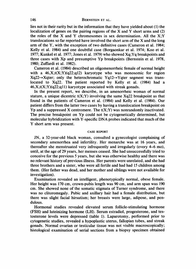
JN, a 32-year-old black woman, consulted a gynecologist complaining ofsecondary amenorrhea and infertility. Her menarche was at 16 years, andthereafter she menstruated very infrequently and irregularly (every 4-6 mo),until, at the age of 29 years, her menses ceased. She had unsuccessfully tried toconceive for the previous 5 years, but she was otherwise healthy and there wasno relevant history of previous illness. Her parents were unrelated, and she hadthree brothers and a sister, who were all fertile and had had 15 children amongthem. (Her father was dead, and her mother and siblings were not available forinvestigation).Examination revealed an intelligent, phenotypically normal, obese female.
Her height was 170 cm, crown-pubis length was 90 cm, and arm span was 190cm. She showed none of the somatic stigmata of Turner syndrome, and therewas no clitoromegaly. Pubic and axillary hair had a female distribution, butthere was slight facial hirsutism; her breasts were large, adipose, and pen-dulous.Hormonal studies revealed elevated serum follicle-stimulating hormone
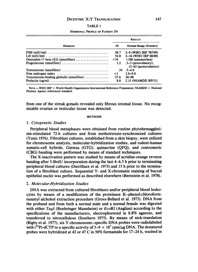
(FSH) and luteinizing hormone (LH). Serum estradiol, progesterone, and tes-tosterone levels were depressed (table 1). Laparotomy, performed prior tocytogenetic studies, revealed a hypoplastic uterus, fallopian tubes, and streakgonads. Normal ovarian or testicular tissue was not visible macroscopically;histological examination of serial sections from a biopsy specimen obtained
146 BERNSTEIN ET AL.
DICENTRIC X;Y TRANSLOCATION
TABLE I
HORMONAL PROFILE OF PATIENT JN
RESULTS
HORMONE JN Normal Range (Females)
FSH (mIU/ml) ....................................... 58.7 2-9 (WHO IRP 78/549)LH (mIU/ml) ............. 54.8 2-16 (WHO IRP 68/40)Oestradiol-17 beta (E2) (pmollliter). <74 <100 (amenorrhea)Progesterone (nmol/liter) ............. ................. 1.2 .5-5 (preovulatory);
15-65 (postovulatory)Testosterone (nmol/liter) ............. ................. .54 .5-4.0Free androgen index ................ ................. <1 2.0-9.0Testosterone-binding globulin (nmol/liter) ....... ........ 27.6 30-90Prolactin (ng/ml) ..................................... 8.0 2.15 (NIAMDD RP/1 I)
NOTE.-WHO IRP = World Health Organization International Reference Preparation; NIAMDD = NationalPituitary Agency referenced standard.
from one of the streak gonads revealed only fibrous stromal tissue. No recog-nizable ovarian or testicular tissue was detected.
METHODS
1. Cytogenetic StudiesPeripheral blood metaphases were obtained from routine phytohemaggluti-
nin-stimulated 72-h cultures and from methotrexate-synchronized cultures(Yunis 1976). Fibroblast cultures, established from a skin biopsy, were utilizedfor chromosome analysis, molecular-hybridization studies, and rodent-humansomatic-cell hybrids. Giemsa (GTG), quinacrine (QFQ), and centromeric(CBG) banding were performed by means of standard techniques.The X-inactivation pattern was studied by means of acridine-orange reverse
banding after 5-BrdU incorporation during the last 4-6.5 h prior to terminatingperipheral blood cultures (Dutrillaux et al. 1973) and 15 h prior to the termina-tion of a fibroblast culture. Sequential Y- and X-chromatin staining of buccalepithelial nuclei was performed as described elsewhere (Bernstein et al. 1978).
2. Molecular-Hybridization StudiesDNA was extracted from cultured fibroblasts and/or peripheral blood leuko-
cytes by means of a modification of the proteinase K-phenol:chloroform:isoamyl alchohol extraction procedure (Gross-Bellard et al. 1973). DNA fromthe proband and from both a normal male and a normal female was digestedwith either TaqI (Boehringer Mannheim) or EcoRI (Anglian) according to thespecifications of the manufacturers, electrophoresed in 0.8% agarose, andtransferred to nitrocellulose (Southern 1975). By means of nick-translation(Rigby et al. 1977), six Y chromosome-specific DNA probes were radiolabeledwith (32P)-dCTP to a specific activity of 3-9 x i07 cpm/,ug DNA. The denaturedprobes were hybridized at 42 or 47 C in 50% formamide for 17-24 h, washed in
147

BERNSTEIN ET AL.
-4 E~gEsHIrn.
2 poPm1cw - + -
2 pOPI incvS -_ -
3 pOP 10/A IWZ 4/A + + -
PDP31 O(W1 4 4 -
Csmuu. DPM W D/Z 3 + + _
5
a_ pDP10/B DiZ 4/B + + -
7 pV431-Hk*A MZ 2 + + -
FIG. 1.-DNA-hybridization probes used to test for the presence (+) or absence (-) of Y-specific restriction fragments (see fig. 4). The deletion intervals to which these loci have beenassigned are numbered 1-7. No implication as to cytologic distances is intended.
*According to Page 1986; Vergnaud et al. 1986.
0.1 x standard saline citrate (SSC), 0.1% sodium dodecyl sulfate at tempera-tures of 55-70 C, and autoradiographed with 3M X-ray film, type XD.The probes described below have been assigned to a deletion map initially
published by Vergnaud et al. (1986) and recently updated (Page 1986); aschematic representation of this deletion map is shown in figure 1.Probe pDP132 (D. C. Page, unpublished data) detects highly homologous
sequences on the X and Y chromosomes (locus DXYS23). At high stringency,pDP132 detects a Y-specific TaqI fragment of 4.4 kb, a marker for deletioninterval 1 on the short arm of the Y chromosome.Probe pDP61 (D. C. Page, unpublished data) is a subclone derived from
plasmid 115 (Geldwerth et al. 1985), and it detects highly homologous se-quences on the X and Y chromosomes (DXYS8). At high stringency, pDP61detects a Y-specific TaqI fragment of 2.1 or 2.6 kb, a marker for interval 2 onthe short arm of the Y chromosome.Probe pDP105 (D. C. Page, unpublished data) defines multiple Y-specific loci
(DYZ4). At low stringency, pDP1O5 detects many Y-specific TaqI fragments.We scored for the presence or absence of TaqI fragments of 2.5 kb (pDP105/A)and 5.2 kb (pDP105/B). Fragment pDP105/A is a marker for interval 3, on theshort arm, whereas pDP105/B is a marker for interval 6, on the long arm ofthe Y chromosome.
148W.

DICENTRIC X;Y TRANSLOCATION
Probe pDP31 detects highly homologous sequences on the X and Y chromo-somes (DXYSI) (Page et al. 1982, 1984). At high stringency, pDP31 detects aY-specific TaqI fragment of 15 kb, a marker for interval 4A, on the short arm.Probe pDP97 (D. C. Page, unpublished data) is a subclone derived from
cosmid Y97 (Wolfe et al. 1985). At high stringency, it detects a repeated Y-specific EcoRI fragment of 5.5 kb (DYZ3), a marker for the centromere of the Ychromosome and for interval 4B (D. C. Page, unpublished data).Probe pY431-HinfA (K. Smith, personal communication) detects highly re-
peated, Y-specific sequences (DYZ2). We scored for the presence, at lowstringency, of Y-specific fragments on EcoRI digests, a marker for interval 7 onthe long arm of the Y chromosome.
3. Somatic-Cell HybridizationRodent-human hybrids were constructed from JN's fibroblasts and from the
rodent cell lines RAG(HGPRT -), Cl.ID(TK -), and B82(TK -). Fusion wasachieved with 50% polyethylene glycol (PEG; mol. wt. 1,500). Fused hybridswere maintained in hypoxanthine-aminopterin-thymidine (HAT) medium for 1-2 days after fusion, until hybrid colonies were isolated. Unfused humanfibroblasts were eliminated with ouabain. Five of 11 clones isolated from theRAG-human cross (JNR) were continuously maintained in HAT medium forretention of the X chromosome.
RESULTS
1. Cytogenetic StudiesPeripheral blood chromosome analysis of G-banded metaphases revealed a
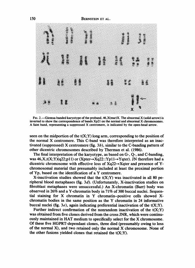
normal autosomal complement and an abnormal X chromosome in 122 of 127cells analyzed [46,Xmar(X)] (fig. 2). A 45,X karyotype was found in five (3.9%)of the 127 cells. All 58 fibroblast metaphases analyzed had a 46,X,mar(X)karyotype. The abnormal X was noticeably shorter than its normal homologue,and the G-banding pattern of the mar(X) did not match that of the normal X.However, when the mar(X) was inverted, the G-bands on the abnormal Xqcorresponded to those on the normal Xp. A faint band on the mar(X), usuallyvisible as two chromatid dots, corresponded to the position of the normal Xcentromere but could not be clearly identified as centromeric heterochromatinby means of G-banding. The G-band-negative region between bands Xp2l andXq21 on the mar(X) was consistently shorter than that on the normal X, possi-bly owing to the presence of a suppressed X centromere (figs. 2, 3a).On the distal portion of the abnormal Xp, adjacent to the primary centro-
meric constriction, Q-banding showed the brilliant fluorescence characteristicof Yql2--qter. The mar(X) was thus identified as a t(X;Y), and the primarycentromeric constriction corresponded to that of a Y-chromosome centromere(fig. 3c).C-banding confirmed both the presence of distal Yq heterochromatin and
that the primary centromeric constriction belonged to the Y chromosome.Another, unconstricted, dark C-band (not clearly visible on G-banding) was
149
BERNSTEIN ET AL.
FIG. 2.-Giemsa-banded karyotype of the proband, 46,X(mar)X. The abnormal X (solid arrow) isinverted to show the correspondence of bands Xp2l on the normal and abnormal X chromosomes.A faint band, representing a suppressed X centromere, is indicated by the open-head arrow.
seen on the midportion of the t(X;Y) long arm, corresponding to the position ofthe normal X centromere. This C-band was therefore interpreted as an inac-tivated (suppressed) X centromere (fig. 3b), similar to the C-banding pattern ofother dicentric chromosomes described by Therman et al. (1986).The final interpretation of the karyotype, as based on G-, Q-, and C-banding,
was 46,X,t(X;Y)(q22;pl 1) or (Xpter- Xq22::Yp 1-*Yqter). JN therefore had adicentric chromosome with effective loss of Xq22- Xqter and presence of Y-chromosomal material that presumably included at least the proximal portionof Yp, based on the identification of a Y centromere.
X-inactivation studies showed that the t(X;Y) was inactivated in all 80 pe-ripheral blood metaphases (fig. 3d). (Unfortunately, X-inactivation studies onfibroblast metaphases were unsuccessful.) An X-chromatin (Barr) body wasobserved in 26% and a Y-chromatin body in 71% of 300 buccal nuclei. Sequen-tial staining for X chromatin in Y chromatin-positive cells showed X-chromatin bodies in the same position as the Y chromatin in 24 informativebuccal nuclei (fig. 3e), again indicating preferential inactivation of the t(X;Y).
Further indirect confirmation of the nonrandom inactivation of the t(X;Y)was obtained from five clones derived from the cross JNR, which were continu-ously maintained in HAT medium to specifically select for the X chromosome.Of these five HGPRT-dependant clones, three died (presumably owing to lossof the normal X), and two retained only the normal X chromosome. None ofthe other fusions yielded clones that retained the t(X;Y).
150

DICENTRIC X;Y TRANSLOCATION
a b
cell-i3
cell-
cell-i ii Am
cell-iv AE
FIG. 3.-a, Giemsa-banded partial karyotypes of the normal X (left) and the t(X;Y) (right). b,Centromeric banding pattern of the t(X;Y), showing dark Yql2 heterochromatin, the primarycentromeric constriction of the Y centromere, and the dark band of the suppressed X centromere(arrowed). c, Quinacrine-banded partial karyotypes of the normal X and the t(X;Y), showing thebrilliant fluorescence of Yq12. d, Acridine-orange reverse banding after 5-BrdU incorporationduring the late S-phase; the whole t(X;Y) is inactivated except for two fluorescent pericentric bandsadjacent to the suppressed X centromere in cells i-iii (arrowed). e, Sequential quinacrine andcresyl-violet staining of buccal epithelial nuclei, showing associated X- and Y-chromatin bodies(arrowed).
2. Molecular-Hybridization StudiesSouthern blot analysis was performed using each of the six probes described
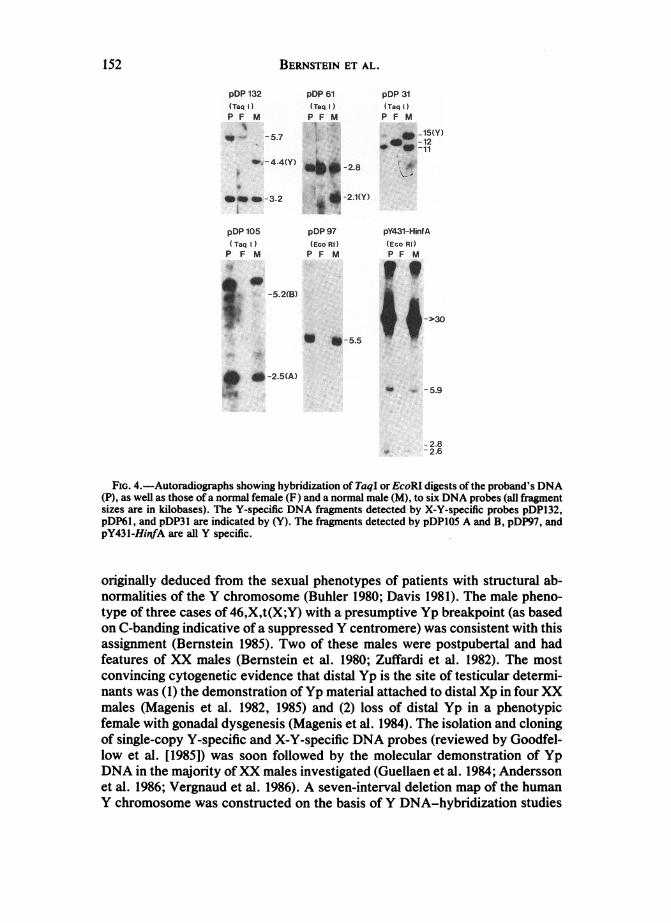
above. The results are shown in figures 1 and 4. Probes pDP1O5, pDP31,pDP97, and pY431-HinfA showed a normal XY male pattern in the proband,but the Y-specific fragments detected by pDP61 (2.1 kb) and pDP132 (4.4 kb)were absent.Probe pDP3 1, which detects X-linked allelic fragments of either 11 or 12 kb,
showed double intensity of the 1-kb fragment when compared with the 15-kbY-specific fragment in the proband. Similarly, when hybridized to the pro-band's DNA, probe pDP61 detected the 2.8-kb X-specific band at an intensityequal to that in normal females.
DISCUSSION
The phenotypic sex of 46,X,t(X;Y) individuals is determined by the interac-tion of several factors, the first of which is the physical presence or absence oftestis determining-factor (TDF) genes on their t(X;Y) chromosome. The lo-calization of a TDF or of factors on Yp (and possibly on proximal Yq) was
151
BERNSTEIN ET AL.
pDP 132 pDP 61 pDP 31(Taq )TqI) {TMIP F10 P F M P f M
-5.7 _ _-12
_-~~~~~~~~~11-4.4 T'Y - 8
-3.2 _- 2.1(Y)
pDP 105 pW 97 pY431-HkA(TaqI (hEo RI) (Eeo Rl)P F 1 P F M P F M
h wing hrid n of T
pDP~l,and pDP l ar idctdb(Y. Th frget deece by pDlAadBpP7n
pY43l-HinfAareallYspecific.
| e _ I~~-5.5I
normalit~~_SieofteYcrms e (Buhe 190 Dai91. TE mal phno
_Y'<~~ ~ ~ ~~~5
onGC.-bandingidicgativeoig yriiatoofasprseYcntrqlomrewaRdgssconsithentwith' this(,assignenasth(ersteioanora1985). Twoadnofrthes males werepostxDApuobert(al fandmhadfiesatresi iofbX ales) (Bern-steini etA fag.m1980 Zuffaredi et al. 1982).iThoes mostconvIn-inng cytoenalsetiic.iec htdsa pi h ieo etclrdtrioriginansw (1)ucedfthedm sai o Yphmatyeria atached to distaXifura Xnormalesigenistet al. 1982,o1985 and (28ls ofvis Yp in maphenotfemale withreegonad dysgenesis (Manis eta.p 1984). Theiation an cloin
of sibnglecop Y-spcaivelc an X-Y-prsedYcifceNAtroberws(reviewedtbyiGoodfellowignental [1985])twas soon fwollowefb thee moleculare deonstpbrtlatindofaYDNatuein the majorit ofBXXmaesinvestigated980 (Guellaen et al. 1984; Andermsso
fetmal. 1986; VegonaudadygnssMet al.1986)4svn-neradheleotionmapofth humanin
Y chromosome was constructed on the basis of Y DNA-hybridization studies
152

DICENTRIC X;Y TRANSLOCATION
(Page 1986; Vergnaud et al. 1986). The presence of certain Yp DNA sequencesin most XX males (Vergnaud et al. 1986) and the absence of the same Ypsequences in some XY females (Disteche et al., in press) provided strongevidence that TDF was located in deletion interval 1 of Yp (Page 1986).
In the present case, Y chromosome-specific DNA probes, some of whichhave been detected in XX males, were hybridized to the DNA of the proband inan attempt to show the extent of Yp-specific DNA sequences in the X;Y trans-location. Four of these probes detect sequences on Yp, one (pDP105) detectssequences on Yp and Yq, and pY431-HinfA detects highly repeated sequenceson the distal portion of Yq.The probes used have been ordered on the Y chromosome by means of
deletion mapping (Page 1986; Vergnaud et al. 1986). They represent a means ofindependently determining, albeit at a gross level, those portions of the Ychromosome present or absent from the patient's genome. Probe pDP132,which detects a Y-specific DNA sequence commonly present in XX males,(interval 1 as defined by Vergnaud et al. [1986] and Page [1986]) was not presentin the proband. The Y-specific sequence detected by probe pDP61, a sequencethat is also present in many XX males (interval 2), was also absent in theproband, indicating that the distal portion of Yp, containing probes pDP132 andpDP61, is deleted. Probes pDP105, pDP31, and pDP97, which are also presentin some XX males, all showed the typical Y-specific DNA fragments. As ex-pected, pY431-HinfA gave the usual Y-specific DNA fragments after digestionwith EcoRI. Thus, independently of the chromosome-banding studies, theDNA studies suggest the presence of the long arm, centromere, and proximalshort arm (intervals 3-7) and the absence of the distal short arm (intervals 1 and2). Given the female phenotype of JN, these DNA studies are consistent withthe presence of TDF in interval 1 of Yp (Page 1986; Vergnaud et al. 1986).
X-specific dosage effects were seen with X-Y probes pDP31 and pDP61. TheX-specific fragment detected by pDP31, a fragment that has been localized toXql3-q21 (Page et al. 1984), was present at double the intensity of the Y-specific fragment, confirming the cytogenetic observation that Xql3-q21 hadnot been deleted in the translocation event. Probe pDP61 shows an X-specificband equal in intensity to that of normal females, indicating that the X-specificsequence is not located on Xq22-*Xqter, the portion of Xq deleted in JN.Because only one streak gonad was biopsied, it was not possible to establish
whether the other streak gonad contained any primitive testicular tissue. Theimportance of removing both streaks was stressed to JN, because of the in-creased risk of malignancy in her dysgenetic gonads.
Screening of more than 300 cells yielded no cytogenetic evidence of mosai-cism for an intact Y chromosome. The phenotype could have been modified by45,X mosaicism, but only a minority of peripheral blood cells and not a singlefibroblast metaphase showed loss of the t(X;Y). The percentage of 45,X cells inother tissues, especially the gonads, is unknown.The Xq22 breakpoint of this translocation is also of interest. The phenotype
of JN-and of the patients of Cameron et al. (1984) and Kelly et al. (1984),patients who also had an Xq22 breakpoint-differs from that of previously
153
154 BRSENE Ldescribed females with a 46,X,t(X;Y) karyotype and Xp;Yq breakpoints(Bernstein 1985). All three women had a stature >150 cm, and two of them hadnormal breast development, associated with oligomenorrhea in Cameron etal.'s (1984) 17-year-old patient and with oligomenorrhea followed by secondaryamenorrhea in 32-year-old JN. The 17-year-old female described by Kelly et al.(1984) had primary amenorrhea and no breast development. Deletions, inver-sions, or translocations affecting the critical region Xql3-)Xq27 are almostalways associated with varying degrees of gonadal dysgenesis, but stature isusually normal and somatic features of Turner syndrome are minimal (Therman1983). These patients' normal height contrasts with the short stature that isboth associated with Xp deletions and found to be a characteristic feature of allt(X;Y) cases involving Xp (Bernstein 1985).The t(X;Y) in JN is unique, in that the clear cytological demonstration of a Y
centromere indicates that at least the proximal portion of Yp must be present;but, unlike the three males previously reported (Bernstein et al. 1978, 1980;Zuffardi et al. 1982) as having a 46,X,t(X;Y)(p22;pl 1) karyotype, she had afemale phenotype. On the basis of cytogenetic studies alone, one could nothave excluded the possibility that TDF genes were, in fact, present but nonran-domly inactivated. However, the molecular studies show that Y-chromosomeinterval 1, which contains TDF (Page 1986; Vergnaud et al. 1986), is in factdeleted.The findings in JN illustrate the complex nature of sex determination and
emphasize the need for a multidisciplinary approach in the investigation ofunusual chromosome abnormalities, if the phenotype-genotype interrelation-ships are to be understood.
ACKNOWLEDGMENTSWe wish to thank Dr. C. Polon for referring the patient, Dr. K. Smith for providing
probe pY431-HinfA, Dr. K. Margolius for reviewing the gonadal histology, G. R. Turn-bull for hormonal studies, M. Dos Santos and J. Borthwick for technical help, and Y.Descy for photographic assistance. This work was supported in part by grant HD 20059from the National Institutes of Health.
REFERENCES
Andersson, M., D. C. Page, and A. de la Chapelle. 1986. Chromosome Y-specific DNAis transferred to the short arm of the X chromosome in human XX males. Science233:786-788.
Bernstein, R. 1985. X;Y translocations and their manifestations. Pp. 171-206 in A. A.Sandberg, ed. The Y chromosome. Part B: Clinical aspects of Y chromosome abnor-malities. Alan R. Liss, New York.
Bernstein, R., M. R. Pinto, M. Almeida, S. M. Solarsh, J. Meck, and T. Jenkins. 1980.X;Y translocation in an adolescent mentally normal phenotypic male with features ofhypogonadism. J. Med. Genet. 17:437-443.
Bernstein, R., J. Wagner, J. Isdale, G. T. Nurse, A. B. Lane, and T. Jenkins. 1978. X-Ytranslocation in a retarded phenotypic male. J. Med. Genet. 15:466-474.
Borgaonkar, D. S., B. M. Sroka, and M. Flores. 1974. Y-to-X translocation in a girl.Lancet 1:68-69.
Buhler, E. M. 1980. A synopsis of the human Y chromosome. Hum. Genet. 55: 145-175.
154 BERNSTEIN ET AL.

DICENTRIC X;Y TRANSLOCATION
Cameron, I. T., K. E. Buckton, and D. T. Baird. 1984. X-Y translocation: a case report.Hum. Genet. 67:457-459.
Davis, R. M. 1981. Localisation of male determining factors in man: a thorough reviewof structural anomalies of the Y chromosome. J. Med. Genet. 18:161-195.
Disteche, C. M., M. Casanova, H. Saal, C. Friedman, V. Sybert, J. Graham, H.Thuline, D. C. Page, and M. Fellous. 1986. Small deletions of the short arm of the Ychromosome in 46,XY females. Proc. Natl. Acad. Sci. USA 83:7841-7844.
Dutrillaux, B., C. Laurent, J. Couturier, and J. Lejeune. 1973. Coloration des chromo-somes humains par l'acridine orange apres traitement par le 5 bromodeoxyuridine. C.R. Hebdomadaires Seances Acad. Sci. 276:3179-3181.
Fryns, J. P., and H. van den Berghe. 1983. Y-X translocations in man. Pp. 245-251 inA. A. Sandberg, ed. Cytogenetics of the mammalian X chromosome. Part B: Xchromosome anomalies and their clinical manifestations. Alan R. Liss, New York.
Geldwerth, D., C. Bishop, G. Guellaen, M. Koenig, G. Vergnaud, J. L. Mandel, and J.Weissenbach. 1985. Extensive DNA sequence homologies between the human Y andthe long arm of the X chromosome. EMBO J. 4:1739-1743.
Goodfellow, P., S. Darling, and J. Wolfe. 1985. The human Y chromosome. J. Med.Genet. 22:329-344.
Gross-Bellard, M., P. Oudet, and P. Chambon. 1973. Isolation of high-molecular-weightDNA from mammalian cells. Eur. J. Biochem. 36:32-38.
Guellaen, G., M. Casanova, C. Bishop, D. Geldwerth, G. Andre, M. Fellous, and J.Weissenbach. 1984. Human XX males with Y single-copy DNA fragments. Nature307:172-173.
Jones, H. W., Jr., J. M. Rary, J. A. Rock, and D. Cummings. 1979. The role of the H-Yantigen in human sexual development. Johns Hopkins Med. J. 145:33-43.
Kelly, T. E., S. S. Wachtel, L. Cahill, V. M. Barnabei, K. Willson-Suddath, and H. E.Wyandt. 1984. X;Y translocation in a female with streak gonads, H-Y phenotype, andsome features of Turner's syndrome. Cytogenet. Cell Genet. 38:122-126.
Koo, G. C., S. S. Wachtel, K. Krupen-Brown, L. R. Mittl, W. R. Breg, M. Genel, I. M.Rosenthal, D. S. Borgaonkar, D. A. Miller, R. Tantravahi, R. R. Schreck, B. F.Erlanger, and 0. J. Miller. 1977. Mapping the locus of the H-Y gene on the human Ychromosome. Science 198:940-942.
Kunkel, L. M., K. D. Smith, S. H. Boyer, D. S. Borgaonkar, S. S. Wachtel, 0. J.Miller, W. R. Breg, H. W. Jones, Jr., and J. M. Rary. 1977. Analysis of human Y-chromosome-specific reiterated DNA in chromosome variants. Proc. Natl. Acad. Sci.USA 74:1245-1249.
Magenis, R. E., M. G. Brown, T. Donlon, S. B. Olson, R. Sheehy, and D. Tomar. 1985.Structural aberrations of the Y chromosome, including the nonfluorescent Y: cy-tologic origin and consequences in the Y chromosome. Pp. 537-574 in A. A. Sand-berg, ed. The Y chromosome. Part A: Basic characteristics of the Y chromosome.Alan R. Liss, New York.
Magenis, R. E., M. L. Tochen, K. P. Holahan, T. Carey, L. Allen, and M. G. Brown.1984. Turner syndrome resulting from partial deletion of Y chromosome short arm:localization of male determinants. J. Pediatr. 105:916-919.
Magenis, R. E., M. J. Webb, R. S. McKean, D. Tomar, L. J. Allen, H. Kammer, D. L.Van Dyke, and E. Lovrien. 1982. Translocation (X;Y)(p22.33;pll.2) in XX males:etiology of male phenotype. Hum. Genet. 62:271-276.
Page, D. C. 1986. Sex reversal: deletion mapping the male-determining function of thehuman Y chromosome. In Molecular biology of Homo sapiens. Cold Spring HarborSymp. Quant. Biol. 51.
Page, D. C., B. de Martinville, D. Barker, A. Wyman, R. White, U. Francke, and D.Botstein. 1982. Single-copy sequence hybridizes to polymorphic and homologous locion human X and Y chromosomes. Proc. Natl. Acad. Sci. USA 79:5352-5356.
Page, D. C., M. E. Harper, J. Love, and D. Botstein. 1984. Occurrence of a transposi-
155
tion from the X-chromosome long arm to the Y-chromosome short arm during humanevolution. Nature 311:114-123.
Rigby, P. W. J., M. Dieckman, C. Rhodes, and P. Berg. 1977. Labelling deoxyribonu-cleic acid to high specific activity in vitro by nick translation with DNA polymerase. J.Mol. Biol. 113:237-251.
Ross, J. B., P. W. Allderdice, L. J. Shapiro, J. Aveling, A. Eales, and D. Simms, Jr.1985. Familial X-linked ichthyosis, steroid sulfatase deficiency, mental retardation,and nullisomy for Xp22.3-pter. Arch. Dermatol. 121:1524-1528.
Southern, E. M. 1975. Detection of specific sequences among DNA fragments separatedby gel electrophoresis. J. Mol. Biol. 98:503-517.
Speevak, M., B. Clifford, D. M. Cox, and A. G. W. Hunter. 1985. Detection at am-niocentesis of a maternally inherited X;Y translocation. Clin. Genet. 27:595-599.
Therman, E. 1983. Mechanisms through which abnormal X-chromosome constitutionsaffect the phenotype. Pp. 159-173 in A. A. Sandberg, ed. Cytogenetics of the mam-malian X chromosome. Part B: X chromosome anomalies and their clinical manifesta-tions. Alan R. Liss, New York.
Therman, E., C. Trunca, E. M. Kuhn, and G. E. Sarto. 1986. Dicentric chromosomesand the inactivation of the centromere. Hum. Genet. 72:191-195.
Vergnaud, G., D. C. Page, M.-C. Simmler, L. Brown, F. Rouyer, B. Noel, D. Botstein,A. de la Chapelle, and J. Weissenbach. 1986. A deletion map of the human Y chromo-some based on DNA hybridization. Am. J. Hum. Genet. 38:109-124.
Wegner, S., R. Muneer, and 0. Rennert. 1984. An X;Y translocation, t(X;Y)(p22;ql 1) inthree generations (abstract). Am. J. Hum. Genet. 36, suppl.:117S.
Wolfe, J., S. M. Darling, R. P. Erickson, I. W. Craig, V. J. Buckle, P. W. J. Rigby,H. F. Willard, and P. N. Goodfellow. 1985. Isolation and characterization of analphoid centromeric repeat family from the human Y chromosome. J. Mol. Biol.182:477-485.
Yunis, J. J. 1976. High resolution of human chromosomes. Science 191:1268-1270.Zuffardi, O., P. Maraschio, F. Lo Curto, U. Muller, A. Giarola, and L. Perotti. 1982.The role of Yp in sex determination: new evidence from X/Y translocations. Am. J.Med. Genet. 12:175-184.
156 BERNSTEIN ET AL.