Embed Size (px)
Citation preview

Auditory System Physiology

Ear components
• 3 parts:
external ear
middle ear
inner ear


External ear The external ear funnels sound waves to the external
auditory meatus. In some animals, the ears can be moved like radar antennas to seek out sound. From the meatus, the external auditory canal passes inward to the tympanic membrane (eardrum)

Middle ear
The middle ear is an air-filled cavity in the temporal bone that opens via the auditory (eustachian) tubeinto the nasopharynx and through the nasopharynx to the exterior.
The tube is usually

The three auditory ossicles, the malleus, incus, and stapes, are located in the middle ear.
The manubrium (handle of the malleus) is attached to the back of the tympanic membrane. Its head is attached to the wall of the middle ear, and its short process is attached to the incus, which in turn articulates with the head of the stapes.
Its foot plate is attached by an annular ligament to the walls of the oval window
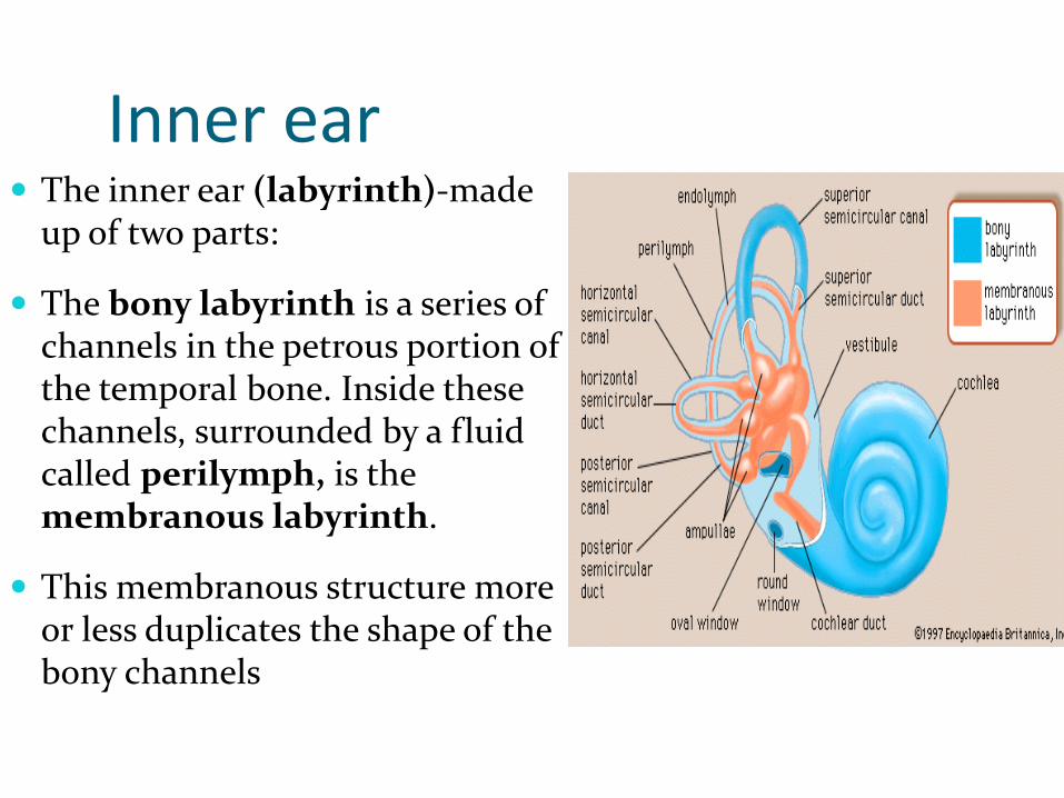
Inner ear The inner ear (labyrinth)-made
up of two parts:
The bony labyrinth is a series of channels in the petrous portion of the temporal bone. Inside these channels, surrounded by a fluid called perilymph, is the membranous labyrinth.
This membranous structure more or less duplicates the shape of the bony channels


Path of sound external canal
vibrates eardrum
vibration moves through ossicles
Malleus
Incus
Stapes
stapes vibrates oval window of cochlea
creates pressure wave in the fluid inside

Membranous labyrinth 3 semicircular channels oriented perpendicular to each
other and following the 3D planes of space
Utricle and saccule= vestibule
Cochlea
The semicircular channels, utricle and saccule-balance- they contain the vestibular receptors
Cochlea has a hearing function- auditory receptor (Corti organ)


Inner ear The membranous labyrinth is filled with a fluid called
endolymph K+ RICH
The bony labyrinth is filled with a fluid called perilymph
There is no communication between the spaces filled with endolymph and those filled with perilymph.

Cochlea The cochlear portion of the
labyrinth is a coiled tube which in humans is 35 mm long and makes 2.5 turns.
The basilar membrane and Reissner's membrane divide it into three chambers (scalae)
The upper scala vestibuli and the lower scala tympani contain perilymph and communicate with each other at the apex of the cochlea through a small opening called the helicotrema

At the base of the cochlea, the scalavestibuli ends at the oval window, which is closed by the footplate of the stapes.
The scala tympani ends at the round window, a foramen on the medial wall of the middle ear that is closed by the flexible secondary tympanic membrane.
The scala media is continuous with the membranous labyrinth and does not communicate with the other two scalae. It contains endolymph.

The cochlea is made up of three canals wrapped around a bony axis, the modiolus. These canals are: the scala tympani (3), the scala vestibuli (2) and the scalamedia (or cochlear duct) (1).
The scalae tympani and vestibule are filled with perilymph (in blue) and are linked by a small opening at the apex of the cochlea called the helicotrema. The triangular scalamedia, situated between the scalae vestibuli and tympani is filled with endolymph (in green)


Corti organ
Located on the basilar membrane
It contains the hair cells which are the auditory receptors.
The hair cells are arranged in four rows: three rows of outer hair cells and one row of inner hair cells medial to the tunnel of Corti
There are 20,000 outer hair cells and 3500 inner hair cells in each human cochlea


The tips of the hair cells go through the reticular membrane
Then they inbed in the thin tectorial membrane
At the basis of the receptor cell- dendrites of the first order neuron located in the modeolus= spiral ganglion of Corti 90-95% of the afferent fibresleave from the inner hair cells (they travel through the Corti tunnel)
only 5-10% innervate the more numerous outer hair cells

Inner/outer hair cells
Inner hair cells- sound perception
Outer hair cells- sound amplifiers-mechanic response to stimulation (vibration) otoacoustic emissions (can be registered from the external meatus with microphones) hearing loss screening in babies


Efferent system
The efferent auditory fibers are originated from many different sites in the central nervous system. From the superior olivary complex, they are projected to the cochlea through two different tracts:
the medial olivocochlear tract, which comprises large myelinated neurons that innervate predominantly the outer hair cells
lateral olivocochlear tract, with unmyelinated neurons, that synapses with the inner hair cells.
Function- change sensitivity of the receptor cells


Sound transmission
The ear converts sound waves in the external environment into action potentials in the auditory nerves.
The waves are transformed by the eardrum and auditory ossicles into movements of the footplate of the stapes.
These movements set up waves in the fluid of the inner ear.
The action of the waves on the organ of Cortigenerates action potentials in the nerve fibers.

Sound transmission Sound wave.... stapes oval window perilymph in
the scala vestibuli helicotrema perilymph in
Wave in the perilymph transmits to endolymph in the scala media
Basilar membrane vibrates- resonant structure-deflected in response to waves- deformation is a traveling wave from basis to apex
APICAL CILIA OF HAIR CELLS- DEFLECTIONDEPOLARISATION
http://www.youtube.com/watch?v=1JE8WduJKV4

Basilar membrane Unique structure
Differs according to area- stiff fibres that are attached firmly to the modeolus, but are “free” at the outer end
Fibres become longer as we approach the helicotrema
Fibres at the basis are more rigid, while the ones at the apex are more flexible
This makes the basilar membrane resonate to different sound pitches!!
Sound travels along the basilar membrane BUT this resonates only in a specific RESONANT point for each of them

Basilar membrane

Frequency coding

Sound frequency- Hz/ Cicles per second

Intensity coding (loudness)
Basilar membrane vibrates directly proportional with sound intensitymore rapid rates of excitation
Spatial summation no of stimulated cells gets higher
Outer cell stimulation only when vibration is very high


Hair cells
The hair cells in the inner ear have a common structure
the kinocilium, is a true but nonmotile cilium,it is one of the largest processes and has a clubbed end.
The other processes are called stereocillia-about 50-70
The membrane potential of the hair cells is about -70 mV.

Hair cells
When the stereocilia are pushed toward the kinocilium, the membrane potential is decreased to about -50 mV.
When the bundle of processes is pushed in the opposite direction, the cell is hyperpolarized.

Signal transduction Very fine processes called tip links tie the tip
of each stereocilium to the side of its higher neighbor, and at the junction there appear to be mechanically sensitive cation channels in the higher process.
When the shorter stereocilia are pushed toward the higher, the open time of these channels increases. K+ is the most abundant cation in endolymph receptor potentialtriggers Ca2+ enterance via specific channels neurotransmitter release and produce depolarization.
Neurotransmitter- probably glutamate, which initiates depolarization of neighboring afferent neurons.


Auditory pathway
Dendrites of the neurons in the Spiral ganglion are located at the basis of the sensorial hair cells
Spiral ganglion in the modiolus. Ganglion is formed by proper bipolar nerve cells. Their myelinated axons run together to form the acoustic nerve, which unites with the vestibular nerve to form the VIIIth cranial nerve. Myelinated dendrites lose their sheaths as they perforate the bone and pass to the organ of Corti, terminating hair cells.


1st order neuron- Cortiganglion
2nd order neuron- dorsal and ventral cochlear nuclei in the pons
3rd order neuron- inferior colliculus in the midbrain
4th order neuron-geniculate medial nuclei in the methathalamus

Testing

Weber
Hipoacuzie de transmisie
Normal Hipoacuzie neurosenzorială
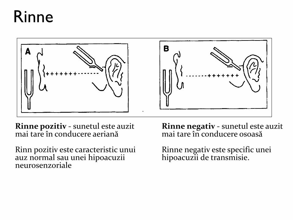
Rinne
Rinne pozitiv - sunetul este auzit mai tare în conducere aeriană
Rinn pozitiv este caracteristic unui auz normal sau unei hipoacuzii neurosenzoriale
Rinne negativ - sunetul este auzit mai tare în conducere osoasă
Rinne negativ este specific unei hipoacuzii de transmisie.

Audiometry

normal
Audiometriy

Transmission
Audiometry

Neurosensorial
Audiometry

mixt
Audiometry

BALANCE

Vestibular system
Vestibular apparatus: utricle, saccule and semicircular ducts.
Sensory cells of vestibular apparatus lie in the ampular cristae of canals and maculaein the utricle and saccule.

Both types of organs- hair cells- special stereocilia over the apical surface and one kinocilium:
In the utricle and saccule, the receptor organ is called macula- stereocillia and kinocilium are embedded in cuticular plate with otoliths (CaCO3 crystals)
In the semicircular ducts, the receptor organs are called ampular cristae- hairs are covered by cupulae, composed of a gelatinous material similar to otolithic membrane but lacking otoliths.

Macula (utricular and saccular)

Cristae ampullaris (semicircular canals)

Utricle and saccule:
– Linear acceleration detection
– Head position (gravity)
Semicircular canals:
-Angular motion

Function of the maculae
detect head position and linear acceleration
otoliths (small calcium carbonate particles) drag on the stereocilia when the head changes position
when the body is in anatomical position: the patch of hair cells in the UTRICLE is nearly horizontal, with the stereocilia oriented vertically
the sensory epithelium is vertical in the SACCULE, with the stereocilia oriented horizontally

when the body is in anatomical position: the patch of hair cells in the UTRICLE is nearly horizontal, with the stereocilia oriented vertically
the sensory epithelium is vertical in the SACCULE, with the stereocilia oriented horizontally
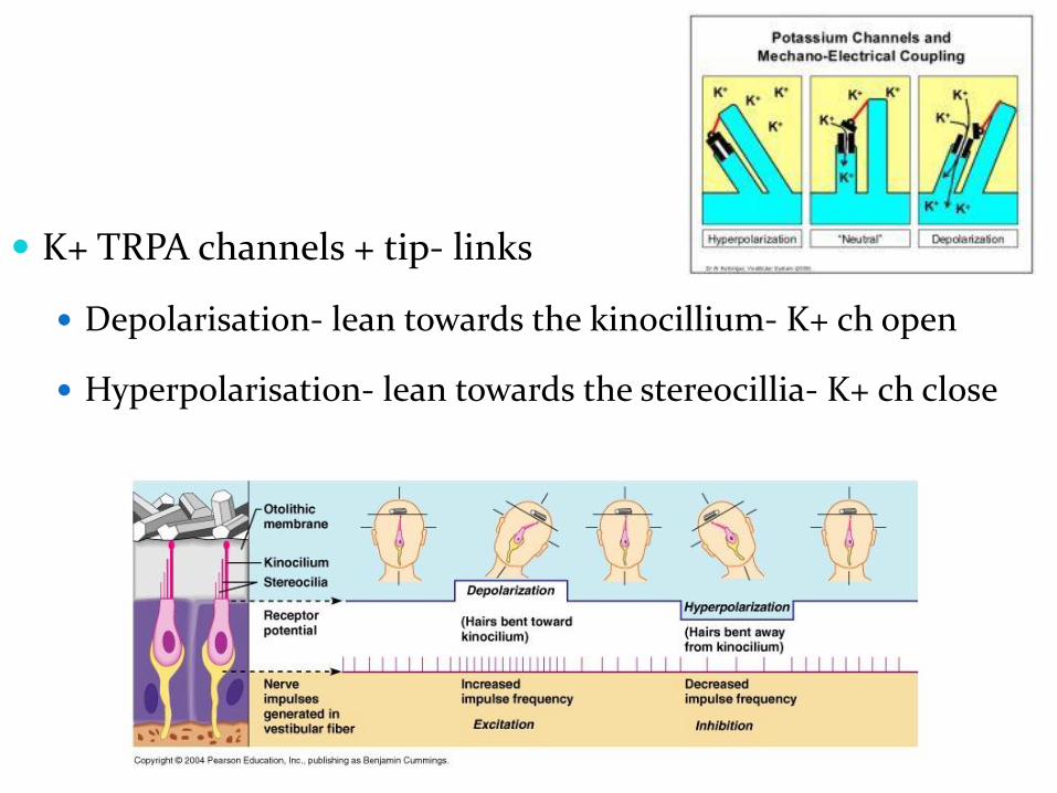
K+ TRPA channels + tip- links
Depolarisation- lean towards the kinocillium- K+ ch open
Hyperpolarisation- lean towards the stereocillia- K+ ch close

orientation of the stereocilia within the sensory epithelium is determined by the STRIOLA, a curved dividing ridge that runs through the middle of the MACULA – in the UTRICLE, the kinocilia are oriented TOWARD the striola, and in the SACCULE they are oriented AWAY from it
in any position, some hair cells will be depolarized and others hyperpolarized in BOTH otolith organs

Signal transduction

Semicircular canals- coding of rotation
Three semicircular canals in each ear
Each canal is oriented in a different plane
Each canal is maximally sensitive to rotations perpendicular to the canal plane

Anterior (superior)- anterior and 45 degrees with the AP plane
Posterior- post and 45 degreees with the AP plane
Horizontal

Function of the cristae detect the rate of head rotation
when the head is initially moved, the endolymph and ampulla (and therefore the hair cells) turn with it in a direction oposing rotation
DEPOLARISATION IN THE KINOCILUM DIRECTION
HYPERPOLARISATION IN THE STEREOCILLIUM DIRECTION


HORIZONTAL CANALS (“no”)
depolarization occurs in the SAME direction as the head movement (LEFT head turn produces depolarization in the LEFT horizontal canal)

ANTERIOR (SUPERIOR) (“YES”) AND POSTERIOR CANALS
anterior canals are located at ~90o to
each other
posterior canals are also located at
~90o to each other
the directionality of the stereocilia is
different in the anterior and posterior
canals the anterior canals have their kinocilium
anterior to the stereocilia
the posterior canals have their kinocilium
posterior to the stereocilia
the natural pairing of A/P canals is:
LEFT ANTERIOR with RIGHT
POSTERIOR
RIGHT ANTERIOR with LEFT
POSTERIOR

Signal transduction
K+ channels in the cillia
When stereocilia are bent towards the kinocilum K+ ch open
Depolarisation of the receptor cell= receptor potential
Ca2+ channels openingmediator release in the synaptic cleft
Action potential on the vestibular pathway

Vestibular pathway
1st order neuron= Scarpa ganglion- axons form the vestibular branch of the VIIIth cranial nerve
2nd order neuron- vestibular nuclei in the medulla oblongata
3rd order neuron- thalamus
Cortical projection= superior temporal gyrus

1. to the thalamus (3rd order neuron)
2. to the paleocerebellum
3. to the spinal chord (vestibulospinal)
4. to the motor nuclei of cranial nerves III,IV,VI