Embed Size (px)
Citation preview

SM Journal of Case Reports
Gr upSM
How to cite this article Wauer U, Sellei R, Zoga E, Braun S and Lorenz D. Atypical Course of an Apocrine Sweat Gland Carcinoma: A very Rare Malignant
Tumor and his Interdisciplinary Treatment. SM J Case Rep. 2017; 3(4): 1052.
OPEN ACCESS
ISSN: 2473-0688
Clinical CourseThe male patient, at that time 67 year old, was operated on in December 2012 with the idea of
an atheroma of the right axilla in an ambulant setting. The unreckoned histopathological result was an incidental finding of an adenocarcinoma G1-2, excised non in sano. A differentiation between a local adenocarcinoma (e.g. sweat gland carcinoma) and a metastasis of another primary tumor wasn`t possible. The patient was referred to our hospital after extensive diagnostics (CT scans of chest and abdomen, colonoscopy, gastroscopy, urological examination). The search for another primary tumor other than the detected sweat gland carcinoma was without any result. So we performed in January 2013 a wide resection and lymph node dissection of the right axilla. The operation and the postoperative course were uneventful, respectively. We proved an apocrine adenocarcinoma of the sweat glands with a maximal size of 1.4 cm and a local lymph node metastasis nearby with a maximal size of 2.7 cm without perinodal tumor infiltration. Microscopically solid adenocarcinomatous assemblies with apocrine differentiation (GCDFP-15 positive) were to be seen. The cores were considerable polymorph, anisochromatic and hyperchromatic; the cytoplasm was eosinophil. Numerous mitoses (>50 mitoses per 10 HPF) were detectable (Figures 1 and 2). Immunohistochemically only a CK 7 expression and a fraction of proliferation of 5 % (Ki 67) was to be seen. Reactions to S100, CK20 and TTF 1 were as negative as the estrogen receptor status.
Case Report
Atypical Course of an Apocrine Sweat Gland Carcinoma: A very Rare Malignant Tumor and his Interdisciplinary TreatmentWauer U1*, Sellei R2, Zoga E3, Braun S4 and Lorenz D1
1Department for General and Oncologic Surgery, Sana Klinikum Offenbach, Germany2Department for Orthopedic and Trauma Surgery, Sana Klinikum Offenbach, Germany3Department for Radiotherapy, Sana Klinikum Offenbach, Germany4Department for General and Oncologic Surgery, Sana Klinikum Offenbach, Germany
Article Information
Received date: Jul 14, 2017 Accepted date: Jul 24, 2017 Published date: Jul 28, 2017
*Corresponding author
U Wauer, Department for General and Oncologic Surgery, Sana Klinikum Offenbach, Starkenburgring 66, 63069 Offenbach, Germany, Tel.: 0698405-3623; Fax: 0698405-4572; Email: [email protected]
Distributed under Creative Commons CC-BY 4.0
Keywords Apocrine; Sweat gland carcinoma; Axilla; Bone metastasis; Lymph node dissection; Immunohistochemical and genetic markers
Abstract
We report on an atypical clinical course of a patient with the very rare diagnosis of an apocrine sweat gland carcinoma with a lymphatic metastasis, metachronous distant metastasis and a now reached survival time of more than four years and give a review about the current literature. There are only a very small number of cases described. The recommendations for diagnostics and treatment of this tumor therefore are not based on prospective randomized studies but upon case reports and on new immunohistochemical and genetic markers.
Figure 1: Histomorphological picture. Still indicated adenoid differentiation of the tumor tissue with middle wide, clear eosinophil cytoplasm (apocrine aspect) with moderate pleomorphism of the cores (haematoxylin/eosin staining, enlargement 100x).
Citation: Wauer U, Sellei R, Zoga E, Braun S and Lorenz D. Atypical Course of an Apocrine Sweat Gland Carcinoma: A very Rare Malignant Tumor and his Interdisciplinary Treatment. SM J Case Rep. 2017; 3(4): 1052. Page 2/3
Gr upSM Copyright Wauer U
The resection was roundly in sano. The Her2/new- status and the presence of genetic markers were not verified. The finally classification of the tumor according to the TNM-system was pT1pN1(1/23) pL0 pV0 pPn0 R0 M0.
The case was discussed in the interdisciplinary tumor board and a radiation of the right axilla to reduce the risk of local recurrence as well as a regular follow-up care was recommended. The radiation was conducted from March to April 2013 as adjuvant percutaneous photon- radiotherapy of the right axillar lymphatic channels with a dosage of 50.4 Gy with a dose fractionation of 5x1.8 Gy per week.
From June 2013 until July 2015 we performed the regularly follow ups. Due to missing established follow up algorithms for this tumor we undertook careful clinical examinations, chestx-ray and an one-time MRI of the right axilla and thoracic wall (August 2013) without pathologic findings.
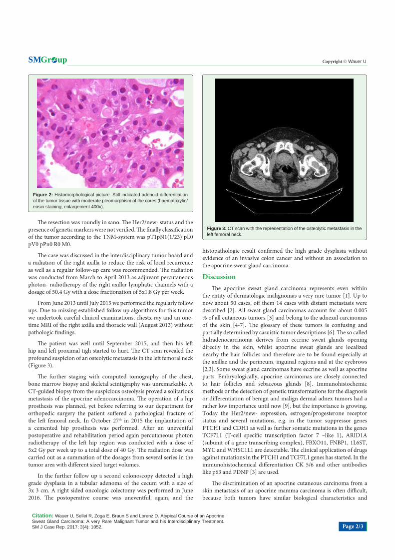
The patient was well until September 2015, and then his left hip and left proximal tigh started to hurt. The CT scan revealed the profound suspicion of an osteolytic metastasis in the left femoral neck (Figure 3).
The further staging with computed tomography of the chest, bone marrow biopsy and skeletal scintigraphy was unremarkable. A CT-guided biopsy from the suspicious osteolysis proved a solitarious metastasis of the apocrine adenocarcinoma. The operation of a hip prosthesis was planned, yet before referring to our department for orthopedic surgery the patient suffered a pathological fracture of the left femoral neck. In October 27th in 2015 the implantation of a cemented hip prosthesis was performed. After an uneventful postoperative and rehabilitation period again percutaneous photon radiotherapy of the left hip region was conducted with a dose of 5x2 Gy per week up to a total dose of 40 Gy. The radiation dose was carried out as a summation of the dosages from several series in the tumor area with different sized target volumes.
In the further follow up a second colonoscopy detected a high grade dysplasia in a tubular adenoma of the cecum with a size of 3x 3 cm. A right sided oncologic colectomy was performed in June 2016. The postoperative course was uneventful, again, and the
histopathologic result confirmed the high grade dysplasia without evidence of an invasive colon cancer and without an association to the apocrine sweat gland carcinoma.
DiscussionThe apocrine sweat gland carcinoma represents even within
the entity of dermatologic malignomas a very rare tumor [1]. Up to now about 50 cases, off them 14 cases with distant metastasis were described [2]. All sweat gland carcinomas account for about 0.005 % of all cutaneous tumors [3] and belong to the adnexal carcinomas of the skin [4-7]. The glossary of these tumors is confusing and partially determined by casuistic tumor descriptions [6]. The so called hidradenocarcinoma derives from eccrine sweat glands opening directly in the skin, whilst apocrine sweat glands are localized nearby the hair follicles and therefore are to be found especially at the axillae and the perineum, inguinal regions and at the eyebrows [2,3]. Some sweat gland carcinomas have eccrine as well as apocrine parts. Embryologically, apocrine carcinomas are closely connected to hair follicles and sebaceous glands [8]. Immunohistochemic methods or the detection of genetic transformations for the diagnosis or differentiation of benign and malign dermal adnex tumors had a rather low importance until now [9], but the importance is growing. Today the Her2/new- expression, estrogen/progesterone receptor status and several mutations, e.g. in the tumor suppressor genes PTCH1 and CDH1 as well as further somatic mutations in the genes TCF7L1 (T-cell specific transcription factor 7 –like 1), ARID1A (subunit of a gene transcribing complex), FBXO11, FNBP1, 1L6ST, MYC and WHSC1L1 are detectable. The clinical application of drugs against mutations in the PTCH1 and TCF7L1 genes has started. In the immunohistochemical differentiation CK 5/6 and other antibodies like p63 and PDNP [3] are used.
The discrimination of an apocrine cutaneous carcinoma from a skin metastasis of an apocrine mamma carcinoma is often difficult, because both tumors have similar biological characteristics and
Figure 2: Histomorphological picture. Still indicated adenoid differentiation of the tumor tissue with moderate pleomorphism of the cores (haematoxylin/eosin staining, enlargement 400x).
Figure 3: CT scan with the representation of the osteolytic metastasis in the left femoral neck.

Citation: Wauer U, Sellei R, Zoga E, Braun S and Lorenz D. Atypical Course of an Apocrine Sweat Gland Carcinoma: A very Rare Malignant Tumor and his Interdisciplinary Treatment. SM J Case Rep. 2017; 3(4): 1052. Page 3/3
Gr upSM Copyright Wauer U
overlapping immunohistochemical profiles. Sweat gland carcinomas with an expression of estrogen receptors impede the demarcation to the latter particularly because all apocrine sweat gland carcinomas express the mamma- carcinoma specific marker GCDFP 15, a glycoprotein. The detection of CEA (CD 66e) and of the EGF receptor in sweat gland carcinomas is in some individual cases possibly suitable for the demarcation to skin metastases of a mamma carcinoma [10-12]. The differences in the estrogen/progesterone receptor status between both tumor entities are statistically significant, however [13]. Further markers like the protein Adipophilin are positive in 36% of the apocrine sweat gland carcinomas, but in 88% of the apocrine mamma carcinoma. In both tumors the detection of the androgen receptor is strongly positive, whereas a remarkable expression of cytoceratine 5/6 and/or mammaglobin is only existent in the apocrine sweat gland carcinoma. The proof of adipophilin, estrogen/progesterone receptor status, Her2/new status, cytoceratine 5/6 and mammaglobin therefore is helpful in the distinction between both tumor entities [13].
The value detecting hormone receptors like estrogen/progesterone receptors consists in a then possible therapy with tamoxifen. Whereas in mamma carcinomas the frequency of expression of these receptors depending on the grading is between 30-90%, the frequency in apocrine sweat gland carcinomas is about 36% [14]. In mamma carcinomas one has to distinguish between the invasive ductal and the invasive lobular carcinoma with higher receptor positivity [15].
Apocrine sweat gland cell carcinomas typically have an aggressive course. In the initial clinical situation often a locally extended tumor stadium exists and after a short interval the disease generalizes by distant metastasis. The tumor spreads in the regional lymph nodes, after that in the bones, lungs, pleura and rarely in visceral organs. The median survival decreases in the presence of lymph node metastasis from 55 to 33 month, in the presence of distant metastasis to 14.5 month [5]. Possible prognostic factors are size of the tumor and histopathological type, affection of lymph nodes and the presentation of distant metastasis [2].
Because of the very low incidence of the tumor are further prospective studies with enough statistical power very unlikely [5]. There exist only case reports or very small case series with obinestadium dependent recommendations for the treatment. These hitherto existing recommendations for apocrine sweat gland carcinomas include local resection of the primary tumor with wide excision margins [7], in the metastatic situation a systemic chemotherapy with carboplatin and paclitaxel [5] or epirubicin and cyclophosphamide i.v. followed by oral fluorouracil [16], the anti estrogen therapy with tamoxifen in cases with positive estrogen receptor status [2,5] and the percutaneous radiotherapies of affected larger lymph node areas in the metastatic stadium [5]. If the Her2/new status is positive, a therapy with trastuzumab is possible. Furthermore new therapeutic possibilities are reviewed with the inhibition of immuno checkpoints by PD-L1 inhibitors analog to the therapy of metastatic melanomas. With a proven mutation in the PTCH1 gen the treatment with vismodegib, a so-called hedgehog-signal inhibitor, is possible as it is in advanced or metastatic basal cell carcinomas [5].
The case of our patient who is currently without symptoms or complaints and has now reached a survival time of 46 month without proven metastasis again, is extraordinary because he had at the time of his primary diagnosis already a regional lymph node
metastasis and the distant metastasis remained solitaire so far [17,18]. The statistically predicted median survival time in his N+ and M+ situation was prolongated by our treatment.
References
1. Chamberlain RS, Huber K, White JC, Travaglino-Parda R. Apocrine gland carcinoma of the axilla: review of the literature and recommendations for treatment. Am J Clin Oncol. 1999; 22: 131-135.
2. Seong MK, Kim EK, Han K, Seol H, Kim HA, Noh WC. Primary apocrine sweat gland carcinoma of the axilla: a report of two cases and a review of the literature. World J Surg Oncol 2015; 13: 59.
3. Floros P, Mikhail M. A rare case of pectoral hidradenocarcinoma and brief review. The Internet Journal of Surgery 2010; 26: 1-4.
4. Kathrotiya PR, Bridge AT, Warren SJ, Do H, Klenk AS, Xu LY, et al. Primary apocrine adenocarcinoma of the axilla. Cutis. 2015; 95: 271-274, 281.
5. Miller DH, Peterson JL, Buskirk SJ, Vallow LA, Ta R, Joseph R, et al. Management of Metastatic Apocrine Hidradenocarcinoma with Chemotherapy and Radiation. Rare Tumors. 2015; 7: 6082.
6. Rütten A, Requena L. Schweißdrüsenkarziome der Haut Hautarzt. 2008; 59: 151-160.
7. Zahid R, Soofi ME, Elmalik H, Junejo K. Primary apocrine carcinoma of the axilla in a male patient: a case report. Clin Case Rep. 2016; 4: 334-337.
8. Cardoso JC, Calonje E. Malignant sweat gland tumours: an update. Histopathology 2015; 67: 589-606.
9. Mentzel T, Rütten A (2014) Hautadnextumoren. Der Pathologe 35:412
10. Kaufmann O. Immunhistochemisch gestützte Tumordiagnostik unter besonderer Berücksichtigung von Metastasen bei unbekanntem Primärtumor. Habilitationsschrift zur Erlangung der Lehrbefähigung für das Fach Pathologie der Medizinischen Fakultät Charité der Humboldt-Universität zu Berlin, vorgelegt am. 2000.
11. Swanson PE, Mazoujian G, Mills SE, Campbell RJ, Wick MR. Immunoreactivity for estrogen receptor protein in sweat gland tumors. Am J SurgPathol 1991; 15: 835-841.
12. Busam, KJ, Tan LK, Granter SR, Kohler S, Junkins-Hopkins J, Berwick M, et al. Epidermal growth factor, estrogene, and progesterone receptor protein expression in primary sweat gland carcinomas and primary and metastatic mammary carcinomas. Mod Pathol 1999; 12: 786-793.
13. Piris A, Peng Y, Boussahmain C, Essary LR, Gudewicz TM, Hoang MP. Cutaneous and mammary apocrine carcinomas have different immunoprofiles. Hum Pathol. 2014; 45: 320-326.
14. Le LP, Dias-Santagata D, Pawlak AC, Cosper AK, Nguyen AT, Selim MA, et al. Apocrine-eccrine carcinomas: molecular and immunohistochemical analyses. PLoS One. 2012; 7: e47290.
15. Truin W, Roumen RMH, Siesling S, van de Vijver KK, Tjan-Heijnen VCG, Voogd AC. Estrogen and progesterone receptor expression levels do not differ between lobular and ductal carcinoma in patients with hormone receptor-positive tumors. Breast Cancer Res Treat. 2017; 164: 133-138.
16. Fujisawa Y, Fujimoto M. Metastatic cutaneous apocrine carcinoma of the axilla successfully treated using systemic chemotherapy with i.v. epirubicin and cyclophosphamide followed by oral fluorinated pyrimidine. J Dermatol. 2014; 41: 280-282.
17. Fernandez-Flores A. Podoplanin immunostaining in cutaneous apocrine carcinoma and in cutaneous metastasis from the breast. Appl Immunohistochem Mol Morphol. 2010; 18: 573-574.
18. Bässler R. Mamma. In: Pathologie 4, Remmele W (Editor). Springer Verlag Berlin Heidelberg. 1997.


![[JCC] 日本心臓病学会 Japanese College of CardiologyLateral portion of plaque Middle portion of plaque Fig. 1 Segmentation of atheroma plaque Plaque was defined as an intima-medial](https://img.dokumen.tips/doc/110x75/60b5ce684caf4a6fbf3e4dec/jcc-oefec-japanese-college-of-lateral-portion-of-plaque-middle.jpg)