Embed Size (px)
Citation preview

Proceedings of the National Academy of SciencesVol. 66, No. 3, pp. 975-982, July 1970
Attachment of Carbohydrate to the Variable Regionof Myeloma Immunoglobulin Light Chains
Harold C. Sox, Jr.,* and Leroy HoodtIMMUNOLOGY BRANCH, NATIONAL CANCER INSTITUTE, BETHESDA, MARYLAND
Communicated by Elvin A. Kabat, April 10, 1970
Abstract. Approximately 15 per cent of the light chains from homogeneousimmunoglobulins in patients with multiple myeloma contain an oligosaccharidegroup. Five human myeloma light chains that contained carbohydrate werestudied. The sequence Asn- -Ser/Thr was at the site of carbohydrate at-tachment in all light chains. The carbohydrate group was attached to theasparagine residue of this triplet sequence which in all five light chains waslocated in the variable region. The occasional occurrence of carbohydrate inmyeloma light chains is seen as the consequence of a variable region mutationcreating an Asn- -Ser/Thr sequence to which carbohydrate is attached byan enzyme capable of recognizing the characteristic triplet sequence.
Introduction. Antibody molecules are glycoproteins with a multichain poly-peptide structure consisting of two identical light chains and two identical heavychains. The functional diversity of antibodies is a result of amino acid sequencevariability in the amino terminal 107 and 117 residues of light and heavy chainsrespectively (the variable region). The amino acid sequence of the remainder ofheavy and light chains (the constant region) is identical in all molecules of agiven class or type.1 An oligosaccharide containing approximately 14 sugarresidues is covalently bound to the constant region of all heavy chains.2 Ap-proximately 15% of the light chains isolated from serum myeloma proteins con-tain an oligosaccharide comparable to that present on all heavy chains.3 Inaddition, a homogeneous light chain with covalently attached carbohydrate hasbeen isolated from the urine of two patients with multiple myeloma.4 We havesought to explain why some but not all myeloma light chains contain carbo-hydrate and report studies of five carbohydrate-containing light chains.
Materials and Methods. Materials: Homogeneous light chains from the urineof patients with multiple myeloma were generously provided by the following persons:Drs. Corrado Baglioni, C. E. Buckley, III, Daniel Ein, Elliott Osserman, and MartinWeigert. Light chains isolated from serum myeloma proteins were the kind gift of Dr.J. T. Harrington, Jr. A kappa light chain from mouse plasmacytoma MPC37 was thegift of Mr. David McKean and Dr. Richard Asofsky.
Screening procedure for carbohydrate in light chains: Homogeneous light chainsfrom urine (Bence-Jones proteins) were tested for hexosamine either in dialyzedconcentrated urine or after purification. Approximately 200 gg light chains were hy-drolyzed in 6 N HCl for 15 hr at 110'C and electrophoresed on paper at 7000 V at pH1.9.5 The color reaction of hexosamine, migrating between alanine and valine, wasmaximal about 1 hr after staining with ninhydrin.
975

976 BIOCHEMISTRY: SOX AND HOOD PROC. N. A. S.
Purification of light chains: Unpurified light chains which seem to contain hex-osamine by the semiquantitative screening assay were precipitated with ammoniumsulfate at 50% saturation and were dialyzed extensively against 0.1 M Tris-Cl,pH 8.0, or 1 N acetic acid. The final step of purification was gel filtration on G-l00Sephadex in 1 N acetic acid or gradient elution from DEAE-cellulose with 0.1-0.3 MTris-Cl, pH 8.2. The light chains which had been isolated from serum myeloma proteinsby gel filtration on G-100 Sephadex required no further purification. All purified lightchains were free of serum-protein contamination as evidenced by immunoelectrophoresisat a protein concentration of at least 10 mg/ml.
Isolation of glycopeptides: Purified light chains (4 mg) were prepared for trypsindigestion by aminoethylation6 and for pronase or subtilisin digestion by heat denatura-tion at 100'C for 30 sec. Enzymatic digestion was performed for 3 hr at 370C by using a1% enzyme-substrate ratio in 0.1 M ammonium bicarbonate. Peptide maps of trypticdigests were prepared by using two systems previously described.78 Glycopeptides stayat the origin in the chromatography dimension and migrate no more than 10 cm in theelectrophoresis dimension. To isolate glycopeptides from pronase or subtilisin digests,chromatography and electrophoresis7 were performed for 48 hr and 90 min respectively.Preparative maps were sprayed with 0.05% ninhydrin in ethanol to localize peptidesthat were eluted with water.Amino acid composition and sequence of glycopeptides: Semiquantitative amino
acid analysis of common region peptides was performed by electrophoresis at pH1.9.15 Glycopeptides were hydrolyzed in sealed, evacuated glass tubes in 6 N HCl for15 hr at 110°C. When tryptophan was suspected, peptides were hydrolyzed in 6 NHCl containing 4% thioglycollic acid.9 The subtractive Edman procedure was used forpeptide sequencing; amino-terminal residues were labeled with dansyl chloride andidentified by high voltage electrophoresis10 or thin-layer chromatography."
Quantitative amino acid analysis was performed using a Beckman model 120C aminoacid analyzer.
Carbohydrate analysis: N-acetylglucosamine and N-acetylgalactosamine werequantitated on an amino acid analyzer after hydrolysis with 4 N HCl in sealed,evacuated tubes for 3 hr at 110°C.4 Destruction during hydrolysis was measured byidentical treatment of amino sugar standards. Neutral sugars were measured by theanthrone reaction with mannose as standard.'2 Sialic acid was determined by the thio-barbituric acid assay with N-acetylneuraminic acid as standard.'3
Results. To obtain material for study, 89 light chains were screened forcarbohydrate (Table 1). For screening homogeneous light chains from theurine of patients with multiple myeloma (Bence-Jones proteins), hexosamine wastaken as the most reliable indicator of carbohydrate since it is usually the carbo-hydrate residue linked directly to the polypeptide chain in immunoglobulinsand least likely to be removed by glycosidases present in the kidney.2 Lightchains containing hexosamine were purified, peptide maps were prepared, and theyield of glycopeptide was measured by amino acid analysis. In five light chains,
TABLE 1. Screening of myeloma light chains for carbohydrate.
Tested Positive* Positive(K/X) (K/X) (%)
Bence-Jones proteinst 71 (31/31) 3 (2/1) 4Light chains from serum myeloma proteins 18 (12/3) 2 (2/0) 11Total 89 (43/34)t 5 (4/1) 6
* These light chains contained approximately 1 mole of glycopeptide/mole of light chain as de-termined by N-acetylglucosamine content and by glycopeptide yield from peptide maps.
t Homogeneous light chains isolated from the urine of patients with multiple myeloma.t Twelve light chains were not typed.

VOL. 66, 1970 BIOCHEMISTRY: SOX AND HOOD 977
TABLE 2. Carbohydrate content of myeloma light chains.*N-acetyl- N-acetyl- Neutral Sialic
glucosamine galactosamine sugar acidHBJ4t 3.4 0.3 6.5 1.3Fult 3.9 1.8 10.0 1.5Mort 2.9 0.9 ND NDDupt 2.3 0.3 ND NDHBJ10t 2.5§ ND ND ND
* The values are expressed as moles sugar residue/mole light chain.t Light chain isolated from the urine of a patient with multiple myeloma (Bence-Jones protein).I Light chain isolated from immunoglobulin G serum myeloma protein.§ This value is based on hydrolysis of the glycopeptide for 15 hr at 110C in 6 N HC1 and is not
corrected for hydrolytic losses.ND, not determined.
the content of amino sugars (three to four residues per light chain molecule,Table 2) was similar to that in heavy chains, indicating that all or nearly all thelight chain molecules in these preparations contained a carbohydrate group.Table 1 shows that three of the 71 Bence-Jones proteins and two of 18 lightchains isolated from serum myeloma proteins contained carbohydrate.Most of the common region peptides from tryptic digests of the five myeloma
light chains were identified and none contained carbohydrate. Two criteriawere used to identify the variable region subgroup14 to which the light chainsbelonged. The subgroup of two kappa chains, HBJ4 and HBJ10, was deter-mined by the amino acid sequence of the amino terminal 20 residues. 15a In athird kappa chain, Mor (a subgroup specific variable region peptide) was iso-lated. All three kappa chains were subgroup I. By both criteria, the lambdalight chain, Ful, was subgroup II.
Table 2 shows the results of quantitative carbohydrate analysis of the fivemyeloma light chains. The two Bence-Jones proteins contained hexosamines,neutral sugars, and sialic acid inamounts comparable to heavy CHOchains. The other light chains were HBJ4 ( H Ala-Ser-Glx-Asn-Val-Ser
not completely characterized. Dup (K) Asx-Glx-Ser-Gly-Thr-Asx-Phe-ThrThe amino acid sequence of the CHO
site of carbohydrate attachment in HBJ10 (K) (Glx,Ile) Asn-....-ThrCHOfive myeloma light chains is shown Mor (K) Val-Asx-Trp-Asx-Trp-Serin Figure 1. Carbohydrate is at- CHO
- Ful()C Se-l-s-e-rtached to asparagine in each case. 1 (X) Cys-Ser -Gly-Asn-Ser -SerIn all five proteins, the glycopeptide FIG. 1.-Amino acid sequences of carbohydratebond was alkali stable as demon- attachment sites in human myeloma light chains.strated by isolation of glycopeptides As described in the text, the site of carbohydratein high yield after prior treatment attachment to Dup is to one or both of the
aspartyl residues in the glycopeptide. In theof the proteins with 0.1 N KOH at light chain Mor, it was not possible to isolate aroom temperature for 24 hr. This glycopeptide containing only one aspartyl residueobservation makes it likely that the despite prolonged digestion with large quantities
of pronase and subtiisin. Thus, the carbohy-glycopeptide bond is a glycosyla- drate attachment site in Mor is assigned bymine linkage between the amide amino acid sequence homology with the othernitrogen of asparagine and the C-i light chain glycopeptides. An aspartyl residue is
designated Asn only if carbohydrate is known un-carbon of N-acetyl-glucosamine as equivocally to be attached at that position.
978 BIOCHEMISTRY: SOX AND HOOD PROC. N. A. S.
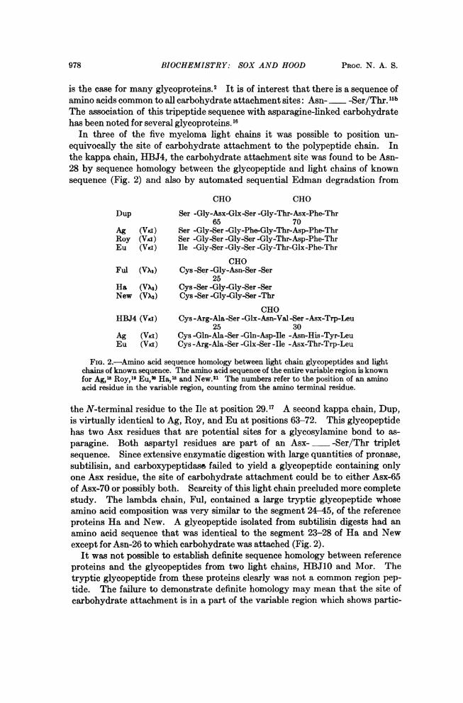
is the case for many glycoproteins.2 It is of interest that there is a sequence ofamino acids common to all carbohydrate attachment sites: Asn- -Ser/Thr. 15bThe association of this tripeptide sequence with asparagine-linked carbohydratehas been noted for several glycoproteins. 16
In three of the five myeloma light chains it was possible to position un-equivocally the site of carbohydrate attachment to the polypeptide chain. Inthe kappa chain, HBJ4, the carbohydrate attachment site was found to be Asn-28 by sequence homology between the glycopeptide and light chains of knownsequence (Fig. 2) and also by automated sequential Edman degradation from
CHO CHO
Dup Ser -Gly-Asx-Glx-Ser -Gly-Thr-Asx-Phe-Thr65 70
Ag (Vri) Ser -Gly-Ser -Gly-Phe-Gly-Thr-Asp-Phe-ThrRoy (Via) Ser -Gly-Ser -Gly-Ser -Gly-Thr-Asp-Phe-ThrEu (VKI) Ile -Gly-Ser -Gly-Ser -Gly-Thr-Glx-Phe-Thr
CHOFul (VX2) Cys -Ser -Gly-Asn-Ser -Ser
25Ha (VX2) Cys -Ser -Gly-Gly-Ser -SerNew (VX2) Cys -Ser -Gly-Gly-Ser -Thr
CHOHBJ4 (Via) Cys -Arg-Ala-Ser -Glx-Asn-Val-Ser -Asx-Trp-Leu
25 30Ag (VKI) Cys -Gln-Ala-Ser -Gln-Asp-Ile -Asn-His-Tyr-LeuEu (VKi ) Cys -Arg-Ala -Ser -Glx-Ser -Ile -Asx-Thr-Trp-Leu
FIG. 2.-Amino acid sequence homology between light chain glycopeptides and lightchains of known sequence. The amino acid sequence of the entire variable region is knownfor Ag,'8 Roy,19 Eu,w Ha,'8 and New.21 The numbers refer to the position of an aminoacid residue in the variable region, counting from the amino terminal residue.
the N-terminal residue to the Ile at position 29.17 A second kappa chain, Dup,is virtually identical to Ag, Roy, and Eu at positions 63-72. This glycopeptidehas two Asx residues that are potential sites for a glycosylamine bond to as-paragine. Both aspartyl residues are part of an Asx- -Ser/Thr tripletsequence. Since extensive enzymatic digestion with large quantities of pronase,subtilisin, and carboxypeptidas& failed to yield a glycopeptide containing onlyone Asx residue, the site of carbohydrate attachment could be to either Asx-65of Asx-70 or possibly both. Scarcity of this light chain precluded more completestudy. The lambda chain, Ful, contained a large tryptic glycopeptide whoseamino acid composition was very similar to the segment 24-45, of the referenceproteins Ha and New. A glycopeptide isolated from subtilisin digests had anamino acid sequence that was identical to the segment 23-28 of Ha and Newexcept for Asn-26 to which carbohydrate was attached (Fig. 2).
It was not possible to establish definite sequence homology between referenceproteins and the glycopeptides from two light chains, HBJ10 and Mor. Thetryptic glycopeptide from these proteins clearly was not a common region pep-tide. The failure to demonstrate definite homology may mean that the site ofcarbohydrate attachment is in a part of the variable region which shows partic-

VOL. 66, 1970 BIOCHEMISTRY: SOX AND HOOD 979
25 CHO 30HBJ4 (Human) Ala-Ser -Glx-Asn-Val-Ser -Aqx-Trp Leu
CHOMOPC46 (Mouse) Ala-Ser -Glx-Asx-Ile -Ser -Asn-Asp-Leu
CHOMPC37 (Mouse) Ala(Ser,Glx, Asx,Val,Ser,Asx)
FIG. 3.-Comparison of amino acid sequence of carbohydrate attachment sitesin mouse and human myeloma light chains. The carbohydrate attachment sitein the light chain secreted by murine plasmacytoma, MOPC46, is taken from apublished sequence of the tryptic glycopeptide.22 The light chain synthesized bymurine plasmacytoma, MPC37, was isolated from a serum myeloma protein(y-2b) by G-100 Sephadex chromatography following reduction and alkylation.The light chain glycopeptide was isolated from a pronase digest by a preparativepeptide map.
ularly extensive amino acid sequence diversity in the myeloma proteins (e.g.,positions 25-35 and 89-96). Indeed, the most probable, though tentative, siteof carbohydrate attachment in both light chains is at position 34 as determinedby comparing the amino acid and codon sequences of the glycopeptides withreference proteins.'8-20The glycopeptides of the human kappa chain, HBJ4, and the mouse kappa
chains, MOPC4622 and MPC37, are compared in Figure 3. The mouse andhuman glycopeptides are nearly identical in amino acid composition or sequence,and the site of attachement of carbohydrate appears to be at the same variableregion position in all three proteins.The various sites of carbohydrate attachment to the variable region of the
five myeloma light chains are summarized schematically in Figure 4. All of theattachment sites contained the common amino acid sequence Asn- __-Ser/
Vuiabe Constant
H2N I 214 COOH107
CHO
HBJ4 (K) 28 1
CHO
Dup (K})65or70
CHO
HBJ1O(K')IMor (IC) (R)
CHO
Ful ( 2I 1
FIG. 4.-Schematic summary of sites of carbohydrate attachment to various human myelomslight chains. The heavy lines in the variable region of the prototype light chain, iwdicae tihelocation of regions of extensive amino acid sequence diversity.
980 BIOCHEMISTRY: SOX AND HOOD PROC. N. A. S.
Thr to which carbohydrate was linked by a glycosylamine bond. The regions ofparticularly extensive diversity of the amino acid sequence are depicted inFigure 4 by heavy lines in the prototype light chain; the site of carbohydrateattachment in HB3J4, Ful, aid probably in H13J10 and Mor lies within theseregions.
Discussion. The primary finding of this study is that all the human myelomalight chains with covalently linked carbohydrate that were examined shared acommon amino acid sequence, Asn- -Ser/Thr, at the site of carbohydrateattachment. This tripeptide sequence apparently serves as an acceptor for anenzyme (N-acetylglucosamine-asparagine transglycosylase) catalyzing the for-mation of a glycosylamine bond between asparagine and N-acetylglucosamine.
All human heavy chains apparently have carbohydrate covalently linked to theconstant region.2 23 A ey, human heavy chain has carbohydrate attached to theAsx residue of an Asx-Ser-Thr triplet at positions 297-299 of the constant region. 20The other y-chain subclasses also have carbohydrate bound to Asx at apparentlyhomologous common region sites.24 Glycopeptides containing Asn and Ser havebeen isolated from at least one common region site on a ,u-chain.25 Althoughless is known of a-chain carbohydrate, there is evidence for asparagine-linkedcarbohydrate. 26'27 There are no Asn- -Ser/Thr triplets in the constant regionof the only completely sequenced heavy chain other than the site of carbohydrateattachment. 20 The available evidence is consistent with the view that allheavy chain constant regions contain asparagine-linked carbohydrate and thusthat all immunoglobulin-forming cells contain the transglycosylase. Thus, lightchains are synthesized in cells containing an enzyme for attaching carbohydrateto an appropriate site on a polypeptide chain.
In this study, glycopeptides from the variable region of human myeloma lightchains contained carbohydrate associated with the triplet, Asn- -Ser/Thr ineach case. We sought to learn if this triplet in the variable region always was asite of carbohydrate attachment by surveying 2700 overlapping amino acidtriplets from complete variable region sequences28 for the characteristic tripletsequence. Three proteins contained the triplet in the variable region but didnot contain variable region carbohydrate. Two were Bence-Jones proteins(Ha and Bo)18 and the third was the heavy chain from a serum macroglobulin(Ou).29 There are at least three possible reasons why carbohydrate might notbe linked to an Asn- -Ser/Thr triplet. First, the triplet could be inaccessibleto the transglycosylase because of the folding of the polypeptide chain. Second,the cells in which the protein was synthesized could lack the transglycosylase,although this seems unlikely for cells forming complete immunoglobulin mole-cules. Finally, the oligosaccharide could be removed from the polypeptide byglycosidases in the kidney.2 There are no data allowing a choice among thesealternatives.Two light chains, HBJ4 and Ful, have carbohydrate attached at or near posi-
tion 28 of the variable region. Furthermore, it seems likely by amino acid andcodon sequence homology that proteins Mlor and HBJ10 have their carbohydrateattached either in this region or possibly in a second region located betweenresidues 89 and 96. In another light chain, the site of carbohydrate attachment
VOL. 66, 1970 BIOCHEMISTRY: SOX AND HOOD 981
is in the segment 25-35 of the variable region.30 The segments 25-35 and 89-96of the light chain variable region are postulated to be directly involved in antigenbinding by two kinds of evidence: first, the extensive amino acid se(uence diver-sity of these regions3"-33 and, second, affinity-labeliiig experiments with a mousemyeloma protein34 35 aiid rabbit antibody,36 both with dinitrophenyl-bindingactivity. Since the oligosaccharide is hydrophilic, the regions of the light chainvariable region that contain carbohydrate are probably on the exterior of themolecule where the carbohydrate can interact with the hydrophilic solvent.In porcine RNase, the oligosaccharide groups do in fact appear to be attachedto the exterior of the protein. 3
It has been demonstrated that at least ten different species of mammals haveAsx- -Ser/Thr at the site of attachment of carbohydrate to the heavy chainconstant region at a site homologous with the human heavy chain.38 Further-more, two mouse kappa chains have variable region carbohydrate attachmentsites virtually identical in position and sequence to the human kappa chainHBJ4 (Fig. 3). Apparently the specificity of the transglycosylase has beenpreserved throughout much of mammalian evolution.The function of carbohydrate in immunoglobulins is unknown although the
remarkable conservation of heavy chain carbohydrate throughout mammalianevolution suggests an important role for carbohydrate attached to heavy chains.The postulated role for carbohydrate in facilitating secretion of immunoglobulinfrom the cell could be served by the oligosaccharide present on all heavy chains.The function of variable region carbohydrate is likewise unknown. It seemsunlikely that carbohydrate plays an important role in antigen binding since theoligosaccharide is a bulky group made up of a limited variety of carbohydrateresidues and cannot be synthesized in a precisely reproducible fashion. Infact, the bulky carbohydrate group attached near the antigen-binding site couldinterfere with antigen binding. Were this the case, one would predict that thevariable regions of light chains from normal serum immunoglobulins should havelittle or no carbohydrate bound near the antigen-binding site, since interactionof antigen with antibody bound to the surface membrane of immunocyte precur-sors is apparently necessary for induction of the immune response.39 Thispossibility is currently being investigated.
This study suggests that if the sequence triplet Asn- -Ser/Thr is notsterically hindered it can act as a carbohydrate acceptor from a transglycosylasewhose primary function is to glycosylate heavy chains and other proteins.This enzyme may respond indiscriminately whenever a polypeptide contains thecharacteristic sequence triplet. Thus, carbohydrate attached to the lightchain variable region may well be an "accident of nature" because of the creationof the sequence triplet Asn-__ -Ser/Thr either by the mutational mechanismthat is responsible for generating antibody diversity or by some other mutationalevent.
We wish to thank Dr. Victor Ginsburg for helpful advice and criticism.* Present address: Department of Medicine, Dartmouth Medical School, Hanover, N.H.t Present address and to whom requests for reprints should be addressed: Division of
Biology, California Institute of Technology, Pasadena, Calif.
982 BIOCHEMISTRY: SOX AND HOOD PROC. N. A. S.
1 Edelman, G. M., and W. E. Gall, Ann. Rev. Biochem., 38, 415 (1969).2 Spiro, R. G., New Engl. J. Med., 281, 991 (1969) is a recent review of glycoprotein struc-
ture and metabolism.' Abel, C. A., H. L. Spiegelberg, and H. M. Grey, Biochemistry, 7, 1271 (1968).4 Edmundson, A. B., F. A. Sheber, K. R. Ely, N. B. Simonds, N. K. Hutson, and J. L.
Rossiter, Arch. Biochem. Biophys., 127, 725 (1968).6 Dreyer, W. J. and E. Bynum, in Methods in Enzymology, ed. C. H. W. Hirs (New York
and London: Academic Press, 1967), vol. 11, p. 32.6 Rafferty, M. A. and R. D. Cole, J. Biol. Chem., 241, 3457 (1966).7 Katz, A. M., W. J. Dreyer, and C. B. Anfinsen, J. Biol. Chem., 234, 2897 (1959).8 Baglioni, C., Biochim. Biophys. Acta, 48, 392 (1961).9 Matsubara, H. and R. M. Sasaki, Biochem. Biophys. Res. Commun., 35, 174 (1969).10 Gray, W. R., Methods in Enzymology, ed. C. H. W. Hirs (New York and London: Aca-
demic Press, 1967), vol. 11, p. 469."1 Woods, K. R. and K. T. Wang, Biochim. Biophys. Acta, 133, 369 (1967).12 Roe, J. H., J. Biol. Chem., 212, 335 (1955).18 Warren, L., J. Biol. Chem., 234, 1971 (1959).14The subgroup designations for immunoglobulin variable regions are those proposed by the
Conference on Nomenclature for Animal Immunoglobulins, Prague, 1969.15 (a) Hood, L., W. R. Gray, B. G. Sanders, and W. J. Dreyer, Cold Spring Harbor Sym-
posia on Quantitative Biology, vol. 32 (1967), p. 133. (b) In glycopeptides from a variety ofproteins, many different amino acids have been found immediately C-terminal to the asparaginelinked to carbohydrate. Therefore, the characteristic triplet is represented in the text asAsn- -Ser/Thr in which the 2 represents any amino acid.
16 Eylar, E. H., J. Theor. Biol., 10, 89 (1965).17 Sox, H. C., Jr. and L. Hood, manuscript in preparation.18 Putnam, F. W., K. Titani, M. Wikler, and T. Shinoda, Cold Spring Harbor Symposta on
Quantitative Biology, vol. 32 (1967) p. 9.19 Hilschmann, N., Z. Physiol. Chemie, 348, 1077 (1967)."* Edelman, G. M., B. A. Cunningham, W. E. Gall, P. D. Gottlieb, U. Rutishauser, and M. J.
Waxdel, these PROCEEDINGS, 63, 78 (1969).2Langer, B., M. Steinmetz-Kayne, and Hilschmann, N., Z. Physiol. Chemie, 349, 945
(1968).22 Melchers, F., Biochemistry, 8, 938 (1969).23 Rosevear, J. W. and E. L. Smith, J. Biol. Chem., 236, 425 (1961).24Grey, H. M. and C. A. Abel, Fed. Proc., 28, 751 (1969).25 Davie, J. M. and C. K. Osterlund, Fed. Proc., 28, 495 (1969).26Abel, C. A. and H. M. Grey, Fed. Proc., 28, 495 (1969).'7In a subclass of a-chains, one of two glycopeptides isolated contained only N-acetylgalacto-
samine (see ref. 26). Presumably, the oligosaccharide in this glycopeptide is linked 0-glyco-sidically to either serine or threonine (see ref. 2).
28 Dayhoff, M. O., Atlas of Protein Sequence and Structure, (Silver Spring, Maryland: Nantional Biomedical Research Foundation, 1969).
29 Wikler, M. H., H. Kohler, T. Shinoda, and F. W. Putnam, Science, 163, 75 (1969).30 Spiegelberg, H. L., C. A. Abel, B. G. Fishkin, and H. M. Grey, Fed. Proc., 29, 703 (1970).81 Kabat, E. A., in Landsteiner Centennial, Dec. 5-7, 1968, Ann. N.Y. Acad. Sci., 169, 43
(1970).3aFranek, F., in Symposium on Developmental Aspects of Antibody Formation and Structure,
eds. J. Sterzl, and H. Riha (New York: Academic Press, in press)."3Wu, T. T. and E. A. Kabat, J. Exp. Med., in press.3' Goetzl, E. J. and H. Metzger, Biochemistry, 9, 1267 (1970).3' Goetzl, E. J. and H. Metzger, manuscript in preparation.36 Singer, S. J., L. I. Slobin, N. 0. Thorpe, and J. W. Fenton, III, Cold Spring Harbor Sym-
posis on Quantitative Biology, vol. 32 (1967), p. 99.'7Jackson, R. L., and C. H. W. Hirs, J. Biol. Chem., 245, 624 (1970).38 Howell, J. W., L. Hood, and B. G. Sanders, J. Mol. Biol., 30, 555 (1967).39 Metzger, H., Ann. Rev. Biochem., 1970, in press.