Embed Size (px)
Citation preview

British Heart gournal, I970, 32, I72.
Atrial parasystole
H. David Friedberg and Leo SchamrothFrom the Cardiac Section, Veterans Administration Center, Wood (Milwaukee), Wisconsin;Marquette School of Medicine, Milwaukee, Wisconsin, U.S.A.; and the University of theWitwatersrand,Johannesburg, South Africa
Two cases of atrial parasystole showing the various manifestations of the arrhythmia are presented.Analysis of the underlying mechanisms shows that atrial parasystolic bigeminy with 'reversed'coupling is a form of escape-capture begeminy, sinus escapes being followed by an ectopic capture ofthe atria. Reasons are given for the rarity of atrial fusion beats. The similarities and differencesbetween atrial and ventricular parasystole are explored. It is suggested that an atrial parasystolicpacemaker may lie within a major atrial preferential conducting pathway, and may consist of acongenitally ectopic fragment of sinus nodal tissue. The clinical significance of the arrhythmia isdiscussed; the associated diseases apparently represent a cross-section of medical ward experience.
Parasystole is a dual rhythm in which theparasystolic pacemaker is protected from theeffects of the dominant, usually faster pace-maker: this protection, which is the essenceof the arrhythmia, is situated within theimmediate vicinity of the parasystolic focus,and is operative throughout its entire cycle(Schamroth, I964). When a parasystolicventricular and a sinus pacemaker coexist,a characteristic arrhythmia appears. Theventricular beats bear no constant relationto the preceding QRS complexes (i.e. thecoupling intervals vary) and indeed may welloccur so late in diastole that they fuse withthe ensuing sinus QRS: the ectopic beats do,however, bear a consistent relation to eachother, so that the interectopic intervals arein simple multiples of the basic ectopic cyclelength. In those rare instances in which the
Received 8 April I969.
parasystolic focus is in the atria, and thus inthe same bi-atrial chamber' as the sinuspacemaker, the resulting disturbance ofrhythm is modified, though the essentialcharacteristics of the arrhythmia are notchanged. In particular, coupling intervalsvary less in atrial parasystole: a fixed bigeminy,closely resembling an extrasystolic bigeminywith nearly constant coupling, may occur.Atrial fusion beats are rare. The manifesta-tions of atrial parasystole will be presentedin this paper and the underlying mechanismsanalysed.
Case reportsCase x The electrocardiogram (Fig. i, a con-tinuous recording of standard lead II) was ob-tained from a 75-year-old man with severeobstructive lung disease. The arrhythmia was1 Electrophysiologically, both atria usually behave as asingle chamber, and will be referred to as the bi-atrialchamber.
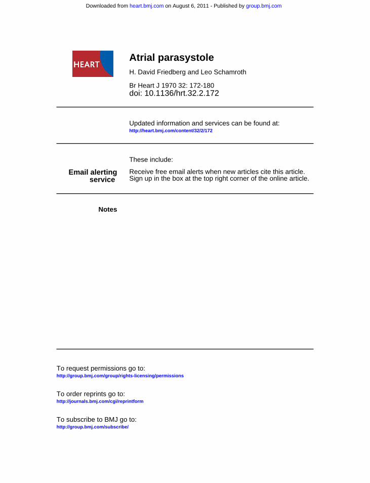
FIG . I Case I. A continuous strip of standard lead II. Premature atrial beats, markedwith a black dot, occur with varying coupling. The interectopic intervals are simplemultiples of I20-124, and thus the rhythm is atrial parasystole.
|1......~~~~~~~~~~~~~~~~~~~~~~~~~~~~~~~~~~~~~~~~~~~~~~~~~~~~~~~~~~~~~~~~~~~~~~~~~~~~~~~~~~~~~~~~~~~~~~~~~~~~~~~~~~~.
< 248_ 2x124 > < 123 > < 369- 3 x 123 > < 120 > <
124 >< 120 >< 248 2x 124 > < 120 >< 246: 2x 123 >
group.bmj.com on August 6, 2011 - Published by heart.bmj.comDownloaded from

Atrial parasystole 173
;~~~~~~~~~~~~~~~~~~~~~~~~~~~~~~~~~~~~~~~~~~~~~~~~~~~~~~~~~~~....N...........~~~~ ~~~~..
... j-F > U'rnM;rn.@A-----FIG. 2 Case x. Strips of standard lead III and aVF. Atrial premature beats occur inbigeminy. The second last P wave in standard lead III arises from AVjunctionalfocus. The last P wave in this lead occurs with a different coupling interval, but maintainsa constant interectopic interval, thus revealing the rhythm to be atrial parasystolic bigeminywith reversed coupling. (See text for discussion.)
present during two weeks only, when a smalldose of digitalis was being given. The basicsinus rhythm is punctuated by atrial prematurebeats. The ectopic P' waves resemble the sinusP waves, but begin more sharply, and rise moresteeply to a higher peak. The premature P'waves are not accurately coupled to their pre-ceding sinus P waves, the coupling intervalsvarying from 421 to 70. The intervals betweenthe P' waves - the interectopic intervals - are allin simple multiples of I20 to 124, therebyindicating that these beats are related to eachother. This combination of inconstant couplingand simply related interectopic intervals is thehallmark of parasystole.
Fig. 2 was recorded two days later, and con-sists of strips of standard lead III and lead aVF.A bigeminy is present, every second beat beingan atrial premature beat. As the coupling inter-vals are nearly constant, this closely resemblesthe commoner extrasystolic atrial bigeminy. In1 All time intervals are expressed in hundredths of asecond, i.e., 22= 0.22 second.
an extrasystolic bigeminy with a regular dominantrhythm and fixed coupling intervals, the inter-ectopic intervals will also be constant. This is notparasystole, but due simply to the constancy ofthe post-extrasystolic pause and the constantcoupling intervals. In Fig. 2, the true situation isrevealed by the last pair of P waves in standardlead III. The first P wave of this pair is inverted,arising from an AV junctional focus, which anti-cipates the sinus impulse. The second P waveof this pair arises from the same atrial focus asthe earlier ones, but now the coupling is con-spicuously different - 54 instead of 32 to 40.The interectopic interval, however, is constantat i i6. It is clear that the atrial focus was notdischarged by the AV junctional impulse, andis parasystolic. It is noteworthy that the para-systolic focus is discharged neither by the sinusimpulse nor by the lower junctional impulse, andis thus protected from below as well as fromabove.The electrocardiogram in Fig. 3 is part of the
same long tracing of standard lead II shown in
FIG. 3 Case I. Part of the same tracing as Fig. i. The sixth P wave in the upperstrip, marked F, is an atrial fusion beat. (See text.) P' in the lower strip isinterpolated (see Fig. 7).
F
* * 124 o 124 0 124 0
es----24-- - -33723x 124 ) - - > - 120--
Pi P P2 P3
0 0* 124 0
-0% .,Al,.
group.bmj.com on August 6, 2011 - Published by heart.bmj.comDownloaded from

174 Friedberg and Schamrothl-4 w
-~~~~~~~~~~~~~~~~~~~~~~~~~~~~~t-I*~--
0 115 0 120 * 1 0 116 118 0
hi :1A:::~.......
7 l W - 4~~~~~~~~~~~~~~~~~~~~~~~~~~~~~~~~~~~------
118 a 117 117 0
FIG.- 4 Case 2. Strips of standard leads II and III. Ectopic P waves with varyingcoupling but constant interectopic intervals occur, showing atrial parasystole. Some ofthe parasystolic P waves are not conducted to the ventricles, some are conducted
with aberrant ventricular conduction, and some are conducted normally.
Fig. i. The parasystolic cycle ranges from 120 to
12.4 (mean 23). The first, third, and eighth P waves
are parasystolic in origin. The beginning of
these P waves rises abruptly, forming a sharp
angle with the baseline. The beginning of the
sixth P wave, marked F, is smoothly rounded, as
is that of the sinus P waves. However, this P
wave is a little taller than the sinus P waves, and
has a slightly steeper descending slope. It is thus
intermediate in form between the sinus and the
ectopic P waves. Furthermore, this P wave begin
o-o6 sec. before the ectopic P' wave is due (asindicated by the half open circle). Therefore, this
P wave results from a fusion between the atrial
parasystolic and the sinus impulses. The second
P' wave in the lower strip is interpolated.
Case 2 The electrocardiogram (Fig. 4) was
recorded during a period of mental confusion
from a 74-year-old man with a history of ischae-
mic heart disease. The arrhythmia disappearedwith rest and sedation, but recurred temporarilyafter he broke his femur some months later. At
no time did he have angina or take digitalis. For
the most part, sinus P waves and ectopic P'
waves alternate in an atrial bigeminy with con-
spicuous variation of the coupling intervals,which range from 36 to 6o. The interectopicintervals range from 115 to i2o; the intersinus
intervals range from 11I4 to 30. In standard lead
III, a supraventricular premature beat from a
different focus (arrow) interrupts the sinus
rhythm, but does not affect the regularity of the
P' series. Some of the P' waves fall so early as
to fail to pass the AV junction, others are con-
ducted with varying degrees of ventricular aber-
ration, and others are conducted normally.
Though the ectopic cycle is a little variable, the
interectopic intervals vary much less than do
the coupling intervals; and all the hallmarks of
atrial parasystole are present.
The electrocardiogram in Fig. 5 was recorded
from Case 2 four months later, shortly after he
had suffered a fracture of the femur. All stripswere recorded at the same session; the bottom
two strips are continuous. P waves from many
different supraventricular foci are present. Some
of these P waves (marked with a dot) closelyresemble the parasystolic P' waves shown in
Fig. 4.
In the three upper strips, the interectopicintervals are a simple multiple of 98 (range 94 to
ioo). In the bottom strip, the interectopicintervals are I45, 148, and i44. These are not a
simple multiple of 98. All the figures, however,are simple multiples Of 49 ± i. An atrial para-
systole is therefore present, the basic cycle lengthof which is 49 ± i, and which is complicated byan exit block. If every second impulse reaches the
atrial muscle to inscribe a P' wave, the apparent
cycle length is 98 2, and 2:'i exit block is
present. If every third impulse reaches the atria
and is manifest, the apparent cycle length is
147±3, and 3: i exit block is present.
Mechanisms
(a) Parasystolic bigemiiny: 'reversed'
coupling. In atrial parasystole, both the
protected atrial pacemaker and the unpro-
tected sinus pacemaker are situated in the
same bi-atrial chamber. As a result, the
sinus node is vulnerable to, and will fre-
quently be prematurely discharged by., the
ectopic impulse. The sinus cycle will be
consequently reset,, while the parasystolic
cycle continues undisturbed. This is illus-
trated in Fig. 6., in which the 'build-up' of
the sinus impulse toward a threshold is pic-tured above a representation of events at the
atrial level. In Diagram I of Fig. 6, the para-
systolic impulse Ei reaches the sinus node
and aborts the immature impulse there at
point Sdi., interrupting and resetting the
II
group.bmj.com on August 6, 2011 - Published by heart.bmj.comDownloaded from

Atrial parasystolk 175
VF
*9 9 * 99 * 0
________----_------_---- _-- 297
09;*O * 94 O* I198-98-- 196 --
*98 100 - 100 *- - -- ---- 294-
o 145 * 148 * 146
FIG. 5 Case 2. Strips of lead aVF and standard lead II. The bottom two strips arecontinuous. P' waves are indicated by a black dot. The interectopic intervals are atfirst in multiples of 98 ± 2, and change abruptly to 147 ± 3, indicating atrialparasystole with varying exit-block. (See text for discussion.)
sinus cycle. The recharge of the sinus im-pulse now begins anew, which on reachingthreshold, discharges spontaneously at pointS2, after the expiry of the sinus cycle (sc).Now, if the duration of the ectopic cycle (ec)is a little greater than the sinus cycle plusthe refractory period of the sinus pacemaker(rp), the next ectopic impulse (E2) will againdischarge the sinus pacemaker (at point Sd2)and the sinus cycle is again reset. In thismanner, the sinus discharge is causallyrelated and linked to the protected ectopicdischarge, and the bigeminy is perpetuated(see also Fig. 8B). This is a form of escape-capture bigeminy (Bradley and Marriott,I958; Schamroth and Dubb, I965), but onein which a sinus escape is followed by anectopic capture of the atria.The time of appearance of the sinus P
waves is thus dependent on, and coupled to,the parasystolic P' waves. This is the reverseof the situation in an extrasystolic arrhythmia,in which the ectopic complexes are dependenton, and coupled to, the dominant beats. Asimilar 'reversed' coupling may occasionallyappear during ventricular parasystole. This
occurs if the ventricular impulse is com-pletely conducted retrogradely through theAV junction, atria, and SA junction to dis-charge the sinus node. Such complete retro-grade conduction is the exception. The rela-tively slowly conducting AV junction willusually protect the sinus node and effectivelyprevent the maintenance of a bigeminy.
Atrial parasystolic bigeminy thus dependsupon a fortuitous arithmetical relation be-tween the rates of the two centres of impulseformation, of which one is protected and theother is vulnerable. Once begun, the bige-miny tends to persist. The bigeminy may beinterrupted and the true nature of thearrhythmia revealed under the followingcircumstances. (i) If there is sufficient sinusarrhythmia to make the ectopic impulse soearly that it falls during the absolute refrac-tory period of the atria induced by a sinusimpulse (see also Interpolation below); (2) ifa different ectopic impulse discharges thesinus node and further dislocates its rhythm(see Fig. 2); (3) if the parasystolic impulsesuffers exit-block and fails to leave its focusand invade the atria.
group.bmj.com on August 6, 2011 - Published by heart.bmj.comDownloaded from

176 Friedberg and Schamroth
'a- - -sc ..... ....
----- sc- - ...
.
I.
Sdl Sd2 Sd3_~~~= L~~~~.1
El E2
- ec----- --. ..
FIG. 6 I. A diagram to show the mechanism of atrial parasystolic bigeminy. The uppersaw-tooth line represents the 'build-up' of the sinus impulse towards threshold.A = atrial level; SI, S2, and S3 are sinus impulses; Ei, E2, and E3 are ectopic impulses;rp = the refractory period of the sinus node; sc = the duration of the sinus cycle;ec = the ectopic cycle. (See text.)II. Interpolation of an atrial premature beat. The conventions are the same as in I. Ectopicimpulse E2 invades the atria before the end of the refractory period of the sinus node, sothat the sinus cycle is not reset. S3 then occurs when anticipated, and an ectopic beatis sandwiched between two sinus beats. Note: The AVjunctional and ventricular levelshave not been drawn. Conduction delay across the sino-atrial junction is shown in Fig. 7.
FIG. 7 A diagram to show the characteristic allorhythmia of an interpolated atrialpremature beat. PI, P', P2, and P3 refer to the P waves so marked in the lower stripof Fig. 3 (see text). S= sinus node; S-A = sino-atrial junction; A = atrial level.AVjunctional and ventricular levels have not been drawn.
Si S2 S3
S 7 I I
S-A
A I
P1 P' P2 P3
I
group.bmj.com on August 6, 2011 - Published by heart.bmj.comDownloaded from

Atrial parasystole rL77
(b) Interpolation If the ectopic impulsefalls early in the sinus cycle, at a time whenthe atria have recovered excitability but whenthe sinus node is still refractory, the ectopicimpulse will fail to reach and reset the sinuspacemaker. The spontaneous discharge ofthe sinus node then occurs when anticipated.Interpolation of the ectopic impulse betweentwo sinus impulses results. This is illustratedin Diagram II of Fig. 6, where impulseE2 is interpolated between S2 and S3. Thisphenomenon is evident electrocardiographic-ally in the lower strip of Fig. 3. Incompletepenetration of the S-A junction (the junc-tional delay area between the S-A node andthe surrounding atrial myocardium) by theectopic impulse may render it partiallyrefractory to the next sinus impulse. Thisproduces a characteristic disturbance of PPintervals (Langendorf et al., I962): the inter-val between a pair of sinus P waves enclosingan interpolated atrial ectopic beat is longerthan the intersinus interval. The subsequentPP interval is foreshortened. This is illus-trated in Fig. 7, a diagram of the conductionsequence of the interpolated parasystolicimpulse in the lower strip of Fig. 3. Im-pulses SI, S2, and S3 are regular consecutivesinus impulses resulting in P waves PI, P2,and P3, respectively. P' is the parasystolicimpulse which penetrates the S-A junctionbut fails to reach and reset the sinus pace-maker. This renders the junction partiallyrefractory. Conduction of S2 through the SAjunction is therefore prolonged, and theinscription of P2 is delayed. But for theeffect of P', P2 would have been inscribedas indicated by the dotted line. The nextsinus impulse S3 - P3 is not so delayed. P2is thus shifted towards P3, producing thecharacteristic allorhythmia of interpolation.A strictly analogous variation of RR inter-vals occurs with an interpolated ventricularparasystolic or extrasystolic beat (Schamroth,(i967b).
(c) Atrial fusion Ventricular fusion beatshave certain characteristics which lead toeasy recognition (Marriott, Schwartz, andBix, I962). Fusion is suspected if an ectopicQRS is due near the time of a sinus QRS,and an intermediate QRS is seen. Changesin QRS configuration are easily detectedbecause of the finely inscribed, detailed, andcomplex QRS deflections. Thus, very smalldegrees of fusion may be deduced from subtlechanges in the direction of the initial orterminal QRS vectors, or in the T wave.Detection is further facilitated by the factthat the two complexes concerned are usually
conspicuously different; and because therelatively slowly conducting AV junctioneffectively isolates the two rhythms, couplingintervals vary greatly, and end-diastolicectopic beats are common.
Several factors militate against the occur-rence and the recognition of atrial fusionbeats during atrial parasystole.i) Lack of P wave detail The P wave is lessdetailed, less complex, and coarser than theQRS complex. Subtle changes are thereforemore difficult to detect. Furthermore, the Pwaves from the sinus and the atrial pace-maker are not usually conspicuously different,so that the recognition of fusion by changesin shape alone is very difficult. Nor are atrialrepolarization waves - Ta waves - sufficientlydistinct to help. In Fig. 3, atrial fusion ispostulated because the P wave concerned hasan initial vector typical of the sinus P wavesin that lead, and is of intermediate heightand terminal slope, and because the timingis correct.2) Limited duration of opportunity for fusionIn the sixth beat of Fig. 3, probable atrialfusion is recognized when the ectopic P'wave was due oo6 sec. after the onset of thesinus P wave. Yet, in the ninth beat of theupper strip of Fig. i, fusion did not occurthough the ectopic P' wave was due o-o8 sec.after the start of the P wave. This suggeststhat within o-o8 sec., the sinus impulse hasspread to the tissues surrounding the atrialparasystolic focus, i.e. this is the conductiontime from the sinus to the ectopic focus.Therefore, in this case, if the sinus fires first,the time available for possible fusion is notgreater than o0o7 sec. Should the ectopicfocus discharge first, a similar limit for theduration of opportunity for fusion exists.While this is not known, it should also beabout o0o7 sec., but may well be less. Inboth these cases, and in many of the othersreported, the sinus and parasystolic P wavesare similar, with some differences in theirinitial contour, but with little difference intheir terminal contour. Thus, once theectopic impulse has inscribed the initialpart of the P wave, the contribution of thesinus impulse to the shape of this wave maynot be recognizable. Moreover, the time ofappearance of the more variable sinus im-pulse may not be known. Though a sinusimpulse may fuse in the atria with an earlierappearing ectopic impulse, this fusion maywell not be diagnosable.3) Resetting of sinus node As shown above,the presence of two centres of impulse forma-tion in the same bi-atrial chamber facilitates
group.bmj.com on August 6, 2011 - Published by heart.bmj.comDownloaded from

178 Friedberg and Schamroth
FIG. 8 Relating the possibility of atrial fusion to the resetting of the sinus cycle.Impulse formation in the S-A node (S) and the ectopic focus (e) is shown by the black dots.The refractory period of the sinus node is represented by the stippled area. The sinus impulsedoes not penetrate into the parasystolic focus. The rates of the two pacemakers are assumedto be moderately steady. (A) The parasystolic cycle is a littk longer than the sinus cycle;occasional parasystolic beats appear. (B) The parasystolic cycle exceeds the swn of the sinus cycleand the sinus refractory period; parasystolic escape-capture bigeminy results. (C) Theparasyitolic cycle equals two sinus cycles less the last coupling interval; atrial fusion results(marked f). (D) The parasystolic cycle equals the sinus cycle; repetitive fusion results.
the resetting of the unprotected sinus node.The frequency of this resetting and theresulting arrhythmic disturbance is depen-dent on the relation of the two cycle lengths.This is diagrammatically illustrated in Fig. 8.This theoretical analysis is based on theassumption that the two rates remain moder-ately steady.
a) If the parasystolic cycle exceeds thesinus cycle but is less than the sinus cycleplus the refractory period of the sinusnode (Diagram A of Fig. 8), there will beoccasional 'captures' of the atria by theectopic impulse.b) If the parasystolic cycle exceeds thesum of the sinus cycle plus the ensuingrefractory period of the sinus node (Dia-gram B of Fig. 8), parasystolic bigeminy -
escape-capture bigeminy - of the atriawill ensue.c) If the parasystolic cycle is shorter thanthe sinus cycle (not illustrated), the ectopicimpulses anticipate every sinus dischargeand thus usurp complete control of theatria, resulting in an uncomplicated ectopicatrial rhythm - an atrial parasystolictachycardia.The aforementioned relation with the re-
setting of the sinus cycle militates against theoccurrence of atrial fusion complexes. Thesewill only occur under the following rare
circumstances.
a) If the parasystolic cycle equals two, or any
integral number of sinus cycles, less the lastcoupling interval, both impulses will simul-
A
B
Sl...,...,.. ........ .....-~~ ~ ~ ~ ~ .... ... ...I...
C f D~~ ~ff
B I.... ....................................... ............tt...l . .......
... . .. .. .
... . .. .. . .......
group.bmj.com on August 6, 2011 - Published by heart.bmj.comDownloaded from

Atrial parasystole 179
taneously invade the atria and fuse (DiagramC of Fig. 8).b) If the parasystolic cycle equals the sinuscycle, or any integral number of sinus cycles,fusion will recur regularly (Diagram D ofFig. 8).The aforementioned principles may be
modified under the following special circum-stances: (a) in the presence of sinus arrhyth-mia; (b) in the presence of exit-block; (c) inthe presence of a variable ectopic cycle; and(d) if the sinus cycle is reset by another -different - ectopic impulse.
Fusion between atrial parasystolic andsinus impulses then is rare because (i) it isdifficult to recognize, (2) the coupling inter-vals vary little, (3) there is only a short periodof opportunity for fusion, and (4) becausethe mathematical relations between a pro-tected and an unprotected centre of impulseformation in the same chamber militateagainst fusion except in unusual circum-stances. Scherf, Yildiz and De Armas (I959)commented that such fusion is rare. Theonly clear cases are those of Vedoya (I944,Case 5) and of Katz, Eschelbacher, andStrauss (I937).
(d) Exit-block The parasystolic dischargewill activate the surrounding myocardiumwhenever the myocardium is not refractoryafter activation by the other pacemaker.Occasionally, however, calculation revealsthat, though the myocardium is responsiveat the time of the parasystolic discharge,activation does not occur. This has beenexplained on the basis of exit-block - aphenomenon in which an impulse is con-fined to its focus of generation by a 'block'at the ectopic-myocardial junction. Exit-block is also present if the calculated ectopiccycle length is less than the cycle of thedominant pacemaker, or if it is less than theduration of any post-ectopic pause; forwithout exit-block an ectopic tachycardiawould be present.There is a further circumstance in which
exit-block may be diagnosed. Should a con-stant 2: I exit-block change abruptly to onewith a 3: I ratio, the apparent commondenominator of the interectopic intervalswill change to one and a half times itself.Similarly, a change from a 3: I to a 4: Iconduction ratio will result in an increase ofone-third in the apparent ectopic cyclelength. That is to say, if the largest commondenominator in a continuous recordingchanges abruptly to one with which it has asimple fractional relation, exit-block with achange in conduction ratio may be diagnosed.
Thus, in Fig. 4, an abrupt increase in thecommon denominator from 98 tO I47 (i.e.,il times 98) alows the diagnosis of a basicectopic cycle length of 49 with 2:I exit-block changing abruptly to 3: i exit-block.
DiscussionIncidence Well-documented cases of atrialparasystole are rare. In a search of the pub-lished material, Eliakim (I965) could findonly I5 reported instances andaddedone more.To these may be added that of Attinger(I940), two of Holzmann (I960), two ofMoulopoulos and Sideris (I967), and Desh-pande (I968). The two reported here makea total of 24 cases. This is, then, a very muchrarer arrhythmia than either ventricular orjunctional parasystole. Exit-block has beenpresent in the cases of Katz et al. (1937),Holzmann (I96o), Deshpande (I968), andin one of the two in this paper, a total of fourcases. Though figures for the frequency ofexit-block in other forms of parasystole arenot available, exit-block does not appear tobe less common in atrial parasystole.
Clinical significance Most cases of atrialparasystole have been reported as curiosities,and a review of well-substantiated cases doesnot reveal any particular clinical association.It has been stressed that parasystole is asign of a diseased heart and is associatedwith digitalis therapy (Faltitschek and Scherf,1932; Scherf et al., 1959). On the other hand,Jervell (1932), Vedoya (I944), and Eliakim(i965) reported cases of atrial parasystolewithout heart disease. Certainly, patientswithout heart disease are much less likelyto have long electrocardiographic strips takenand have atrial parasystole diagnosed. Theassociation of parasystole and heart diseasemust be regarded as not definitely proven.Digitalis is often incriminated, but in noreported case is the appearance and regres-sion of the arrhythmia so clearly related tothe drug as in Case i above. Case 2 exempli-fies the apparent provocation of the arrhyth-mia by an acute psychotic episode and by anacute injury, in the absence of digitalis andnot necessarily related to ischaemic symp-toms.
Site of the parasystolic pacemaker Ofthe reported cases in which the parasystolicfocus is clearly atrial (as opposed to junc-tional, coronary sinus, or atrio-nodal), theP' waves of many bear a remarkable resem-blance to one another. These include thosereported by Jervell (I932), Attinger (I940),Scherf et al. (I959), Chung, Walsh, and
group.bmj.com on August 6, 2011 - Published by heart.bmj.comDownloaded from

18o Friedberg and Schamroth
Massie (i964, Cases I, 3, and 4), Moulo-poulos and Sideris (i967), and Deshpande(I968), and the two reported here. In allthese cases the P' wave arises more abruptlyto a higher peak than the sinus P wave, whilethe later portions of the two waves are simi-lar. Further, the P-R and the P'-R intervalsare equal or nearly so.Now, there is considerable evidence that
intra-atrial conduction and internodal conduc-tion occur via definite pathways, and are notradial, as was formerly thought (James,I966; Merideth and Titus, I968). It seemspossible that the atrial parasystolic focuslies within a major atrial conducting path-way, and is close to the sinus node. The'family resemblance' and the rarity of atrialparasystole suggest that in these patientsthe focus is a portion of sinus tissue a littledistance from the main node itself. This'ectopic sinus tissue' may be an embryonalrest. Interestingly, even the sinus node itselfmay be parasystolically protected (Scham-roth, I967a). If an atrial parasystole doesreflect a congenital anatomical variant, thenthe clinical features of this condition wouldreflect a cross-section of hospital practice,which does seem to be the case.
ReferencesAttinger, E. (I940). Zur Pathologie des Vorhof-
rhythmus und der P. Zacke. Schweizerische medi-zinische Wochenschrift, 70, 782.
Bradley, S. M., and Marriott, H. J. L. (I958). Escape-capture bigeminy. Report of a case of A-V dis-sociation initiated by 2:i S-A block with result-ing bigeminal rhythm. American Journal ofCardiology, I, 640.
Chung, Koo-Young, Walsh, T. J., and Massie, E.(I964). Atrial parasystole. American Journal ofCardiology, 14, 255.
Deshpande, S. Y. (I968). Atrial parasystole with exitblock and post-parasystolic P and T wave changes.Diseases of the Chest, 54, I62.
Eliakim, M. (I965). Atrial parasystole. Effect of caro-tid sinus stimulation, Valsalva maneuver andexercise. American journal of Cardiology, I6, 457.
Faltitschek, F., and Scherf, D. (I932). KlinischerBeitrag zur Parasystoliefrage. Wiener Archiv furinnere Medizin, 23, 269.
Holzmann, M. (I960). Beitrag zur Kenntnis derVorhof-Parasystolie. Cardiologia (Basel), 36, 223.
James, T. N. (I966). Anatomy of the conduction sys-tem of the heart. In The Heart, p. 64I. Ed. byJ. W. Hurst and R. B. Logue. McGraw-Hill,New York.
Jervell, A. (1932). Ein Fall von Vorhofparasystolie.Acta Medica Scandinavica, 79, 239.
Katz, L. N., Eschelbacher, J. L., and Strauss, S.(I937). An unusual case of auricular parasystoleshowing 'exit' block. American Heart journal, 14,571I.
Langendorf, R., Lesser, M. E., Plotkin, P., and Levin,B. D. (I962). Atrial parasystole with interpolation;observations on prolonged sinoatrial conduction.American Heart Journal, 63, 649.
Marriott, H. J. L., Schwartz, N. L., and Bix, H. H.(I962). Ventricular fusion beats. Circulation, 26,88o.
Merideth, J., and Titus, J. L. (I968). The anatomicatrial connections between sinus and A-V node.Circulation, 37, 566.
Moulopoulos, S. D., and Sideris, D. A. (I967). Timerelation between two pacemakers in atrial para-systole. British Heart journal, 29, 758.
Schamroth, L. (I964). The definition of parasystole.Cardiologia (Basel), 44, 37.
- (I967a). Sinus parasystole. American Journal ofCardiology, 20, 434.
- (I967b). Interpolated extrasystoles. South Afri-can MedicalJournal, 41, 919.
-, and Dubb, A. (I965). Escape-capture bigeminy.Mechanisms in S-A block, A-V block, and re-versed reciprocal rhythm. British Heart Journal,27, 667.
Scherf, D., Yildiz, M., and De Armas, D. (1959).Atrial parasystole. American Heart Journal, 57,507.
Vedoya, R. (I944). Parasystolia. A. L6pez, BuenosAires.
group.bmj.com on August 6, 2011 - Published by heart.bmj.comDownloaded from
doi: 10.1136/hrt.32.2.172 1970 32: 172-180Br Heart J
H. David Friedberg and Leo Schamroth Atrial parasystole
http://heart.bmj.com/content/32/2/172Updated information and services can be found at:
These include:
serviceEmail alerting
Sign up in the box at the top right corner of the online article.Receive free email alerts when new articles cite this article.
Notes
http://group.bmj.com/group/rights-licensing/permissionsTo request permissions go to:
http://journals.bmj.com/cgi/reprintformTo order reprints go to:
http://group.bmj.com/subscribe/To subscribe to BMJ go to:
group.bmj.com on August 6, 2011 - Published by heart.bmj.comDownloaded from


![Dysrhythmias (002) [Read-Only] - Aventri · Atrial AV node Ventricular Classification of Rhythm Abnormalities Supraventricular Atrial origin Atrial fibrillation Atrial flutter Atrial](https://img.dokumen.tips/doc/110x75/5f024baa7e708231d4038f22/dysrhythmias-002-read-only-aventri-atrial-av-node-ventricular-classification.jpg)