Embed Size (px)
Citation preview

Atlas of Animal Anatomy and Histology


Péter Lőw • Kinga Molnár • György Kriska
Atlas of Animal Anatomy and Histology

Péter Lőw Department of Anatomy, Cell and Developmental Biology Institute of BiologyEötvös Loránd University Budapest Hungary
Kinga Molnár Department of Anatomy, Cell and Developmental Biology Institute of BiologyEötvös Loránd University Budapest Hungary
György Kriska Group for Methodology in Biology Teaching Institute of Biology Eötvös Loránd University Budapest Hungary
Danube Research Institute Centre for Ecological ResearchHungarian Academy of Sciences Budapest Hungary
Additional material to this book can be downloaded from http://extras.springer.com
ISBN 978-3-319-25170-7 ISBN 978-3-319-25172-1 (eBook) DOI 10.1007/978-3-319-25172-1
Library of Congress Control Number: 2016931371
Springer Cham Heidelberg New York Dordrecht London © Springer International Publishing Switzerland 2016 This work is subject to copyright. All rights are reserved by the Publisher, whether the whole or part of the material is concerned, specifi cally the rights of translation, reprinting, reuse of illustrations, recitation, broadcasting, reproduction on microfi lms or in any other physical way, and transmission or information storage and retrieval, electronic adaptation, computer software, or by similar or dissimilar methodology now known or hereafter developed. The use of general descriptive names, registered names, trademarks, service marks, etc. in this publication does not imply, even in the absence of a specifi c statement, that such names are exempt from the relevant protective laws and regulations and therefore free for general use. The publisher, the authors and the editors are safe to assume that the advice and information in this book are believed to be true and accurate at the date of publication. Neither the publisher nor the authors or the editors give a warranty, express or implied, with respect to the material contained herein or for any errors or omissions that may have been made.
Printed on acid-free paper
Springer International Publishing AG Switzerland is part of Springer Science+Business Media (www.springer.com)
General professional reviewer
Zsolt Kovács Department of Zoology Faculty of Sciences and TechnologyUniversity of West Hungary Szombathely Hungary

v
The purpose of this book is to provide an introduction to comparative anatomy and histology for biology undergraduates and for all those who are interested in the internal structure of ani-mals. The information is presented in the form of colour photographs of step-by-step dissec-tion stages integrated with histological sections of actual organs. A specialty of this atlas is that it contains only high-quality, accurate, and attractive photographs, not idealised line drawings. Dissection plays an important part in understanding the anatomy of an animal, and this book has been designed to make full use of the wealth of information made available through dis-section. The accompanying text aims to outline the evolutionary and functional aspects of the anatomy revealed in the photographs. Our book encourages and facilitates active and self- directed learning by the students so that instructors can teach more effectively and effi ciently. This manual emphasises dissection procedures that preserve as many structures as possible for later review of the entire specimens. Every effort has been made to give clear, lucid descrip-tions and instructions, and enough background material has been included to create interest in and understanding of the subject matter.
The animals dissected in this book have been chosen as representative examples of six invertebrate phyla and four classes of vertebrates. This book offers step-by-step illustrations and instructions for dissecting a roundworm, earthworm, snail, mussel, crayfi sh, cockroach, crucian, frog, chicken, and rat. The types included are commonly studied in undergraduate zoology courses. They can be used also as a guide to dissection of other animals in the same group. Dissections range from beginning to advanced and discuss the digestive, circulatory, respiratory, excretory, reproductive, and nervous systems. Skeletal material of vertebrate animals is also included to show the supporting framework of the body and its development during evolution.
Another valuable aspect of this atlas is that it features large-size, full-colour histological micrographs, with labels and legends that draw attention to details of microanatomy of the most important organs. The histological descriptions follow the anatomical pictures and expla-nation of an actual organ, and they are highlighted with a coloured background. In this way, students can correlate microscopic structures with the gross composition. Clear histological explanations give details of how tissues are structured and how they work. Students will learn to recognise different types of tissues easily. The detailed photographs enable the reader to gather microanatomical knowledge even in the lack of prepared light microscopic sections or microscopic facilities.
The digital annex of the book includes slide-shows and interactive tests that can be used to check the knowledge. A special item of the software is a stereoscopic (3D) application enabling to visualize three-dimensional (anaglyph) pictures on a monitor or by a projector. Anaglyph pictures should be viewed through red-cyan glasses. The slide-shows are also available on-line at http://bszm.elte.hu/anatomy/ , optimized for mobile browsers.
Budapest, Hungary Péter Lőw Kinga Molnár György Kriska
Pref ace

vii
It is always important to perform a dissection in an appropriate lab under the guidance of an experienced instructor. Do not do anything uncertainly; wait for specifi c instructions in the lab. Dissection is both a skill and an art. A good dissection requires time and patience. Always prepare for a dissection in advance, learn the structures you want to fi nd, and work deliberately. Make small cuts and do not remove a piece of tissue unless you know what it is. Each dissec-tion chapter in this book includes background information about the sample animal, availabil-ity and proper, species-specifi c anaesthesia of the animal.
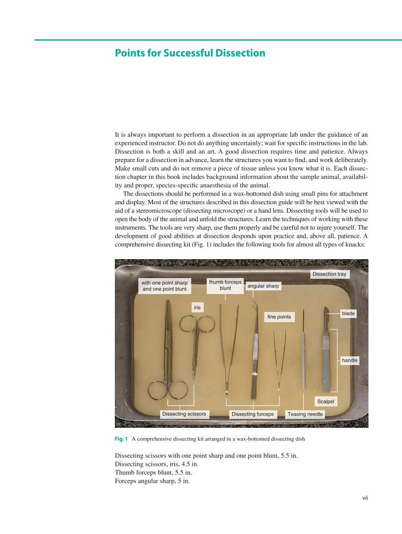
The dissections should be performed in a wax-bottomed dish using small pins for attachment and display. Most of the structures described in this dissection guide will be best viewed with the aid of a stereomicroscope (dissecting microscope) or a hand lens. Dissecting tools will be used to open the body of the animal and unfold the structures. Learn the techniques of working with these instruments. The tools are very sharp, use them properly and be careful not to injure yourself. The development of good abilities at dissection desponds upon practice and, above all, patience. A comprehensive dissecting kit (Fig. 1 ) includes the following tools for almost all types of knacks:
Dissecting scissors with one point sharp and one point blunt, 5.5 in. Dissecting scissors, iris, 4.5 in. Thumb forceps blunt, 5.5 in. Forceps angular sharp, 5 in.
Points for Succe ssful Dissection
with one point sharpand one point blunt
thumb forcepsblunt angular sharp
iris
fine points
Dissection tray
blade
Scalpel
Teasing needleDissecting forcepsDissecting scissors
handle
Fig. 1 A comprehensive dissecting kit arranged in a wax-bottomed dissecting dish

viii
Forceps fi ne points, 4.5 in. Teasing needle straight with metal chuck Scalpel handle No. 4 Scalpel blade No. 22
Although these instruments serve the requirements of nearly all kinds of dissecting situa-tions for special tasks and fi ne, elaborate work, we recommend some further tools (Fig. 2 ):
Bone rongeurs (Adson, Blumenthal, or Friedman type), 6 in. Micro forceps Dissecting scissors, micro iris (McPherson-Vannas), straight, sharp, 4.5 in. Dissecting scissors, angled sharp, 4.5 in.
Finally, it is well worth to use dissecting pins (insect pins) to position parts as you proceed with your examination of the specimen, so that you have a clearer view of the structure and organisation of the organism.
Keep in mind that dissecting does not mean “to cut up”; in fact, it means “to expose to view”. Careful dissecting techniques will be needed to observe all the structures and their connections to other structures. You will not need to use a scalpel very often. On the contrary to popular belief, a scalpel is not the best tool for dissection. Scissors are better because the point of the scissors can be pointed upwards to prevent damaging organs underneath. Always raise structures to be cut with your forceps before cutting, so that you can see exactly what is underneath and where the incision should be made. Never cut more than is absolutely necessary to expose a part. Sometimes the so-called blunt dissection is the most appropriate when you only tear connective tissue struc-tures with forceps to reveal an underlying compact organ and do not cut anything.
When completed, clean up your dissection. Dispose of your materials according to the directions from your instructor. Pour your excess liquid into the sink and wrap the body parts in a paper towel before throwing them in the carcass container. Never dispose the body parts into ordinary communal waste. Immediately after use, rinse instruments under warm or cool running water to remove all blood, body fl uids, and tissue. Dried soils may damage the instru-ment surface and make cleaning very diffi cult. Do not use hot water as this will coagulate proteinous substances. Clean up your work area and wash your hands before leaving the lab.
Micro forceps
micro iris angled sharp
Dissecting scissors
Bone rongeurs
Fig. 2 Special dissection instruments for meticulous tasks
Points for Successful Dissection

ix
Histological Sections
Histology is the study of the microscopic anatomy of cells and tissues of animals (or plants). During the routine procedure, the organs are fi xed to prevent decay and embedded in paraffi n (paraplast) to give support for cutting very thin (2–5 μm thick) sections. The sections are placed onto microscope slides and stained with histological stains, then covered with a cover-slip and mounting medium for preservation. Histological slides are examined with light microscope.
Histological Stains
Histological stains are used to increase the typically minor differences in light refraction of biological samples. The procedure is based on the variances in binding of histological stains by tissue and cell components.
HE (haematoxylin – eosin) stain: It provides a general overview – haematoxylin stains the nucleic acids, and eosin stains the cytosol and the extracellular matrix. Azan (azocarmine – aniline blue) stain: It provides a general overview – azocarmine stains the cell nucleus and the cytoplasm, aniline blue stains the connective tissue matrix and fi bres and some mucous secret. PAS (Periodic acid-Schiff reaction) stain: This reaction is used to detect structures containing a high proportion of carbohydrate macromolecules (glycoproteins, glycolipids, and polysac-charides). The reaction gives a purple- magenta colour typically in mucus gland cells, connec-tive tissue, and basement membrane.
Semithin Sections
Plastic (epoxy resin) embedding is commonly used in the preparation of material for electron microscopy. Semithin sections (0.8–1 μm) are cut using glass knives. The sections are stained with toluidine blue and examined using a light microscope.
Histologic al Methods


xi
Here we explain compass points of anatomy. Many of these are taken from Latin or Greek languages, and each has a very specifi c meaning. It is really important to understand the basic terms, which are used throughout the anatomical and histological descriptions.
Frontal plane: It is a vertical plane at right angle to median plane. If you draw a line from one ear to another from above the head and then divide the whole body along this line, the plane formed will be frontal plane. It is also known as coronal plane. Median or mid-sagittal plane: This is the plane which divides the body into equal right and left halves. Oblique plane: Any plane other than the above described planes will be oblique plane. Sagittal plane: It is any plane parallel to the median plane. This plane divides the body into unequal right and left halves. Transverse plane: It is the horizontal plane of the body. It is perpendicular to both frontal and median planes.
Directional terms describe the positions of structures relative to other structures or locations in the body:
Anterior: Towards the head end (e.g. the oesophagus is located anterior to the stomach) Caudal: Away from the head, towards the tail end of the body Cranial: Towards the head end of the body Distal: Away from or farthest from the middle line of an organism or from the point of attach-ment (e.g. the hand is located at the distal end of the forearm) Dorsal: Towards the back or upper part of the animal Inferior: Lower Lateral: Situated at the side away from the midline of the body (e.g. the little toe is located at the lateral side of the foot) Longitudinal: Lengthwise; along the length of the body Medial: Towards the midline of the body (e.g. the middle toe is located at the medial side of the foot) Median: Along the middle of the long axis Periferal: Referring to parts away from the centre Posterior: Facing towards the tail end (e.g. the pelvic girdle is located on the posterior end of the backbone) Proximal: Towards or nearest to the middle line of the organism or the point of origin of a part (e.g. the proximal end of the femur joins with the pelvic girdle) Sagittal: along or parallel with the middle plane of the body Superfi cial: On or near the surface Superior: Upper Transverse: Lying across or between or at right angles to the longitudinal axis Ventral: Towards the abdominal surface
Important Tec hnical Terms


xiii
The authors wish to thank the many people who helped in the preparation of this book. In particular, thanks are due to Dr. György Csikós, Viktor Kis, Sarolta Pálfi a, and Zsolt Pálfi a of the Department of Anatomy, Cell and Developmental Biology, Eötvös University, Budapest, Hungary for their prepared specimens. We are indebted not only to them but also to Dr. Zsolt Kovács (Department of Zoology, Faculty of Sciences and Technology, University of West Hungary, Szombathely, Hungary) who revised the manuscript. Any inaccuracies remaining are, of course, our own responsibility. We are grateful for the excellent technical assistance to Eszter Papp. We thank Monika Truszka for the fi rst-rate histological work from embedding to outstanding sections and brilliant staining. We thank András Barta for creating the on-line ver-sion of the slide-shows. We would also like to thank the cheerful and hardworking production team at Springer Verlag whose encouragement sustained us on several occasions.
P. Lőw K. Molnár G. Kriska
Acknowledgements


xv
Part I Invertebrates
1 Examination of a Hydra . . . . . . . . . . . . . . . . . . . . . . . . . . . . . . . . . . . . . . . . . . . . . 3
2 Examination of a Planarian . . . . . . . . . . . . . . . . . . . . . . . . . . . . . . . . . . . . . . . . . . 7
3 Dissection of a Roundworm ( Ascaris suum ) . . . . . . . . . . . . . . . . . . . . . . . . . . . . . 11
4 Dissection of the Earthworm ( Lumbricus terrestris ) . . . . . . . . . . . . . . . . . . . . . . . 27
5 Dissection of a Snail ( Helix pomatia ) . . . . . . . . . . . . . . . . . . . . . . . . . . . . . . . . . . . 49
6 Dissection of a Freshwater Mussel ( Anodonta anatina ) . . . . . . . . . . . . . . . . . . . . 79
7 Dissection of a Crayfi sh ( Astacus astacus ) . . . . . . . . . . . . . . . . . . . . . . . . . . . . . . . 101
8 Dissection of a Cockroach ( Blaberus sp.) . . . . . . . . . . . . . . . . . . . . . . . . . . . . . . . . 139
Part II Vertebrates
9 Dissection of the Crucian ( Carassius carassius ) . . . . . . . . . . . . . . . . . . . . . . . . . . 173
10 Dissection of a Frog ( Rana sp.) . . . . . . . . . . . . . . . . . . . . . . . . . . . . . . . . . . . . . . . . 213
11 Dissection of a Chicken ( Gallus domesticus ) . . . . . . . . . . . . . . . . . . . . . . . . . . . . . 265
12 Dissection of the Rat ( Rattus norvegicus ) . . . . . . . . . . . . . . . . . . . . . . . . . . . . . . . . 325
Bibliography . . . . . . . . . . . . . . . . . . . . . . . . . . . . . . . . . . . . . . . . . . . . . . . . . . . . . . . . . . . 401
Index . . . . . . . . . . . . . . . . . . . . . . . . . . . . . . . . . . . . . . . . . . . . . . . . . . . . . . . . . . . . . . . . . 403
Contents