Embed Size (px)
Citation preview

Upgrade Info
ATLAS 2D - Extreme Field of View ImagingATLAS 2D : Large Area Imaging for Unrivaled Productivity

Upgrade Info
Contact: Dr. Ulrich Kohl-Roscher
Date: December 2013
Introduction
The ATLAS system is an integrated package comprising hard-
ware and software that can automatically acquire large field
of view images and image mosaics without compromising
resolution. In combination with any scanning electron micro-
scope from Carl Zeiss, it enables quick and efficient imaging
of large-area specimens with nanometre resolution.
With its adaptive 16-bit scan generator and dual super-sam-
pling signal acquisition, ATLAS acquires individual images up
to one gigapixel in size (32k x 32k) at up to sixteen bits per
pixel.
By integrating into the SmartSEM software for microscope
control and calling upon its rich suite of automation fea-
tures, ATLAS allows for the automated acquisition of an
image spanning extremely large fields of view. With suitable
specimens, unattended operation can acquire multi-image
montages exceeding one terapixel in size.
With these capabilities, ATLAS can capture images of regions
of centimetre scale at nanometre resolution with just a few
hours of work. The continuous zoom function of the built-in
image viewer allows you to enlarge the final image from
a complete overview down to nanometre resolution. As a
result, ATLAS separates studying the specimen from the task
of acquiring the image.
ATLAS offers users a new degree of productivity.
ATLAS 2D - Extreme Field of View ImagingATLAS 2D : Large Area Imaging for Unrivaled Productivity
Availability
The instrument is available for:
• EVO series
• 15xx series
• SUPRA series
• ULTRA series
• MERLIN series
• CrossBeam series
Benefits
• Produce images into the terapixel range
By stitching single images each as large as 32k X 32k
pixels (1 gigapixel), ATLAS allows for the imaging of areas
into the range of square centimetres at nanometre reso-
lutions.
• Reduce time operating the microscope
By automating acquisition and first-pass stitching,
ATLAS allows users to get to data analysis faster.
• Acquire images for later exploration
By imaging larger areas of potential interest, ATLAS re-
duces the need to return to the lab for follow up acquisi-
tions.
• Distribute results easily
By exporting the final stitched image to HTML, ATLAS
makes results universally compatible and accessible using
the feature-rich ATLAS browser-based viewer that can run
from a local hard drive, over your intranet, or across the
Internet.
ATLAS meets the demand for efficient, cost-effective methods of examining a steadily rising number of speci-
mens with constantly increasing sizes, using resolutions in the nanometre range.
2
Upgrade Info
Operation
The ATLAS system includes:
- Acquisition and scan generator hardware
- Workstation class PC & LCD
- The ATLAS software
- The VE-Viewer software
Upgrade Path
ATLAS will run on any Zeiss microscope that has the fol-
lowing:
- SmartSEM API options.
- SmartSEM V05.04 or later.
The retrofit must be performed by an authorised Carl Zeiss
Microscopy service engineer. For further information,
contact:
Part Ordering no.
ATLAS for non-MERLIN Systems
ATLAS PC, ATLAS Hardware, Smart
EXTIF Switch Box, Necessary Cables,
licenses.
347823-9060-
000
ATLAS for MERLIN Systems
ATLAS PC, ATLAS Hardware, Smart
EXTIF Switch Box, Necessary Cables,
licenses.
347823-9063-
000
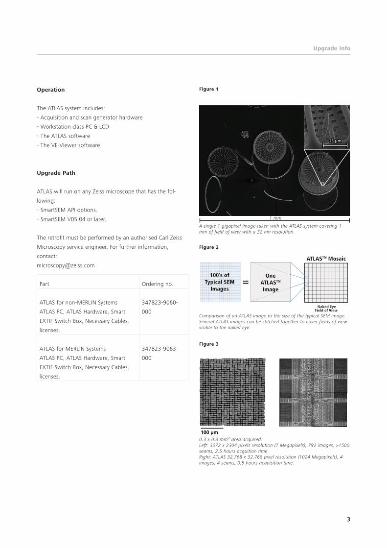
Figure 1
A single 1 gigapixel image taken with the ATLAS system covering 1 mm of field of view with a 32 nm resolution.
1 mm
3
Figure 2
Comparison of an ATLAS image to the size of the typical SEM image. Several ATLAS images can be stitched together to cover fields of view visible to the naked eye.
OneATLASTM
Image
100’s ofTypical SEM
Images
ATLASTM Mosaic
Naked EyeField of View
Figure 3
0.3 x 0.3 mm² area acquired. Left: 3072 x 2304 pixels resolution (7 Megapixels), 792 images, >1500 seams, 2.5 hours acquition time. Right: ATLAS 32,768 x 32,768 pixel resolution (1024 Megapixels), 4 images, 4 seams, 0.5 hours acquisition time.
100 µm
1 mm

EN_4
3_01
1_02
7 | C
Z-11
/201
3 |
Subj
ect
to c
hang
e in
des
ign
and
scop
e of
del
iver
y an
d as
a r
esul
t of
ong
oing
tec
hnic
al d
evel
opm
ent.
| ©
Car
l Zei
ss M
icro
scop
y G
mbH
Carl Zeiss Microscopy GmbH 07745 Jena, Germany [email protected] www.zeiss.com/microscopy