Embed Size (px)
Citation preview
At Birth, Tarsiers Lack a PostorbitalBar or Septum
TIMOTHY D. SMITH,1,2* VALERIE B. DELEON,3 AND ALFRED L. ROSENBERGER4
1School of Physical Therapy, Slippery Rock University, Slippery Rock, Pennsylvania, USA2Department of Anthropology, University of Pittsburgh, Pittsburgh, Pennsylvania, USA
3Center for Functional Anatomy and Evolution, Johns Hopkins University School ofMedicine, Baltimore, Maryland, USA
4Department of Anthropology and Archaeology, Brooklyn College, CUNY, Brooklyn,New York, USA
ABSTRACTAmong primates, partial or complete posterior closure of the orbit has
been widely accepted as a shared derived characteristic justifying an exclu-sive tarsier-anthropoid clade, while some regard the tarsier lateral orbit asan elaborated postorbital bar (POB). To test these competing hypotheseswhile minimizing the confounding effect of tarsier orbital hypertrophy, wecompared tarsiers and other primates at early (fetal and newborn) agesusing dissection, micro-CT scans and soft tissue histology. Our findings dem-onstrate unanticipated variation in the anatomy and development of thezygomaticofrontal (ZFA) articulation, which forms the orbit’s lateral frame-work. Tarsiers uniquely exhibit a combination of two features: absence of apre- and peri-natal frontal spur to join with the zygomatic to form the ZFA;and, the spur’s substitution by an elaborate ligament, which envelops theeye laterally as an expansive postorbital membrane (POM) that mergeswith the anterolateral fontanelle of the lateral cranial vault. In lacking afrontal spur, tarsiers are distinct from strepsirhines, while the ligamentousstructure of the POM distinguishes its ZFA from that of anthropoids, whichis a typical facial suture at and prior to birth. The POM of tarsiers may bethought of as an accessory fontanelle, a structural compromise that providesflexible stability and spatial separation of bones while anticipating rapidpostnatal growth of an enormously enlarged eye. We regard the tarsierPOM as part of a neomorphic eyeball hypertrophy complex, and reject thehypothesis of derived homology of the postorbital septa of adult tarsiers andanthropoids on histological, developmental and functional grounds. AnatRec, 00:000–000, 2013. VC 2013 Wiley Periodicals, Inc.
Key words: Anthropoidea; Haplorhini; homology; postorbitalseptum; tarsiiforms; ocular hypertrophy; Tarsius
The orbit of adult primates is well supported laterallyby the articulation of the frontal and zygomatic bones.In extant strepsirhines (lemurs, lorises, and allies) and
Paleogene euprimates (adapiforms, fossil tarsiiforms,and primates of modern aspect), each of these bonesprojects toward one another with orbital processes,
Additional Supporting Information may be found in theonline version of this article.
Grant sponsor: NSF; Grant number: BCS-0959438.
Grant sponsor: Pennsylvania State System of Higher Education.
*Correspondence to: Timothy D. Smith, School of PhysicalTherapy, Slippery Rock University, Slippery Rock, PA, USA.E-mail: [email protected]
Received 15 August 2012; Accepted 21 November 2012.
DOI 10.1002/ar.22648Published online in Wiley Online Library(wileyonlinelibrary.com).
THE ANATOMICAL RECORD 00:00–00 (2013)
VVC 2013 WILEY PERIODICALS, INC.

forming a postorbital bar (POB). While all extant prima-tes possess a functionally similar supportive structure(e.g., the lateral border of the human eye socket), onlytwo groups exhibit a posterolateral extension of bonebehind the eye: tarsiers and anthropoids (Pocock, 1918;Cartmill, 1980; Rosenberger et al., 2008). For nearly 100years this character has been interpreted as a sharedderived feature supporting the central phylogenetic di-chotomy of crown euprimate evolution (Pocock, 1918;Szalay and Delson, 1979; Fleagle, 1999): the distinctionbetween strepsirhines and haplorhines (tarsiers, anthro-poids, and allies) and, more specifically, the sister-grouprelationship between tarsiers and anthropoids to theexclusion of many fossil tarsiiformes.
Among anthropoids, an effectively complete postorbitalseptum (POS) is produced by sheets of the zygomatic andalisphenoid that form extensive sutural contacts with oneanother, in addition to the zygomatic’s contact with thefrontal. In tarsiers, the extent of postorbital closure is farless and restricted to the upper part of the framework,leaving a relatively wide open confluence between theorbit and the temporal fossa behind it (see electronic Sup-porting Information Fig. S1). Here, too, the zygomaticand frontal meet to form a tripartite lateral orbitalmosaic with the addition of a slip arising from the ali-sphenoid. While these particular similarities have beeninterpreted as homologies having the same overall func-tional explanation (Cartmill, 1980), to insulate the eyefrom mechanical interference generated by the adjacenttemporalis muscle lying behind the orbit, they also pres-ent something of a paradox, for tarsiers have the largesteyes of all mammals relative to their body size (Polyak,1957) while those of anthropoids are relatively small bycomparison with other primates (Ross and Kirk, 2007).As a consequence of these proportional differences, thegross anatomy of their adult orbits differs and this has
led to a fundamental disagreement. Some authors viewthe lateral orbital mosaic of tarsiers as a homologous ver-sion of the anthropoid POS, either preconfiguring theevolution of complete orbital closure or representing avestige of closure that was lost in connection with eyeballhypertrophy (Cartmill, 1980). Others regard the tarsier’slateral mosaic as little more than an elaborated postorbi-tal bar (Simons and Rasmussen, 1989; Rosenberger et al.,2008), modified to accommodate enormous eyeballs.
It is widely acknowledged that the derived orbitalmorphologies of tarsiers and anthropoids evolveddirectly or step-wise from a basal condition resembling asimple ring-like postorbital bar, like those found inEocene fossil primates and modern strepsirhines (Hersh-kovitz, 1977; Cartmill, 1980; Rosenberger et al., 2008).However, absent paleontological evidence of the transfor-mation, this model is inferred entirely from adultmorphology. Here we present the first comparativeassessment of perinatal development of this region inlight of recent work documenting profound differencesbetween tarsiers and other primates in the timing of eyegrowth (Jeffery et al., 2007; Cummings et al., 2012). Theenormous tarsier eye grows disproportionately more inthe postnatal period (�66% in diameter) compared withanthropoids (�38–50% in diameter) and quickly outpa-ces bony growth surrounding the eye (Cummings et al.,2012); in adults more than half the globe falls outsidethe margin of the orbit (Schultz, 1940). Moreover, a com-parison of orbital diameter in fetal, 0-day-old and 6-day-old tarsiers suggests that the growth rate of the orbitaccelerates rapidly in the perinatal timeframe (Cum-mings et al., 2012). To disentangle the morphologicalinfluence of hypertrophic eyeball expansion, we investi-gated late fetal and perinatal development of thezygomaticofrontal articulation (ZFA) at stages prior tothe divergent postnatal orbital growth rates observed in
TABLE 1. Species, ages, and selected measurements of the specimens under study
Spec. no. Species SourceAge in days(if known)
Craniallength (mm) CRL (mm) Figure reference
P96 Tarsius syrichta DLC Fetal 24.85 48.73 3, 5E-G, S3P94 T. syrichta DLC 0 29.45 68 3A-C, 4B, 5A-D,
H, 10, S3P98 T. syrichta DLC 6 29.15 ? 5I, S3013 T. bancanus LM 1 31.7 ? 1, 2, 4AP696 Cheirogaleus medius DLC 0 24.38 54.81 1P888 Microcebus murinus DLC 1 ? 40.86 S2P385 Mirza coquereli DLC 0 25.52 60.32 7P2502 Lemur catta DLC 1 40.25 96.29 1P6154 Propithecus verrauxi DLC Fetal D �102.53 S2OG101 Otolemur garnettii DLC 0 36.02 82.09 63019 Galago senegalensis LN 0 ? 64.93 1CJV177 Callithrix jacchus WNPRC Perinatal ? ? 1CP11 Cebuella pygmaea DWA Perinatal ? ? S2SG10 Saguinus geoffroyi CMZ Fetal 21.25 55 8, 9, S5SG11 Saguinus geoffroyi CMZ 0 35.56 100 1Ss5033 Saimiri boliviensis SAPRC Late fetal
(Stillborn)41.66 125 1, S4
Sq101 Saimiri boliviensis SAPRC 0 53.18 132 S4Alouatta3 Alouatta seniculus DWA Fetal 32.68 81.49 8, S5Alouatta2 Alouatta seniculus DWA Fetal 45.54 118.47 112-17-01B Tupaia glis DMC 1? 23.06 ? 1
Source: CMZ, Cleveland Metropark Zoo; DLC, Duke Lemur Center; DMC, Duke Medical Center; DWA, Dallas WorldAquarium; LM, collection of Lawrence Martin; LN, collection of Leanne Nash; SAPRC, South Alabama Primate ResearchCenter; WNPRC, Wisconsin National Primate Research Center.?, measurement unavailable.
2 Smith et al.

tarsiers and anthropoids, before the tarsier eye attainsthe extreme ectopic position found in adults.
MATERIALS AND METHODS
Cadaveric specimens of tarsiers (Tarsius syrichta andT. bancanus) at late fetal to infant ages were studied bygross dissection, microcomputed tomographic (microCT)imaging, and histology (Table 1). These specimens were
compared to seven species of strepsirhines, five speciesof anthropoids, and the tree shrew (Tupaia glis). Thecomparative sample was selected to reveal morphologicalpatterns in primates with varying eye sizes at birth(Cummings et al., 2012), with Tupaia selected to repre-sent a model of an equally if not more primitive versionof the primate POB.
The specimens were CT scanned prior to dissection orhistological processing. Most were scanned using a Scanco
Fig. 1. Osteology of the zygomaticofrontal articulation (ZFA) in new-born primates. The ZFA of all newborn strepsirhines (A–D) is a com-plete or nearly complete postorbital bar (POB), formed by postorbitalprocesses of the frontal (fr) and zygomatic (z) bones. The POB isincomplete in cheirogaleids at birth (D). In Tarsius bancanus (E), thereis no direct articulation at the ZFA. The spur-like postorbital process of
the frontal is lacking, leaving a gap between the frontal and the post-orbital process of the zygomatic (*). The ZFA of anthropoids is fullyestablished in an adult-like configuration at birth (F–H) in that a zygo-maticofrontal suture is fully formed and a postorbital flange of the zy-gomatic (open arrows) extends towards the calvaria.
At Birth, Tarsiers Lack A Postorbital Bar or Septum 3

vivaCT 75 scanner (55 kVp, 20.5 lm reconstructed voxelsize) at Northeastern Ohio Medical University(NEOMED). Two specimens were microCT scanned atJohns Hopkins University using a dedicated small animalSPECT/CT imaging system (X-SPECTVR, Gamma Medica-Ideas, Northridge, CA) (55 kVp, 142 lA, 50 mm spatial re-solution; 33 mm minimum reconstructed voxel size).Surface reconstructions of skulls were produced for eachimage volume using Amira 5.3 software (Visage Imaging,GmbH).
First, surface reconstructions were examined to estab-lish the extent of postorbital suture formation for thePOB or POS. Next, all tarsiers, a selected strepsirrhine(Otolemur) and anthropoids (Saguinus, Alouatta) weredissected to examine non-osseous connective tissues inthe postorbital region. An Olympus SZ40 dissectionmicroscope was used to study most of the specimens; forT. bancanus a Leica MZ75 dissecting microscope wasused. To establish the relationship between bone andsoft tissue structures (e.g., the eye), CT reconstructionsof bone (Fig. 1E) were compared to dissection photo-graphs (Fig. 2). Illustrations were made aftersuperimposing CT-based skull reconstructions with dis-section photographs and CT slices windowed to showbone tissue and the position of the eyeball (the latterwas seen as pixels in lower densities compared to bone).
The two infant (postnatal) tarsiers were paraffin em-bedded, and sectioned at 10 to 12 mm in the coronalplane. The other species had been partially sectioned for
previous studies (Dennis et al., 2004; Smith et al., 2010).For the present study, the remainder of these paraffinblocks was sectioned through the postorbital region inthe same manner as described for the infant tarsiers.Every 5th section was mounted and stained with eitherhematoxylin-eosin or Gomori trichrome. Selected sec-tions were stained using Verhoeff ’s hematoxylin methodfor identifying elastic fibers (Humason, 1979). Using aLeica photomicroscope, the tissues were examined at 253 26303 to describe histological structure of the ZFA.Suture morphology was assessed with respect to stagesof development published by Pritchard et al. (1956).
RESULTS
Our reconstructions reveal four configurations of theZFA at birth in our comparative sample. In thenewborn Tupaia glis, a gracile, incomplete postorbitalbar is present; the orbital process of the frontal is espe-cially small and partially separated from the rest of thefrontal bone (Fig. 1A). In most strepsirhines, the POB isfully established at birth, with sutural contact evident atthe ZFA. The POB is established at some point prena-tally, as is clear in the fetal sifaka (Propithecus verrauxi,see Supporting Information Fig. S2A). The tips of the zy-gomatic and frontal orbital processes that join at theZFA may be slender (e.g., Galago, Fig. 1B) or wider, flat,overlapping elements (e.g., Lemur, Fig. 1C). One excep-tion to this pattern is seen in perinatal dwarf and mouse
Fig. 2. Dissection of the lateral border of the orbit (right side) in Tarsius bancanus (1-day-old), showing(A,B) the relationship of the orbital process of the zygomatic bone (*) to the eye, with the temporalis mus-cle (Te) dissected away. In A and B, the periorbita (p) is intact. (C) Most of the periorbita is removed,except where it joins denser connective tissue (open arrows) between the orbital process of the zygo-matic and the frontal bone (fr). (D) This forms a flexible syndesmosis.
4 Smith et al.

lemurs (cheirogaleids), where no sutural contact occursbetween the orbital processes of the frontal and zygo-matic bones (Fig. 1D; and see Supporting InformationFig. S2B). They resemble Tupaia glis in that zygomaticand frontal orbital processes are developed but the POBis incomplete. Similarly, in the tarsier there is also nodirect articulation at the ZFA (Fig. 1E; and see Support-ing Information Fig. S2C). Unlike the strepsirhines andtree shrews, however, the spur-like orbital process of thefrontal is lacking, leaving a substantial gap between thefrontal bone and the orbital process of the zygomatic.The frontal bone has a limited extension that reachesaway from the orbital rim toward the orbital process ofthe zygomatic, either as a flat, short plate (Fig. 1E), or aslight prominence (see Supporting Information Fig. S2C).The most extensive zygomaticofrontal articulation isseen in newborn anthropoids. The anthropoid postorbitalregion is characterized by the apposition of frontal and
zygomatic bones and a fully formed, linearly expansivesuture at the ZFA (Fig. 1F–H; and see Supporting Infor-mation Fig. S2D). In addition, the orbit, even in fetalstages, is enclosed posterolaterally by a large lamina ofbone extending from the orbital process of the zygomaticto meet the braincase at the alisphenoid, thus formingthe complete POS (Figs. 1F–H; and see Supporting Infor-mation Fig. S1D). This posterior lamina of the zygomaticis not present in either strepsirhines or tarsiers (Fig.1A–E; and see Supporting Information Figs. S2A–C).
Dissection of fetal and infant tarsiers reveals a thick,expansive postorbital membrane (POM) spanning thegap between the frontal and zygomatic bones (Figs. 2and 3). This fan-shaped ligament is continuous with athinner periorbital membrane at its anterior and poster-oinferior limits. Anterosuperiorly, the tarsier POMextends along the orbital rim of the frontal bone(Figs. 2C, 3E, and 4). Deep to the temporalis muscle, the
Fig. 3. (A–C) Dissection of the lateral border of the orbit (Or) in Tar-sius syricta (0-day-old), emphasizing the relationship of the orbital pro-cess of the zygomatic bone (*) with the frontal bone (fr). (D,E) similardissection in a late fetal T. syrichta. In (C and E) The temporalis muscle(Te) is dissected away, revealing that the orbital process of the zygo-
matic bone does not form a suture with the frontal bone. Instead, athick membrane (open arrows) spans the distance between these twobones. p, parietal bone. Figs. A–C are the basis for the illustration inFig. 4.
At Birth, Tarsiers Lack A Postorbital Bar or Septum 5
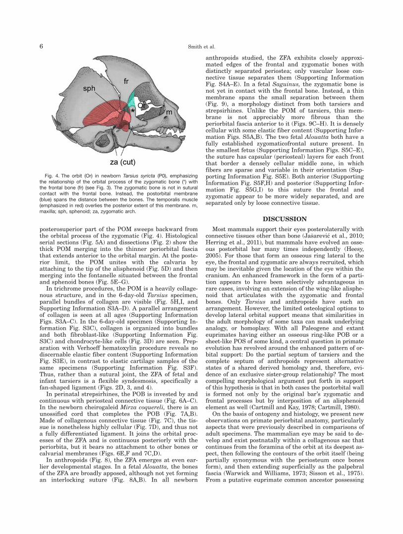
posterosuperior part of the POM sweeps backward fromthe orbital process of the zygomatic (Fig. 4). Histologicalserial sections (Fig. 5A) and dissections (Fig. 2) show thethick POM merging into the thinner periorbital fasciathat extends anterior to the orbital margin. At the poste-rior limit, the POM unites with the calvaria byattaching to the tip of the alisphenoid (Fig. 5D) and thenmerging into the fontanelle situated between the frontaland sphenoid bones (Fig. 5E–G).
In trichrome procedures, the POM is a heavily collage-nous structure, and in the 6-day-old Tarsius specimen,parallel bundles of collagen are visible (Fig. 5H,I, andSupporting Information S3A–D). A parallel arrangementof collagen is seen at all ages (Supporting InformationFigs. S3A–C). In the 6-day-old specimen (Supporting In-formation Fig. S3C), collagen is organized into bundlesand both fibroblast-like (Supporting Information Fig.S3C) and chondrocyte-like cells (Fig. 3D) are seen. Prep-aration with Verhoeff hematoxylin procedure reveals nodiscernable elastic fiber content (Supporting InformationFig. S3E), in contrast to elastic cartilage samples of thesame specimens (Supporting Information Fig. S3F).Thus, rather than a sutural joint, the ZFA of fetal andinfant tarsiers is a flexible syndesmosis, specifically afan-shaped ligament (Figs. 2D, 3, and 4).
In perinatal strepsirhines, the POB is invested by andcontinuous with periosteal connective tissue (Fig. 6A–C).In the newborn cheirogaleid Mirza coquereli, there is anunossified cord that completes the POB (Fig. 7A,B).Made of collagenous connective tissue (Fig. 7C), the tis-sue is nonetheless highly cellular (Fig. 7D), and thus nota fully differentiated ligament. It joins the orbital proc-esses of the ZFA and is continuous posteriorly with theperiorbita, but it bears no attachment to other bones orcalvarial membranes (Figs. 6E,F and 7C,D).
In anthropoids (Fig. 8), the ZFA emerges at even ear-lier developmental stages. In a fetal Alouatta, the bonesof the ZFA are broadly apposed, although not yet formingan interlocking suture (Fig. 8A,B). In all newborn
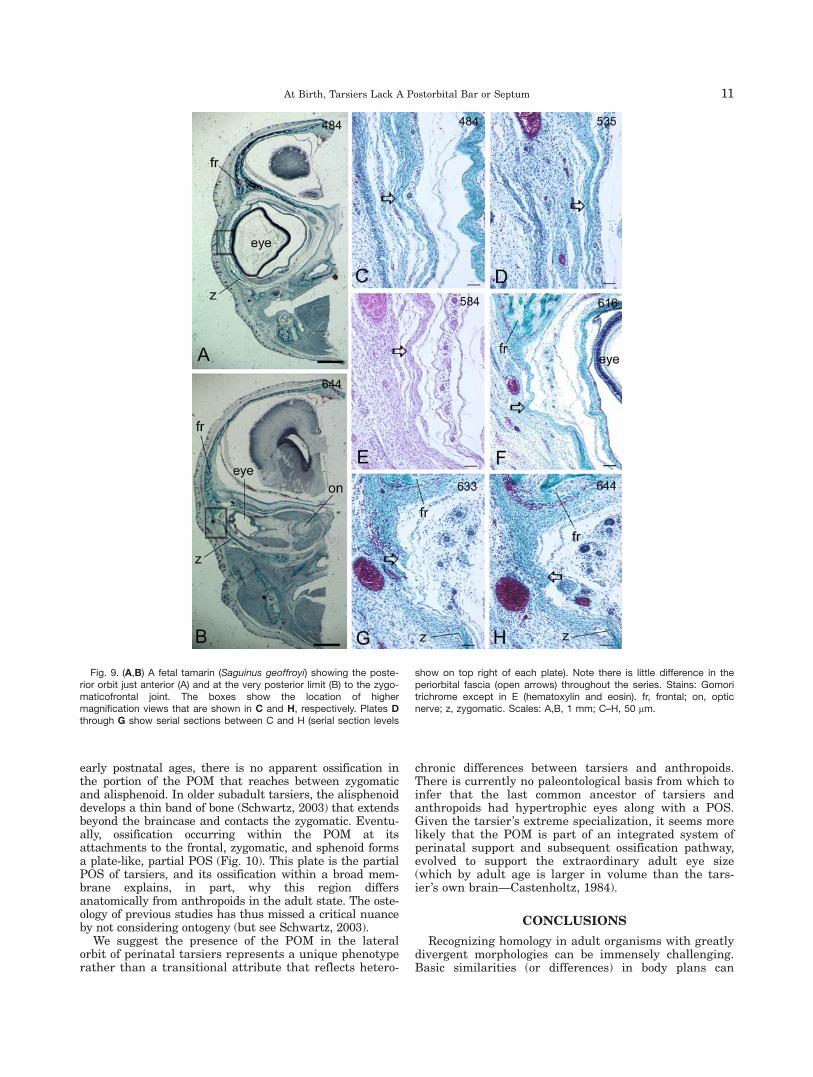
anthropoids studied, the ZFA exhibits closely approxi-mated edges of the frontal and zygomatic bones withdistinctly separated periostea; only vascular loose con-nective tissue separates them (Supporting InformationFig. S4A–E). In a fetal Saguinus, the zygomatic bone isnot yet in contact with the frontal bone. Instead, a thinmembrane spans the small separation between them(Fig. 9), a morphology distinct from both tarsiers andstrepsirhines. Unlike the POM of tarsiers, this mem-brane is not appreciably more fibrous than theperiorbital fascia anterior to it (Figs. 9C–H). It is denselycellular with some elastic fiber content (Supporting Infor-mation Figs. S5A,B). The two fetal Alouatta both have afully established zygomaticofrontal suture present. Inthe smallest fetus (Supporting Information Figs. S5C–E),the suture has capsular (periosteal) layers for each frontthat border a densely cellular middle zone, in whichfibers are sparse and variable in their orientation (Sup-porting Information Fig. S5E). Both anterior (SupportingInformation Fig. S5F,H) and posterior (Supporting Infor-mation Fig. S5G,I) to this suture the frontal andzygomatic appear to be more widely separated, and areseparated only by loose connective tissue.
DISCUSSION
Most mammals support their eyes posterolaterally withconnective tissues other than bone (Ja�sarevic et al., 2010;Herring et al., 2011), but mammals have evolved an osse-ous postorbital bar many times independently (Heesy,2005). For those that form an osseous ring lateral to theeye, the frontal and zygomatic are always recruited, whichmay be inevitable given the location of the eye within thecranium. An enhanced framework in the form of a parti-tion appears to have been selectively advantageous inrare cases, involving an extension of the wing-like alisphe-noid that articulates with the zygomatic and frontalbones. Only Tarsius and anthropoids have such anarrangement. However, the limited osteological options todevelop lateral orbital support means that similarities inthe adult morphology of some taxa can mask underlyinganalogy, or homoplasy. With all Paleogene and extanteuprimates having either an osseous ring-like POB or asheet-like POS of some kind, a central question in primateevolution has revolved around the enhanced pattern of or-bital support: Do the partial septum of tarsiers and thecomplete septum of anthropoids represent alternativestates of a shared derived homology and, therefore, evi-dence of an exclusive sister-group relationship? The mostcompelling morphological argument put forth in supportof this hypothesis is that in both cases the postorbital wallis formed not only by the original bar’s zygomatic andfrontal processes but by interposition of an alisphenoidelement as well (Cartmill and Kay, 1978; Cartmill, 1980).
On the basis of ontogeny and histology, we present newobservations on primate periorbital anatomy, particularlyaspects that were previously described in comparisons ofadult specimens. The mammalian eye may be said to de-velop and exist postnatally within a collagenous sac thatcontinues from the foramina of the orbit at its deepest as-pect, then following the contours of the orbit itself (beingpartially synonymous with the periosteum once bonesform), and then extending superficially as the palpebralfascia (Warwick and Williams, 1973; Sisson et al., 1975).From a putative euprimate common ancestor possessing
Fig. 4. The orbit (Or) in newborn Tarsius syricta (P0), emphasizingthe relationship of the orbital process of the zygomatic bone (*) withthe frontal bone (fr) (see Fig. 3). The zygomatic bone is not in suturalcontact with the frontal bone. Instead, the postorbital membrane(blue) spans the distance between the bones. The temporalis muscle(emphasized in red) overlies the posterior extent of this membrane. m,maxilla; sph, sphenoid; za, zygomatic arch.
6 Smith et al.

a POB, one might envision expansion of the bar’s postero-lateral rim by ossification that follows a fascial plane, theperiorbital membrane. It is precisely this collagenoussheet that is thought to create a “path” for expansion ofthe postorbital septum (Cartmill, 1980); the periorbitaalong that path is thought to become periosteum of theorbit. However, in mammals without a POS, the perior-bita is partially a nonperiosteal membrane that separatessoft tissues of the orbital and temporal fossae (Sissonet al., 1975; Cartmill, 1980).
In some mammals, a cord-shaped ligament exists inplace of an osseous postorbital bar. It may be a fibrocar-tilaginous ligament in at least some cases (Ja�sarevicet al., 2010). The connective tissue cord that unites theincomplete POB of cheirogaleids at the ZFA is a tran-sient postorbital ligament. The cord is narrow, and itsimply ties the frontal and zygomatic processes of thedeveloping POB. One might interpret the POM oftarsiers as another example of a transient postorbital
ligament that is sheet-like in its form, thus taking onthe character of a broader membrane. Postnatal delay ofzygomaticofrontal ossification in tarsiers could then betaken as an archaic euprimate phenomenon shared withsome strepsirhines, either primitively or via parallelism.Indeed, while formation of the POB is generally a pro-gressive prenatal process concurrent with eye growth(Cummings et al., 2012; Ramaswami, 1957), small strep-sirhines like the cheirogaleids, with their allometricallyenlarged eyes (Kay and Kirk, 2000), may also have agap in the POB through late fetal and even early post-natal ontogeny, ostensibly allowing for rapid postnatalgrowth of the eye. However, their resemblance appearsto be homoplasious for reasons that also bear on theadult morphology. The tarsier POM is anteroposteriorlyfar more expansive than the strepsirhine POB and itsconnective tissue precursor. It connects both facial andneurocranial bones, while the strepsirhine POB and itsmembranous ZFA bridge are restricted to the facial
Fig. 5. (A–D): Histological sections of the same tarsier shown in Fig.4 (contralateral side), from anterior to posterior, showing the soft tissuemembranes spanning the distance from the frontal bone (fr) to the zy-gomatic bone (z) and sphenoid bone (sph). At all levels, the orbitalprocess of the zygomatic bone is widely separated from the frontalbone. At its anterior limit, the periorbita (p, B) reflects outward fromthe bones deep to the skin over the eye. The periorbita is continuouswith a thick ligament posteriorly (open arrows, B–D). This flat ligament,the postorbital membrane, connects the tip of the orbital process ofthe zygomatic (*) with the frontal bone (B,C). The posterior extent ofthis membrane merges into the anterolateral fontanelle. The anteriorextent of this fontanelle intervenes between the sphenoid and frontalbones (open arrow, D). At these posterior levels, the temporalis mus-cle (Te) attaches (C,D). Insets: approximate level of each coronal sec-
tion. (E–G) Histology of lateral orbit in late fetal T. syrichta, showingthe gap between the orbital process of the zygomatic bone (*), andthe frontal bone (fr) bridged by a thick postorbital membrane (openarrows). Posterior to the zygomatic, the membrane thins slightly, hasan inferior attachment to the alisphenoid (sph) and converges towardthe periosteal layer of the dura mater (pd). Together, the postorbitalmembrane and periosteal dura merge into the anterolateral fontanelle(alf). (H,I) High magnification view of the postorbital membrane in P0(H: T. syrichta) and P6 (I: T. syrichta) tarsiers, showing thickness withnumerous rows of parallel collagen fibers, organized into bundles inFig. 5I. at, auditory tube lumen; E, eye; np, nasopharynx; npm, naso-pharyngeal meatus; oa, opening of auditory tube; sp, soft palate.Stains: A–D, H,I, Gomori trichrome; E–G, hematoxylin and eosin. Scalebars: A (applies to A–D), 1 mm; E–G, 150 mm.; H, 50 mm; I, 20 mm.
At Birth, Tarsiers Lack A Postorbital Bar or Septum 7

skeleton. In other words, in tarsiers the POM matchesthe spatial arrangement of the lateral orbit of adult tars-iers, including its connection to the calvaria. This lastpoint is critical, because it corresponds with a prominentosseous feature that also argues for nonhomology of thelateral orbit in tarsiers and strepsirhines: the spur-likezygomatic process of the frontal bone typical of postorbi-tal bars, precociously present in strepsirhines(Ramaswami, 1957) and common among other mammalswith a POB (Heesy, 2005), is absent in tarsiers (Fig. 1E).It has either been lost or suppressed and modifieddevelopmentally.
Could the tarsier POM represent an unusually thick-ened portion of the periorbita that presages thehaplorhine POS? Our findings suggest that it does not.The zygomaticofrontal suture is not formed until sometime after the first postnatal week in Tarsius syrichta; itis delayed in T. bancanus as well. Instead, the postero-lateral border is completed by the POM. Smallextensions of the frontal bone also indicate ossificationoccurs within the POM (e.g., Fig. 1E, and see SupportingInformation Fig. S2C). Anthropoids, in contrast, have afully formed osseous postorbital septum well establishedat birth (Schwartz, 2003), even in small species with
Fig. 6. Dissection of the orbital region in Otolemur garnettii. (A) Skinand eye removed, temporalis muscle undisturbed. (B) The thin postor-bital bar (pb) is emphasized after resection of the temporalis m. (C)The periorbita (p) is partially stripped aside. (D) The periorbita is inti-mately connected with sutural margins of the zygomaticofrontalsuture. After the connective tissue is fully stripped from the post or-bital bar, gracile tips of the orbital process of the frontal bone (opF)and orbital process of the zygmomatic bone (*) remain. A small portion
of the zygomatic bone broke away upon removal of the periorbita. Inhistological sections of the same specimen (contralateral size), thefrontal (fr) and zygomatic bones form a suture (small arrow, E). Poste-rior to the suture, the connective tissue between these bones thins(open arrow, Fig. F) and disappears, forming no association with thecalvaria. Or, orbit; Te, temporalis muscle. Stains: E,F, Gomori tri-chrome. Scale bars: E, 500 mm; F, 200 mm.
8 Smith et al.

Fig. 7. In Mirza coquereli (A, CT reconstruction) the postorbital baris incomplete at birth, with a small gap between the orbital process ofthe zygomatic (*) and the orbital process of the frontal bone (opF). Dis-section of the same specimen reveals a cord-like ligament completes
the postorbital bar (open arrow, B). Histological examination revealsthat the postorbital bar of Mirza is preceded by a transient, thick liga-ment (C,D). fr, frontal bone. Stains: C, D, Gomori trichrome. Scalebars: C, 100 mm; D, 50 mm.
At Birth, Tarsiers Lack A Postorbital Bar or Septum 9

relatively large eyes (Fig. 1H). Furthermore, the ZFA ofanthropoids is histologically distinct from that of thetarsier at birth. All anthropoids studied thus far exhibita typical facial suture at the ZFA at the time of birth.This suture develops between two facial bones with dis-tinctly separated periostea (Sperber, 2001). At theperinatal age, only loose connective tissue separatesthem (Supporting Information Fig. S3), as previouslydescribed for immature facial sutures (Pritchard et al.,1956; Sperber, 2001). In contrast, the tarsier POM is aligamentous joint. As a fan-shaped sheet, its role is tosupport the large eye posterolaterally before bony sup-port is fully formed. The possibility that there is somefibrocartilage in the POM would be in keeping with pre-vious descriptions of the postorbital ligament of therabbit (Ja�sarevic et al., 2010).
To propose that the tarsier configuration evolved intoan anthropoid-like facial suture would require a transi-tion between distinct joint types with differentmechanical functions and biological roles in the twogroups. The POM of tarsiers is unique among extant pri-mates, representing a morphology that is distinct fromany stage of development in anthropoids. Its structurerelates to the function of enclosing protruding orbitalcontents, similar to the purpose of the more typical post-orbital ligament of some other mammals that lack anosseous septum (Ja�sarevic et al., 2010). In the case ofinfant tarsiers, it is most likely that the adaptive evolu-tion of this tissue arrangement was driven bymechanical requirements associated with a uniquelylarge pulse of eyeball growth, which occurs soon afterbirth (Cummings et al., 2012). The POM of tarsiers hasmore in common with a neurocranial fontanelle, an ex-pansive collagenous intersection of more than twocalvarial bones (Enlow and Hans, 1996; Opperman,2000), than a facial suture. In this sense, it can bethought of as an “accessory fontanelle,” a spatial separa-tor and structural compromise that provides flexiblemechanical stability while anticipating rapid postnataleye growth.
Thus, Tarsius possesses neither a POB nor a POS atbirth. It is neither a suture as seen in late fetal andnewborn anthropoids; nor is it like the thin membranousprecursor sheet (periorbita) that precedes suture forma-tion in prenatal anthropoids; nor is it the same as theconnective tissue cord that transiently attaches the zygo-matic and frontal orbital processes in perinatalcheirogaleids (e.g., Fig. 7D). By extension, our observa-tions indicate that the entire postnatal trajectory of POSdevelopment differs between anthropoids and tarsiers.Closure of the lateral orbital mosaic in anthropoids iscompleted by inward and posterior growth of a lamina offthe orbital process of the zygomatic, whereas in tarsiersit is primarily an outward extension derived from thefrontal bone. In tarsier specimens spanning fetal and
Fig. .8
Fig. 8. (A) Dissection of lateral orbit in a late fetal howler monkey(Alouatta seniculus). Here, the orbital process of the zygomatic bone(*) is in direct contact with the frontal bone (fr). This bone is alsoexpanded posteriorly with broad proximity to the frontal, as shown af-ter resection of the temporalis muscle (B). Note the broad contactbetween the frontal and zygomatic bones. (C) A fetal Saguinus has azygomatic process that is not fully in contact with the frontal bone,although the postorbital lamina does extend posteriorly. za, zygomaticarch. Rulers 5 mm.
10 Smith et al.
early postnatal ages, there is no apparent ossification inthe portion of the POM that reaches between zygomaticand alisphenoid. In older subadult tarsiers, the alisphenoiddevelops a thin band of bone (Schwartz, 2003) that extendsbeyond the braincase and contacts the zygomatic. Eventu-ally, ossification occurring within the POM at itsattachments to the frontal, zygomatic, and sphenoid formsa plate-like, partial POS (Fig. 10). This plate is the partialPOS of tarsiers, and its ossification within a broad mem-brane explains, in part, why this region differsanatomically from anthropoids in the adult state. The oste-ology of previous studies has thus missed a critical nuanceby not considering ontogeny (but see Schwartz, 2003).
We suggest the presence of the POM in the lateralorbit of perinatal tarsiers represents a unique phenotyperather than a transitional attribute that reflects hetero-
chronic differences between tarsiers and anthropoids.There is currently no paleontological basis from which toinfer that the last common ancestor of tarsiers andanthropoids had hypertrophic eyes along with a POS.Given the tarsier’s extreme specialization, it seems morelikely that the POM is part of an integrated system ofperinatal support and subsequent ossification pathway,evolved to support the extraordinary adult eye size(which by adult age is larger in volume than the tars-ier’s own brain—Castenholtz, 1984).
CONCLUSIONS
Recognizing homology in adult organisms with greatlydivergent morphologies can be immensely challenging.Basic similarities (or differences) in body plans can
Fig. 9. (A,B) A fetal tamarin (Saguinus geoffroyi) showing the poste-rior orbit just anterior (A) and at the very posterior limit (B) to the zygo-maticofrontal joint. The boxes show the location of highermagnification views that are shown in C and H, respectively. Plates Dthrough G show serial sections between C and H (serial section levels
show on top right of each plate). Note there is little difference in theperiorbital fascia (open arrows) throughout the series. Stains: Gomoritrichrome except in E (hematoxylin and eosin). fr, frontal; on, opticnerve; z, zygomatic. Scales: A,B, 1 mm; C–H, 50 mm.
At Birth, Tarsiers Lack A Postorbital Bar or Septum 11

become obscured under a host of adaptations in theadult (Gould, 1977). It is precisely this dilemma that hascreated a challenge when comparing the orbits of tars-iers and anthropoids. In such comparisons, the mostinformative evidence to distinguish between homologyand analogy may be developmental.
In considering developmental evidence, we are mind-ful that ontogeny itself is subject to selection, andphenotypes that are structurally homologous mayemerge by different mechanisms (Hall, 1999). However,the late prenatal/early postnatal morphology of tarsiersappears to be adapted to a unique ontogenetic trajectory.In other words, the structure at birth is functionallywell designed for tarsiers but not for any known anthro-poids (living or fossil). These results, detailing profoundgross and histological differences between tarsiers andanthropoids at birth, are consistent with the notion thattarsiers evolved a novel postorbital morphology that cor-responds with their specialized, enormously enlargedeyes. The evolution of enormously large eyes in tarsierspresents an instance where selection for an intenselygrowth-dependent adult morphology profoundly influen-ces the ontogenetic program and resultant morphologyof functionally related structures; here, the lateral or-bital mosaic. The tarsier postorbital region ismorphologically, histologically and ontogenetically distin-guished from that in tree shrews, strepsirhines andanthropoids as follows: (i) prenatal loss or suppression ofthe orbital process of the frontal bone (which itself couldbe a haplorhine synapomorphy); (ii) its replacement by aunique POM that makes the ZFA functionally analogousto a fontanelle; (iii) postnatal development and ossifica-tion of what may be a secondary orbital process of thefrontal bone within the POM; and, (iv) a different mech-anism and timing for fusion of the zygomatic with thealisphenoid. In sum, this suggests the entirety of the lat-eral orbital mosaic of Tarsius is a neomorphic structure.
ACKNOWLEDGEMENTS
The authors thank CJ Vinyard and JW Young for provid-ing CT scans of the specimens studied. Thanks to JB Ros-sie, one anonymous reviewer, and CF Ross for providing avaluable critique of an earlier version of our manuscript.AE Bruening helped with histological preparation of oneof the specimens, and DM DeNicola generously providedchemicals for one of our histochemical procedures. Wethank AMNH for access to specimens and logistical sup-port. They are grateful to many individuals and institu-tions for preserving invaluable specimens, including CJBonar of Dallas World Aquarium, EH Less, G Fuller andveterinary staff at the Cleveland Metroparks zoo, and SZehr and Duke Lemur Center veterinary staff. Finally, weare immensely grateful to B Demes and the faculty at theDepartment of Anatomical Sciences, SUNY Stony Brook,for making a neonatal Tarsius bancanus for dissectionand CT scanning. This is DLC publication number 1237.
LITERATURE CITED
Cartmill M. 1980. Morphology, function, and evolution of theanthropoid postorbital septum. In: Ciochon RL, Chiarelli AB, edi-tors. Evolutionary biology of the New World monkeys and conti-nental drift. New York: Plenum. p 243–274.
Cartmill M, Kay RF. 1978. Cranio-dental morphology, tarsier affin-ities, and primate suborders. In: Chivers D, Joysey J, editors.Recent advances in primatology. Vol.3. Evolution. London: Aca-demic Press. p 205–214.
Castenholtz A. 1984. The eye of Tarsius. In: Niemitz C, editor. Biol-ogy of tarsiers. Stuttgart: Gustav-Fisher Verlag. p 303–318.
Cummings JR, Muchlinski MN, Kirk EC, Rehorek SJ, DeLeon VB,Smith TD. 2012. Eye size at birth in prosimian primates: life his-tory correlates and growth patterns. PLoS One 7:e36097.
Dennis JC, Smith TD, Bhatnagar KP, Bonar CJ, Burrows AM, Mor-rison EE. 2004. Expression of neuron-specific markers by thevomeronasal neuroepithelium in six species of primates. Anat Rec281:1190–1199.
Enlow D, Hans M. 1996. Essentials of facial growth. Philadelphia:W.B. Saunders.
Fleagle JG. 1999. Primate adaptation and evolution. San Diego:Academic Press.
Gould SJ. 1977. Ontogeny and phylogeny. Cambridge: Belknap Press.Hall BK. 1999. Evolutionary developmental biology. AH Dordrecht:
Kluwer Academic.Heesy CP. 2005. Function of the mammalian postorbital bar. J Mor-
phol 264:363–380.Herring SW, Rafferty KL, Liu J, Lemme ML. 2011. Mastication and
the postorbital ligament: dynamic strain in soft tissues. IntegrComp Biol 51:297–306.
Hershkovitz P. 1977. Living New World monkeys (Platyrrhini) with anintroduction to primates. Vol. 1. Chicago: University of Chicago Press.
Humason G. 1979. Animal tissue techniques. San Francisco: W.H.Freeman and Co.
Ja�sarevic E, Ning J, Daniel AN, Menegaz RA, Johnson JJ, StackMS, Ravosa MJ. 2010. Masticatory loading, function, and plastic-ity: a microanatomical analysis of mammalian circumorbital soft-tissue structures. Anat Rec 293:642–650.
Jeffery N, Davies K, K€ockenberger W, Williams S. 2007. Craniofa-cial growth in fetal Tarsius bancanus: brains, eyes and nasalsepta. J Anat 210:703–722.
Kay RF, Kirk EC. 2000. Osteological evidence for the evolution ofactivity pattern and visual acuity in primates. Am J Phys Anthro-pol 113:235–262.
Opperman LA. 2000. Cranial sutures as intramembranous growthsites. Devel Dynam 219:472–485.
Pocock RI. 1918. On the external characters of the lemurs and Tar-sius. Proc Zoolog Soc Lond 1918:19–53.
Fig. 10. Comparison of a newborn Tarsius syrichta (A) to an adultTarsius bancanus (B, based on AMNH # 106649). Note that the orbitalprocess of the zygomatic (*) remains spatially separated from the cal-varia in the adult and, unlike anthropoids, only meets the braincasethrough a bridge formed by processes that grow outward from theorbit postnatally. The only nonsize related or proportional change inthis specimen involves the development of a small medial flange thatgrows inward to meet the alisphenoid (open arrow, B).
12 Smith et al.

Polyak S. 1957. The vertebrate visual system. Chicago: Universityof Chicago Press.
Pritchard JJ, Scott JH, Girgis FG. 1956. The structure and develop-ment of cranial and facial sutures. J Anat 90:73–86.
Ramaswami LS. 1957. The development of the skull in the slenderloris, Loris tardigradus lydekkerianus. Cabr Acta Zoolog Stock-holm 38:27–68.
Rosenberger AL, Hogg R, Wong SM. 2008. Rooneyia, postorbital clo-sure, and the beginnings of the age of Anthropoidea. In: SargisEJ, Dagosto M, editors. Mammalian evolutionary morphology: atribute to Frederick S. Szalay. Dordecht: Springer. p 325–346.
Ross CF, Kirk EC. 2007. Evolution of eye size and shape in prima-tes. J Hum Evol 52:294–313.
Schultz AH. 1940. The size of the orbit and of the eye in primates.Am J Phys Anthropol 26:398–408.
Schwartz JH. 2003. How close are the similarities between Tarsiusand other primates. In: Wright PC, Simons EL, Gursky S, editors.
Tarsiers: past, present, and future. New Jersey: Rutgers Univer-sity Press. p 50–96.
Simons EL, Rasmussen DT. 1989. Cranial anatomy of Aegyptopithe-cus and Tarsius and the question of the tarsier-anthropoideanclade. Am J Phys Anthropol 79:1–23.
Sisson S, Daniels Grossman D, Getty R. 1975. Sisson and Gross-man’s the anatomy of the domestic animals. New York:Saunders.
Smith TD, Rossie JB, Cooper GM, Carmody KA, Schmieg RM,Bonar CJ, Mooney MP, Siegel MI. 2010. The maxillary sinus inthree genera of New World monkeys: factors that constrain sec-ondary pneumatization. Anat Rec 293:91–107.
Sperber GH. 2001. Craniofacial development. Hamilton: BC Decker.Szalay FS, Delson E. 1979. Evolutionary history of the primates.
New York: Academic.Warwick R, Williams PL. 1973. Grays anatomy. 35th ed. New York:
Saunders.
At Birth, Tarsiers Lack A Postorbital Bar or Septum 13