Embed Size (px)
Citation preview

Asymmetric unwrapping of nucleosomal DNApropagates asymmetric opening anddissociation of the histone coreYujie Chena,1, Joshua M. Tokudaa,1, Traci Toppingb, Steve P. Meisburgera,2, Suzette A. Pabita, Lisa M. Glossb,3,and Lois Pollacka,3
aSchool of Applied and Engineering Physics, Cornell University, Ithaca, NY 14853; and bSchool of Molecular Biosciences, Washington State University,Pullman, WA 99164
Edited by Taekjip Ha, Johns Hopkins University, Baltimore, MD, and approved December 6, 2016 (received for review July 8, 2016)
The nucleosome core particle (NCP) is the basic structural unit forgenome packaging in eukaryotic cells and consists of DNA woundaround a core of eight histone proteins. DNA access is modulatedthrough dynamic processes of NCP disassembly. Partly disassembledstructures, such as the hexasome (containing six histones) and thetetrasome (four histones), are important for transcription regulationin vivo. However, the pathways for their formation have beendifficult to characterize. We combine time-resolved (TR) small-angleX-ray scattering and TR-FRET to correlate changes in the DNAconformations with composition of the histone core during salt-induced disassembly of canonical NCPs. We find that H2A–H2B his-tone dimers are released sequentially, with the first dimer beingreleased after the DNA has formed an asymmetrically unwrapped,teardrop-shape DNA structure. This finding suggests that the octa-some-to-hexasome transition is guided by the asymmetric unwrap-ping of the DNA. The link between DNA structure and histonecomposition suggests a potential mechanism for the action of pro-teins that alter nucleosome configurations such as histone chaper-ones and chromatin remodeling complexes.
contrast variation SAXS | FRET | nucleosomes | hexasome | time resolved
Genome access is highly regulated through the hierarchicalorganization of proteins and nucleic acids within the cell
nucleus. The nucleosome core particle (NCP) is the first level of thishierarchy (1) and contains two dimers of H2A–H2B histones and an(H3–H4)2 tetramer that is assembled as a dimer of dimers. Aroundthis symmetric octamer core, ∼146 base pairs of dsDNA are wrap-ped in ∼1.7 superhelical turns (1, 2). The NCP structure physicallyimpedes access to DNA, but is dynamically modulated by numerousmechanisms: posttranslational modification (PTM) of histones, in-corporation of histone variants, DNA sequence-dependent effects,and the actions of extrinsic protein factors (e.g., histone chaperones,ATP-dependent remodeling complexes, and histone PTM bindingproteins) (3, 4).Studies of the intrinsic properties and dynamics of NCPs are
critical for understanding how nuclear machinery gains DNA ac-cess in vivo (3, 5, 6). Insight into the nature of partially unfoldedNCP structures has been gleaned from in vitro studies of NCPassembly and disassembly. Intermediate species with partiallyunwrapped DNA (5, 7), disrupted histone–histone interfaces (8, 9),and dissociation of one (hexasomes) or two (tetrasomes) H2A–H2B dimers have been reported (10–12). Some of these NCP in-termediates have been directly connected to chromatin functionin vivo. For example, the hexasome is formed by the action ofRNA Pol II (13) and the essential histone chaperone FACT (14).In addition to equilibrium studies, the kinetics of nucleosome
assembly and disassembly have been characterized by bulk andsingle-molecule methods, including Förster resonance energytransfer (FRET) (7, 8, 15–17), atomic force microscopy (AFM)(9, 18), force spectroscopy (19–21), and small-angle X-ray scat-tering (SAXS) (10, 22). Many studies focused primarily on specificDNA–histone contacts and local conformational changes. Few, if
any studies, use complementary methods to directly compare, onsimilar kinetic time scales, the structural changes of the DNA andhistone components of the NCP. A major gap in our understand-ing of NCP disassembly arises from our limited knowledge ofthe coordination between DNA conformation and histone corecomposition.Because the NCP protein–DNA complex is stabilized predomi-
nantly by polycation–polyanion interactions, the in vitro equilibriumand kinetic properties can be manipulated by ionic solvent condi-tions. NaCl has been widely used to study partially assembled, bi-ologically relevant NCP species that are marginally populated underphysiological conditions (5, 16, 23, 24). The use of recombinanthistones and the Widom 601 DNA sequence (selected for its abilityto form stable, well-positioned nucleosomes) (25, 26) allows pro-duction of large amounts of homogeneous NCPs (601-NCP) forbiophysical studies. Fig. 1 shows the NaCl-induced disassemblypathway for 601-NCPs (7, 8, 23, 24). Whereas the various speciesshown in Fig. 1 have been detected at equilibrium, much less isknown about the kinetics of NCP disassembly, including the rele-vant transition times and pathways between states, or the potentialfor coordination between DNA unwrapping and disruption ofhistone–histone interfaces.Our recent time-resolved SAXS (TR-SAXS) study of salt-
induced NCP dissociation revealed asymmetric DNA releasefrom the histone octamer (22). In kinetic jumps from ∼0–1.9 M
Significance
Nucleosomes are fundamental protein–DNA structures throughwhich eukaryotes package and organize DNA inside the nucleus.Nucleosomes are disassembled to gain access to the critical in-formation stored in DNA. Here, we describe a new experimentalapproach that characterizes the kinetics of nucleosome disassem-bly and the synergy between DNA conformation and proteincomponents. Using NaCl to disrupt electrostatic interactions, weidentify kinetic pathways and transient intermediates that revealhow DNA unwrapping and protein dissociation are linked in thismacromolecular complex. These dynamic structures may providenew insight into the regulation of DNA access during transcription,replication, and repair.
Author contributions: Y.C., J.M.T., T.T., L.M.G., and L.P. designed research; Y.C., J.M.T.,T.T., S.P.M., S.A.P., and L.P. performed research; Y.C., J.M.T., T.T., and L.M.G. analyzeddata; and Y.C., J.M.T., L.M.G., and L.P. wrote the paper.
The authors declare no conflict of interest.
This article is a PNAS Direct Submission.
Data deposition: The data reported in this paper have been deposited in the Small AngleScattering Biological Data Bank (SASBDB), https://www.sasbdb.org/ (accession nos.SASDBS7 and SASDBT7).1Y.C. and J.M.T. contributed equally to this work.2Present address: Department of Chemistry, Princeton University, Princeton, NJ 08544.3To whom correspondence may be addressed. Email: [email protected] or [email protected].
This article contains supporting information online at www.pnas.org/lookup/suppl/doi:10.1073/pnas.1611118114/-/DCSupplemental.
334–339 | PNAS | January 10, 2017 | vol. 114 | no. 2 www.pnas.org/cgi/doi/10.1073/pnas.1611118114

NaCl, a transient intermediate was observed with the DNA in a“J”-shaped conformation bound to a disrupted histone core. Weapplied contrast variation TR-SAXS to focus solely on the dy-namic changes in DNA conformation (22). Information aboutthe histone proteins was restricted to the “endpoint states” ofintact or completely dissociated octamer.Here, we report the coupling of TR-SAXS studies of DNA
conformational changes with time-resolved FRET (TR-FRET)studies of H2A–H2B dimer dissociation during salt-induced NCPdisassembly. Two conditions are characterized here: completeNCP disassembly following rapid increase from low salt (∼0) to1.9 M NaCl (as in ref. 22), and partial disassembly upon increaseto 1.2 M NaCl. The latter condition allowed observation of DNAconformations that facilitate release of the H2A–H2B dimers. Thecombination of TR-SAXS and TR-FRET provides insights intothe conformational dynamics of open intermediate and hexasomeformation (Fig. 1), with important implications for the biologicalfunction of the NCP in regulating DNA accessibility.
ResultsDNA Unwrapping at 1.9 M NaCl Visualized by TR-SAXS. TR-SAXS withcontrast variation was used to monitor the DNA conformationsduring complete NCP disassembly upon the rapid shift from ∼0–1.9 M NaCl by stopped-flow mixing (for SAXS profiles, see Fig.S1A). In standard SAXS measurements, both protein and DNAcontribute to the scattering. Interpretation of these SAXS profilesis challenging and requires knowledge of how each component isdistributed. Through contrast variation, scattering from the DNAis isolated by matching the electron density of the solvent to thatof the protein. As illustrated in Fig. 2, this condition is achievedthrough the addition of 50% sucrose to the buffer. This contrast-matched condition allows for unambiguous analysis of the DNAconformation because only the DNA contributes to the SAXSsignal. Sucrose is an effective contrast additive because it negli-gibly affects electrostatics (27) and NCP stability (22).Because previous equilibrium and time-resolved SAXS studies
of NCP disassembly revealed the presence of multiple structures,an ensemble optimization method (EOM) was applied to identifyensembles of DNA conformations that best recapitulate the SAXSprofiles (22, 28, 29). An overview of this experimental strategy issummarized in Fig. 3. A pool of 9,182 nucleosomal DNA structureswas generated with varying degrees of DNA unwrapping to createa large conformational space. For each time point, a genetic al-gorithm selected a subset of structures (or “ensemble”) that yieldsa theoretical SAXS profile that best fits the SAXS data (for details,seeMaterials and Methods; for χ2 values and example fit, see Fig. S2
A and B). The optimized ensemble consists of representativestructures that closely approximate the conformations in solution.To visualize the ensembles, we calculated size distributions (ra-
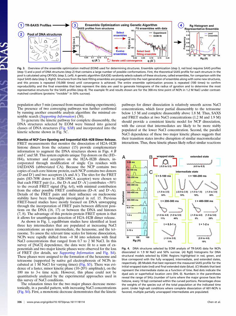
dius of gyration, Rg) of the structures selected by EOM for eachtime point (Fig. 4A; for initial DNA pool, see Fig. S2C). Those withan Rg of ∼50 Å represent mostly wrapped DNA. The diminishingamplitude of this peak with time corresponds to the disruption ofthe canonical NCP structure and the population of other confor-mations. Population of a state with an Rg near 140 Å (the Rg fora fully extended DNA) represents complete disassembly. Struc-tures with intermediate values of Rg (between the peaks at 50 Åand 140 Å) represent intermediate states. The presence of multiplepeaks at each time point suggests heterogeneous populations ofNCP structures. The dominant conformations selected by EOM forthe fully wrapped and extended states are shown in Fig. 4B and themajor conformations selected for the intermediate states are shownin Fig. 4C. Within 100 ms, approximately half of the NCPs containnearly symmetrically unwrapped DNA in a “U” shape, a quarter ofthe DNAs are fully wrapped, and a quarter of the DNAs are fullyunwrapped. Between 100 and 500 ms, the partially wrapped in-termediate states become more asymmetric with most of thepopulation exhibiting J-shaped structures. A small fraction form“teardrop” structures, where one end of the DNA remains wrap-ped around the histone core, whereas the other is extended. After2 s, the nucleosomal DNA is predominantly unwrapped.
NCP Dissociation at 1.2 M NaCl Visualized by TR-SAXS. To better char-acterize transient, asymmetric DNA species, we applied the sameapproach to study NCP disassembly at a final NaCl concentration of1.2 M (for SAXS profiles, see Fig. S1B). This lower salt concen-tration limits disassembly to tetrasome species (tetramer dissociationoccurs above 1.4 M NaCl, see Fig. 1) (15, 24) and captures DNAconformations associated with release of the H2A–H2B dimers.Compared with 1.9 M NaCl, NCP unwrapping at 1.2 M NaCl
was significantly slower and was incomplete within our 10-s mea-surement window. The DNA ensembles selected by EOM (Fig.S3A) are quantified in the Rg histograms shown in Fig. 5A. A fullyunwrapped state (Rg ∼140 Å) appears after 300 ms, but comprisesonly about 8% of the ensemble. In a majority of the NCPs, theDNA remains partially wrapped with Rgs that range from 50 Å to90 Å. For the first 200 ms of the reaction, these partially wrappedspecies vary in size and shape; after 300 ms these structures con-verge to one with an Rg of 76 Å (circled in red). Fig. 5B showsrepresentative structures for the major populations for the first300 ms, together with proposed pathways for the time-dependentevolution of DNA unwrapping. After 300 ms, the asymmetricteardrop-shaped DNA is the predominant species. As highlightedin Fig. 5 A and B, DNA reaches this structure through two majorpathways. In pathway I, the teardrop forms directly as DNAunwraps asymmetrically. In pathway II, DNA initially unwrapssymmetrically, but one end rewraps to form the asymmetric in-termediate. The teardrop is relatively stable at 1.2 M NaCl andrepresents 80% of the population at 300 ms and 36% of the
Fig. 1. A schematic of NaCl-dependent disassembly for NCPs containing the601-DNA (15), based on equilibrium studies ([NCP] ≥ 25 nM). At physiologicalionic strength, NCP configurations reflect local dynamics [i.e., DNA breathing(6), and formation of an open intermediate (8)]. Above 0.5 M NaCl, H2A–H2Bdimers begin to dissociate, allowing the formation of hexasomes and tetra-somes (23). Above 1.4 M NaCl, (H3–H4)2 tetramers begin to dissociate, allowingfor complete disassembly (24). Although dimer dissociation is reversible, tet-rasomes are the minimal configurations required to maintain a wrapped DNAstructure.
Fig. 2. Contrast variation SAXS isolates structural information for the DNAcomponent of NCPs. (A) Color scale bar with typical electron density values forsolvent (water), protein, and DNA. (B and C) NCP structures (PDB 1AOI) shownin buffers with electron densities that vary depending on the presence of 0%(B) or 50% (C) sucrose. We used contrast variation SAXS to monitor DNAconformations during NCP disassembly induced with a salt jump.
Chen et al. PNAS | January 10, 2017 | vol. 114 | no. 2 | 335
BIOPH
YSICSAND
COMPU
TATIONALBIOLO
GY
population after 5 min (assessed from manual mixing experiments).The presence of two converging pathways was further confirmedby running another ensemble analysis algorithm: the minimal en-semble search (Supporting Information) (30).To generate the kinetic pathway for complete disassembly, the
DNA structures selected by EOM were binned into generalclasses of DNA structures (Fig. S3B) and incorporated into thekinetic scheme shown in Fig. 5C.
Kinetics of NCP Core Opening and Sequential H2A–H2B Dimer Release.FRET measurements that monitor the dissociation of H2A–H2Bhistone dimers from the octamer (15) provide complementaryinformation to augment the DNA structures shown in Figs. 4 Band C and 5B. This system exploits unique Trp donors on the (H3–H4)2 tetramer and acceptors on the H2A–H2B dimers, in-corporated through modification of single Cys residues withIAEDANS (abbreviated CA). Because the NCP contains twocopies of each core histone protein, each NCP contains two donors(D and D′) and two acceptors (A and A′). The sites for the FRETpairs (H3-78W donor to H2B-109CA acceptor) were chosen sothat each FRET pair (i.e., the D–A and D′–A′) contributes ∼50%to the overall FRET signal (Fig. 6A), with minimal contributionfrom the other possible FRET combinations (D–A′ and D′–A).Details of the FRET pairs and their influence on nucleosomestability have been thoroughly investigated in ref. 15. PreviousFRET-based studies have mostly focused on DNA unwrappingthrough the incorporation of FRET pairs between different posi-tions on the DNA (16, 17) or between the DNA and histones(7, 8). The advantage of this protein–protein FRET system is thatit allows for unambiguous detection of H2A–H2B dimer release.As shown in Fig. 1, equilibrium studies have identified at least
three key intermediates that are populated at increasing NaClconcentrations: an open intermediate, the hexasome, and the tet-rasome. To assess the relevant time scales for histone dissociation,NCPs were rapidly shifted from ∼0 M into solutions with finalNaCl concentrations that ranged from 0.7 to 2 M NaCl. In thissurvey of [NaCl] dependence, the data were fit to a sum of ex-ponentials and two major kinetic phases were observed for the lossof FRET (for details, see Supporting Information and Fig. S4).These phases were assigned to the formation of the hexasome andtetrasome (supported by native gel electrophoresis of NCPs in-cubated at 1 M NaCl for varying times) (Fig. S5). There was evi-dence of a faster, minor kinetic phase (10–20% amplitude), on the100 ms to 3-s time scale. However, this phase could not bequantitatively analyzed by the experimental approaches used inthis survey of NaCl conditions.The relaxation times for the two major phases decrease mono-
tonically, in a parallel pattern, with increasing NaCl concentrations(Fig. S4). First, a monotonic decrease demonstrates that the kinetic
pathways for dimer dissociation is relatively smooth across NaClconcentrations, which favor partial disassembly to the tetrasomebelow 1.5 M and complete disassembly above 1.8 M. Thus, SAXSand FRET studies at two NaCl concentrations (1.2 M and 1.9 M)should provide a consistent kinetic model for NCP dissociation,with the caveat that intermediates are likely to be more stablypopulated at the lower NaCl concentration. Second, the parallelNaCl dependence of these two major kinetic phases suggests thattheir transition states involve disruption of similar macromolecularinteractions. Thus, these kinetic phases likely reflect similar reactions
Fig. 3. Overview of the ensemble optimization method (EOM) used for determining structures. Ensemble optimization (step 3, red box) requires SAXS profiles(step 1) and a pool of DNA structures (step 2) that contains a large number of possible conformations. First, the theoretical SAXS profile for each structure in thepool is calculated using CRYSOL (step 3, Left). A genetic algorithm (GAJOE) randomly selects subsets of these structures, called ensembles, for comparison with theinput SAXS data (step 3, Right). Structures from the best-fitting ensembles are propagated into the next generation of ensembles along with some new structures,and this process is repeated (10,000 times) until convergence is achieved. The entire ensemble optimization process is repeated (100 times) to confirmreproducibility and the final ensembles that best represent the data are used to generate histograms of the radius of gyration and to determine the mostrepresentative structures for the SAXS profiles (step 4). The example fit and results shown are for the 300-ms time point of NCPs in 1.2 M NaCl under contrast-matched conditions (proteins “invisible” in 50% sucrose).
Fig. 4. DNA structures selected by EOM analysis of TR-SAXS data for NCPsdissociated in 1.9 M NaCl and 50% sucrose. (A) Rg(t) histograms for DNAstructural models selected by EOM. Regions highlighted in red, green, andblue correspond with the fully wrapped, intermediate, and extended states,respectively. (B) Models that best represent the measured SAXS profile for theinitial wrapped state (red) and final extended state (blue). (C) Models that bestrepresent the intermediate states as a function of time. Red dots indicate thedyad axis or superhelical location zero (SHL 0). Numbers in the parenthesesreveal the range of SHLs (number of turns where the major groove faces thehistone, every 10 bp) contained within the curved portions. Percentages showthe weights of the species out of the total population at the indicated timepoint. Under high-salt conditions where complete dissociation of 601-NCPs isfavored, multiple partially unwrapped intermediates are populated.
336 | www.pnas.org/cgi/doi/10.1073/pnas.1611118114 Chen et al.

in a sequential mechanism (e.g., dissociation of the first andthen second H2A–H2B dimer). This conclusion is supported byanalysis of the relative FRET amplitudes from multiple FRETpairs described below.To better characterize the nature of the kinetic phases, espe-
cially the fastest, low-amplitude kinetic phase, larger datasets werecollected as a function of final NCP concentration (25–250 nM), at1.2 M and 1.9 M NaCl, using a combination of manual andstopped-flow mixing to monitor reactions from 10 ms to 1,000 s.Datasets at different NCP concentrations were globally fit to threekinetic phases. These results are shown in Fig. 6B and are sum-marized in Table 1 (for details, see Supporting Information andFig. S6 A and B). The relative amplitudes for the three kineticphases provide insight into the conformational changes associatedwith each phase as shown in Fig. 6C. The relatively small ampli-tude of the fastest phase (loss of ∼20% of the FRET signal at both1.2 M and 1.9 M) is consistent with a conformational change thatopens the dimer–tetramer interfaces, rather than full dissociationof an H2A–H2B dimer. The larger amplitudes for the slowerphases are consistent with dimer dissociation. Surprisingly, therelative amplitudes for the slower, dissociation phases are unequal(∼30% and ∼50%). This amplitude pattern is consistent with asequential formation of an asymmetric open intermediate, inwhich only one dimer–tetramer interface is disrupted, followed bydissociation of this H2A–H2B dimer to form the hexasome andthen dissociation of the second dimer to form the tetrasome(Fig. 6C). A detailed comparison of this model with that expectedfor a symmetric formation of the open intermediate is shown inFig. S7A.To verify the kinetic model presented in Fig. 6C, kinetic param-
eters were measured using a second FRET pair (H4-60W donor toH2A-108CA acceptor, Fig. S7B). Despite measuring different in-teractions, the relaxation times from the second FRET pair (H4–H2A NCP) agree well, and the inequality of the amplitudes for thehexasome and tetrasome phases is consistent with the two dimersbeing released through different pathways (Table 1, Fig. S7, andSupporting Information). Table 1 also provides kinetic parametersdetermined from the SAXS data collected in the absence of su-crose (for details, see Fig. S8). The reasonable agreement of re-laxation times determined by FRET and SAXS is highlighted inthe overlay shown in Fig. S6C.
DiscussionAlthough nucleosome disassembly is crucial for DNA access, thedynamics of this process is largely unexplored. This study com-bines knowledge of the DNA conformations monitored by SAXSwith insight into histone configurations reported by FRET to
Fig. 5. DNA structures selected by EOM for NCPs dissociated in 1.2 M NaCl. (A) Rg histograms from DNA models selected by EOM that best represent the SAXSdata. Red and green arrows highlight two pathways through which DNA structures change before settling into a prominent peak after 300 ms (circled in red).(B) DNA models selected by EOM before (t = 0) and after mixing into 1.2 M NaCl (20 ms, 100 ms, 200 ms, and 300 ms). Green and red arrows highlight two majorpathways through which DNA unwraps to form the teardrop DNA structure. Black arrows showminor pathways. Red dots indicate the dyad axis (SHL 0). Numbersin parentheses reveal the range of SHLs (number of turns) contained within the curved portions. Percentages shown are the weights of the species out of the totalpopulation at the indicated time point. Under moderate salt conditions that favor partial disassembly, the majority of structures unwrap symmetrically andasymmetrically before converging into the teardrop structure. (C) Kinetic scheme for complete disassembly with pathways inferred from prominent DNAstructures selected by EOM (Fig. S3).
Fig. 6. NCP FRET pairs and the histone configurations observed. (A) FRET pairswith H3-78W donor (green) and H2B-109CysAEDANS acceptor (red). For thisconstruct (H3–H2B NCP), the donor and acceptor on the same face of the NCP(D–A) are close to the Förster radius for this FRET pair (∼20 Å), but the distancefrom the donor to the acceptor on the other NCP face (D–A′) is significantlylonger (∼50 Å) and should contribute less than 1% to the observed FRET signal.The Cβ positions in the 1AOI.pdb structure of the NCP were used to estimatedistances between the FRET pairs. (B) Acceptor fluorescence time course mea-sured for 250 nM NCP in 1.2 M NaCl (blue). The solid black line represents a sumof three first-order exponentials used to determine the relative amplitudes andrelaxation times. To obtain robust values, global fits were used on datasetscollected as a function of NCP concentration (10–250 nM NCP). (C) Histoneconfigurations observed with TR-FRET. Relaxation times (τ) and amplitudes(A) of FRET loss measured at 1.2 M NaCl are reported for each transition.
Chen et al. PNAS | January 10, 2017 | vol. 114 | no. 2 | 337
BIOPH
YSICSAND
COMPU
TATIONALBIOLO
GY

provide details on dynamics and coordination between DNA andhistone proteins during NCP disassembly.
Kinetic Models for DNA Unwrapping. Time-resolved SAXS revealedmultiple pathways through which nucleosomal DNA unwrapsduring salt-induced disassembly. Although small populations ofNCPs (<25% at 1.9M and <10% at 1.2 M) dissociate at a rate thatexceeds our limit of detection, in the majority of cases, NCPsprogressively unwrap from the ends with rates that increase as afunction of [NaCl]. Following a jump to high salt (1.9 M NaCl),complete disassembly is achieved within 10 s. The jump to a lowerfinal salt concentration (1.2 M NaCl) reveals significantly slowerkinetics, with a majority of the DNAs remaining partially wrapped.Under both conditions, the DNA unwraps to form asymmetricconformations (J and teardrop shapes). These observations areconsistent with previous studies of nucleosomes containing thenonpalindromic 601-DNA sequence, where asymmetric nucleo-some stability is observed with the 5′ end showing a greater bindingaffinity than the 3′ end (26). Asymmetric unwrapping may be ageneralizable feature of DNA sequences with asymmetric affini-ties, as observed for both the 601- and 5S-DNA sequences (22).The slower kinetic responses at 1.2 M NaCl reveal two pathways
to form the teardrop DNA. In addition to asymmetric unwrapping(pathway I), the detection of symmetric unwrapping and rewrap-ping of one end to form the same conformation (pathway II)highlights the potential for coordination between the DNA ends.These results are consistent with that reported by Ngo et al. usingforce-FRET spectroscopy and Monte Carlo simulations (21).Under low tension (<5 pN), they report that both DNA endsunwrap and rewrap synchronously, but further opening of one endstabilizes the rewrapping of the other end in a manner that isdirected by the local flexibility of the DNA. They suggest that theunwrapping of one end may help stabilize the wrapping of theother through an overall reduction in electrostatic repulsion. In-terestingly, the rewrapping observed in our work is observed underconditions where electrostatic interactions should be effectivelyscreened (1.2 M NaCl). One possible explanation is that therewrapping is facilitated by the histone tails.
For some of the asymmetric models determined in this study,the curved portion of the DNA that remains in contact with thehistone core appears shifted away from the dyad and closer tothe entry/exit sites compared with canonical structures. Thisconformation may depend on a sliding of the histone–DNAcontacts (31). One intriguing possibility is that the partial DNAunwrapping may help facilitate nucleosome sliding.
Structures and Pathways of Hexasome Formation Suggest DNA-Directed NCP Disassembly. The integrated results from SAXSand FRET at 1.2 M NaCl provide insight into the transient speciespopulated by 601-NCPs. The time-resolved DNA populations(classified in Fig. S3) were globally fitted to the kinetic schemeshown in Fig. 5C to obtain relaxation times (Fig. S9). Theseglobally fitted populations are shown as a function of time in Fig.7A, along with the expected populations of the histone configu-rations based on the H3–H2B NCP FRET data in Table 1. Thecomplete kinetic scheme of NCP disassembly at 1.2 M is presentedin Fig. 7B (for details, see Supporting Information).The DNA rapidly unwraps from the histone octamer to form the
teardrop DNA, which is the dominant conformation on the 0.2- to1-s timescale. This teardrop conformation forms appreciably fasterthan the 2–30 s required for the asymmetric opening that disruptsan interface between the (H3–H4)2 tetramer and one of the H2A–H2B dimers and subsequent dissociation of the first dimer to formthe hexasome. Such a state, containing partially unwrapped DNA,but a full complement of histone proteins, is completely consistentwith results of Li and Widom (6), suggesting that contacts betweenthe DNA and H2A–H2B dimers are disrupted by conformationaldynamics observed under physiological conditions, yet other con-tacts prevent immediate release of one of the heterodimers. Thus,the teardrop DNA precedes changes in the histone octamer con-formation, suggesting that the unwrapped DNA end acts like agate to expose the proximal H2A–H2B dimer for release. TheDNA further unwraps and releases the remaining dimer to formthe tetrasome. This picture is in full agreement with equilibriumstudies reported by Böhm et al. (8).
Table 1. Comparison of relaxation times and relative amplitudes from two FRET pairs and singular value decomposition analysis of TR-SAXS data in the absence of sucrose
Relaxation times (s)/relative amplitude (%)
Data
1.2 M NaCl 1.9 M NaCl
τopen/Aopen τhexasome/Ahexasome τtetrasome/Atetrasome τopen/Aopen τhexasome/Ahexasome τtetrasome/Atetrasome
H3-78W to H2B-109CA 2.3 (0.5)/20 (4) 27 (3)/33 (4) 288 (30)/47 (6) 0.22 (0.07)/23 (7) 1.6 (0.3)/26 (5) 8.1 (0.8)/50 (7)H4-60W to H2A-108CA 2.6 (0.6)/23 (3) 29 (3)/47 (3) 188 (38)/30 (5) 0.06 (0.03)/10 (8) 1.5 (0.2)/58 (14) 3.7 (0.7)/32 (16)SAXS 2.7 14 — 0.21 1.2 —
The errors associated with the kinetic parameters are indicated in parentheses.
Fig. 7. TR-FRET and TR-SAXS analyses reveal hexasome formation at 1.2 M NaCl. (A) Predicted populations of DNA conformational states (black lines) and histoneconfiguration states (blue lines) based on the kinetic rates determined for NCPs at 1.2 M NaCl from the kinetic analysis of EOM models (Figure 6A, see details inFig. S9) and TR-FRET measurements (Table 1), respectively. (B) NCP disassembly pathway determined from TR-SAXS with histone configurations informed by TR-FRET. Black numbers reflect the SAXS relaxation times (inverse of rates in Fig. S9C). Blue numbers reflect the FRET relaxation times (Table 1). The curved blackarrow represents a minor pathway. For simplicity, histone orientations were centered on the dyad when possible.
338 | www.pnas.org/cgi/doi/10.1073/pnas.1611118114 Chen et al.

This work suggests an intriguing mechanism for NCP remod-eling in which DNA conformation facilitates the reconfiguration ofthe histone core. Although the asymmetric nature of the DNAunwrapping and subsequent dimer dissociation observed here wasdirected by the asymmetry of the tightly positioning 601 sequence,this mechanism may be exploited by gene regulatory proteins as ageneral strategy to exchange (32) or modify (33) one H2A–H2Bdimer while simultaneously protecting the other. The combinedSAXS and FRET approach used in this work is readily adaptableto test the hypothesis that, in addition to direct interaction with thehistone core, key partner proteins, such as chromatin remodelersor histone chaperones, affect the composition of the histone coreby interacting with and altering nucleosomal DNA conformation.
Materials and MethodsNCP Production and Reconstitution. Previously described procedures were usedfor the expression and purification of recombinant Xenopus laevis histones(15, 34, 35), production of the 149-bp DNA derived from the Widom 601 se-quence (15, 25) and their reconstitution into NCPs (22). Unless noted other-wise, experiments were conducted with the following buffer: 20 mM Tris-Cl(pH 7.5), 0.1 mM EDTA, and 0.1 mM DTT.
TR-SAXS Experiments.All TR-SAXS experiments were conducted using a BiologicSFM-400 stopped flow mixer at BioCAT Sector 18 at Advanced Photon Source(APS). The experimental procedures and SAXS image analysis are described indetail in Supporting Information.
Ensemble Optimization Method (EOM). Ensembles of DNA structures that bestrecapitulate the measured TR-SAXS profiles were selected using the programGAJOE v2.0 (28, 29). The DNA pool included 9182 structural models generatedusing PyMol (expanded from 32 models in ref. 22). Details on model
generation are described in Supporting Information. The q-range used forGAJOE fitting was 0.006–0.2 Å−1. Rg histograms of selected DNA models wereaveraged from 100 independent repeats of the genetic algorithm. Parametersettings: number of harmonics = 50, maximum s-value = 0.25, number ofpoints = 101, number of generations = 10000, number of ensembles = 200,ensemble size fixed = no, maximum/minimum number of curves per en-semble = 1, constant subtraction = no, number of times genetic algorithmrepeated = 100.
The reliability and uniqueness of the solutions achieved by the genetic al-gorithm depend on two interdependent factors (1): the size and diversity ofthe pool (which needs to contain an ensemble that fits the data well, e.g., χ2 ≤2) and (2) the number of generations and iterations of the algorithm (toprovide sufficient sampling and evolution to find the best fitting ensemble).The solutions obtained for a given SAXS profile using the DNA pool and pa-rameter settings described above consistently converged to give nearly iden-tical ensembles (with 0- to 2-bp variations).
TR-FRET Experiments. A previous paper described the design of a FRET systemto monitor interactions in the NCP specifically between the H2A–H2B dimers,with Cys-AEDANS acceptors, and the (H3–H4)2 tetramer, with Trp donors(15). See details in Supporting Information.
ACKNOWLEDGMENTS. We thank Srinivas Chakravarthy, Weifeng Shang,and Richard Heurich for experimental and technical assistance at BioCAT(Sector 18ID) Advanced Photon Source. SAXS research was supported byNational Institutes of Health (NIH) Grants EUREKA R01-GM088645 andR01-GM085062 (to L.P.). FRET research was supported by NIH GrantGM073787 (to L.M.G.). SAXS data were acquired at the BioCAT beamlineof the Advanced Photon Source, a US Department of Energy (DOE) Officeof Science User Facility operated for the DOE Office of Science by ArgonneNational Laboratory under Contract No. DE-AC02-06CH11357. BioCAT is sup-ported by Grant 9 P41 GM103622 from the National Institute of GeneralMedical Sciences (NIGMS) of the NIH.
1. Andrews AJ, Luger K (2011) Nucleosome structure(s) and stability: Variations on atheme. Annu Rev Biophys 40:99–117.
2. Luger K, Mäder AW, Richmond RK, Sargent DF, Richmond TJ (1997) Crystal structureof the nucleosome core particle at 2.8 A resolution. Nature 389(6648):251–260.
3. Bell O, Tiwari VK, Thomä NH, Schübeler D (2011) Determinants and dynamics of ge-nome accessibility. Nat Rev Genet 12(8):554–564.
4. Luger K (2006) Dynamic nucleosomes. Chromosome Res 14(1):5–16.5. Tims HS, Gurunathan K, Levitus M, Widom J (2011) Dynamics of nucleosome invasion
by DNA binding proteins. J Mol Biol 411(2):430–448.6. Li G, Widom J (2004) Nucleosomes facilitate their own invasion. Nat Struct Mol Biol
11(8):763–769.7. Li G, Levitus M, Bustamante C, Widom J (2005) Rapid spontaneous accessibility of
nucleosomal DNA. Nat Struct Mol Biol 12(1):46–53.8. Böhm V, et al. (2011) Nucleosome accessibility governed by the dimer/tetramer in-
terface. Nucleic Acids Res 39(8):3093–3102.9. Miyagi A, Ando T, Lyubchenko YL (2011) Dynamics of nucleosomes assessed with
time-lapse high-speed atomic force microscopy. Biochemistry 50(37):7901–7908.10. Arimura Y, Tachiwana H, Oda T, Sato M, Kurumizaka H (2012) Structural analysis of
the hexasome, lacking one histone H2A/H2B dimer from the conventional nucleo-some. Biochemistry 51(15):3302–3309.
11. Mazurkiewicz J, Kepert JF, Rippe K (2006) On the mechanism of nucleosome assemblyby histone chaperone NAP1. J Biol Chem 281(24):16462–16472.
12. Zlatanova J, Bishop TC, Victor JM, Jackson V, van Holde K (2009) The nucleosomefamily: Dynamic and growing. Structure 17(2):160–171.
13. Kireeva ML, et al. (2002) Nucleosome remodeling induced by RNA polymerase II: Lossof the H2A/H2B dimer during transcription. Mol Cell 9(3):541–552.
14. Belotserkovskaya R, et al. (2003) FACT facilitates transcription-dependent nucleosomealteration. Science 301(5636):1090–1093.
15. Hoch DA, Stratton JJ, Gloss LM (2007) Protein-protein Förster resonance energytransfer analysis of nucleosome core particles containing H2A and H2A.Z. J Mol Biol371(4):971–988.
16. Hazan NP, et al. (2015) Nucleosome core particle disassembly and assembly kineticsstudied using single-molecule fluorescence. Biophys J 109(8):1676–1685.
17. Park YJ, Dyer PN, Tremethick DJ, Luger K (2004) A new fluorescence resonance energytransfer approach demonstrates that the histone variant H2AZ stabilizes the histoneoctamer within the nucleosome. J Biol Chem 279(23):24274–24282.
18. Katan AJ, Vlijm R, Lusser A, Dekker C (2015) Dynamics of nucleosomal structuresmeasured by high-speed atomic force microscopy. Small 11(8):976–984.
19. Brower-Toland BD, et al. (2002) Mechanical disruption of individual nucleosomesreveals a reversible multistage release of DNA. Proc Natl Acad Sci USA 99(4):1960–1965.
20. Hall MA, et al. (2009) High-resolution dynamic mapping of histone-DNA interactionsin a nucleosome. Nat Struct Mol Biol 16(2):124–129.
21. Ngo TTM, Zhang Q, Zhou R, Yodh JG, Ha T (2015) Asymmetric unwrapping of nu-cleosomes under tension directed by DNA local flexibility. Cell 160(6):1135–1144.
22. Chen Y, et al. (2014) Revealing transient structures of nucleosomes as DNA unwinds.Nucleic Acids Res 42(13):8767–8776.
23. Oohara I, Wada A (1987) Spectroscopic studies on histone-DNA interactions. I. Theinteraction of histone (H2A, H2B) dimer with DNA: DNA sequence dependence. J MolBiol 196(2):389–397.
24. Oohara I, Wada A (1987) Spectroscopic studies on histone-DNA interactions. II. Threetransitions in nucleosomes resolved by salt-titration. J Mol Biol 196(2):399–411.
25. Lowary PT, Widom J (1998) New DNA sequence rules for high affinity binding tohistone octamer and sequence-directed nucleosome positioning. J Mol Biol 276(1):19–42.
26. Chua EYD, Vasudevan D, Davey GE, Wu B, Davey CA (2012) The mechanics behindDNA sequence-dependent properties of the nucleosome. Nucleic Acids Res 40(13):6338–6352.
27. Blose JM, et al. (2011) Effects of a protecting osmolyte on the ion atmosphere sur-rounding DNA duplexes. Biochemistry 50(40):8540–8547.
28. Bernadó P, Mylonas E, Petoukhov MV, Blackledge M, Svergun DI (2007) Structuralcharacterization of flexible proteins using small-angle X-ray scattering. J Am ChemSoc 129(17):5656–5664.
29. Tria G, Mertens HDT, Kachala M, Svergun DI (2015) Advanced ensemble modelling offlexible macromolecules using X-ray solution scattering. IUCrJ 2(Pt 2):207–217.
30. Pelikan M, Hura GL, Hammel M (2009) Structure and flexibility within proteins asidentified through small angle X-ray scattering. Gen Physiol Biophys 28(2):174–189.
31. Bowman GD (2010) Mechanisms of ATP-dependent nucleosome sliding. Curr OpinStruct Biol 20(1):73–81.
32. González PJ, Palacián E (1989) Interaction of RNA polymerase II with structurally al-tered nucleosomal particles. Transcription is facilitated by loss of one H2A.H2B dimer.J Biol Chem 264(31):18457–18462.
33. Yen K, Vinayachandran V, Pugh BF (2013) SWR-C and INO80 chromatin remodelersrecognize nucleosome-free regions near +1 nucleosomes. Cell 154(6):1246–1256.
34. Banks DD, Gloss LM (2003) Equilibrium folding of the core histones: The H3-H4 tet-ramer is less stable than the H2A-H2B dimer. Biochemistry 42(22):6827–6839.
35. Gloss LM, Placek BJ (2002) The effect of salts on the stability of the H2A-H2B histonedimer. Biochemistry 41(50):14951–14959.
36. Svergun DI (1992) Determination of the regularization parameter in indirect-transform methods using perceptual criteria. J Appl Cryst 25(4):495–503.
37. Macke TJ, Case DA (1998) Modeling unusual nucleic acid structures. MolecularModeling of Nucleic Acids, eds Leontis NB, Santalucia J (American Chemical Society,Washington, DC), pp 379–393.
38. Beechem JM (1992) Global analysis of biochemical and biophysical data.Methods Enzymol210(2):37–54.
Chen et al. PNAS | January 10, 2017 | vol. 114 | no. 2 | 339
BIOPH
YSICSAND
COMPU
TATIONALBIOLO
GY