Embed Size (px)
Citation preview

The Journal of Neuroscience, March 1994, 14(3): 1677-l 667
Astroglial Modulation of Transient Potassium Current Development in Cultured Mouse Hippocampal Neurons
Rui-Lin Wu and Michael E. Barish
Division of Neurosciences, Beckman Research institute of the City of Hope, Duarte, California 91010
Hippocampal neurons exhibit three voltage-gated potassium currents, two transient currents and a delayed rectifier, that influence numerous aspects of electrogenesis including ac- tion potential duration and accommodation to sustained de- polarization. These currents, termed A-, D-, and K-currents, respectively, can be distinguished based on kinetics, steady state inactivation characteristics, and sensitivity to 4-aminopyridine (see Wu and Barish, 1992b). We have com- pared the voltage-gated potassium currents in voltage- clamped pyramidally shaped cultured hippocampal neurons growing on or touching glial fibrillary acidic protein-express- ing astroglia (termed on-glia or touching-glia neurons, re- spectively) with those in similar neurons growing directly on a coated glass substrate (termed off-glia neurons). We ob- served differences in the wave forms of total potassium cur- rent that correlated with the extent of astroglial contact. After 5-7 d in culture, A-current amplitude in off-glia neurons was approximately 19% of that of neurons growing in the normal (for culture) on-glia configuration. D-current amplitude tend- ed to be larger in these off-glia neurons. Neurons in contact with astroglia had greater membrane area than off-glia neu- rons. Comparison of current densities (current at a fixed voltage normalized to capacitance and expressed in units of pA/pF) indicated that A-currents were the major com- ponent of transient potassium current in on- and touching- glia neurons, while D-currents were more dominant in off- glia neurons.
Astroglia influenced membrane currents by a surface- or extracellular matrix-associated mechanism, rather than by free diffusion of a soluble factor, as differences were ob- served between closely adjacent neurons on the same cov- erslip. Living glia were required, as potassium currents in neurons grown on dried or methanol-fixed glia resembled those of off-glia neurons. On-glia neurons in cultures treated with an RNA synthesis inhibitor [DRB (5,6-dichloro-l-&o- ribofuranosylbenzimidazole)] for 5-7 d had reduced whole- cell capacitance and A-current amplitude. This effect was localized to DRB actions on underlying astroglia, not on the neurons.
Action potentials elicited by current injection varied with astroglial contact. In on-glia neurons with relatively larger
Received Feb. 26, 1993; revised Aug. 24, 1993; accepted Aug. 31, 1993. We thank Dr. R. Hammerschlag for assistance with measurement of RNA
synthesis, Drs. G. Laurent and K. J. Seymour-Laurent for comments on the manuscript, and Ms. J. Flanagan for assistance in its preparation. This work was supported by grants from the NSF (IBN9121751) and NIH (NS23857). M.E.B. is an Established Investigator of the American Heart Association.
Correspondence should be addressed to Michael E. Barish at the above address.
Copyright 0 1994 Society for Neuroscience 0270-6474/94/ 14 1677- 11$05.00/O
A-currents a delay was seen in the onset of firing after de- polarization. In contrast, action potentials in off-glia neurons rose smoothly after initiation of depolarization.
We conclude that astrocytes modulate the appearance of transient potassium currents in hippocampal pyramidal neu- rons by inducing development of A-current. Increased A-cur- rent amplitude was associated with an increase in membrane area; this observation suggests the possibility that glia pro- mote insertion of A-current-rich membrane. Signals appear to pass from astroglia to neurons by contact or short-range diffusion. We propose astroglial-induced plasticity of A-cur- rent as a mechanism for long-term modulation of hippocam- pal neuron excitability.
[Key words: A-current, D-current, K-current, potassium currents, neuronal development, ion channels, astroglia, neuron-glia interactions]
The activation of transient potassium currents (Hagiwara et al., 1961; Connor and Stevens, 1971a; Neher, 1971; for reviews, see Rogawski, 1985; Rudy, 1988) in hippocampal neurons (Gus- tafsson et al., 1982; Segal and Barker, 1984; Segal et al., 1984; Zbicz and Weight, 1985; Halliwell et al., 1986; Nakajima et al., 1986; Neumann et al., 1987; Storm, 1988; Ficker and Heine- mann, 1992; Wu and Barish, 1992b) influences both action potential wave forms and patterns of repetitive firing (Segal et al., 1984; Halliwell et al., 1986; Nakajima et al., 1986; Storm, 1987, 1988). Rodent hippocampal neurons express two tran- sient potassium currents, termed A- and D-currents, that can be separated based on kinetics, voltage dependence, and phar- macological sensitivities (Storm, 1988; Ficker and Heinemann, 1992; Wu and Barish, 1992b). Despite their importance as reg- ulators of excitability, little is known about the control of the appearance of these transient potassium currents during devel- opment.
The appearance of A-currents in hippocampal neurons may be regulated by influences that vary during the initiation and growth of neurons in dissociated cell culture. This period often consists of plating onto an inert substrate followed by astroglial proliferation, neurite growth, and neuronal maturation. In hip- pocampal CA 1 pyramidal neurons of acute slices from mature rat (Storm, 1988), or acutely dissociated neurons from mouse embryos (15-l 6 d gestation; Wu and Barish, 1992b), or neurons isolated from embryonic rat hippocampus and grown for more than 1 week in culture (Ficker and Heinemann, 1992), A-cur- rents comprise greater than 70% of total peak transient potas- sium current. In contrast, in embryonic mouse hippocampal neurons cultured on polylysine-coated glass for 2-3 d, or in cultured embryonic rat hippocampal neurons examined within 4 d of dissociation, A-currents comprise less than half of total

1678 Wu and Barish * Glial Modulation of Potassium Current Development
transient potassium current (Ficker and Heinemann, 1992; Wu and Barish, 1992b). This pattern is seen in measures of absolute current amplitudes, and in current densities (current amplitudes normalized to membrane area measured as whole-cell capaci- tance). The relative deficit of A-current in neurons after short periods in culture suggests regulation associated with loss of the in vivo environment.
This report is part of an effort to identify potential regulatory influences on potassium currents. We here present evidence indicating that, in culture, contact with astroglia supports ap- pearance of A-type transient potassium currents in developing hippocampal neurons. Specifically, we show that the presence ofA-current during neuronal growth in culture is correlated with the extent of contact with astroglia, that only living astroglia are competent to support A-currents, and that this ability is blocked by inhibition of astroglial RNA synthesis during the first 24 hr ofculture. These experiments thus suggest that astroglia actively participate in developmental regulation of transient potassium current during neuronal differentiation.
Some of these results have recently been presented in abstract form (Wu and Barish, 1992a).
Materials and Methods Preparation of cultures. Procedures for preparation of cultured hippo- campal neurons from mouse embryos at 15-l 6 d gestation were based on classical descriptions (Banker and Cowan, 1977) and are described in detail in a previous publication (Wu and Barish, 1992b). Briefly, dissociated hippocampal cells were grown in serum-containing medium on 12-mm-diameter glass coverslips coated with poly-o-lysine and in some cases laminin. They were treated with antimitotic (cytosine ara- binoside or fluorodeoxyuridine) at 24 hr, which resulted in appearance of an incomplete astroglial monolayer after 5-7 d. Cell density was greatest in central regions of coverslips because cells were applied to the coverslips as a bubble, and medium was added after the cells had settled onto the substrate. Thus, on-glia neurons tended to be found toward the centers of the cover slips, while off-glia neurons were found closer to the edges.
Electrophysiological measurements. Whole-cell patch gigaohm seal techniques were standard (Hamill et al., 1981). Recordings were made using an Axopatch 1B amplifier and ~CLAMP acquisition and analysis software (both from Axon Instruments). Leak and capacitative currents were subtracted on line by a combination of analog compensation in the amplifier and a P/-8 digital subtraction scheme in the acquisition software. In the latter, currents recorded during eight voltage steps of one-eighth amplitude and opposite sign as the test pulse were summed and added to the experimental trace. Total membrane capacitance was determined by integrating capacity transients recorded immediately af- ter entering the whole-cell configuration during 10 mV depolarizations from -80 mV. Input resistance was determined from the steady state current recorded during these same depolarizations after capacity tran- sients had settled.
For recording voltage-gated potassium currents under voltage clamp, the internal solution contained (in mM) 65 KCI, 65 KF, 1 CaCI,, 2 MgCI,, 11 EGTA, 10 HEPES at pH 7.3, and the external solution [based on-Hanks’ balanced salt solution (BSS)] contained (in mM) 140NaC1, 5.8 KCl. 1.8 CaCl,. 1 MaCI,. 4.2 NaHCO,. 5.5 glucose. 15 HEPES at pH 7.3.‘This internal soh&m blocked a&at&r of Ca currents and Ca-dependent currents (R.-L. Wu and M. E. Barish, unpublished ob- servations). External solutions contained tetrodotoxin (TTX; 0.5-l PM)
to block transient and sustained sodium currents (for review, see Brown et al., 1990). Under our recording conditions all voltage-gated potassium currents could be accounted for by the combination of the two transient currents (A- and D-currents), and a delayed rectifier-like current (K- current) showing minimal inactivation (Wu and Barish, 1992b). These voltage-gated potassium currents have properties similar to those re- cently reviewed by Storm (1990). For recording action potentials under current clamp, the internal solution contained 124 mM KC1 and 6 mM KF with other components unchanged, and the external solution was identical except that TTX was eliminated. Voltages were corrected for liquid junction potentials between internal and external solutions.
As in our previous investigation (Wu and Barish, 1992b), recordings were made from neurons identified as pyramidal based on the presence of a triangular perikaryon with one prominent apical dendrite and sev- eral shorter basilar dendrites (see also Banker and Cowan, 1979; Krieg- stein and Dichter, 1983). Examples of the neurons studied are shown in Figure 1. Astroglia were identified during experiments by their ap- pearance, under these serum-containing growth conditions, as thin flat cells underlying neurons. These cells were selectively stained by antibody against glial fibrillary acidic protein (GFAP), a marker of astroglia (Big- nami et al., 1972). Recordings were made at room temperature.
The analysis of transient potassium currents would be complicated by inadequate control of membrane voltage. However, two character- istics of the A-currents recorded from these cells indicated that space clamp was sufficient for this investigation. First, the forms of A-current Z-V relations (see Fig. 4) and steady state inactivation curves (see Fig. 6) were similar for on- and off-glia neurons; the differences observed were quantitative rather than qualitative. Second, in our previous in- vestigation of transient potassium currents during first 3 d of culture (Wu and Barish, 1992b), we did not observe changes in the voltage dependencies oftransient potassium current activation and inactivation during an early period of neurite outgrowth. As one might expect that inadequate space clamp would manifest itself as shifts in voltage de- pendence correlated with cell age and membrane capacitance, we con- cluded that an adequate space clamp was attained.
Measurement of RNA synthesis. Mixed neuron-glia cultures (24 hr in vitro) were employed. Experimental cultures were pretreated with DRB (5,6-dichloro-1-P-o-ribofuranosylbenzimidazole; 32 FM) for 2.5 hr be- fore ‘H-uridine (100 pCi/ml) was added to both control and experi- mental cultures. After 12 hr under normal growth conditions, cultures were rinsed in Hanks’ BSS (5 min), treated with trichloroacetic acid (5%) and dissolved in TS-1 tissue solvent before measurement of in- corporated radioactivity by scintillation.
DRB was obtained from Sigma and diluted in ethanol to make a 32 mM stock, which was stored at -20°C. This stock was diluted 1: 1000 into experimental cultures.
Statistical tests. Significance was assessed using two-tailed t test for unpaired data. Results were considered significant at p < 0.05.
Results
We have previously reported that the macroscopic transient potassium currents (A-current and D-current) in pyramidally shaped hippocampal neurons dissociated at embryonic day 15- 16 and placed into culture changed markedly during the first 3 d in vitro (Wu and Barish, 1992b). During this initial period in cell culture, when neurons have relatively little contact with glial cells, the proportion of total transient potassium current identified as A-current declined from greater than 80% imme- diately after dissociation to less than 40%. In the present study we have examined neurons in culture for 5-7 d, a period during which neurons come into contact with, and start to grow on, an expanding astroglial monolayer covering 30-60% of the sub- strate. We observed that during this period the presence of A- and D-currents in neurons depended on the extent of contact with underlying or nearby astroglial cells. In the sections that follow (1) the voltage-gated potassium currents in neurons grow- ing on or touching astroglia are compared to those growing on the coated glass substrate, (2) living versus fixed or dried glia are evaluated as substrates for appearance of neuronal potas- sium currents, and (3) the consequences of inhibition of RNA synthesis in neurons and glia on the development of potassium currents are described.
Potassium currents in on-, touching-, and off-glia neurons The micrographs in Figure 1 illustrate pyramidally shaped neu- rons from the same coverslip considered to be on-glia, touching- glia, or off-glia neurons for the purposes of this investigation. Two examples are given for each case. Off-glia neurons had no

The Journal of Neuroscience, March 1994, 14(3) 1679
Off-91 ia Touching-glia On-g1 ia
Figure 1. Phase-contrast micrographs illustrating the appearances of neurons growing on the polylysine/laminin-coated glass substrate (Oflgliu), neurons with somata on the substrate but with neurites growing across glia (Touching-gliu), and neurons growing with somata on top of glia (On- g/b). The extent of glial contact varied considerably for the touching-glia class of neurons. In the upper touching-glia panel, the contact of the small process with the adjacent glial cell was sufficient to alter expression of transient potassium currents. Neurons were photographed from the same coverslip, and had been in culture for 72 hr. They were treated with an antimitotic at 24 hr. Scale bar, 50 pm.
detectable contact with any non-neuronal cells, while on-glia neurons had their cell bodies and virtually all of their neuritic field on glial cells. Touching-glia neurons had their cell bodies on the noncellular substrate, and some contact between neurites and glia. As illustrated by the two examples of touching-glia neurons, the extent of glial contact for neurons in this category could vary considerably.
The flat non-neuronal cells in these cultures were predomi- nately astroglia, as cultures were grown in serum-containing medium that promotes proliferation of immature astroglia, and these non-neuronal cells displayed immunoreactivity against GFAP (Bignami et al., 1972).
To provide an indication of the relative health of on-, touch- ing-, and off-glia neurons, we compared their resting electrical properties (Fig. 2). While whole-cell capacitance (used as an index of membrane area) increased with degree of neuron-as- troglia contact, and input resistance was smaller in the larger on-glia neurons, there was no significant difference in input conductance density at - 80 mV (conductance was normalized to capacitance, noted as specific conductance and expressed as pS/pF) between these classes of neurons. Thus, while on-glia neurons were larger than touching- or off-glia neurons, as ex- pected given the known growth-promoting activities of glial monolayers (Noble et al., 1984; Fallon, 1985; Pixley et al., 1987) to the extent that resting conductance is an indicator of cell health the three classes of neurons were equivalent.
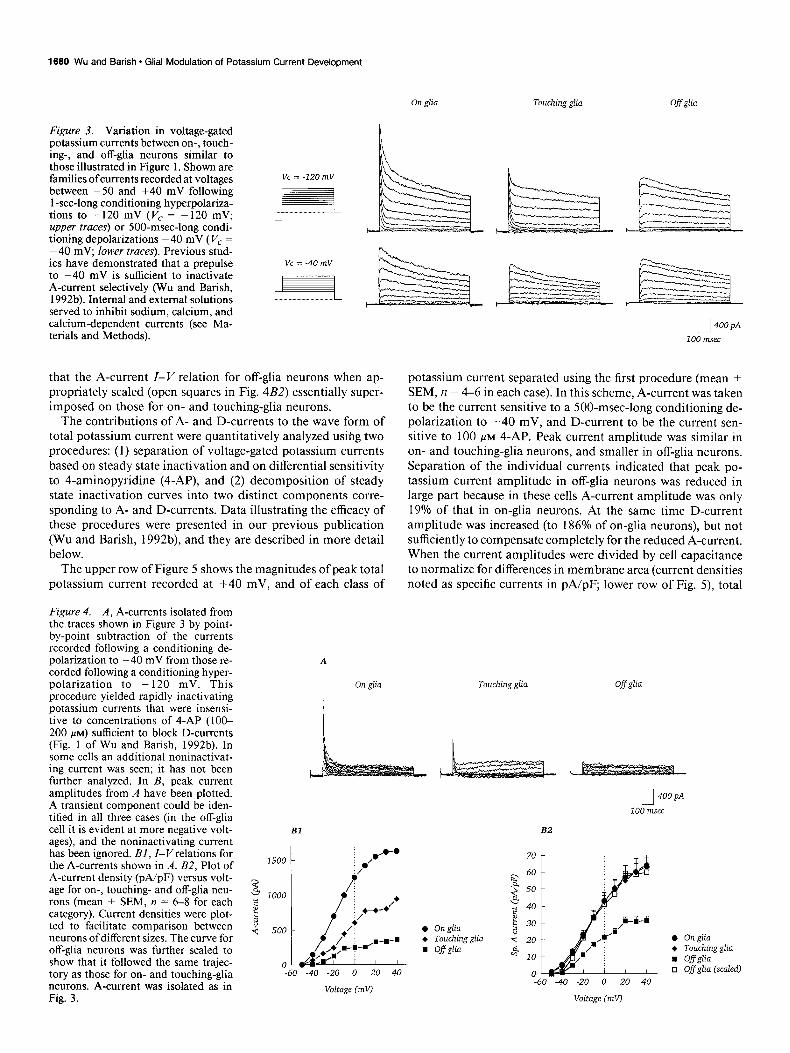
The wave forms of potassium currents recorded from on-, touching-, and off-glia neurons showed characteristic differ- ences. As illustrated by the traces shown in Figure 3, when recordings were made following conditioning hyperpolariza- tions to - 120 mV, a fast transient component present in on- glia neurons was reduced in touching-glia neurons and absent from off&a neurons (upper traces). In contrast, recordings made from the same cells following a conditioning depolarization to -40 mV lacked this rapidly relaxing component (lower traces). Since steady state inactivation of A-current in mouse hippo-
campal neurons is essentially complete at -40 mV (Wu and Barish, 1992b), the fast transient component of potassium cur- rent that varied with astroglial contact was considered to be A-current.
Isolation of the A-currents from these neurons as the transient currents sensitive to a conditioning depolarization to -40 mV revealed a fast inactivating current that was largest in the on- glia neuron (Fig. 4A). Consideration of the current-voltage (Z- I’) relations for these cells (Fig. 4BZ) and for a population of cells (Fig. 4B2) indicated that the A-current activation threshold was between -50 and -40 mV regardless of the extent of as- troglial contact. Further, the shapes of the A-current Z-V rela- tions were similar for the three classes of cells. Note in particular
Contact with glia
Figure 2. Resting properties of on-, touching, and off-glia neurons studied after 5-6 d in culture. Input resistance was determined from the steady state current measured during 10 mV excursions from the holding potential (-80 mV). Whole-cell capacitance, used as an index of membrane area, was determined by integrating the increment in capacity transient seen on passing from the on-cell to the whole-cell recording configuration. Resting conductance density is the ratio of input conductance near the holding potential to the whole-cell capacitance, and is noted as specific conductance. Data are mean + SEM; n = 8 for on-glia, 6 for touching-glia, and 6 for off-glia neurons; for off-glia versus on-glia, p < 0.05 for input resistance and cell capacitance, not significant for specific input conductance.
1660 Wu and Barish l Glial Modulation of Potassium Current Development
Figure 3. Variation in voltage-gated potassium currents between on-, touch- ing-, and off-glia neurons similar to those illustrated in Figure 1. Shown are families ofcurrents recorded at voltages between -50 and +40 mV following 1 -set-long conditioning hyperpolariza- tions to -120 mV (V, = -120 mV; upper traces) or 500-msec-long condi- tioning depolarizations -40 mV ( Vc = -40 mV, lower traces). Previous stud- ies have demonstrated that a prepulse to -40 mV is sufficient to inactivate A-current selectively (Wu and Barish, 1992b). Internal and external solutions served to inhibit sodium, calcium, and calcium-dependent currents (see Ma- terials and Methods).
Vc = -120 mV
.pt
Vc = -40 mV
On glia Touchingglia Off glia
that the A-current Z-V relation for off-glia neurons when ap- propriately scaled (open squares in Fig. 4B2) essentially super- imposed on those for on- and touching-glia neurons.
The contributions of A- and D-currents to the wave form of total potassium current were quantitatively analyzed using two procedures: (1) separation of voltage-gated potassium currents based on steady state inactivation and on differential sensitivity to 4-aminopyridine (4-AP), and (2) decomposition of steady state inactivation curves into two distinct components corre- sponding to A- and D-currents. Data illustrating the efficacy of these procedures were presented in our previous publication (Wu and Barish, 1992b), and they are described in more detail below.
The upper row of Figure 5 shows the magnitudes of peak total potassium current recorded at +40 mV. and of each class of
J 400 pA
100 msec
Figure 4. A, A-currents isolated from the traces shown in Figure 3 by point- by-point subtraction of the currents recorded following a conditioning de- polarization to -40 mV from those re- corded following a conditioning hyper- polarization to - 120 mV. This procedure yielded rapidly inactivating potassium currents that were insensi- tive to concentrations of 4-AP (loo- 200 PM) sufficient to block D-currents (Fig. 1 of Wu and Banish, 1992b). In some cells an additional noninactivat- ing current was seen; it has not been further analyzed. In B, peak current amplitudes from A have been plotted. A transient component could be iden- tified in all three cases (in the off-glia cell it is evident at more negative volt- ages), and the noninactivating current has been ignored. BI, I-V relations for the A-currents shown in A. B2, Plot of A-current density (pA/pF) versus volt- age for on-, touching- and off-glia neu- rons (mean + SEM, n = 6-8 for each category). Current densities were plot- ted to facilitate comparison between neurons ofdifferent sizes. The curve for off-glia neurons was further scaled to show that it followed the same trajec- tory as those for on- and touching-glia neurons. A-current was isolated as in Fig. 3.
A
On glia Touching glia Offglia
potassium current separated using the first procedure (mean ? SEM, n = 4-6 in each case). In this scheme, A-current was taken to be the current sensitive to a SOO-msec-long conditioning de- polarization to -40 mV, and D-current to be the current sen- sitive to 100 ,u~ 4-AP. Peak current amplitude was similar in on- and touching-glia neurons, and smaller in off-glia neurons. Separation of the individual currents indicated that peak po- tassium current amplitude in off-glia neurons was reduced in large part because in these cells A-current amplitude was only 19% of that in on-glia neurons. At the same time D-current amplitude was increased (to 186% of on-glia neurons), but not sufficiently to compensate completely for the reduced A-current. When the current amplitudes were divided by cell capacitance to normalize for differences in membrane area (current densities noted as specific currents in pA/pF, lower row of Fig. 5) total
J 400 pA
100 lmec
Bl B2
1500 I
Voltage (mV)
0 Onglia + Touching glia n Ofiglia
i2 60
a” 50
4 p 40
g 30
4 20
t+ 10
0 -60 -40 -20 0 20 40
l Onglia + Touching glia n Ofglia q Ofglia (scaled]

The Journal of Neuroscience, March 1994, 14(3) 1661
Peak current A-CUiTf?ld D-current K-current
80
Figure 5. Peak potassium current, and A-, D-, K-currents, isolated from on-, touching-, and off-glia neurons. The up- per row shows absolute current ampli- tude (PA); in the lower row these same data have been normalized to whole-
I- cell capacitance (pA/pF). A-current was isolated as the rapidly inactivating cur- rent sensitive to a conditioning depo- larization to -40 mV. D-current was isolated as the more slowly inactivating current sensitive to block by 100 PM 4-AP. K-current was taken to be the noninactivating current insensitive to both conditioning depolarization and 4-AP. Data are mean ? SEM; n = 4-6 cells in each category. Both A-current
1 amplitude and density (specific current) were significantly reduced in off-glia as compared to on-glia neurons (p < 0.01
- and p < 0.02, respectively). D-current amplitude and density were increased in off-glia as compared to on-glia neu- rons, but only the change in current density reached significance (p < 0.05).
1 60
40
20
0
8
potassium current density did not differ significantly between the three classes of neurons. However A-current density was highest in on-glia neurons, and D-current density was highest in off-glia neurons. K-current density was similar in all three classes of neurons.
Comparison of the current amplitudes with the current den- sities in Figure 5 suggests that while total potassium current density remained constant, the presence of additional mem- brane in on-glia neunons (capacitance data in Fig. 4) was ac- companied by a disproportionate increase in A-current ampli- tude (see Discussion).
Illustrated in Figure 6 is separation of the contributions of A- and D-currents to total relaxing current in the three classes of neurons evaluated using the second procedure based on differ- ences in steady state inactivation curves. Since the half-inacti- vation voltage for A-current is approximately 75 mV negative to that of D-current (Wu and Barish, 1992b), plots of total inactivating current availability versus voltage (III,,,) show two inflection points. The proportion of the total inactivation curve attributable to A- or D-currents was determined from these inactivation curves by using a least-squares regression routine to fit two Boltzmann relations with fixed parameters appropriate for A- and D-currents. Since adequate fits were obtained by varying only the relative power of the two Boltzmann relations, we concluded that the steady state inactivation properties of the three classes of cells could be described by an additive mixture of those for A- and D-currents. The relative contributions of A- and D-currents obtained using this procedure were similar to those obtained for the contributions of A- and D-currents to total potassium current using pharmacological separation as in Figure 5 (see Fig. 6 caption).
Variation of potassium currents with substrate: living versus nonliving astroglia
Five substrates were compared to determine if living astroglia were required for support of the large A-currents characteristic of on-glia neurons. In these experiments, substrates were treated
as described below immediately before addition of freshly dis- sociated hippocampal cells, and currents were recorded from neurons after 3-4 d of growth. At this time, most neurons plated directly onto coated glass will not be in contact with coplated astroglia, and thus their A-current amplitudes will be at a min- imum (see below; Wu and Barish, 1992b). The substrates uti- lized were the following.
On-glia. Astroglial monolayers were grown from embryonic hippocampal cells following the same procedures as described
Offglia
Figure 6. Voltage dependence of steady state inactivation of transient potassium currents in on-, touching-, and off-glia neurons (mean + SEM, n = 6 in each category), and separation of the contributions of A- and D-currents to the total. In each case, the data (symbols) were fit to the sum of two Boltzmann relations representing A- and D-current. Each is indicated by a dashed line, and the sum by a solid line. Boltzmann relations were of the form Z/I,,,,, = I/{1 + exp[(V - V,,,)/k]}. For each category only the relative contributions of the two Boltzmann relations to the total were varied. Boltzmann relation parameters for A-current were V,,2 = -75 mV, k = 12, and for D-current were V,,, = - 1 mV, k = 12. The contribution of the A-current to the total relaxing current was 0.82 + 0.03 for on-glia, 0.63 + 0.06 for touching-glia, and 0.34 -+ 0.06 for off-glia neurons (mean * SEM; on-glia vs touching-glia, p < 0.02; on-glia vs off-glia, p < 0.000 1). When separation of A-current was by sensitivity to conditioning depolarization (as in Figs. 4, 5) the con- tribution of A-current to maximum peak current was 0.73 ? 0.03 in on-glia neurons, while in off-glia neurons this value was 0.23 & 0.05 (mean + SEM; numbers derived from data in Figure 5; on-glia vs off- glia, p i 0.0001).

1662 Wu and Barish * Glial Modulation of Potassium Current Development
Contmf with glia Time of DRB application
Figure 7. Variation with substrate of whole-cell capacitance, input resistance, and resting conductance density measured near the holding potential (-80 mV). Astroglia were treated with distilled water, dried, or fixed with methanol, as described in Results. On- and off-glia prep- arations served as positive and negative controls. Recordings were made 3-4 d after dissociation and plating. Data are mean + SEM (n = 4-12 cells for each category). Although trends in the data are evident (as compared to off-glia neurons, neurons on dried or fixed glial substrates may have membrane areas similar to those of on-glia neurons but have input resistances more similar to those of off-glia neurons), differences in resting electrical properties in these experiments did not reach sta- tistical significance.
Figure 9. Effects of RNA synthesis inhibition with DRB on whole- cell capacitance, input resistance, and resting conductance density mea- sured near the holding potential. DRB (32 PM) was applied 16-24 hr (labeled 24 hr) or 72-84 hr (labeled 72 hr) after plating, and recordings were made after 5-8 d in culture. Measurements are mean -t SEM, n = 1 l-34 cells in each category. DRB affected resting properties only when applied within 24 hr of plating; within this time window both reduction in whole-cell capacitance and increase in input resistance were significant (p < 0.0001 and p < 0.05, respectively), but there was no significant change in resting input conductance density.
for neurons under Materials and Methods, except that mitotic inhibitors were not added until monolayers were formed. Neu- rons were killed by brief exposure to ice-cold medium (Huettner and Baughman, 1986). Monolayers were used after 7-10 d.
dti,O-glia. Astroglial monolayers (prepared as above) were immersed in distilled water for 30 set to 2 min to release any susceptible surface components. This treatment did not lyse the glia. Cells were added after the return to normal medium.
Dry-glia. Astroglia monolayers were dried in air overnight. Cells in medium were plated directly onto the dried cells.
MeOH-glia. Astroglia monolayers were rapidly fixed in meth- anol (MeOH; 10 min at -20°C) and air dried. As with dried glia, cells were plated directly onto the fixed cells.
Off-gliu. Cells were plated directly onto polylysine- or lami- nin/polylysine-coated glass.
Neurons grown on the nonliving substrates (dry-, MeOH-,
Figure 8. Substrate-induced variation in peak potassium current, and A-, D- and K-currents, presented as absolute current (PA, upper row) and as current density (pA/pF, lower row). Currents were separated based on prepulse in- activation for A-current and sensitivity to 100 KM 4-AP for D-current. Data are mean & SEM, n = 4-12 cells in each category. Both A-current amplitude and A-current density were significantly re- duced in neurons grown on nonliving substrates (dry-, MeOH-, and off-glia neurons) as compared to on-glia neu- rons (p < 0.0 l-0.02 in all cases). D-cur- rent amplitude and density in neurons grown on nonliving substrates tended to be larger than those of on-glia neu- rons (in most instances), but these dif- ferences did not reach statistical signif- icance except for dry-glia neurons (p < 0.04 for absolute amplitude and p < 0.02 for specific amplitude).
Peak current
2000 1 T T
3 1500 4 2 pi 1000 5
u 500
0
140 r T
100 f
80 3.0
and off-glia neurons) tended to be smaller than those grown on the living glia (on- and dH,O-glia) and to have larger input resistances (Fig. 7) although these differences did not reach statistical significance. The resting conductance densities (at - 80 mV) of the five classes of neurons were similar.
Analysis of potassium currents recorded from on-, dH,O-, dry-, MeOH-, and off-glia neurons, and separated based on voltage dependence and pharmacology into A-, D-, and K-cur- rents, is shown in Figure 8. Peak current amplitudes were similar for neurons grown on all substrates except coated glass (off-glia), for which they were smaller. A-current amplitudes were large for neurons on living substrates (on- and dH,O-neurons), and significantly smaller in those grown on nonliving substrates (dry-, MeOH-, and off-glia neurons). D-current amplitudes tended to be larger in neurons grown on nonliving substrates, but these differences did not reach statistical significance. When current
A-current D-current K-current
Substrate treatment

The Journal of Neuroscience, March 1994, 14(3) 1663
Peak current A-current
80
60
Time of DRB application
D-current K-current
amplitudes were normalized to whole-cell capacitance, A-cur- rent density was significantly greater in neurons grown on living substrates, while D-current density tended to be larger for non- living substrates. K-current amplitude and density appeared to be relatively unaffected by substrate.
Inhibition of RNA synthesis DRB, an inhibitor of mRNA production (Sehgal et al., 1976), was applied to the cultures to examine the possibility that the in vitro development of A-current was transcriptionally regu- lated. In control experiments, DRB reduced incorporation of 3H-uridine in mixed neuron-glia cultures to approximately 15% of control values (n = 3; 32 pM DRB). We confirmed that in DRB-treated cultures, as in normal control cultures, the flat non- neuronal cells forming the underlying monolayer after 4-6 d in culture were GFAP-expressing astroglia (data not shown).
Pyramidally shaped neurons could be readily identified in DRB-treated cultures. When quantitative indices of membrane area and conductance were examined, on-glia neurons grown in the presence of DRB had smaller whole-cell capacitance and higher input resistance, but only when DRB was added to cul- tures within the first 24 hr after plating (Fig. 9). No effects were seen when DRB was added at 72 hr or later. Resting conductance density (at -80 mV) was similar for control and DRB-treated neurons regardless of when the inhibitor was applied.
DRB affected the development of potassium currents only when applied during the initial 24 hr period, but was without effect when added after 72 hr (Fig. 10). DRB reduced the am- plitude of peak total potassium current by selectively affecting A-current amplitude, as both D- and K-current amplitudes were similar in control and experimental cultures. As with other treat- ments that reduced membrane area and A-current amplitude, the net effect of DRB was to reduce A-current density and in- crease D-current density.
In principle DRB could have acted on glia or neurons, or both. To sort out these possibilities two complementary exper- iments were performed. To affect neurons differentially, normal
- wl
Figure IO. Effects of RNA synthesis inhibition with DRB on total potassi- um current, and A-, D-, and K-cur- rents, presented as absolute current (PA, upper row) and as current density (pA/ pF, lower row). DRB was applied as for Figure 9. Currents were separated based on prepulse inactivation for A-current and sensitivitv to 100 UM 4-AP for D-current. Data are mean k SEM, n = 1 l-34 cells. Both A-current amplitude and A-current density were significant- ly reduced in neurons exposed to DRB during the initial 24 hr period (p < O.OOOl), but not in neurons exposed to DRB after 72 hr. D-current amplitude was not significantly affected by DRB, but its density was increased because of the inhibitor-induced decrease in mem- brane area (p < 0.003). Neither K-cur- rent amplitude nor its density was sig- nificantly affected by DRB.
glial monolayers were prepared, embryonic hippocampal cells were plated onto these monolayers, and DRB was added after 16 hr. To affect astroglia differentially, glial monolayers were grown in the presence of DRB, the DRB was removed, and hippocampal cells were plated onto the treated glia. In both cases potassium currents were recorded 5-8 d after addition of neurons. The results of these experiments are summarized in Figure 11. For the first variant, normal glia and DRB-treated neurons, peak current density and A-, D- and K-current den- sities (separated based on voltage dependence and sensitivity to 4-AP) were similar in control and experimental cultures (Fig. 11AI). Further, the numbers of cells expressing low (~30 pA/ pF), intermediate (30-50 pA/pF), or high (>50 pA/pF) A-cur- rent densities were similar in control and experimental cultures (Fig. 1 lA2). In contrast, for the second variant, DRB-treated glia and normal neurons, A-current density was reduced in neu- rons grown on treated glia (Fig. 11BI). More significantly, the distribution of neurons showing low, medium, and high A-cur- rent densities was shifted toward lower densities (Fig. 1 lB2). Taken together, comparison of the results presented in Figure 11, A2 and B2, suggests that inhibition of RNA synthesis in underlying astroglia was responsible for reduction in A-current amplitude in contacting neurons.
It is not clear why A-currents in neurons grown on the DRB- treated glia were not reduced to the same extent as when both neurons and glia were treated with DRB (as in the experiment? presented in Fig. 10). One possibility is that because the hip- pocampal cells added to the treated astroglia were a mixture of neurons, glia, and their undifferentiated pluripotent precursors (see Temple and Raff, 1985) neurons may have made contact with astroglia not exposed to DRB. An additional but less likely possibility is reversal of DRB inhibition after termination of astroglial exposure. Although DRB is reported to be a reversible inhibitor, we did not observe an increase in A-current amplitude after removal of DRB from cultures exposed beginning at 24 hr after plating (data not shown). Consistent with this observation, 3H-uridine incorporation into glia exposed to DRB for 3 weeks increased by only 59% (n = 2) after removal of DRB (data not

1664 Wu and Barish * Glial Modulation of Potassium Current Development
Al Peak current
A2
0.75 d 8 6 c 0.50 .z k
8 0.25
60
Normal glia + DRB-treated neurons DRB-treated glia + normal neurons
A-current D-current K-current Bl Peak current A-current
Sp. A-current (pA/pF] Sp. A-current (pA/pF)
m Control
&$$j DRB-treated neurons
D-current
Figure 11. Effects of selective treatment of neurons or of astroglial substrates with RNA synthesis inhibitor on the appearance of total potassium current, and of A-, D-, and K-currents. A, Potassium current densities after treatment of neurons with DRB. Cultures of normal hippocampal astroglia were allowed to form confluent monolayers (approximately 7 d in culture). After washing, 25 x 10’ hippocampal cells (a mixture of neurons, glia, and undifferentiated pluripotent precursor cells) were plated onto these monolayers. DRB (32 PM) was added 1 hr after plating, and currents were recorded after 7 d in culture. A-, D-, and K-currents were separated based on sensitivity to conditioning depolarization or to 100 PM 4-AP. Data are mean * SEM, II = 5-6 cells. Al, Neither peak total potassium current density, nor A-, D-, or K-current densities were significantly affected by DRB treatment. A2, There was no effect of DRB on the proportions of cells exhibiting low (< 30 pA/pF), intermediate (30-50 pA/pF), or high (>50 pA/pF) A-current densities (same cells as in Al). B, Potassium current densities after treatment of glia with DRB. Cultures of hippocampal astroglia were exposed to 32 PM DRB beginning 24 hr after plating, and allowed to form confluent monolayers (approximately 7-10 d in culture). After washing, 25 x lo3 hippocampal cells were added, and currents were recorded after 3-5 d in culture. Data are for n = 7-21 cells. BI, A-current density was reduced in neurons grown on DRB-treated glia, but this difference was only marginally significant @ < 0.08). There was no significant difference in peak, D-, or K-current densities. B2, DRB treatment of glia shifted the distribution of A-current densities towards lower values along the scale of low, intermediate, or high A-current density (same cells as in BI).
B2
60
K-current
m Control
B DRB-treated glia
shown; based on the extent of inhibition of 3H-uridine incor- poration by DRB discussed above, one would expect complete reversal to increase 3H-uridine incorporation by 600-700%).
Relation between potassium currents (as afected by glial contact) and excitability Action potentials recorded under current clamp were affected by the presence of an astroglial substrate, as shown in Figure 12. While contact with astroglial cells may affect multiple species of ionic currents, examination of action potentials will begin to provide an indication of the relationship between transient po- tassium current expression and electrogenesis. The voltage- and current-clamp recordings shown were made from the same neu- rons. The neuron labeled on-glia exhibited substantial A-current and little D-current (upper traces). Membrane voltage recording during depolarizing current injections (middle and lower traces) showed a notch and a delay before the appearance of the first action potential. This notch reflects the activation of A-current, as it is removed by applications of 4-AP sufficient to block A-current (as shown for the neuron labeled on-glia (+4-AP);
see also Connor and Stevens, 197 1 b). The sustained firing of this on-glia neuron reflects a balance between sustained inward current and in this case minimal D-current. In contrast, the neuron labeled off-glia exhibited only D-current, and during current injections membrane voltage reached threshold without a notch or delay. The firing of this neuron may have been trun- cated by activation of D-current. The neuron not exposed to DRB but grown on DRB-treated (and washed) glia (neuron labeled DRB-treated glia) showed minimal A-current, and a pattern of action potential generation similar to that of an off- glia rather than an on-glia neuron.
Discussion Overview The results presented indicate that A-current amplitude in cul- tured hippocampal neurons was increased by contact with as- troglia (Figs. 3-5). This modulation required membrane-mem- brane contact or other short-range interaction rather than free diffusion of a soluble factor, as currents could differ between neighboring cells on the same cover slip (Fig. 1). Variation in

The Journal of Neuroscience, March 1994. 14(3) 1666
On-&a Off-glia
I
1 15OpA
700 pA L 100 msec
DRB-treated glia On-glia (+4-M)
Figure 12. Variations in action potential generation related to contact with astroglial cells and the presence of A- or D-currents. Upper row, Recordings made under voltage clamp (without TTX present externally). Middle and lower rows, Current-clamp recordings made from the same neurons on two different time scales. Recordings in the first two columns were obtained from typical on- and off-glia neurons. The recording in the third column was obtained from a neuron grown on an astroglial monolayer nretreated with the RNA synthesis inhibitor DRB (32 PM for 3 weeks). The recordings in the fourth column were obtained from an on&a neuron, and illustrate the consequences for action potential generation of A-current inhibition with 3 mM 4-AP. The records presented are typical of nine on-glia and six off-glia neurons studied under current clamp.
+3mM4-AP -
A-current was not accompanied by shifts in the voltage depen- dencies of activation or inactivation (Figs. 4, 6). Enhanced A-current amplitude was supported only by living astroglia, not by dried or fixed glia (Fig. 8) and could be blocked by exposure to RNA synthesis inhibitor during the first 24 hr after dissoci- ation and plating (Figs. 10, 11). Astroglial induction ofA-current was accompanied by increase in membrane area (Figs. 2, 7, 9). We conclude that astroglia express on their surface one or more factors that regulate both neuronal growth and appearance of A-type transient potassium current channels. Enhancement of A-current amplitude may occur by preferential insertion of A-channel-rich membrane, although the increases in current and capacitance may not necessarily be linked. We believe that astroglia are acting directly on neurons to influence the ap- pearance of A-current, although we cannot exclude the possi- bility that the astroglia act indirectly by facilitating interactions between on-glia neurons. We suggest astroglia-to-neuron sig- naling and potassium current modulation as a mechanism for regulation of hippocampal neuron growth and excitability.
Modulation of transient potassium current development by non-neuronal cells may not be unique to hippocampal neurons. For chick ciliary ganglion neurons, Dourado and Dryer (1992a,b) concluded based on comparisons of age-matched acutely dis- sociated and cultured neurons that extrinsic factor(s) potentially derived from target cells were required for normal A-current development. For rat superior cervical ganglion neurons, McFarlane and Cooper (199 1, 1992, 1993) found that devel- opment of two forms of A-current differing in inactivation ki-
netics (noted in their study as ABr, and A,,,,) required factor(s) present in vivo but not in cultures free of satellite cells. Further, the development of these A-current variants in culture could be influenced by non-neuronal cells. The similarity of these ob- servations to the results presented here is striking. While the precise mechanisms of transient potassium current modulation may differ between hippocampal neurons and peripheral neu- rons, given the role of A-currents in establishing the repetitive firing properties of neurons, its regulation within a small region of excitable tissue by non-neuronal or target cells may be a significant mechanism for modulation ofthe signaling properties of groups of neurons.
D$erential regulation of A-current
The regulation of A-current described here differs from previ- ously described modulations of transient potassium currents in hippocampal neurons. Other studies have not separated tran- sient potassium currents into subtypes, and have been concerned with rapid changes following neurotransmitter receptor acti- vation or perturbations of intracellular ion concentrations (Na- kajima et al., 1986; Atkins et al., 1990; Saint et al., 1990; Chen and Wong, 199 1). In contrast, the regulation observed here ap- pears to involve developmental appearance of A-currents under astroglial control, and to be associated and potentially linked to increases in neuronal membrane area.
In some instances a decrease in A-current was associated with a compensatory increase in D-current amplitude, particularly in off-glia neurons (Fig. 5) and in neurons grown on dried glia

1888 Wu and Barish - Glial Modulation of Potassium Current Development
(Fig. 8). However, in other cases this pattern was not seen, for generation, this pattern means that the relative contributions of example, in DRB-treated neurons (Fig. 10) and in some off-glia A- and D-currents to overall potassium current density could neurons (Fig. 8). While the behavior of D-current was much vary considerably with astroglial contact. Because A- and D-cur- more variable than that of A-current, the data suggest reciprocal rents differ in voltage dependencies and activation and inacti- regulation of A- and D-currents may occur with the absence of vation kinetics (Storm, 1988; Ficker and Heinemann, 1992; Wu the signals promoting A-current resulting in enhancement of and Barish, 1992b), they make different contributions to the D-current.
Nonuniform distribution of channels underlying A- and D-currents In each of the experimental series in which A-current amplitude was regulated-on- versus touching- versus off-glia neurons, living versus nonliving glial substrates, and control versus DRB- treated cultures-contact with glial cells was associated with increased membrane area and greater than proportional aug- mentation of A-current amplitude. Consider, for example, the data concerned with on-, touching-, and off-glia neurons pre- sented in Figures 2 and 5.Off-glia neurons had a mean A-current amplitude 19% of that in on-glia neurons, while mean whole- cell capacitance was 70% of that of on-glia neurons. Thus, 8 1% of the total A-current in on-glia neurons was associated with a 30% differential in membrane area, and one possibility is that the additional A-current was localized to this membrane. While the link of increased A-current with membrane area was not rigid-for example in one series touching-glia neurons were sig- nificantly smaller than on-glia neurons but had A-currents of similar amplitude-it was seen in the majority of cases. As morphological studies have shown that astroglia induce neurite growth in hippocampal neurons (Noble et al., 1984; Fallon, 1985; Pixley et al., 1987), one interpretation of the present re- sults is that the membrane added as a consequence of contact with astroglia may be enriched in A-current. However, an al- ternative hypothesis is that astroglial contact promotes insertion of both membrane and A-current channels throughout the cell, but that these are unrelated processes.
A mechanism of astroglial-induced addition of A-current- rich membrane could contribute to regional variation in hip- pocampal neuron excitability (Spencer and Kandel, 1961; Schwartzkroin, 1977; Wong et al., 1979; Wong and Stewart, 1992). Preferential insertion of particular types of sodium, cal- cium, or potassium channels in regions of dendritic, somatic, or axonal membranes has been observed (Angelides et al., 1988; Blaxter et al., 1989; Jones et al., 1989; Westenbroek et al., 1989; Ahlijanian et al., 1990; Sheng et al., 1992). Of particular interest is the observation of Sheng et al. (1992) that two potassium channel polypeptides segregate to either dendrites and somas, or to axons and terminals, of hippocampal neurons. Since the products of the mRNAs encoding the two peptides Kvl.4 and Kv4.2 form inactivating potassium channels when injected into Xenopus oocytes, it is tempting to speculate that the channels formed by these peptides could be those underlying the A- and D-currents described here.
overall pattern of action potential generation. Connor and Ste- vens (197 la,b) indicated that during sustained stimulation A-current would manifest itself as a delay during the slow de- polarizing approach toward threshold (see also Fig. 12). In con- trast, D-current is thought to have stronger effects on the width of the hippocampal action potential and on the pattern of sus- tained repetitive firing (Storm, 1988; Wu and Barish, 1992b). Therefore, variation in the relative proportions of the two cur- rents in the membrane should result in changes in electrical behavior. The results presented in Figure 12 are consistent with this notion, and suggest that the extent of A-current induction by glia will influence the trajectory of voltage toward threshold while regulation of D-current will affect action potential width and repetitive firing. More detailed analyses of the effects of variation in A-current amplitude and membrane area, and of the consequences of changes in the contribution of D-current to the total potassium current wave form, will require construc- tion of quantitative models of hippocampal neuron excitability incorporating these two distinct transient potassium currents.
Conclusions
Signals that pass from glia to neurons may involve long-range interactions employing soluble factors (Banker, 1980) or more short-range interactions dependent on cell-cell contact or lim- ited diffusion of soluble factors. These glia-derived signals have multiple influences on neuronal development and functioning. During early development glia guide neuronal migration by mechanisms involving cell-cell contact (for review, see Rakic, 198 1, 1990), and they are a source of multiple growth factors (for review, see Giulian et al., 1988). In mature brain, glia in- fluence neuronal excitability by buffering extracellular potassi- urn and amino acid neurotransmitters (for review, see Hertz, 1979; Walz, 1989), and by continued production of growth fac- tors and cytokines active on neurons (for review, see Benveniste, 1992; Martin, 1992). Our present work further suggests that by modulating expression of transient potassium currents and the characteristics of action potential generation astroglia may in- fluence use-dependent Ca2+ entry and accumulation in hippo- campal neurons. By this mechanism glia may participate in long- term regulation of synaptic efficacy in the hippocampus.
Consequences for hippocampal neuron excitability
A variety of manipulations that blocked the ability of astroglial cells to support A-currents resulted in reductions of both A-cur- rent amplitude and membrane area that were accompanied by either no change or increase in D-current amplitude. When these amplitude data were normalized against membrane area (mea- sured as whole-cell capacitance), the changes in average densities of A- and D-currents per unit membrane area became approx- imately reciprocal. Considered in the context of action potential
References Ahlijanian MK, Westenbroek RE, Catterall WA (1990) Subunit struc-
ture and localization of dihydropyridine-sensitive calcium channels in mammalian brain, spinal cord, and retina. Neuron 4:819-832.
Akins PT, Surmeier DJ, Kitai ST (1990) Muscarinic modulation of a transient K+ conductance in rat neostriatal neurons. Nature 344:240- 242.
Angelides KJ, Elmer LW, Loftus D, Elson E (1988) Distribution and lateral mobility of voltage-dependent sodium channels in neurons. J Cell Biol 106:191 l-1925.
Banker GA (1980) Trophic interactions between astroglial cells and hippocampal neurons in culture. Science 209:809-8 10.
Banker GA, Cowan WM (1977) Rat hippocampal neurons in dispersed cell culture. Brain Res 126:397-425.
Banker GA, Cowan WM (I 979) Further observations on hippocampal neurons in dispersed cell culture. J Comp Neurol 187:469-494.
Benveniste EN (1992) Inflammatory cytokines within the central ner- vous system: sources, function and mechanisms of action. Am J Phys- iol 263:Cl-C16.

The Journal of Neuroscience, March 1994, f4(3) 1687
Bignami A, Eng LF, Dahl D, Uyeda CT (1972) Localization of the glial fibrillary acidic protein in astrocytes by immunofluorescence. Brain Res 43:429-435.
Blaxter TJ, Carlen PL, Niesen C (1989) Pharmacological and anatom- ical separation of calcium currents in rat dentate granule neurones in vitro. J Physiol (Lond) 4 12:93-l 12.
Brown DA, Gliwiler BH, Griffith WH, Halliwell JV (1990) Membrane currents in hippocampal neurons. Prog Brain Res 83:141-160.
Chen QX, Wong RKS (199 1) Intracellular Ca’+ suppressed a transient potassium current in hippocampal neurons. J Neurosci 11:337-343.
Connor JA, Stevens CF (197 la) Voltage clamp studies of a transient outward membrane current in gastropod neural somata. J Physiol (Lond) 213:21-30.
Connor JA, Stevens CF (197 1 b) Prediction of repetitive firing behav- iour from voltage clamp data on an isolated neurone soma. J Physiol (Lond) 213:31-53.
Dourado MM, Dryer SE (1992a) Changes in the electrical properties of chick ciliary ganglion neurones during embryonic development. J Physiol (Lond) 449:41 l-428.
Dourado MM, Dryer SE (1992b) Extrinsic factors influence the ex- pression of K+ currents in chick ciliary ganglion neurons. Sot Neu- rosci Abstr 18:4 1.
Fallon JR (1985) Preferential outgrowth of central nervous system neurites on astrocytes and Schwann cells as compared with nonglial cells in vitro. J Cell Biol 100: 198-207.
Ficker E, Heinemann U (1992) Slow and fast transient potassium currents in cultured rat hippocampal cells. J Physiol (Lond) 445:43 l- 455.
Giulian D, Vaca K, Johnson B (1988) Secreted peptides as regulators of neuron-glia and glia-glia interactions in the developing nervous system. J Neurosci Res 2 1:487-500.
Gustafsson B, Galvan M, Grafe P, Wigstrom H (1982) A transient outward current in a mammalian central neurone blocked by 4-aminopyridine. Nature 299:252-254.
Hagiwara S, Kusano K, Saito N (196 1) Membrane changes of Onch- idium nerve cell in potassium-rich media. J Physiol (Lond) 155:470- 489.
Halliwell JV, Othman IB, Pelchen-Matthews A, Dolly JO (1986) Cen- tral action of dendrotoxin: selective reduction of a transient K con- ductance in hippocampus and binding to localized acceptors. Proc Nat1 Acad Sci USA 83~493-497.
Hamill OP, Marty A, Neher E, Sakmann B, Sigworth F (198 1) Im- proved patch-clamp techniques for high-resolution current recordings from cells and cell-free membrane patches. Pfluegers Arch 391:85- 100.
Hertz L (1979) Functional interactions between neurons and astro- cytes. I. Turnover and metabolism of putative amino acid transmit- ters. Prog Neurobiol 13:277-323.
Huettner JE, Baughman RW (1986) Primary culture of identified neu- rons from the visual cortex ofpostnatal rats. J Neurosci 6:3044-3060.
Jones OT, Kunze DL, Angelides KJ (1989) Localization and mobility of w-conotoxin-sensitive Ca2+ channels in hippocampal CA 1 neurons. Science 244:1189-l 193.
Kriegstein AR, Dichter MA (1983) Morphological classification of rat cortical neurons in cell culture. J Neurosci 3: 1634-l 647.
Martin DL (1992) Synthesis and release of neuroactive substances by glial cells. Glia 5:8 l-94.
McFarlane S, Cooper E (1991) Non-neuronal cells influence the ex- pression of K currents on cultured SCG neurons. Sot Neurosci Abstr 17~778.
McFarlane S, Cooper E (1992) Postnatal development of voltage-gated K currents on rat sympathetic neurons. J Neurophysiol 67:1291- 1300.
McFarlane S, Cooper E (1993) Extrinsic factors influence the expres- sion of voltage-gated K currents on neonatal rat sympathetic neurons. J Neurosci 13:2591-2600.
Nakajima Y, Nakajima S, Leonard RJ, Yamaguchi K (1986) Acetyl- choline raises excitability by inhibiting the fast transient potassium
current in cultured hippocampal neurons. Proc Nat1 Acad Sci USA 83:3022-3026.
Neher E (197 1) Two fast transient current components during voltage clamp on snail neurons. J Gen Physiol 58:36-53.
Neumann RE, Wadman WJ, Wong RKS (1987) Outward currents of single hippocampal cells obtained from the adult guinea-pig. J Physiol (Lond) 393:331-353.
Noble M, Fok-Seang J, Cohen J (1984) Glia are a unique substrate for the in vitro growth of central nervous system neurons. J Neurosci 4:1892-1903.
Pixley SKR, Nieto-Sampedro M, Cotman CW (1987) Preferential adhesion of brain astrocytes to laminin and central neurites to astro- cytes. J Neurosci Res 18:402-406.
Rakic P (198 1) Neuronal-glial interaction during brain development. Trends Neurosci 4: 184-l 87.
Rakic P (1990) Principles of neural cell migration. Experientia 46: 882-891.
Rogawski MA (1985) The A-current: how ubiquitous a feature of excitable cells is it? Trends Neurosci 8:2 14-2 19.
Rudy B (1988) Diversity and ubiquity of K channels. Neuroscience 251729-749.
Saint DA, Thomas T, Gage PW (1990) GABA, agonists modulate a transient potassium current in cultured mammalian hippocampal neurons. Neurosci Lett 118:9-l 3.
Schwartzkroin PA (1977) Further characteristics of hippocampal CA 1 cells in vitro. Brain Res 128:53-68.
Segal M, Barker JL (1984) Rat hippocampal neurons in culture: po- tassium conductances. J Neurophysiol 51: 1409-1433.
Segal M, Rogawski MA, Barker JL (1984) A transient potassium con- ductance regulates the excitability of cultured hippocampal and spinal neurons. J Neurosci 4:604-609.
Sehgal PB, Dame11 JE Jr, Tamm I (1976) The inhibition by DRB (5,6- dichloro- 1 -P-o-ribofuranosylbenzimidazole) of hnRNA and mRNA production in HeLa cells. Cell 9:473-480.
Sheng M, Tsaur M-L, Jan YN, Jan LY (1992) Subcellular segregation of two A-type K+ channel proteins in rat central neurons. Neuron 9:27 l-284.
Spencer WA, Kandel ER (196 1) Electrophysiology of hippocampal neurons. J Neurophysiol 24:272-285.
Storm JF (1987) Action potential repolarization and a fast after-hy- perpolarization in rat hippocampal pyramidal cells. J Physiol (Lond) 385:733-759.
Storm JF (1988) Temporal integration by a slowly inactivating K+ current in hippocampal neurons. Nature 336:379-38 1.
Storm JF (1990) Potassium currents in hippocampal pyramidal cells. Prog Brain Res 83:161-187.
Temple S, Raff MC (1985) Differentiation of a bipotential glial pro- genitor cell in single cell microculture. Nature 313:223-225. -
Walz W (1989) Role of elial cells in the reeulation of the brain mi- croenvironment. Prog N&robiol 33:309-313.
Westenbroek RE, Merrick DK, Catterall WA (1989) Differential sub- cellular localization of the R, and R,, Na+ channel subtypes in central neurons. Neuron 3:695-704.
Wong RKS, Stewart M (1992) Different firing patterns generated in dendrites and somata of CA 1 pyramidal neurones in guinea-pig hip- pocampus. J Physiol (Lond) 457:675-687.
Wong RKS, Prince DA, Basbaum AI (1979) Intradendritic recordings from hippocampal neurons. Proc Nat1 Acad Sci USA 76:986-990..
Wu R-L. Barish ME (1992a) Glial modulation of transient notassium current expression in cultured mouse hippocampal neurons. Sot Neu- rosci Abstr 18: 150 1.
Wu R-L, Barish ME (1992b) Two pharmacologically and kinetically distinct transient potassium currents in cultured mouse hippocampal neurons. J Neurosci 1212235-2246.
Zbicz KI, Weight FF (1985) Transient voltage and calcium-dependent outward currents in hippocampal CA3 pyramidal neurons. J Neu- rophysiol 53:1038-1058.