Embed Size (px)
Citation preview

Instructions for use
Title Astaxanthin improves the developmental competence of invitro-grown oocytes and modifies the steroidogenesis ofgranulosa cells derived from bovine early antral follicles
Author(s) Abdel-Ghani, M. A.; Yanagawa, Y.; Balboula, A. Z.; Sakaguchi, K.; Kanno, C.; Katagiri, S.; Takahashi, M.; Nagano,M.
Citation Reproduction, Fertility and Development, 31(2), 272-281https://doi.org/10.1071/RD17527
Issue Date 2019-01
Doc URL http://hdl.handle.net/2115/73394
Type article (author version)
File Information Reproduction, Fertility and Development_31(2)_272_281.pdf
Hokkaido University Collection of Scholarly and Academic Papers : HUSCAP

1
Astaxanthin improves the developmental competence of in vitro grown oocytes and 1
modifies the steroidogenesis of granulosa cells derived from bovine early antral follicles 2
3
4
M. A. Abdel-GhaniAB, Y. YanagawaA♯, A. Z. BalboulaC, K. SakaguchiD, C. KannoD, S. 5
KatagiriA, M. TakahashiC, M. NaganoA* 6
7
A Laboratory of Theriogenology, Department of Clinical Sciences, Faculty of Veterinary 8
Medicine, Hokkaido University, Sapporo 060-0818, Japan 9
B Department of Theriogenology, Faculty of Veterinary Medicine, Assuit University, Assuit, 10
71515, Egypt 11
C Laboratory of Animal Genetics and Reproduction, Research Faculty of Agriculture, 12
Hokkaido University, Sapporo 060-8589, Japan 13
D Laboratory of Theriogenology, Graduate School of Veterinary Medicine, Hokkaido 14
University, Sapporo 060-0818, Japan 15
16
17
♯ Equal contribution to the first author 18
*Correspondence: Masashi Nagano (e-mail: [email protected]) 19
TEL.&FAX: +81-11-706-5232 20
21

2
Abstract 22
We investigated the influence of Astaxanthin (Ax), which exhibits strong antioxidant activity, 23
during in vitro growth (IVG) on the developmental competence of oocytes and 24
steroidogenesis of granulosa cells derived from early antral follicles. Bovine oocyte-25
cumulus-granulosa complexes collected from early antral follicles were cultured for 12 days 26
in the presence (500 µM) or absence (control) of Ax. Viability of oocytes and antrum 27
formation in granulosa cell layer during IVG culture were greater in the presence of Ax than 28
in its absence (P<0.05). Regardless of Ax treatment, estradiol-17β production increased 29
during IVG culture; however, progesterone production was significantly lower in the 30
presence of Ax than in its absence (P<0.05). Reactive oxygen species levels were lower in 31
Ax-treated oocytes than in controls after IVG (P<0.05). Although the nuclear maturation and 32
cleavage rates did not differ regardless of Ax treatment, Ax treatment led to weaker cathepsin 33
B activity in oocytes and better blastocyst rates than those in controls (P<0.05). Accordingly, 34
Ax treatment during IVG increased total cell numbers in blastocysts (P<0.05). These results 35
indicate that Ax supplementation to IVG medium improves the quality of bovine oocytes due 36
to its antioxidative effects on growing oocytes and its suppression of the luteinization of 37
granulosa cells. 38
39
Additional Keywords: astaxanthin, cathepsin B, early antral follicle, IVG, ROS generation 40
41

3
Introduction 42
43
Assisted reproductive technologies (ARTs) including in vitro maturation (IVM), the in vitro 44
fertilization (IVF) of oocytes, and in vitro culture (IVC) for the production of transferable 45
embryos are uniquely desirable for improving the breeding of animals and also for the 46
perpetuation programs of wild and endangered species. Moreover, the isolation and IVC of 47
ovarian follicles to obtain fertilizable oocytes and further embryo development have been 48
regarded as a propitious strategy to strive against infertility issues (Hansen 2014; 49
Szamatowicz 2016; Kushnir et al. 2017). 50
The follicle features a fitting microenvironment that is responsible for confirming the 51
production of oocytes with high quality and integrity, allowing its final growth, capacitation, 52
and the nuclear and cytoplasmic maturation needs of the female gamete until it is released 53
for fertilization into the uterine tubes (Hennet and Combelles 2012). In growing follicles, the 54
concentration of estradiol-17β (E2) increases in conjunction with follicular development, 55
while elevations in progesterone (P4) concentrations are accompanied by follicular degeneration 56
(Kruip and Dieleman 1985). However, oocyte-cumulus-granulosa complexes (OCGCs) have 57
been shown to produce large amounts of P4 during in vitro growth (IVG) cultures (Yang et 58
al. 2016; Sakaguchi et al. 2017), similar to degenerating bovine follicles (Kruip and Dieleman 59
1985). Previous studies have documented and focused on the establishment of a suitable 60
system for the IVG of bovine OCGCs obtained from early antral follicles or mimicking in 61
vivo development of bovine follicles; however, success has been limited (Makita and Miyano 62

4
2015; Huang et al. 2016; Makita et al. 2016; Sakaguchi et al. 2017). Therefore, further 63
research is needed in order to improve IVG systems. 64
Astaxanthin (Ax) is a red-orange carotenoid pigment that is present in fishery products 65
such as salmon, shrimp, and crab (Kuraji et al. 2016). Previous studies reported that Ax 66
possesses a wide range of biological functions including the control of lipid peroxidation, 67
anti-inflammatory and anti-tumor activities, the scavenging of reactive oxygen species 68
(ROS), and as a hydroxyl radical antioxidant (Namekawa et al. 2010; Fassett and Coombes 69
2011; Kuraji et al. 2016). Moreover, Ax feeding was beneficial to improve chicken egg yolk 70
color; egg quality during storage and it also improved the carcass traits and meat quality of 71
pigs (Yang et al. 2006). Besides, there is an increase in the number of corpora lutea (CL), 72
implantation sites and fetuses, and a decrease in the percentage of stillborn kits among minks 73
fed Ax (Hansen et al. 2014). Furthermore, Ax exhibits more powerful antioxidant activity 74
than vitamin C, vitamin E, and β-carotene; the antioxidant activity of Ax was shown to be 75
100- to 500-fold greater than that α-tocopherol and 15-fold greater than those of other 76
carotenoids (Naguib 2000). The antioxidant effects of Ax on the developmental competence 77
of in vitro-produced bovine embryos have been attributed to the induction of antioxidant 78
genes and suppression of apoptotic genes (Jang et al. 2010). Additionally, it is soluble in 79
lipids and, thus, is incorporated into cell membranes and reduces DNA damage (Kuraji et al. 80
2016). Ax added to the IVC medium of embryos improved bovine embryonic development 81
impaired by heat stress (Namekawa et al. 2010), and its supplementation effectively 82
promoted the maturation, fertilization, and development of oocytes exposed to heat stress 83
during IVM in pigs (Do et al. 2015). Therefore, Ax is also assumed to be beneficial during 84

5
IVG; however, the effects of Ax on bovine OCGCs obtained from early antral follicles during 85
IVG culture currently remain unknown. A previous study reported that oxidative stress 86
induced apoptosis in mouse granulosa cells (Weng et al. 2016). Therefore, the antioxidative 87
effects of Ax are expected to improve the steroid hormone environment for OCGCs. 88
Cathepsin B (CTSB) is a lysosomal cysteine protease found in many types of cells such 89
as bovine oocytes (Balboula et al. 2010). A relationship has been reported between CTSB 90
activity and apoptosis, in which CTSB was found to induce the apoptotic pathway by 91
activating caspases, and the inhibition of CTSB during IVM significantly improved the 92
developmental competence of bovine COCs as well as the quality of their embryos (Balboula 93
et al. 2010). Moreover, the activity of CTSB inversely correlated with the developmental 94
competence of bovine oocytes, and, thus, CTSB activity may be a useful marker for oocytes 95
and embryos of inferior quality (Balboula et al. 2013). Although the role of CTSB activity 96
has been elucidated in bovine oocytes, its activity in bovine oocytes derived from IVG and 97
the effect of Ax on CTSB activity currently remains unclear. 98
In the present study, the effects of Ax supplementation during IVG on the growth 99
parameters of OCGCs (survivability, antrum formation in the granulosa cell layer, and 100
diameter of oocytes), the quality of oocytes after IVM (CTSB activity), maturation, the 101
further embryonic development of bovine oocytes obtained from early antral follicles, and 102
the production of steroid hormones from granulosa cells were investigated. 103
104
Materials and methods 105
106

6
Chemicals 107
All the chemicals used in the present study were purchased from Sigma-Aldrich (St. Louis, 108
MO, USA), unless otherwise stated. 109
110
Collection of OCGCs from early antral follicles and IVG 111
Bovine ovaries (n=158) were obtained from a local abattoir. They were transported to the 112
laboratory within 6 to 10 hours of collection in a plastic bag at 20°C. After three washes in 113
physiological sterile saline, sliced ovarian cortex tissues (<1 mm thick) were prepared using 114
a surgical blade (No. 11) and stored in TCM199 (Invitrogen; Grand Island, NY, USA) 115
supplemented with 0.1% polyvinyl alcohol, 25 mM HEPES, 10 mM sodium bicarbonate, and 116
50 mg/mL gentamicin sulfate (isolation medium, pH 7.4, at 37°C), as described elsewhere 117
(Huang et al. 2013; Huang 2014). Under a stereomicroscope, early antral follicles (0.5-1 mm 118
in diameter) were dissected from the sliced ovarian tissues using a surgical blade (No. 20) 119
(Fig. 1A). OCGCs were isolated from follicles using a pair of fine forceps and those with a 120
normal appearance were individually cultured in 96-well culture plates (Falcon 353872, 121
Becton Dickinson, Franklin Lakes, NJ, USA) with 200 µL of growth medium for 12 days at 122
39°C in humidified air with 5% CO2. Growth medium consisted of HEPES-buffered 123
TCM199 (Invitrogen) supplemented with 0.91 mM sodium pyruvate, 10 ng/mL 124
androstenedione, 5% fetal calf serum (FCS; Invitrogen), 4 mM hypoxanthine, 4% 125
polyvinylpyrrolidone (PVP; MW 360,000), and 50 mg/mL gentamicin sulfate. At the onset 126
of the IVG culture, OCGCs were photographed under an inverted microscope (CK 40, 127
Olympus, Tokyo, Japan) attached to a CCD camera (Moticam 2000, Shimadzu Rika 128

7
Corporation, Tokyo, Japan), and the diameters of oocytes (excluding the zona pellucida) were 129
assessed using software (Motic Images Plus 2.2s, Shimadzu) (Fig. 1B). During the IVG 130
culture, half (100 µL) of the growth medium was replaced by the same amount of fresh 131
medium every 4 days. In the Ax-treated group, 500 µM of Ax was added to IVG medium. 132
IVG medium without Ax was used as a control. The dose of Ax was selected according to a 133
previous study, in which 500 µM Ax achieved the highest developmental competence in 134
bovine IVF embryos (Jang et al. 2010). 135
136
Evaluation of OCGC growth 137
The viability of OCGCs, antrum formation in the granulosa cell layer, and diameter of 138
oocytes were employed as OCGC growth parameters in the present study. OCGC growth 139
parameters were measured before and after the IVG culture. The survivability of OCGCs was 140
evaluated by their morphological appearance according to previously reported criteria 141
(Huang et al. 2013). Oocytes were considered to be viable when completely enclosed by a 142
healthy granulosa cell layer at the end of IVG, and isolated oocytes had a cytoplasm with a 143
normal appearance and several layers of cumulus cells (Fig. 1C). OCGCs were considered to 144
be abnormal and/or dead when oocytes were denuded by a scattering cumulus and granulosa 145
cells and/or had an abnormal appearance (Fig. 1D). 146
147
IVM of in vivo-grown and IVG oocytes 148
In vivo-grown oocytes (approximately 120 μm in diameter) were collected from antral 149
follicles (2–8 mm in diameter) as described previously (Huang et al., 2013; Huang 2014) and 150

8
submitted them to IVM. Briefly, cumulus-oocyte complexes (COCs) were incubated in 50-151
μL droplets of IVM medium (approximately 10 COCs per droplet) and were then covered 152
with paraffin oil for 22 h at 39°C in a humidified atmosphere with 5% CO2 (Takahashi and 153
Kanagawa 1998a). COCs derived from OCGCs after IVG were cultured individually in 154
microwell plates (Mini Trays 163118; NUNC, Roskilde, Denmark) filled with 6 mL of 155
maturation medium (Nagano et al. 2013). Maturation medium consisted of HEPES-buffered 156
TCM199 supplemented with 0.2 mM sodium pyruvate, 0.02 units/mL FSH (from the porcine 157
pituitary), 1 mg/mL E2, 10% FCS, and 50 mg/mL gentamicin sulfate at 39°C for 22 h under 158
5% CO2 in air. 159
160
Evaluation of oocyte nuclear maturation 161
Following IVM, oocytes were denuded from cumulus cells by vortexing and were then 162
stained with 1% aceto-orcein. In order to evaluate nuclear maturation, their nuclear status 163
was classified as germinal vesicle (GV; Fig. 2A), germinal vesicle breakdown (GVBD), 164
metaphase I (M I), and metaphase II (M II; Fig. 2B). based on observations under a phase-165
contrast microscope (Nagano et al. 2006). Before fixation after IVM, the diameter of each 166
denuded oocyte was measured and regarded as the oocyte diameter after the IVG culture. 167
168
IVF and IVC 169
IVF using frozen semen was performed according to a previously described procedure 170
(Takahashi and Kanagawa 1998a) with slight modifications. Briefly, motile sperm (5 × 10⁶ 171
sperm/mL) separated by a Percoll gradient (45% and 90%) were incubated with COCs in a 172

9
100-µL droplet (approximately 10 COCs per droplet) of modified Brackett and Oliphant 173
isotonic medium (Brackett and Oliphant 1975) containing 3 mg/mL fatty acid-free BSA and 174
2.5 mM theophylline (Takahashi and First 1992) at 39°C for 18 h in a humidified atmosphere 175
of 5% CO2, 5% O2, and 90% N2. IVC of inseminated oocytes (presumptive zygotes) was 176
performed as previously described (Takahashi and Kanagawa 1998b). Briefly, after an 177
incubation with sperm, presumptive zygotes were freed from cumulus cells by vortexing and 178
washing three times in culture medium. Cumulus-free zygotes were cultured for 6 days in 179
30-µL droplets (approximately 20 zygotes per droplet) of culture medium at 39 °C under 5% 180
CO2, 5% O2, and 90% N2. Culture medium consisted of modified synthetic oviduct fluid 181
containing 1 mM glutamine, 12 essential amino acids for basal medium Eagle, seven non-182
essential amino acids for minimum essential medium, 10 µg/mL insulin, and 5 mM glycine, 183
5 mM taurine, 1 mM glucose, and 3 mg/mL fatty acid-free BSA. Cleavage (Fig. 2C) and 184
blastocyst (Fig. 2D) rates were assessed after 2 days (approximately 30 h) and 6 days 185
(approximately 150 h) of IVC, respectively. The total cell numbers of blastocysts obtained 186
after 6 days of IVC were counted using an air-drying method (Takahashi and First 1992). 187
188
Evaluation of ROS generation after IVG oocytes 189
After 12 days of the IVG culture, COCs derived from OCGCs were transferred and incubated 190
in Petri dishes at 37 °C for 15 min in the dark in 500 μL Dulbecco’s phosphate-buffered 191
saline (DPBS) supplemented with 10 μM 2’,7’-dichlorodihydrofluorescein diacetate 192
(DCFHDA) and 10% FCS, as described previously (Huang et al. 2016). Hoechst 33342 was 193
added at a concentration of 25 µg/ml to detect nuclei, and incubated under the same culture 194

10
conditions for a further 10 min. After washing three times in DPBS, stained oocytes were 195
mounted on glass slides with coverslips and examined under a fluorescence microscope using 196
an excitation filter of 495 nm to observe ROS generation (Fig. 3A) and an excitation filter of 197
365 nm to detect nuclei (DMi8, LEICA Co., Wetzlar, Germany). In order to compare the 198
fluorescent intensity of the captured images of COCs (area of oocytes), the average of total 199
fluorescence emissions (pixels) was examined by the image analyzing software ImageJ 1.38e 200
(LISTSERV, NIH, MD, USA). All images were taken precisely at the same parameters for 201
all groups. 202
203
Evaluation of CTSB activity after IVM oocytes 204
The detection of CTSB activity in COCs derived from OCGCs was performed using the 205
Magic red CTSB detection kit (P 6133; Immunochemistry Technologies LLC, Bloomington, 206
MN, USA) according to the manufacturer’s instructions and as previously described 207
(Balboula et al. 2010). Briefly, COCs after IVM were incubated in 500 μL DPBS with 2 μL 208
of the reaction mix in a 4-well dish (176740 Nunc, Thermo Fisher Scientific, Roskilde, 209
Denmark) in a humidified atmosphere of 5% CO2 at 38.5 °C for 20 min. Hoechst 33342 was 210
added at a concentration of 25 µg/ml to detect nuclei, and incubated under the same culture 211
conditions for a further 10 min. After rinsing in DPBS containing 3 mg/ml PVP, stained fresh 212
COCs were mounted onto a glass slide with a coverslip, and examined under the fluorescence 213
microscope (LEICA). An excitation filter of 365 nm was used to detect nuclei, while an 214
excitation filter of 550 nm was applied to observe CTSB activity. CTSB activity images of 215

11
oocytes were captured and analyzed by ImageJ software (NIH) (Fig. 3A). All images were 216
taken precisely at the same parameters for all groups. 217
218
Hormonal assay 219
The culture medium (100 µL) at 4, 8, and 12 days of the IVG culture was collected and frozen 220
at −30 °C until the P4 and E2 assays using a competitive double antibody enzyme 221
immunoassay, as previously described (Yanagawa et al. 2015). Briefly, samples (n=61 and 222
36 for the Ax and control groups, respectively) were subjected to 2- to 2000-fold serial 223
dilutions with assay buffer (145 mM NaCl, 40 mM Na2HPO4, and 0.1% BSA (w/v), pH 7.2). 224
Diluted samples (20 µL) were incubated with the primary antisera and HRP-labeled hormone 225
(100 µL each) in the wells of a 96-well microplate (Costar 3590, Corning, NY, USA) coated 226
with the secondary antiserum at 4 °C for 16-18 h. The primary antisera used for the E2 and 227
P4 assays were anti-estradiol-17β-6-CMO-BSA (FKA 204, Cosmo Bio, Tokyo, Japan) and 228
anti-progesterone 3-CMO-BSA (KZ-HS-P13, Cosmo Bio, Tokyo, Japan), respectively. Goat 229
anti-rabbit serum (111-005-003, Jackson Immune Research, PA, USA) was used as 230
secondary antiserum. After washing all wells four times with more than 300 µL of washing 231
buffer (0.05% Tween 80), 150 µL of TMB solution (5 mM citric acid, 50 mM Na2HPO4, 500 232
mM UHP, 1 mM TMB, and 2% DMSO) was added to each well and incubated at 37 °C for 233
40 min. The absorbance of the solution in the wells was measured at 450 nm using a 234
microplate reader (Model 550, Bio-Rad Laboratories, Tokyo, Japan) after stopping the 235
chromogenic reaction with 50 µL of 4 N H2SO4. All samples were assayed in triplicate. Assay 236

12
sensitivities were 7.1 pg/well for E2 and 11.2 pg/well for P4. The inter- and intra-assay 237
coefficients of variation were 5.1 and 4.0% for E2 and 10.1 and 3.9% for P4, respectively. 238
E2 and P4 production during each period (days 4-8 and days 8-12) was calculated using 239
the following formula (Sakaguchi et al., 2017): Steroid hormone production (ng) = 0.2 (mL) 240
× concentration at the end of the period (ng/mL) - 0.1 (mL) × concentration at the start of the 241
period (ng/mL). 242
243
Experimental design 244
245
Experiment 1 246
A total of 692 OCGCs (14 replicates) were used to evaluate the effects of the presence or 247
absence of Ax on oocyte viability and antrum formation after 12 days of IVG. Oocyte 248
diameters were measured before and after the IVG culture; measurements of cumulus-249
denuded oocytes (6-7 replicates, n = 110 for Ax and n = 76 for control) derived from 296 250
OCGCs (10 to 20 oocytes per replicate) after IVM were used as the diameters after IVG. In 251
addition, a total of 51 oocyte derived from 89 OCGCs (6 replicates) were used to evaluate 252
the effect of Ax addition to IVG medium on the nuclear status immediately after IVG. 253
254
Experiment 2 255
In order to evaluate the effects of Ax on ROS generation by IVG oocytes, 44 viable IVG 256
oocytes derived from 65 OCGCs were used to examine ROS generation (3 replicates; 5-8 257
oocytes per replicate). In order to evaluate the quality of oocytes after IVM, CTSB activity 258

13
(3 replicates) was investigated using 49 IVG oocytes derived from 82 OCGCs (6 to 8 oocytes 259
per replicate). 260
261
Experiment 3 262
In order to investigate oocyte developmental competence, IVF was performed using 154 IVG 263
oocytes derived from 249 OCGCs (4 replicates; 20 oocytes per replicate). 264
265
Experiment 4 266
In order to investigate steroidogenesis in granulosa cells during the IVG culture, E2 and P4 267
production during each period (days 0-4, 4-8, and 8-12) was calculated in the Ax-treated and 268
control groups. 269
270
Statistical analysis 271
All statistical analyses were performed using JMP software version 11.0.0 (SAS Institute, 272
Cary, NC, USA). The effects of Ax on oocyte diameters, cleavage and blastocyst rates, and 273
blastocyst cell numbers were analyzed using a one-way ANOVA followed by Turkey-274
Kramer’s HSD as a post hoc test. The effects of Ax on the viability and antrum formation of 275
OCGCs, and the oocyte nuclear status were analyzed by the chi-squared test. E2 and P4 276
production in the Ax and control groups was compared by a one-way ANOVA followed by 277
Turkey-Kramer’s HSD as a post hoc test. Differences of P < 0.05 were considered to be 278
significant. 279
280

14
Results 281
282
Effects of the Ax treatment on OCGC growth parameters after IVG. 283
The results for the growth parameters of OCGCs before and after the IVG culture in the 284
presence and absence of the Ax treatment are shown in Fig. 4. OCGC viability decreased in 285
a day-dependent manner in both groups. However, OCGC viability during the IVG culture 286
was significantly higher in the Ax-treated group than in the control (P < 0.05). The percentage 287
of antrum-forming OCGCs increased in a day-dependent manner in both groups and was 288
significantly higher in the Ax-treated group than in the control (P < 0.05). The diameters of 289
oocytes were significantly larger (P < 0.05) than those before the IVG culture in both groups; 290
however, no significant difference was observed in oocyte diameters between the Ax and 291
control groups (P > 0.05). 292
293
Effects of the Ax treatment on ROS and CTSB activities 294
ROS and CTSB activities in IVG oocytes were greater in the control group than in the Ax-295
treated group (P < 0.01; Fig. 3B). 296
297
Effects of the Ax treatment before and after IVG on the nuclear maturation of IVG oocytes 298
and their subsequent development 299
No significant differences were noted between meiotic stages before IVM (Table 1). Most of 300
IVG oocytes (approximately 90%) were arrested at GV stage regardless of Ax addition. 301
However, after IVM, the percentage of oocytes at the M II stage was slightly higher in the 302

15
Ax group than in the control group (P = 0.05) and the percentage of oocytes progressed 303
beyond GVBD stage were higher in Ax-treated group than the control group (P < 0.05; Table 304
2). In vivo-grown oocytes showed higher maturation rate than both group of IVG oocytes 305
(Table 2). No significant differences were noted in cleavage rates after IVF between the 306
groups (P > 0.05; Table 3), but cleavages rate in both IVG groups were lower than that in in 307
vivo-grown oocytes. The blastocyst rate based on inseminated oocytes was significantly 308
higher in the Ax-treated group than in the control group (P < 0.05). The mean cell number in 309
blastocysts was significantly larger in the Ax-treated group than in the control group (P < 310
0.05). However, these values were higher in in vivo-grown oocytes than in both IVG groups. 311
312
Hormone production 313
The results of steroidogenesis by OCGCs were shown in Fig. 5. Throughout the IVG culture 314
period, the production of P4 was greater in the control group than in the Ax-treated group (P 315
< 0.05); however, the production of E2 between days 0-4 was higher in the Ax-treated group 316
than in the control group (P < 0.05). E2 production was lower in the Ax-treated group than in 317
the control group between days 4-8 (P < 0.05), but was similar between days 8-12. 318
319
Discussion 320
321
The supplementation of culture medium with antioxidants has been shown to protect oocytes 322
and embryos from the detrimental effects of heat and oxidative stress (Jang et al. 2010). 323
Oxidative stress is identified as an imbalance between the production and neutralization of 324

16
ROS that may occur because of excess ROS production and/or a deficiency in antioxidant 325
mechanisms (Combelles et al. 2009). The generation of ROS in in vitro culture media has 326
harmful effects on oocytes, embryo quality, the post-fertilization development of embryos, 327
and assisted reproduction outcomes (Das et al. 2006). ROS are highly reactive with complex 328
cellular molecules including lipids, proteins, and DNA, and produce significant malfunctions 329
such as enzyme inactivation, the loss of membrane integrity, mitochondrial abnormalities, 330
and DNA fragmentation (Agarwal A 2005). ROS also induce development blocks and the 331
retardation of mammalian preimplantation embryos (Guérin et al. 2001). The present study 332
showed that the Ax treatment during IVG significantly decreased ROS production by oocytes 333
after 12 days of IVG to lower than that in IVG media without Ax. This result indicates that 334
supplementation with Ax mitigates the deleterious effects of IVG long incubation-induced 335
ROS on the growth parameters and subsequent development of bovine embryos. 336
Ax elicits strong antioxidant effects on cellular, lipid peroxidation, and embryonic 337
development (Jang et al. 2010). Combelles et al. (2009) reported that in order for an embryo 338
to acquire developmental competence, it is vitally important for antioxidants to be stored in 339
oocytes (as mRNA transcripts or proteins) through their growth and maturation stages. 340
Furthermore, Ax is soluble in lipids, so it is incorporated into the cell membrane and reduces 341
DNA damage (Kuraji et al. 2016) as demonstrated by decreased ROS production after IVG 342
and improved developmental competence of oocytes. Therefore, Ax-improving effects 343
during IVG may be attributed to its antioxidant activity. Moreover, the supplementation of 344
Ax to maturation medium improved oocyte maturation, fertilization, and developmental 345
competence after fertilization under normal or heat stress conditions (Do et al. 2015). 346

17
Namekawa et al. (2010) reported that the improving effects of Ax appeared to be due to its 347
ability to alter the expression of stress-related genes. 348
Our results showed that the number of oocytes at the M II stage was slightly higher in the 349
IVG group treated with Ax than in the control group, and the blastocyst rate and cell numbers 350
in blastocysts were significantly higher in the IVG group treated with Ax. These results 351
suggest that Ax improved the quality of oocytes during IVG, as reflected by enhanced 352
cytoplasmic maturation and developmental competence. The reduction observed in CTSB 353
activity in the Ax-treated group in the present study indicated an improvement in cytoplasmic 354
maturation. Successful IVM requires not only nuclear, but also cytoplasmic maturation 355
(Combelles et al. 2009). Success has been achieved in terms of nuclear maturation in vitro; 356
however, cytoplasmic maturation is delayed, reflecting asynchrony between nuclear and 357
cytoplasmic maturation (Combelles et al. 2009). The accomplishment of nuclear changes to 358
produce a M II oocyte is not recognized as developmental competence and does not reflect 359
the molecular and structural maturity of an oocyte, which is sometimes termed cytoplasmic 360
maturation (Trounson et al. 2001). CTSB is a lysosomal cysteine protease of the papain 361
enzyme family that is involved in the induction of apoptosis, degradation of the extracellular 362
matrix, and catabolism of intracellular proteins (Balboula et al. 2010). Previous studies 363
indicated that an inverse relationship exists between CTSB activity and the quality of bovine 364
oocytes and embryos (Balboula et al. 2010). The artificial inhibition of CTSB with the 365
specific inhibitor E-64 improved the developmental competence of preimplantation embryos 366
and increased the total number of good quality embryos by attenuating apoptosis (Balboula 367
et al. 2013). The present study showed that CTSB activity was significantly weaker in MII 368

18
oocytes treated with Ax during IVG than in those in the control group, indicating that Ax 369
supplementation has the ability to improve the quality of oocytes. This result suggests that 370
the significant increases observed in the blastocyst rate and embryonic quality are due to the 371
promotion of oocyte cytoplasmic maturation. 372
The results obtained between days 4 to 8 and days 8 to 12 showed that P4 production was 373
significantly lower when Ax was added, indicating that the production of P4 by granulosa 374
cells was inhibited by the supplementation of IVG media with Ax. Although E2 production 375
decreased between days 4 to 8 compared with days 0 to 4 regardless of Ax treatment, E2 376
production in Ax-treated group significantly increased from days 8-12 as OCGCs grew 377
during IVG similar to the in vivo development of dominant follicles (Kruip and Dieleman 378
1985). P4 is generally regarded as a suppressor of follicle growth and inhibits mitosis in small 379
granulosa cells as well as follicle development (Peluso and Pappalardo 1998), as observed 380
by the positive correlation reported between circulating P4 levels and delayed antral follicle 381
development in rats (Buffler and Roser 1974) and monkeys (Ting et al. 2015). Additionally, 382
in atretic follicles, granulosa cells luteinized and their P4 production increased (Jolly et al. 383
1994). Moreover, OCGCs that produced mature oocytes secreted slightly larger amounts of 384
E2 and less P4 than OCGCs that produced immature oocytes, and OCGCs with antrums 385
produced more E2 and less P4 than those without antrums, similar to follicles that grew in 386
vivo (Sakaguchi et al. 2017). Endo et al. (2013) suggested that E2 secretion was closely 387
related to OCGC developmental competence, that E2 itself induced OCGCs to secrete E2, and 388
that OCGCs forming antrums exhibited similar levels of gene expression to healthy follicles 389
that grew in vivo. Sakaguchi et al. (2017) reported that although antrum formation in the 390

19
granulosa cell layer was related to the steroidogenesis of OCGCs, a relationship did not exist 391
between oocyte maturation and antrum formation in the granulosa cell layer. On the other 392
hand, the synthesis of P4 is a feature of the OCGC luteinization process (Murphy 2000). 393
Based on these findings, Ax was shown to promote antrum formation and E2 production with 394
the suppression of P4 production in the present study, suggesting its effects on anti-395
luteinization and subsequent steroidogenesis, similar to healthily growing follicles in vivo. In 396
our previous study (Sakaguchi et al., 2017), E2 production decreased between days 8 and 12, 397
but increased in the present study even though we used basically the same IVG medium. In 398
the previous study, we used 50 μg/ml ascorbic acid 2-glucoside as an antioxidant instead of 399
Ax. Since ascorbic acid 2-glucoside produces glucose during cultures, we speculate that the 400
glucose produced may have exerted harmful effects on E2 production by granulosa cells. 401
Recently, Kamada et al. (2017) reported that low concentration (0.1 to 10 nM) of Ax 402
increased the P4 production of luteal cells, but high concentrations (1,000 nM) of Ax 403
suppressed P4 production of luteal cells. And they speculated that the effect of Ax on P4 404
production was not caused by antioxidative function. We used high concentration (500 µM) 405
of Ax for IVG and did not examine the function of Ax in the present study. In further study, 406
we should examine the mechanism of enhanced E2 production and suppressed P4 production 407
by Ax in detail. 408
In conclusion, the present results demonstrated that Ax supplementation improved the 409
growth parameters and developmental competence of bovine oocytes derived from early 410
antral follicles after IVG by suppressing ROS generation during IVG. Lower CTSB activity 411
in IVG oocytes after IVM may indicate improvements in cytoplasmic maturation. 412

20
Furthermore, the inhibition of P4 production without the suppression of E2 production 413
between days 8 to 12 of IVG culture suggests that the luteinization of granulosa cells during 414
IVG cultures is inhibited by the antioxidative effects of Ax. Further investigation is needed 415
to clarify the function of Ax on steroidogenesis during IVG. 416
417
Acknowledgments 418
This study was supported by JSPS KAKENHI Grant Number JP16K08043 to M. Nagano. 419
420
References 421
Agarwal, A., and Sharma R. (2005). Oxidative stress and its implications in female infertility 422
- a clinician's perspective. Reprod. Biomed. Online 11, 641-650. 423
424
Balboula, A.Z., Yamanaka, K., Sakatani, M., Hegab, A.O., Zaabel, S.M., and Takahashi, M. 425
(2010). Intracellular cathepsin B activity is inversely correlated with the quality and 426
developmental competence of bovine preimplantation embryos. Mole. Reprod. Dev. 77, 427
1031-1039. 428
429
Balboula, A.Z., Yamanaka, K., Sakatani, M., Kawahara, M., Hegab, A.O., Zaabel, S.M., and 430
Takahashi, M. (2013). Cathepsin B activity has a crucial role in the developmental 431
competence of bovine cumulus–oocyte complexes exposed to heat shock during in vitro 432
maturation. Reproduction 146, 407-417. 433

21
434
Brackett, B.G., and Oliphant, G. (1975). Capacitation of Rabbit Spermatozoa in vitro. Biol. 435
Reprod. 12, 260-274. 436
437
Buffler, G., and Roser, S. (1974). New data concerning the role played by progesterone in 438
the control of follicular growth in the rat. Acta Endocrinol. (Copenh) 75, 569-578. 439
440
Combelles, C.M.H., Gupta, S., and Agarwal, A. (2009). Could oxidative stress influence the 441
in-vitro maturation of oocytes? Reprod. Biom. Online 18, 864-880. 442
443
Das, S., Chattopadhyay, R., Ghosh, S., Goswami, S.K., Chakravarty, B.N., and Chaudhury, 444
K. (2006). Reactive oxygen species level in follicular fluid—embryo quality marker in IVF? 445
Hum. Reprod. 21, 2403-2407. 446
447
Do, L.T.K., Luu, V.V., Morita, Y., Taniguchi, M., Nii, M., Peter, A.T., and Otoi, T. (2015). 448
Astaxanthin present in the maturation medium reduces negative effects of heat shock on the 449
developmental competence of porcine oocytes. Reprod. Biol. 15, 86-93. 450
451

22
Endo, M., Kawahara, M. R., Cao, F., Kimura, K., Kuwayama, T., Monji, Y., and Iwata, H. 452
(2013). Estradiol supports in vitro development of bovine early antral follicles. Reproduction 453
145, 85-96. 454
455
Fassett, R.G., and Coombes, J.S. (2011). Astaxanthin: A Potential Therapeutic Agent in 456
Cardiovascular Disease. Marine Drugs 9, 447-465. 457
458
Guérin, P., El Mouatassim, S., and Ménézo, Y. (2001). Oxidative stress and protection 459
against reactive oxygen species in the pre-implantation embryo and its surroundings. Hum. 460
Reprod. Update 7, 175-189. 461
462
Hansen, P.J. (2014). Current and future assisted reproductive technologies for mammalian 463
farm animals. Adv. Exp. Med. Biol. 752, 1-22. 464
465
Hennet, M. L., and Combelles, C.M.H. (2012). The antral follicle: a microenvironment for 466
oocyte differentiation. Int. J. Dev. Biol. 56, 819 - 831. 467
468
Huang, W. (2014). 'Studies on in vitro maturation/fertilization/development and 469
mitochondrial activity of in vitro-grown bovine oocytes derived from early antral follicles.' 470

23
(Doctoral dissertation). Hokkaido University Collection of Scholarly and Academic Papers, 471
https://eprints.lib.hokudai.ac.jp/dspace/bitstream/2115/58152/1/Huang_Weiping.pdf 472
473
Huang, W., Kang, S.-S., Nagai, K., Yanagawa, Y., Takahashi, Y., and Nagano, M. (2016). 474
Mitochondrial activity during pre-maturational culture in in vitro-grown bovine oocytes is 475
related to maturational and developmental competences. Reprod. Fertil. Dev. 28, 349-356. 476
477
Huang, W., Nagano, M., Kang, S.-S., Yanagawa, Y., and Takahashi, Y. (2013). Effects of 478
in vitro growth culture duration and prematuration culture on maturational and 479
developmental competences of bovine oocytes derived from early antral follicles. 480
Theriogenology 80, 793-799. 481
482
Jang, H., Ji, S., Kim, Y., Lee, H., Shin, J., Cheong, H., Kim, J., Park, I., Kong, H., and Park, 483
C. (2010). Antioxidative Effects of Astaxanthin against Nitric Oxide‐Induced Oxidative 484
Stress on Cell Viability and Gene Expression in Bovine Oviduct Epithelial Cell and the 485
Developmental Competence of Bovine IVM/IVF Embryos. Reprod. Domest. Anim. 45, 967-486
974. 487
488
Jolly, P.D., Tisdall, D.J., Heath, D.A., Lun, S., and McNatty, K.P. (1994). Apoptosis in 489
bovine granulosa cells in relation to steroid synthesis, cyclic adenosine 3',5'-monophosphate 490

24
response to follicle-stimulating hormone and luteinizing hormone, and follicular atresia. Biol. 491
Reprod. 51, 934-44. 492
493
Kamada, H., Akagi, S., and Watanabe, S. (2017). Astaxanthin increases progesterone 494
production in cultured bovine luteal cells. J. Vet. Med. Sci. 79, 1103-1109. 495
496
Kruip, T.A., and Dieleman, S.J. (1985). Steroid hormone concentrations in the fluid of bovine 497
follicles relative to size, quality and stage of the oestrus cycle. Theriogenology 24, 395-408. 498
499
Kuraji, M., Matsuno, T., and Satoh, T. (2016). Astaxanthin affects oxidative stress and 500
hyposalivation in aging mice. J. Clin. Bioch. Nut. 59, 79-85. 501
502
Kushnir, V.A., Barad, D.H., Albertini, D.F., Darmon, S.K., and Gleicher, N. (2017). 503
Systematic review of worldwide trends in assisted reproductive technology. Reprod. Biol. 504
End. 15, 1-9. 505
506
Makita, M., and Miyano, T. (2015). Androgens promote the acquisition of maturation 507
competence in bovine oocytes. J. Reprod. Dev. 61, 211-217. 508
509

25
Makita, M., Ueda, M., and Miyano, T. (2016). The fertilization ability and developmental 510
competence of bovine oocytes grown in vitro. J. Reprod. Dev. 62, 379-384. 511
512
Murphy, B.D. (2000). Models of Luteinization. Biol. Reprod. 63, 2-11. 513
514
Nagano, M., Kang, S.-S., Koyama, K., Huang, W., Yanagawa, Y., and Takahashi, Y. (2013). 515
In vitro maturation system for individual culture of bovine oocytes using micro-volume 516
multi-well plate. Jap. J.Vet. Res. 61, 149-154. 517
518
Nagano, M., Katagiri, S., and Takahashi, Y. (2006). ATP content and 519
maturational/developmental ability of bovine oocytes with various cytoplasmic 520
morphologies. Zygote 14, 299-304. 521
522
Naguib, Y.M.A. (2000). Antioxidant Activities of Astaxanthin and Related Carotenoids. J. 523
Agr. F. Chem. 48, 1150-1154. 524
525
Namekawa, T., Ikeda, S., Sugimoto, M., and Kume, S. (2010). Effects of Astaxanthin-526
containing Oil on Development and Stress-related Gene Expression of Bovine Embryos 527
Exposed to Heat Stress. Reprod. dom. anim. 45, 387-391. 528
529

26
Peluso, J.J., and Pappalardo, A. (1998). Progesterone mediates its anti-mitogenic and anti-530
apoptotic actions in rat granulosa cells through a progesterone-binding protein with gamma 531
aminobutyric acidA receptor-like features. Biol. Reprod. 58, 1131-1137. 532
533
Sakaguchi, K., Huang, W., Yang, Y., Yanagawa, Y., and Nagano, M. (2017). Relationship 534
between in vitro growth of bovine oocytes and steroidogenesis of granulosa cells cultured in 535
medium supplemented with bone morphogenetic protein-4 and follicle stimulating hormone. 536
Theriogenology 97, 113-123. 537
538
Szamatowicz, M. (2016). Assisted reproductive technology in reproductive medicine - 539
possibilities and limitations. Ginekol. Pol. 87, 820-823. 540
541
Takahashi, Y., and First, N.L. (1992). In vitro development of bovine one-cell embryos: 542
Influence of glucose, lactate, pyruvate, amino acids and vitamins. Theriogenology 37, 963-543
978. 544
545
Takahashi, Y., and Kanagawa, H. (1998a). Effect of oxygen concentration in the gas 546
atmosphere during in vitro insemination of bovine oocytes on the subsequent embryonic 547
development in vitro. J.Vet. Med. Sc. 60, 365-367. 548
549

27
Takahashi, Y., and Kanagawa, H. (1998b). Effects of Glutamine, Glycine and Taurine on the 550
development of in vitro fertilized bovine zygotes in a chemically defined medium. J. Vet. 551
Med. Sc. 60, 433-437. 552
553
Ting, A.Y., Xu, J., and Stouffer, R.L. (2015). Differential effects of estrogen and 554
progesterone on development of primate secondary follicles in a steroid-depleted milieu in 555
vitro. Hum. Reprod. 30, 1907-1917. 556
557
Trounson, A., Anderiesz, C., and Jones, G. (2001). Maturation of human oocytes in vitro and 558
their developmental competence. Reproduction 121, 51-75. 559
560
Weng, Q., Liu, Z., Li, B., Liu, K., Wu, W., and Liu, H. (2016). Oxidative Stress Induces 561
Mouse Follicular Granulosa Cells Apoptosis via JNK/FoxO1 Pathway. PLoS ONE 11, 562
e0167869. 563
564
Yanagawa, Y., Matsuura, Y., Suzuki, M., Saga, S., Okuyama, H., Fukui, D., Bando, G., 565
Nagano, M., Katagiri, S., Takahashi, Y., and Tsubota, T. (2015). Accessory corpora lutea 566
formation in pregnant Hokkaido sika deer (Cervus nippon yesoensis) investigated by 567
examination of ovarian dynamics and steroid hormone concentrations. J. Reprod. Dev. 61, 568
61-6. 569

28
Yang, Y. X., Kim, Y. J., Jin, Z., Lohakare, J. D., Kim, C. H., Ohh, S. H., Lee, S. H., Choi . 570
J. Y., and Chae B. J. (2006). Effects of dietary supplementation of astaxanthin on production 571
performance, egg quality in layers and meat quality in finishing pigs. Asian. Aust. J. Anim. 572
Sci. 19, 1019-1025. 573
574
Yang, Y., Kanno, C., Huang, W., Kang, S.-S., Yanagawa, Y., and Nagano, M. (2016). Effect 575
of bone morphogenetic protein-4 on in vitro growth, steroidogenesis and subsequent 576
developmental competence of the oocyte-granulosa cell complex derived from bovine early 577
antral follicles. Reprod. Biol. End. 14, 1-8. 578
579
580
581
29
Figure Legends 582
583
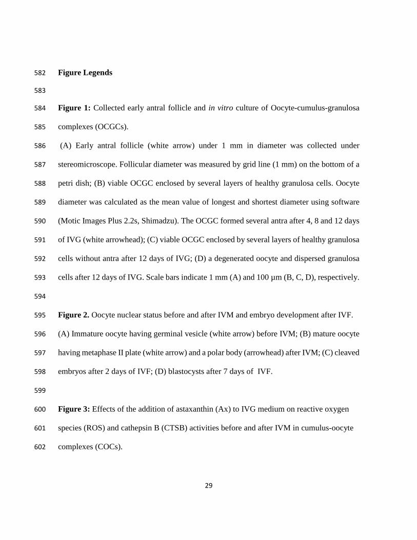
Figure 1: Collected early antral follicle and in vitro culture of Oocyte-cumulus-granulosa 584
complexes (OCGCs). 585
(A) Early antral follicle (white arrow) under 1 mm in diameter was collected under 586
stereomicroscope. Follicular diameter was measured by grid line (1 mm) on the bottom of a 587
petri dish; (B) viable OCGC enclosed by several layers of healthy granulosa cells. Oocyte 588
diameter was calculated as the mean value of longest and shortest diameter using software 589
(Motic Images Plus 2.2s, Shimadzu). The OCGC formed several antra after 4, 8 and 12 days 590
of IVG (white arrowhead); (C) viable OCGC enclosed by several layers of healthy granulosa 591
cells without antra after 12 days of IVG; (D) a degenerated oocyte and dispersed granulosa 592
cells after 12 days of IVG. Scale bars indicate 1 mm (A) and 100 µm (B, C, D), respectively. 593
594
Figure 2. Oocyte nuclear status before and after IVM and embryo development after IVF. 595
(A) Immature oocyte having germinal vesicle (white arrow) before IVM; (B) mature oocyte 596
having metaphase II plate (white arrow) and a polar body (arrowhead) after IVM; (C) cleaved 597
embryos after 2 days of IVF; (D) blastocysts after 7 days of IVF. 598
599
Figure 3: Effects of the addition of astaxanthin (Ax) to IVG medium on reactive oxygen 600
species (ROS) and cathepsin B (CTSB) activities before and after IVM in cumulus-oocyte 601
complexes (COCs). 602

30
(A) Quantification of the relative fluorescence intensity of ROS and CTSB activity. (B) 603
ROS and CTSB were detected as green and red fluorescence, respectively. The relative 604
fluorescence intensities of ROS and CTSB were measured using 44 and 49 COCs (3 605
replicates each), respectively. 606
** Asterisk indicates a significant difference between the groups (P < 0.01). 607
608
Figure 4: Viabilities, antrum formation by oocyte-cumulus-granulosa complexes (OCGCs) 609
during IVG cultures, and diameters of IVG oocytes obtained from early antral follicles in the 610
presence or absence of astaxanthin (Ax). 611
Percentages of viability and antrum formation were calculated based on the pooled data of 612
all replicates. 613
* Asterisk indicates a significant difference between experimental groups on the same days 614
(P < 0.05). 615
a,b,c Different letters indicate a significant difference between the days of the IVG culture in 616
the control group (P < 0.05). 617
x,y,z Different letters indicate a significant difference between the days of the IVG culture in 618
the group treated with Ax (P < 0.05). 619
620
Figure 5: Production of progesterone (P4) and estradiol-17β (E2) from OCGCs in the 621
presence or absence of astaxanthin (Ax) during the IVG culture. 622
* Asterisk indicates a significant difference between the experimental groups (P < 0.05). 623

31
a,b Different letters indicate a significant difference between the days of the IVG culture in 624
the control group (P < 0.05). 625
x,y,z Different letters indicate a significant difference between the days of the IVG culture in 626
the Ax-treated group (P < 0.05). 627
628

32
Table 1 629
Nuclear status of bovine in vitro-grown oocytes obtained from early antral follicles before maturation. 630
Treatment No. of oocytes
(replicates)
% meiotic stage
GV GVBD M I M II Deg GVBD-M II
Control 27 (3) 92.6 0 0 0 7.4 0
Ax 24 (3) 95.8 0 0 0 4.2 0
GV, germinal vesicle; GVBD, germinal vesicle breakdown; MI, metaphase I; MII, metaphase II, Deg; 631
degeneration. 632
633
634
635
Table 2 636
Effects of astaxanthin (Ax) during IVG culture on meiotic resumption after IVG in oocytes obtained 637
from early antral follicles. 638
639
Oocyte Ax No. of oocytes
(replicates)
% meiotic stage
GV GVBD M I M II Deg GVBD-M II
In vivo-grown - 66 (3) 4.5 1.5a 7.6a 84.8a 1.5a 93.9a
IVG 0 µM 76 (6) 9.2 9.2b 30.3b 38.2b 13.2b 77.6a
500 µM 110 (7) 3.6 6.4b 30.9b 52.7b 6.4b 90.0b
GV, germinal vesicle; GVBD, germinal vesicle breakdown; M I, metaphase I; M II, metaphase II, 640
Deg; degeneration. 641 ab Different superscripts within a column indicate a significant difference between groups (P < 0.05). 642
643
644
645
646

33
Table 3 647
Effects of astaxanthin (Ax) during IVG culture on cleavage and blastocyst production rates and cell 648
numbers in blastocysts derived from IVG oocytes. 649
Oocyte Ax No. of oocytes
(replicates) % cleavage % blastocyst Cell numbers in
blastocysts (n)
In vivo-grown - 87 (4) 80.8 ± 3.5a 42.9 ± 10.0a 154.8 ± 14.0a (37)
IVG 0 µM 73 (4) 58.9 ± 13.1b 16.4 ± 4.1c 107.4 ± 7.1c (12)
500 µM 81 (4) 65.9 ± 9.4b 23.6 ± 2 .2b 126.1 ± 11.1b (19)
Values are mean ± S.D. 650
Percentages of blastocysts are based on inseminated oocytes. 651 abc Different superscripts within a column indicate a significant difference between groups (P < 0.05). 652
653
(A)
D0 D4 D8 D12
(B)
(C) (D)
Figure 1
D12 D12

(D)
Figure 2
(C)
(A) (B)

Control Astaxanthin0.0
0.2
0.4
0.6
0.8
1.0
1.2
**
Rel
ativ
e R
OS
inte
nsity
Control Astaxanthin0.0
0.2
0.4
0.6
0.8
1.0
1.2
**
Rel
ativ
e C
TSB
act
ivity
inte
nsity
Figure 3
Control
Astaxanthin
(A)
(B)
ROS Dark field CTSB Bright field

0
20
40
60
80
100
ControlAstaxanthin
a
b
x yz
* **
c
OC
GC
via
bilty
(%
)
4 8 120
20
40
60
80
100
ab
*c
**
x
yz
IVG culture (days)
Antru
m fo
rmat
ion
(%)
Day 0 After IVM0
20
40
60
80
100
120
ControlAstaxanthin
Xayb
Ooc
yte
diam
eter
(µm
)
Figure 4

Day 0-
4
Day 4-
8
Day 8-
1202468
101214161820 Control
Astaxanthin
*a
*b
*a
xy yP 4
pro
duct
ion
(ng/
wel
l)
0-4 4-8 8-120.00.51.01.52.02.53.03.54.04.55.0
*b
z
y
aab
In vitro culture (days)
E 2 p
rodu
ctio
n (n
g/w
ell)
*x
Figure 5