Embed Size (px)
Citation preview
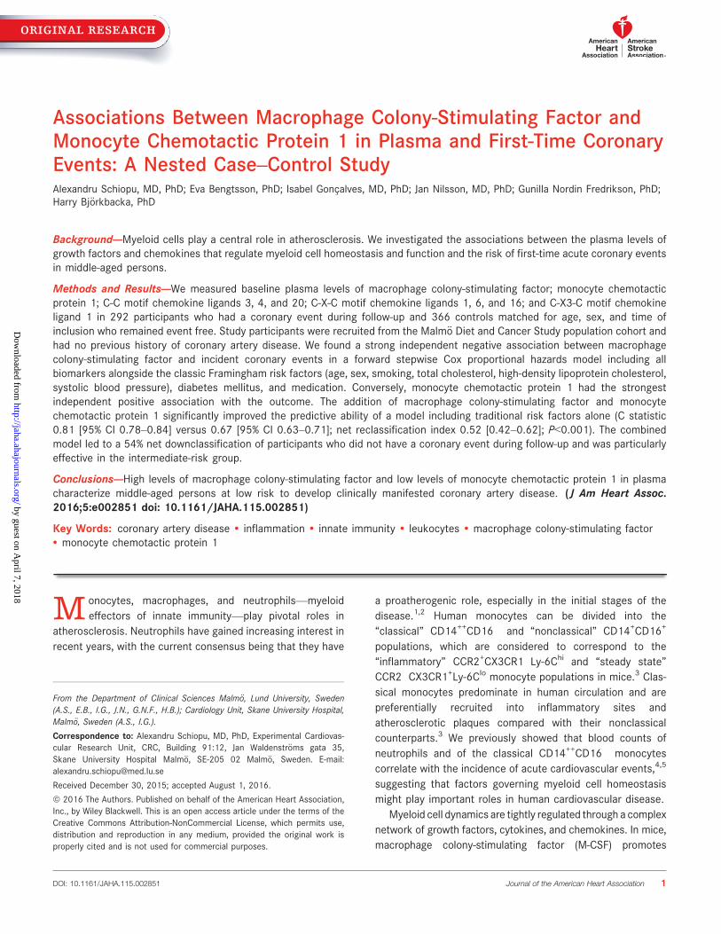
Associations Between Macrophage Colony-Stimulating Factor andMonocyte Chemotactic Protein 1 in Plasma and First-Time CoronaryEvents: A Nested Case–Control StudyAlexandru Schiopu, MD, PhD; Eva Bengtsson, PhD; Isabel Gonc�alves, MD, PhD; Jan Nilsson, MD, PhD; Gunilla Nordin Fredrikson, PhD;Harry Bj€orkbacka, PhD
Background-—Myeloid cells play a central role in atherosclerosis. We investigated the associations between the plasma levels ofgrowth factors and chemokines that regulate myeloid cell homeostasis and function and the risk of first-time acute coronary eventsin middle-aged persons.
Methods and Results-—We measured baseline plasma levels of macrophage colony-stimulating factor; monocyte chemotacticprotein 1; C-C motif chemokine ligands 3, 4, and 20; C-X-C motif chemokine ligands 1, 6, and 16; and C-X3-C motif chemokineligand 1 in 292 participants who had a coronary event during follow-up and 366 controls matched for age, sex, and time ofinclusion who remained event free. Study participants were recruited from the Malm€o Diet and Cancer Study population cohort andhad no previous history of coronary artery disease. We found a strong independent negative association between macrophagecolony-stimulating factor and incident coronary events in a forward stepwise Cox proportional hazards model including allbiomarkers alongside the classic Framingham risk factors (age, sex, smoking, total cholesterol, high-density lipoprotein cholesterol,systolic blood pressure), diabetes mellitus, and medication. Conversely, monocyte chemotactic protein 1 had the strongestindependent positive association with the outcome. The addition of macrophage colony-stimulating factor and monocytechemotactic protein 1 significantly improved the predictive ability of a model including traditional risk factors alone (C statistic0.81 [95% CI 0.78–0.84] versus 0.67 [95% CI 0.63–0.71]; net reclassification index 0.52 [0.42–0.62]; P<0.001). The combinedmodel led to a 54% net downclassification of participants who did not have a coronary event during follow-up and was particularlyeffective in the intermediate-risk group.
Conclusions-—High levels of macrophage colony-stimulating factor and low levels of monocyte chemotactic protein 1 in plasmacharacterize middle-aged persons at low risk to develop clinically manifested coronary artery disease. ( J Am Heart Assoc.2016;5:e002851 doi: 10.1161/JAHA.115.002851)
Key Words: coronary artery disease • inflammation • innate immunity • leukocytes • macrophage colony-stimulating factor• monocyte chemotactic protein 1
M onocytes, macrophages, and neutrophils—myeloideffectors of innate immunity—play pivotal roles in
atherosclerosis. Neutrophils have gained increasing interest inrecent years, with the current consensus being that they have
a proatherogenic role, especially in the initial stages of thedisease.1,2 Human monocytes can be divided into the“classical” CD14++CD16� and “nonclassical” CD14+CD16+
populations, which are considered to correspond to the“inflammatory” CCR2+CX3CR1�Ly-6Chi and “steady state”CCR2�CX3CR1+Ly-6Clo monocyte populations in mice.3 Clas-sical monocytes predominate in human circulation and arepreferentially recruited into inflammatory sites andatherosclerotic plaques compared with their nonclassicalcounterparts.3 We previously showed that blood counts ofneutrophils and of the classical CD14++CD16� monocytescorrelate with the incidence of acute cardiovascular events,4,5
suggesting that factors governing myeloid cell homeostasismight play important roles in human cardiovascular disease.
Myeloid cell dynamics are tightly regulated through a complexnetwork of growth factors, cytokines, and chemokines. In mice,macrophage colony-stimulating factor (M-CSF) promotes
From the Department of Clinical Sciences Malm€o, Lund University, Sweden(A.S., E.B., I.G., J.N., G.N.F., H.B.); Cardiology Unit, Skane University Hospital,Malm€o, Sweden (A.S., I.G.).
Correspondence to: Alexandru Schiopu, MD, PhD, Experimental Cardiovas-cular Research Unit, CRC, Building 91:12, Jan Waldenstr€oms gata 35,Skane University Hospital Malm€o, SE-205 02 Malm€o, Sweden. E-mail:[email protected]
Received December 30, 2015; accepted August 1, 2016.
ª 2016 The Authors. Published on behalf of the American Heart Association,Inc., by Wiley Blackwell. This is an open access article under the terms of theCreative Commons Attribution-NonCommercial License, which permits use,distribution and reproduction in any medium, provided the original work isproperly cited and is not used for commercial purposes.
DOI: 10.1161/JAHA.115.002851 Journal of the American Heart Association 1
ORIGINAL RESEARCH
by guest on April 7, 2018
http://jaha.ahajournals.org/D
ownloaded from

maturation of Ly-6Chi into Ly-6Clo monocytes,6 supports thesurvival of Ly-6Clo monocyte and macrophages in blood andtissues,7 and promotes an anti-inflammatory macrophage phe-notypewith important roles in tissue repair and homeostasis.8 Incontrast, granulocyte and granulocyte-macrophage colony-stimulating factors induce proinflammatory activation of neu-trophils, macrophages, dendritic cells, and eosinophils.8,9 Understeady state conditions, only M-CSF can be consistentlymeasured in the circulation, whereas granulocyte and granulo-cyte-macrophage colony-stimulating factors increase andbecome measurable mainly during inflammatory reactions.9
Besides growth factors, a complex network of chemokinescontrol myeloid cell production, trafficking, and function andhave been found to be involved in the pathogenesis ofcardiovascular disease.2,10 Monocyte chemotactic protein 1(MCP-1; also known as C-C motif chemokine ligand 2 [CCL2]) isthe most important regulator of monocyte trafficking and hasbeen shown to have potent proatherogenic properties.2,11,12
Similarly, fractalkine (also known as C-X3-C motif chemokineligand 1 [CX3CL1]), macrophage inflammatory protein 1a (MIP-1a; also known as CCL3), macrophage inflammatory protein 1b(MIP-1b; also known as CCL4), CCL20, C-X-C motif chemokineligand 1 (CXCL1) and CXCL16 promote monocyte and neu-trophil recruitment and survival, supporting atherogenesis andplaque vulnerability in mice.2,13,14 The CXCR1 and CXCR2ligand CXCL6 (granulocyte chemotactic protein 2) is alsoinvolved in neutrophil recruitment,15 but its role in cardiovas-cular disease has not been examined previously.
Much of the knowledge related to myeloid mediators andthe roles they play in cardiovascular disease has been gainedfrom animal studies, and human data are lacking to a largeextent. The aim of our study was to identify whether plasmalevels of M-CSF, MCP-1, CCL3, CCL4, CCL20, CXCL1, CXCL6,CXCL16, and CX3CL1 can predict the incidence of first-timecoronary events and improve the currently used models forcardiovascular risk prediction. To this end, we studied thecorrelations between baseline levels of these proteins andincident coronary events in plasma samples collected from292 middle-aged persons with no previous history of coronaryartery disease who had an acute coronary event during follow-up and 366 sex- and age-matched controls who remainedevent free. Participants were recruited from the Malm€o Dietand Cancer Study (MDC) cohort.
Materials and Methods
Study CohortThe MDC has a population-based prospective epidemiologicalcohort of 28 449 participants enrolled between 1991 and1996.16 Between October 1991 and February 1994, everyother participant was invited to take part in a substudy of the
epidemiology of cardiovascular disease (MDC-CV), yielding acohort of 6103 participants.17 All participants were followedfrom the baseline examination until first hospitalization for anacute coronary event, death, emigration, or June 30, 2009.Incident cases of patients experiencing coronary events wereretrieved by data linkage to the Swedish Hospital DischargeRegister and the Cause of Death Registry of Sweden.Following prior exclusion of 102 participants with prevalentnonfatal myocardial infarction (1.7% of the MDC-CV cohort),402 incident coronary events (6.6% of the MDC-CV cohort)were identified during the follow-up period. A coronary eventwas defined as a nonfatal or fatal myocardial infarction on thebasis of International Classification of Diseases, Ninth Revision(ICD9) code 410 and ICD10 code I21. Death due to ischemicheart disease was defined on the basis of codes 412 and 414(ICD9) or I22, I23, and I25 (ICD10). We matched the incidentcoronary cases with 402 coronary event–free control partic-ipants of the same age, sex, and time of participation in thebaseline examination (�6 months). These matching variableswere selected because of their nonmodifiable nature. Nopaired analyses were used in the analysis of the data. Weexcluded 29 cases and 8 controls (4.6% of the case–controlcohort) because of prevalent revascularization (percutaneouscoronary intervention or coronary artery bypass grafting) orincident revascularization before the first coronary event incases or before the end of the follow-up period in controls.Furthermore, 81 cases and 28 controls (13.6% of the case–control cohort) were excluded because of incomplete clinicaldata or missing plasma samples, yielding a cohort consistingof 292 cases and 366 controls (81.8% of the case–controlcohort). The study design and exclusion details are describedin Figure 1. Although all participants were deemed to beapparently healthy at the time of inclusion, we cannot rule outthe possibility that some participants might have had apotential history of chronic inflammatory conditions such asautoimmune disease, human immunodeficiency virus, cancer,or thrombosis. Because of unavailable information, we wereunable to identify and exclude these participants from thestudy. The study was approved by the regional ethics reviewboard and was conducted in accordance with the Declarationof Helsinki. All participants gave informed written consent.
Baseline AssessmentInformation on baseline characteristics was collected fromself-administered questionnaires and clinical examination.Smoking habits were categorized into never or formersmokers (who quit smoking at least 1 year before theexamination) and current smokers. Diabetes mellitus wasdefined as fasting whole blood glucose >6.1 mmol/L (corre-sponding to a threshold of 7.0 mmol/L in fasting plasmaglucose), self-reported physician diagnosis of diabetes
DOI: 10.1161/JAHA.115.002851 Journal of the American Heart Association 2
M-CSF and MCP-1 and First-Time Coronary Events Schiopu et alORIG
INALRESEARCH
by guest on April 7, 2018
http://jaha.ahajournals.org/D
ownloaded from

mellitus, or use of antidiabetic medication. Blood pressurewas measured twice in the right arm after a 10-minute rest.The average of the 2 measurements was used. Hypertensionwas defined as systolic blood pressure ≥140 mm Hg ordiastolic blood pressure ≥90 mm Hg or the use of bloodpressure–lowering medication. Blood samples were drawnafter overnight fasting. Fasting venous blood glucose, serumcholesterol, low-density lipoprotein cholesterol, high-densitylipoprotein cholesterol, C-reactive protein (CRP), and triglyc-erides were analyzed with standard methods at the clinicallaboratory of Malm€o University Hospital.
Analysis of Myeloid Markers in PlasmaMyeloid markers were analyzed in plasma by the proximityextension assay technique using the Proseek Multiplex
CVD96x96 reagents kit (Olink Bioscience) at the ClinicalBiomarkers Facility, Science for Life Laboratory, in Uppsala,Sweden. Briefly, oligonucleotide-labeled antibody probe pairswere allowed to bind to their respective targets present in theplasma sample. Addition of DNA polymerase led to extensionand joining of the 2 oligonucleotides and formation of apolymerase chain reaction template. Universal primers wereused to preamplify the DNA templates in parallel. Finally, theindividual DNA sequences were detected and quantified usingspecific primers in a microfluidic real-time quantitativepolymerase chain reaction chip (96.96, Dynamic Arrayintegrated fluidic circuit, Fluidigm Biomark; Fluidigm Corp).The chip was run with a Biomark HD instrument.18 Therespective intra- and interassay variations were 7% and 18%for MCP-1, 7% and 12% for M-CSF, 10% and 18% for CCL3, 8%and 12% for CCL4, 8% and 9% for CCL20, 6% and 13% for
All men and women aged45–73 years living in Malmö,
Sweden (n=70 000)
Participating in the baselineexamination of the MDC
cohort (n=28 449)
Coronary events:fatal or nonfatal MI or death
due to ischemic heart diseaseuntil June 2009 (n=402)
Controls matched on age, sexand date of baselineexamination (n=402)
Enrolled in the CVD substudybetween Nov 1991 and Feb 1994
(n=6103)
102 prevalent nonfatal MI excluded
Cases (n=292) Controls (n=366)
12 prevalent revascularizations excluded 0 prevalent revascularizations excluded
17 incident revascularizations occurringbefore the first coronary event excluded
8 incident revascularizations occurringduring follow-up excluded
18 missing plasma samples 2 missing plasma samples
Missing data for total cholesterol (3), HDL(12), diabetes mellitus (1) and smokingstatus (47)
Missing data for total cholesterol (0), HDL (3),diabetes mellitus (0) and smoking status (23)
Figure 1. Study design. Diagram outlining how the matched sample of the case–control cohort wasobtained. *Revascularization indicates coronary artery bypass grafting or percutaneous coronaryintervention. CVD indicates cardiovascular disease; HDL, high-density lipoprotein; MDC, Malm€o Diet andCancer Study; MI, myocardial infarction.
DOI: 10.1161/JAHA.115.002851 Journal of the American Heart Association 3
M-CSF and MCP-1 and First-Time Coronary Events Schiopu et alORIG
INALRESEARCH
by guest on April 7, 2018
http://jaha.ahajournals.org/D
ownloaded from

CXCL1, 8% and 12% for CXCL6, 10% and 14% for CXCL16, and9% and 14% for CX3CL1. Data analysis was performed by apreprocessing normalization procedure using Olink Wizard forGenEx (Multid Analyses). All data are presented as arbitraryunits. General calibrator curves to calculate the approximateconcentrations are available on the Olink website (http://www.olink.com).
StatisticsDifferences in baseline characteristics between the case andcontrol groups were evaluated with Mann–Whitney nonpara-metric tests for nonnormally distributed continuous data andwith Student t tests for normally distributed continuous data.Differences in categorical data were calculated with v2 tests.Nonnormally distributed continuous data are presented asmedians and interquartile ranges, and normally distributedcontinuous data are presented as mean�SD. Nonnormallydistributed variables were natural logarithm transformed, andbiomarker variables were standardized before inclusion intoregression analyses.
Independent associations among myeloid biomarkers,baseline variables, and incident coronary artery disease wereevaluated by calculating partial correlations between eachpair of variables and constructing a partial correlation networkthat included correlations with a Bonferroni-adjusted P<0.05.Calculations were performed in R v. 3.1.1 (R Foundation forStatistical Computing), and the partial correlation networkwas drawn using the yEd Graph Editor software v. 3.14(yWorks GmbH), with a hierarchical layout algorithm.
Cox proportional hazards regression models were used toevaluate risk factor–adjusted hazard ratios and 95% CIs foreach biomarker. We used 2 models for the Cox regressionanalyses. Model A included the traditional risk factors used inthe Framingham risk score (age, sex, smoking, total choles-terol, high-density lipoprotein cholesterol, and systolic bloodpressure) as well as diabetes mellitus, blood pressure–lowering medication, and lipid-lowering medication. Model Bwas additionally adjusted for potential confounders thatdiffered significantly between cases and controls at baseline:diastolic blood pressure, body mass index, triglycerides, low-density lipoprotein, creatinine-based estimated glomerularfiltration rate, CRP, and white blood cell count. The stepwiseforward selection of variables also included MCP-1, M-CSF,CCL3, CCL4, CCL20, CXCL1, CXCL6, CXCL16, CX3CL1, andCRP in both models. P values were adjusted for multipletesting using the Bonferroni method. Plots of the hazardfunction in different groups over time did not indicate that theproportional hazards assumption was violated.
Metrics of risk discrimination were assessed using logisticregression analysis adjusted for matching variables to takethe matched case–control design into account. To estimate
the risk in the original population cohort, the risk modelcalculated from case–control data was adjusted for theincidence rate in the original cohort and for the case–controlratio as described by Huang et al19 and Pencina et al.20
Receiver operating curves and C statistics, net reclassificationindex, and integrated discrimination improvement were cal-culated and used to compare the performance of the modelbased on traditional risk factors with models includingtraditional risk factors and biomarkers for the prediction ofincident coronary events. To be applicable to the case–controldesign, net reclassification index calculations were extended,as suggested by Pencina et al.20 Statistical differences in theareas under the receiver operating characteristic curves werecalculated according to DeLong et al.21
Statistical analyses were carried out using IBM SPSSStatistics v. 22 (IBM Corp) and R v. 3.1.1 (and the R packagespROC, PredictABEL, Rcmdr, and qgraph).
ResultsThe clinical characteristics of the study cohort at baseline aresummarized in Table 1. The case group contained a largerpercentage of participants who were smokers, were over-weight (body mass index ≥25), were diabetic, and hadhypertension compared with the sex- and age-matchedcontrol group. Cases also had higher total cholesterol,triglycerides, low-density lipoprotein cholesterol, CRP, andwhite blood cell counts, whereas high-density lipoproteincholesterol levels were lower than in controls. A higherpercentage of participants within the case group receivedantidiabetic medication at baseline. The median time frombaseline to the occurrence of a coronary event was10.6 years (interquartile range 6.4–13.5 years).
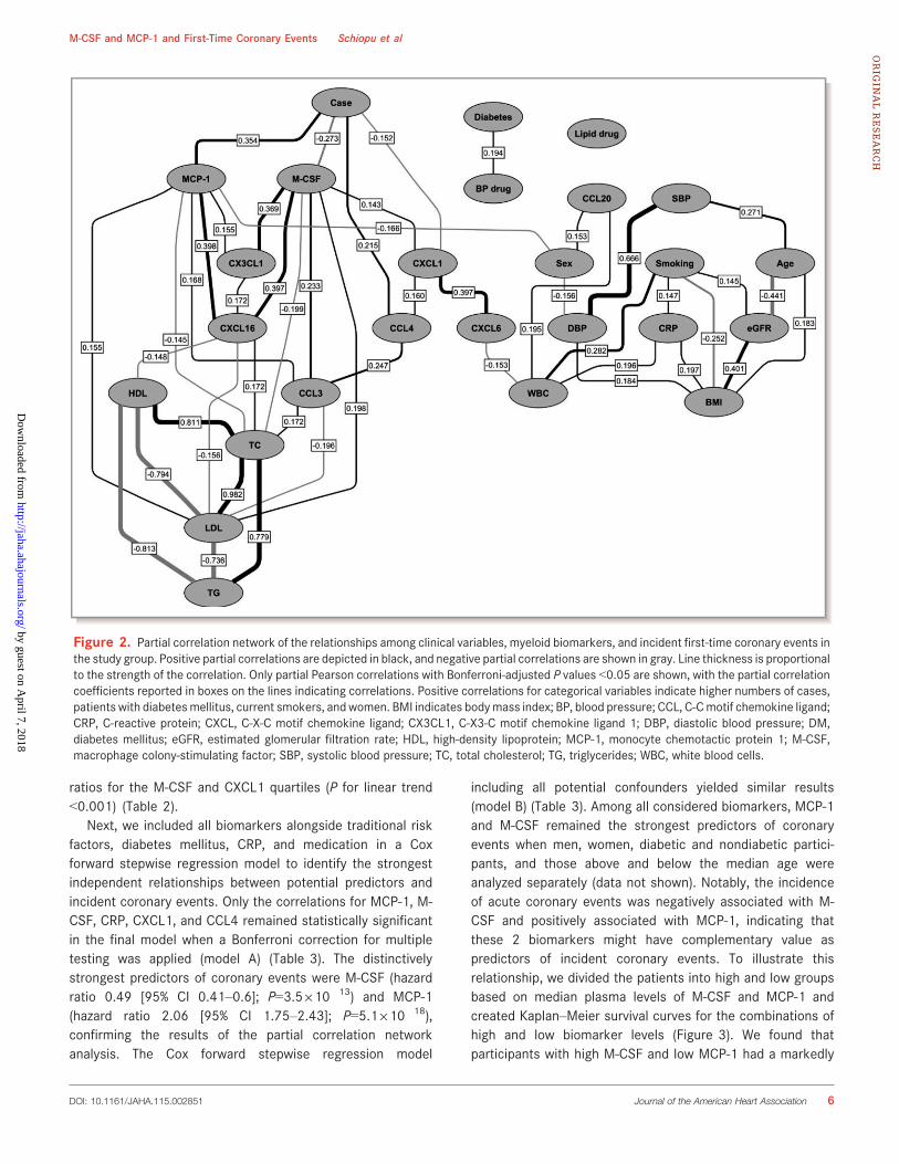
Myeloid Markers in Plasma and Incidence of First-Time Acute Coronary EventsTo concomitantly explore the independent associationsamong myeloid biomarkers, baseline clinical variables, andincident coronary artery disease, partial correlations werecalculated between each pair of variables, controlling for all ofthe others, and a partial correlation network was constructed(Figure 2). M-CSF and CXCL1 showed independent negativeassociations with the case–control variable, whereas MCP-1and CCL4 displayed positive associations (Bonferroni-adjusted P<0.05).
When the 9 biomarkers were tested individually, M-CSF,MCP-1, and CXCL1 were significantly associated withincident coronary events in Cox proportional hazard modelsadjusted for the traditional cardiovascular risk factorsincluded in the Framingham risk score (age, sex, smoking,
DOI: 10.1161/JAHA.115.002851 Journal of the American Heart Association 4
M-CSF and MCP-1 and First-Time Coronary Events Schiopu et alORIG
INALRESEARCH
by guest on April 7, 2018
http://jaha.ahajournals.org/D
ownloaded from

total cholesterol, high-density lipoprotein, systolic bloodpressure) and for blood pressure–lowering medication,lipid-lowering medication, and diabetes mellitus (model A)(Table 2). The associations remained significant followingfurther adjustment for potential confounders that differedbetween cases and controls at baseline: diastolic blood
pressure, body mass index, triglycerides, low-density lipopro-tein, estimated glomerular filtration rate, CRP, and whiteblood cell count (model B) (Table 2). The relationshipsbetween biomarkers and outcome were linear, as demon-strated by gradually increasing hazard ratios for eachincreasing MCP-1 quartile and gradually decreasing hazard
Table 1. Baseline Characteristics of the Case–Control Cohort
Cases (n=292)* Controls (n=366)* P Value†
Age at screening, y 61.8 (57.3–64.9) 61.4 (57.0–64.3) 0.360
Male sex (% male) 172 (58.9%) 217 (59.3%) 0.920
BMI ≥25 (%)‡ 176 (60.5%) 194 (53.2%) 0.060
Current smoker (%) 102 (34.9%) 93 (25.4%) 0.008
Diabetes mellitus (%)§ 49 (16.8%) 27 (7.4%) 1.8910�4
Hypertension (%)|| 229 (78.4%) 249 (68.0%) 0.003
Medication
Antidiabetic (%) 21 (7.2%) 5 (1.4%) 1.4910�4
Lipid-lowering (%) 3 (1.0%) 5 (1.4%) 0.694
Blood pressure-lowering (%) 57 (19.5%) 59 (16.1%) 0.255
Clinical parameters
Systolic blood pressure, mm Hg 150�19 143�18 5910�6
Diastolic blood pressure, mm Hg 91�10 88�9 2.8910�4
Cholesterol, mmol/L 6.3�1.0 6.2�1.1 0.050
Triglycerides, mmol/L 1.3 (1.0–1.9) 1.2 (0.9–1.7) 0.037
HDL, mmol/L 1.2�0.3 1.4�0.4 8910�6
LDL, mmol/L 4.4�1.0 4.2�1.0 0.005
CRP, mg/L 1.9 (0.9–3.4) 1.3 (0.7–2.6) 6.1910�6
White blood cell count, 9109/L 6.4 (5.3–7.4) 5.7 (5.0–6.8) 7.7910�6
eGFR, mL/min/1.73 m2 76�15 75�16 0.894
Myeloid markers
M-CSF, au 530 (440–600) 580 (490–680) 1.3910�7
MCP-1, au 22 (17–29) 18 (15–22) 9.9910�5
CXCL1, au 71 (56–94) 82 (64–114) 1.1910�14
CCL4, au 125 (96–171) 105 (82–146) 8.0910�6
CCL3, au 3.2 (2.9–4.1) 3.2 (2.8–3.9) 0.939
CXCL6, au 119 (89–157) 122 (94–167) 0.308
CXCL16, au 13 (11–16) 12 (10–16) 0.007
CX3CL1, au 45 (37–58) 50 (40–62) 0.003
CCL20, au 52 (39–78) 51 (36–74) 0.453
au indicates arbitrary units; BMI, body mass index; CCL, C-C motif chemokine ligand; CRP, C-reactive protein; CXCL, C-X-C motif chemokine ligand; CX3CL1, C-X3-C motif chemokine ligand1; eGFR, estimated glomerular filtration rate; HDL, high-density lipoprotein; LDL, low-density lipoprotein; MCP-1, monocyte chemotactic protein 1; M-CSF, macrophage colony-stimulatingfactor.*Data are presented as number (percentage of cases/controls) for categorical data, mean�SD for normally distributed continuous variables, and median (interquartile range) fornonnormally distributed variables.†Mann–Whitney test was used for nonnormally distributed data, t test was used for normally distributed data, and v2 test was used for categorical data.‡BMI was calculated as weight/height2 (kg/m2) and categorized as normal weight (BMI <25) and overweight/obese (BMI ≥25).§Positive questionnaire, medication, or glucose ≥6.1 (mmol/L).||Blood pressure ≥140/90 mm Hg or treatment.
DOI: 10.1161/JAHA.115.002851 Journal of the American Heart Association 5
M-CSF and MCP-1 and First-Time Coronary Events Schiopu et alORIG
INALRESEARCH
by guest on April 7, 2018
http://jaha.ahajournals.org/D
ownloaded from
ratios for the M-CSF and CXCL1 quartiles (P for linear trend<0.001) (Table 2).
Next, we included all biomarkers alongside traditional riskfactors, diabetes mellitus, CRP, and medication in a Coxforward stepwise regression model to identify the strongestindependent relationships between potential predictors andincident coronary events. Only the correlations for MCP-1, M-CSF, CRP, CXCL1, and CCL4 remained statistically significantin the final model when a Bonferroni correction for multipletesting was applied (model A) (Table 3). The distinctivelystrongest predictors of coronary events were M-CSF (hazardratio 0.49 [95% CI 0.41–0.6]; P=3.5910�13) and MCP-1(hazard ratio 2.06 [95% CI 1.75–2.43]; P=5.1910�18),confirming the results of the partial correlation networkanalysis. The Cox forward stepwise regression model
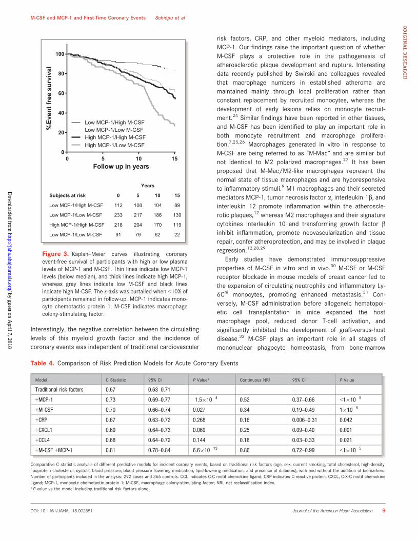
including all potential confounders yielded similar results(model B) (Table 3). Among all considered biomarkers, MCP-1and M-CSF remained the strongest predictors of coronaryevents when men, women, diabetic and nondiabetic partici-pants, and those above and below the median age wereanalyzed separately (data not shown). Notably, the incidenceof acute coronary events was negatively associated with M-CSF and positively associated with MCP-1, indicating thatthese 2 biomarkers might have complementary value aspredictors of incident coronary events. To illustrate thisrelationship, we divided the patients into high and low groupsbased on median plasma levels of M-CSF and MCP-1 andcreated Kaplan–Meier survival curves for the combinations ofhigh and low biomarker levels (Figure 3). We found thatparticipants with high M-CSF and low MCP-1 had a markedly
Figure 2. Partial correlation network of the relationships among clinical variables, myeloid biomarkers, and incident first-time coronary events inthe study group. Positive partial correlations are depicted in black, and negative partial correlations are shown in gray. Line thickness is proportionalto the strength of the correlation. Only partial Pearson correlations with Bonferroni-adjusted P values <0.05 are shown, with the partial correlationcoefficients reported in boxes on the lines indicating correlations. Positive correlations for categorical variables indicate higher numbers of cases,patients with diabetesmellitus, current smokers, and women. BMI indicates bodymass index; BP, blood pressure; CCL, C-Cmotif chemokine ligand;CRP, C-reactive protein; CXCL, C-X-C motif chemokine ligand; CX3CL1, C-X3-C motif chemokine ligand 1; DBP, diastolic blood pressure; DM,diabetes mellitus; eGFR, estimated glomerular filtration rate; HDL, high-density lipoprotein; MCP-1, monocyte chemotactic protein 1; M-CSF,macrophage colony-stimulating factor; SBP, systolic blood pressure; TC, total cholesterol; TG, triglycerides; WBC, white blood cells.
DOI: 10.1161/JAHA.115.002851 Journal of the American Heart Association 6
M-CSF and MCP-1 and First-Time Coronary Events Schiopu et alORIG
INALRESEARCH
by guest on April 7, 2018
http://jaha.ahajournals.org/D
ownloaded from

improved coronary event-free survival compared with the restof the cohort (log-rank P for trend <0.001).
M-CSF and MCP-1 Improve the DiscriminativeAbility of a Traditional Risk Factor Model forCoronary Artery DiseaseTo test whether M-CSF, MCP-1, CXCL1, or CCL4 couldimprove the predictive value of the traditional risk factors for
first-time coronary events, receiver operating characteristiccurves were constructed for binary logistic regression modelsincluding traditional risk factors (age, sex, smoking, totalcholesterol, high-density lipoprotein, systolic blood pressure,and diabetes mellitus), blood pressure–lowering medication,and lipid-lowering medication, as well as M-CSF, MCP-1,CXCL1, CCL4, and CRP alone or in combination (Table 4 andFigure 4). The addition of M-CSF or MCP-1 alone significantlyimproved the discriminative ability of the traditional risk factor
Table 2. Correlations Between Baseline Biomarker Levels and Incident Coronary Events During Follow-up
Biomarker HR* 95% CINominalP Value
CorrectedP Value†
HR vs Q1 (95% CI) LinearTrendP Value
CorrectedP Value†Q1 Q2 Q3 Q4
MCP-1
Model A‡ 1.50 1.33–1.69 2.8910�11 2.8910�10 1 1.14 (0.78–1.65) 1.57 (1.10–2.26) 2.26 (1.62–3.16) 1.1910�7 1.1910�6
Model B§ 1.49 1.32–1.69 2.2910�10 3.7910�9 1 1.12 (0.76–1.63) 1.56 (1.08–2.27) 2.22 (1.56–3.14) 5.4910�7 9.1910�6
M-CSF
Model A 0.67 0.59–0.76 2.5910�10 2.5910�9 1 0.61 (0.45–0.83) 0.51 (0.37–0.69) 0.31 (0.21–0.44) 4.8910�11 4.8910�10
Model B 0.64 0.56–0.73 2.9910�11 5.0910�10 1 0.56 (0.41–0.78) 0.46 (0.34–0.64) 0.27 (0.19–0.40) 8.9910�12 1.5910�10
CXCL1
Model A 0.76 0.68–0.86 1.1910�5 1.1910�4 1 0.83 (0.62–1.13) 0.64 (0.46–0.90) 0.44 (0.31–0.62) 1.2910�6 1.2910�5
Model B 0.76 0.67–0.86 1.5910�5 2.6910�4 1 0.84 (0.62–1.15) 0.61 (0.43–0.86) 0.44 (0.30–0.63) 2910�6 3.4910�5
CX3CL1
Model A 0.84 0.75–0.95 0.005 0.053 1 0.80 (0.58–1.09) 0.69 (0.50–0.95) 0.61 (0.44–0.84) 0.002 0.016
Model B 0.83 0.73–0.94 0.004 0.063 1 0.74 (0.54–1.03) 0.66 (0.48–0.92) 0.58 (0.41–0.82) 0.001 0.020
CCL4
Model A 1.13 1.01–1.27 0.030 0.301 1 1.71 (1.19–2.45) 1.77 (1.23–2.55) 1.75 (1.21–2.53) 0.006 0.060
Model B 1.14 1.01–1.28 0.028 0.476 1 1.65 (1.14–2.39) 1.80 (1.24–2.62) 1.74 (1.19–2.53 0.007 0.111
CCL3
Model A 0.96 0.83–1.11 0.581 1 1 1.22 (0.84–1.78) 0.65 (0.43–0.98) 1.00 (0.68–1.49) 0.331 1
Model B 0.94 0.80–1.11 0.474 1 1 1.17 (0.78–1.73) 0.61 (0.39–0.94) 0.93 (0.61–1.41) 0.184 1
CCL20
Model A 0.91 0.80–1.03 0.140 1 1 1.15 (0.82–1.60) 0.87 (0.62–1.23) 0.85 (0.60–1.21) 0.158 1
Model B 0.91 0.80–1.04 0.165 1 1 1.17 (0.84–1.64) 0.87 (0.61–1.24) 0.88 (0.61–1.26) 0.232 1
CXCL6
Model A 0.93 0.83–1.05 0.244 1 1 0.88 (0.64–1.21) 0.99 (0.72–1.37) 0.77 (0.55–1.07) 0.204 1
Model B 0.94 0.83–1.06 0.321 1 1 0.87 (0.63–1.22) 0.99 (0.71–1.40) 0.80 (0.56–1.13) 0.328 1
CXCL16
Model A 1.12 0.99–1.26 0.068 0.683 1 1.15 (0.82–1.62) 1.05 (0.75–1.48) 1.09 (0.77–1.53) 0.789 1
Model B 1.10 0.97–1.25 0.144 1 1 1.14 (0.81–1.62) 0.97 (0.68–1.39) 1.04 (0.73–1.47) 0.921 1
Number of participants included in the analysis: 292 cases and 366 controls. CCL indicates C-C motif chemokine ligand; CXCL, C-X-C motif chemokine ligand; CX3CL1, C-X3-C motifchemokine ligand 1; HDL, high-density lipoprotein; HR, hazard ratio; MCP-1, monocyte chemotactic protein 1; M-CSF, macrophage colony-stimulating factor; Q, quartile.*Per 1-SD increase in the respective variable.†After Bonferroni correction for multiple testing. Model A: 10 tests, critical cutoff P=0.005. Model B: 17 tests, critical cutoff P=0.003.‡Model A: Cox regression analysis adjusted for age, sex, diabetes mellitus, current smoking, total cholesterol, HDL, systolic blood pressure, blood pressure–lowering medication, and lipid-lowering medication.§Model B: Cox regression analysis adjusted for age, sex, diabetes, current smoking, total cholesterol, HDL, systolic blood pressure, blood pressure–lowering medication, lipid-loweringmedication, diastolic blood pressure, body mass index, triglycerides, low-density lipoprotein, estimated glomerular filtration rate, C-reactive protein, and white blood cell count.
DOI: 10.1161/JAHA.115.002851 Journal of the American Heart Association 7
M-CSF and MCP-1 and First-Time Coronary Events Schiopu et alORIG
INALRESEARCH
by guest on April 7, 2018
http://jaha.ahajournals.org/D
ownloaded from

model for coronary artery disease risk, whereas addition ofCXCL1, CCL4, or CRP alone did not improve discrimination.The addition of MCP-1 to the model already including M-CSFand traditional risk factors further improved discrimination(P=1.1910�10). The model including M-CSF and MCP-1alongside traditional risk factors had a significantly improvedC statistic compared with traditional risk factors alone (0.81[95% CI 0.78–0.85] versus 0.67 [95% CI 0.62–0.70];P=6.6910�13).
We also performed reclassification analyses for 3 prede-fined risk categories for incident coronary events: <10% (lowrisk), 10% to 20% (moderate risk), and >20% (high risk)(Table 5). The addition of M-CSF and MCP-1 significantlyimproved the predictive value of the traditional risk factormodel, with a net reclassification index of 0.52 (95% CI 0.42–0.62; P<0.001) and integrated discrimination improvement of0.18 (95% CI 0.15–0.21; P<0.001). Of 86 participants withlow coronary event risk according to the traditional model, 8(9.3%) were correctly upclassified and 6 (7%) were incorrectlyupclassified after the addition of MCP-1 and M-CSF. Amongparticipants in the intermediate-risk group, 146 were correctlydownclassified and 61 were correctly upclassified (totalcorrect reclassification 207 of 292, 71%) compared with 26incorrectly downclassified and 15 incorrectly upclassified(total incorrect reclassification 41 of 292, 14%). Within thehigh-risk group, we recorded 73 of 280 (26%) with correctdownclassification compared with 50 of 280 (18%) withincorrect downclassification. Taken together, these resultssuggest that addition of M-CSF and MCP-1 to the traditionalcardiovascular risk factors for prediction of first-time coronaryevents is useful mainly in the intermediate-risk population,leading to correct net total reclassification of 166 of 292(57%) participants in this category. Analysis of reclassificationin cases and controls revealed that the addition of M-CSF and
MCP-1 was particularly effective in the control group, leadingto a net downclassification of 54% of participants who did nothave a coronary event during follow-up. In the coronary casegroup, the combined model led to a minimal net downclas-sification of 2% of participants (Table 5).
DiscussionWe are the first to compare the ability of several mediatorsrelated to myeloid cell homeostasis and function (M-CSF,MCP-1, CCL3, CCL4, CCL20, CXCL1, CXCL6, CXCL16, andCX3CL1) to predict first-time acute coronary events in middle-aged persons with no previous history of coronary arterydisease. We identified, for the first time, a strong independentnegative association between M-CSF and coronary arterydisease risk and confirmed the previously described positiverelationship between MCP-1 and coronary events in thispopulation.22,23 Participants at low risk of having acutecoronary events were characterized by high levels of M-CSFand low levels of MCP-1 in plasma. Combining M-CSF andMCP-1 with the classic cardiovascular risk factors included inthe Framingham risk score improved prediction accuracy inthe intermediate-risk group and led to correct net downclas-sification of approximately half of the participants that did nothave a coronary event during follow-up. Nevertheless, themodel did not improve identification of participants whosubsequently developed clinically manifested coronary arterydisease.
M-CSF and Coronary Artery Disease RiskTo our knowledge, this study is the first on the potential roleof plasma M-CSF as a biomarker of cardiovascular risk inpersons with no clinical evidence of coronary artery disease.
Table 3. Independent Predictors of Incident Coronary Events in the Cohort
Step
Model A Model B
Variable HR (95% CI)* Nominal P Value Corrected P Value† Variable HR (95% CI)* Nominal P Value Corrected P Value†
1 MCP-1 2.06 (1.75–2.43) 5.1910�18 9.8910�17 MCP-1 1.93 (1.59–2.34) 3.7910�11 9.4910�10
2 M-CSF 0.49 (0.41–0.6) 3.5910�13 6.7910�12 M-CSF 0.41 (0.33–0.52) 8.2910�14 2.1910�12
3 CRP 1.38 (1.19–1.6) 2.9910�5 5.5910�4 CRP 1.33 (1.14–1.55) 3.5910�4 0.009
4 CXCL1 0.73 (0.63–0.86) 1.1910�5 0.002 CXCL1 0.76 (0.65–0.9) 0.001 0.025
5 CCL4 1.29 (1.11–1.49) 7.0910�4 0.013 CCL4 1.3 (1.12–1.52) 7.0910�4 0.018
Final model of a Cox proportional hazards regression analysis with stepwise forward selection of variables. Only variables with an adjusted P value <0.05 after Bonferroni correction areshown. Number of participants included in the analysis: 292 cases and 366 controls. Variables not retained in the final model A: age, sex, diabetes, current smoking, systolic bloodpressure, total cholesterol, HDL, blood pressure–lowering medication, lipid-lowering medication, CCL3, CCL20, CX3CL1, CXCL6, and CXCL16. Variables not retained in the final model B:age, sex, diabetes mellitus, current smoking, systolic blood pressure, diastolic blood pressure, total cholesterol, blood pressure–lowering medication, lipid-lowering medication, body massindex, low-density lipoprotein, HDL, triglycerides, estimated glomerular filtration rate, white blood cells, CCL3, CCL20, CX3CL1, CXCL6, and CXCL16. CCL indicates C-C motif chemokineligand; CRP, C-reactive protein; CXCL, C-X-C motif chemokine ligand; CX3CL1, C-X3-C motif chemokine ligand 1; HDL, high-density lipoprotein; MCP-1, monocyte chemotactic protein-1; M-CSF, macrophage colony-stimulating factor.*Per 1-SD increase in the respective variable.†After Bonferroni correction for multiple testing. Model A: 19 tests, critical cutoff P=0.003. Model B: 25 tests, critical cutoff P=0.002.
DOI: 10.1161/JAHA.115.002851 Journal of the American Heart Association 8
M-CSF and MCP-1 and First-Time Coronary Events Schiopu et alORIG
INALRESEARCH
by guest on April 7, 2018
http://jaha.ahajournals.org/D
ownloaded from
Interestingly, the negative correlation between the circulatinglevels of this myeloid growth factor and the incidence ofcoronary events was independent of traditional cardiovascular
risk factors, CRP, and other myeloid mediators, includingMCP-1. Our findings raise the important question of whetherM-CSF plays a protective role in the pathogenesis ofatherosclerotic plaque development and rupture. Interestingdata recently published by Swirski and colleagues revealedthat macrophage numbers in established atheroma aremaintained mainly through local proliferation rather thanconstant replacement by recruited monocytes, whereas thedevelopment of early lesions relies on monocyte recruit-ment.24 Similar findings have been reported in other tissues,and M-CSF has been identified to play an important role inboth monocyte recruitment and macrophage prolifera-tion.7,25,26 Macrophages generated in vitro in response toM-CSF are being referred to as “M-Mac” and are similar butnot identical to M2 polarized macrophages.27 It has beenproposed that M-Mac/M2-like macrophages represent thenormal state of tissue macrophages and are hyporesponsiveto inflammatory stimuli.9 M1 macrophages and their secretedmediators MCP-1, tumor necrosis factor a, interleukin 1b, andinterleukin 12 promote inflammation within the atheroscle-rotic plaques,12 whereas M2 macrophages and their signaturecytokines interleukin 10 and transforming growth factor binhibit inflammation, promote neovascularization and tissuerepair, confer atheroprotection, and may be involved in plaqueregression.12,28,29
Early studies have demonstrated immunosuppressiveproperties of M-CSF in vitro and in vivo.30 M-CSF or M-CSFreceptor blockade in mouse models of breast cancer led tothe expansion of circulating neutrophils and inflammatory Ly-6Chi monocytes, promoting enhanced metastasis.31 Con-versely, M-CSF administration before allogeneic hematopoi-etic cell transplantation in mice expanded the hostmacrophage pool, reduced donor T-cell activation, andsignificantly inhibited the development of graft-versus-hostdisease.32 M-CSF plays an important role in all stages ofmononuclear phagocyte homeostasis, from bone-marrow
Table 4. Comparison of Risk Prediction Models for Acute Coronary Events
Model C Statistic 95% CI P Value* Continuous NRI 95% CI P Value
Traditional risk factors 0.67 0.63–0.71 — — — —
+MCP-1 0.73 0.69–0.77 1.5910�4 0.52 0.37–0.66 <1910�5
+M-CSF 0.70 0.66–0.74 0.027 0.34 0.19–0.49 1910�5
+CRP 0.67 0.63–0.72 0.268 0.16 0.006–0.31 0.042
+CXCL1 0.69 0.64–0.73 0.069 0.25 0.09–0.40 0.001
+CCL4 0.68 0.64–0.72 0.144 0.18 0.03–0.33 0.021
+M-CSF +MCP-1 0.81 0.78–0.84 6.6910�13 0.86 0.72–0.99 <1910�5
Comparative C statistic analysis of different predictive models for incident coronary events, based on traditional risk factors (age, sex, current smoking, total cholesterol, high-densitylipoprotein cholesterol, systolic blood pressure, blood pressure–lowering medication, lipid-lowering medication, and presence of diabetes), with and without the addition of biomarkers.Number of participants included in the analysis: 292 cases and 366 controls. CCL indicates C-C motif chemokine ligand; CRP indicates C-reactive protein; CXCL, C-X-C motif chemokineligand; MCP-1, monocyte chemotactic protein 1; M-CSF, macrophage colony-stimulating factor; NRI, net reclassification index.*P value vs the model including traditional risk factors alone.
0 5 10 150
20
40
60
80
100
Follow up in years
%Ev
entf
ree
surv
ival
Low MCP-1/Low M-CSFLow MCP-1/High M-CSF
High MCP-1/Low M-CSFHigh MCP-1/High M-CSF
Years
Subjects at risk 0 5 10 15
Low MCP-1/High M-CSF 112 108 104 89
Low MCP-1/Low M-CSF 233 217 186 139
High MCP-1/High M-CSF 218 204 170 119
Low MCP-1/Low M-CSF 91 79 62 22
Figure 3. Kaplan–Meier curves illustrating coronaryevent-free survival of participants with high or low plasmalevels of MCP-1 and M-CSF. Thin lines indicate low MCP-1levels (below median), and thick lines indicate high MCP-1,whereas gray lines indicate low M-CSF and black linesindicate high M-CSF. The x-axis was curtailed when <10% ofparticipants remained in follow-up. MCP-1 indicates mono-cyte chemotactic protein 1; M-CSF indicates macrophagecolony-stimulating factor.
DOI: 10.1161/JAHA.115.002851 Journal of the American Heart Association 9
M-CSF and MCP-1 and First-Time Coronary Events Schiopu et alORIG
INALRESEARCH
by guest on April 7, 2018
http://jaha.ahajournals.org/D
ownloaded from

production to recruitment, proliferation, and survival in thetissues.33 Perhaps unsurprisingly, because monocytes andmacrophages play a central role in atherogenesis, impairedmonocyte/macrophage function and survival in M-CSF–
deficient Csf1op/op mice and in hyperlipidemic mice treatedwith an M-CSF receptor inhibitor impair monocyte recruitmentand delay atherosclerosis development.34,35 M-CSF receptorblockade, however, had no effects on more advancedatherosclerotic plaques,36 and treatment with recombinanthuman M-CSF suppressed atherogenesis in hyperlipidemicrabbits due to enhanced lipoprotein clearance and cholesterolmetabolism.37,38 Importantly, M-CSF deficiency in LDLR�/
�Csf1+/� mice is associated with increased macrophageapoptosis within the advanced atherosclerotic lesions, whichcould potentially trigger plaque instability and rupture.34
The ability of M-CSF to predict incident coronary eventshas been investigated previously in patients with alreadyestablished, clinically manifested coronary artery disease.Consecutive papers by Rallidis et al identified high plasma M-CSF at hospital admission as a positive predictor of in-hospitaladverse events39 and high M-CSF measured at the 6-weekfollow-up time point as a predictor of long-term negativeprognosis in patients with severe unstable angina.40 Theseresults are supported by similar findings by Saitoh et al in amixed cohort of 142 patients admitted with stable or unstableangina41 and by Ikonomidis et al in 100 stable anginapatients.42 Notably, patients with acute or prevalent myocar-dial infarction or revascularization procedures were excludedfrom these studies, and the incident event rate during follow-up was driven to a large extent by recurrent angina. Incontrast, we reported a negative relationship between circu-lating M-CSF and the incidence of hard coronary events,represented by myocardial infarction and death due toischemic heart disease, in a medium-sized cohort of personswith no previous history of coronary artery disease. The only
Sens
itivi
ty(%
100
80
60
40
20
00 20 40
1 - Specificity (%)
Trad. risk factorsWith MCP-1 and M-CSF
60 80 100
)
Figure 4. Receiver operating characteristiccurves of binary logistic regression models foracute coronary event risk discrimination. Thebroken line represents the model including tradi-tional risk factors (age, sex, total cholesterol, high-density lipoprotein, systolic blood pressure, smok-ing, diabetes mellitus) as well as blood pressure–lowering medication and lipid-lowering medication.The continuous line represents the model withtraditional risk factors, medication, MCP-1, and M-CSF. MCP-1 indicates monocyte chemotactic pro-tein 1; M-CSF indicates macrophage colony-stimu-lating factor.
Table 5. Reclassification of Study Participants Between Risk Categories for Incident Coronary Events After Addition of MCP-1 andM-CSF to a Traditional Risk Factor Model
Without Biomarkers
With MCP-1 and M-CSF Percentage Reclassified
<10% 10–20% >20% Total Total Up Total Down Net
Participants with coronary events
<10% 15 (65%) 3 (13%) 5 (22%) 23 — — —
10–20% 26 (25%) 17 (16%) 61 (58%) 104 — — —
>20% 20 (12%) 30 (18%) 115 (70%) 165 — — —
Total 61 50 181 292 24% (69/292) 26% (76/292) 2% down
Participants without coronary events
<10% 57 (90%) 5 (8%) 1 (2%) 63 — — —
10–20% 146 (78%) 27 (14%) 15 (8%) 188 — — —
>20% 40 (35%) 33 (29%) 42 (37%) 115 — — —
Total 243 65 58 366 6% (21/366) 60% (219/366) 54% down
The number of participants and row percentage are shown. The model without biomarkers included age, sex, current smoking, total cholesterol, high-density lipoprotein cholesterol,systolic blood. Net reclassification index was 0.52 (95% CI 0.42–0.62; P<1910�5) and integrated discrimination improvement was 0.18 (95% CI 0.15–0.21; P<1910�5) after addition ofMCP-1 and M-CSF. MCP-1 indicates monocyte chemotactic protein-1; M-CSF, macrophage colony-stimulating factor.
DOI: 10.1161/JAHA.115.002851 Journal of the American Heart Association 10
M-CSF and MCP-1 and First-Time Coronary Events Schiopu et alORIG
INALRESEARCH
by guest on April 7, 2018
http://jaha.ahajournals.org/D
ownloaded from

other study to date to assess the predictive value of M-CSFfor first-time cardiovascular events revealed a positiverelationship between plasma M-CSF and the incidence ofstroke in a cohort of participants aged 70 years43; however,the relationship could not be confirmed after adjustment fortraditional cardiovascular risk factors in the validation cohortconsisting of men aged 77 years.43 Taken together with theconflicting experimental data, the results of the clinicalstudies performed so far, including ours, suggest that the roleof M-CSF in cardiovascular disease is complex and is probablydependent on the stage of the disease. If studies in othercohorts confirm our results, it is tempting to speculate that inhealthy persons, M-CSF might promote survival of thehomeostatic anti-inflammatory M2 macrophages, maintaininga stable plaque phenotype; however, these mechanisms couldbe disturbed by concurrent factors in patients with advancedatherosclerotic disease, leading to plaque destabilization.Whether M-CSF plays a pathogenic role in this context orwhether the elevated M-CSF levels in these patients are theexpression of an unsuccessful attempt to restore tissuehomeostasis and plaque stability is unclear. Interestingly,previous experimental data demonstrating that M-CSF treat-ment accelerates the healing process after myocardialinfarction support the latter hypothesis.44,45
MCP-1 and Coronary Artery Disease RiskMCP-1 (also known as CCL2) mediates Ly-6Chi monocyterelease from the bone marrow and recruitment of neutrophilsand Ly-6Chi monocytes into inflammatory sites.2 MCP-1 isupregulated in murine and human atherosclerotic lesions andhas been shown to have a potent proatherogenic role inmice.11,12,46 In humans, polymorphisms in the MCP-1promoter are associated with increased plasma MCP-1 andwith a history of coronary events, suggesting active involve-ment of the chemokine in the pathology of coronary arterydisease.47 In cross-sectional studies of large patient cohorts,plasma MCP-1 was directly correlated with the presence ofperipheral artery disease23 and with the severity of coronaryartery disease measured as coronary calcium score.48 Twoindependent prospective studies previously demonstratedthat increasing plasma MCP-1 concentrations are associatedwith elevated risk for first-time acute coronary events.22,23 Inthe work by Herder et al, the relationship between plasmaMCP-1 and incident coronary heart disease narrowly loststatistical significance after adjustment for age, sex, andother cardiovascular risk factors.22 Compared with our study,the authors used a case–cohort design based on a signif-icantly younger population with a shorter follow-up time andcurtailed incident coronary event follow-up at age75 years,22 which can explain the difference in the resultsbetween the studies.
Study LimitationsOur study has some limitations that preclude direct transla-tion of the data into clinical practice. The primary purpose ofthe study was to compare the value of several myeloid cell–related mediators as biomarkers with regard to incidentcoronary events. Consequently, we used a method able tomeasure a large number of parameters in the same plasmasamples at the same time. The results of the analysis areexpressed as relative arbitrary units and cannot be extrapo-lated directly to absolute plasma concentrations. Subse-quently, we cannot propose a cutoff value for either biomarkerthat could be used in the clinic. Moreover, our study cohortwas originally designed as a case-control sample matched forsex, age, and time of inclusion; therefore, interpretation of theresults with regard to unselected populations has to be donewith due caution. To address this issue, we statisticallycompensated our calculations using the method proposed byPencina et al,20 by taking into account event frequency in ourcohort compared with event frequency among the 6103participants in the MDC-CV cohort, from which our cohort wasselected. Last, matching cases and controls on age and sexmay have lessened the influence of these strong riskpredictors in the model based on traditional risk factorsalone, and the C statistic of this model may have beenunderestimated compared with other studies. It is alsopossible that incremental improvements in risk predictionwith the addition of MCP-1 and M-CSF to our traditional riskfactor model may be overestimated.
ConclusionsIn conclusion, we are the first to reveal a strong negativeassociation between plasma M-CSF and the incidence ofcoronary events in humans. We demonstrated that M-CSF andMCP-1 display opposing associations with the risk for first-time coronary events in middle-aged persons with no previoushistory of coronary artery disease. Those with high levels ofM-CSF and low levels of MCP-1 in plasma are at low risk ofdeveloping acute coronary events. The negative relationshipbetween M-CSF and incident acute coronary events requiresfurther confirmation in other cohorts and warrants furtherinvestigation into the potential protective role of M-CSFagainst the development of acute coronary events in humans.
Sources of FundingThis work was supported by grants from the SwedishResearch Council (Stockholm), the Swedish Heart–LungFoundation (Stockholm), the Marianne and Marcus Wallen-berg Foundation (Stockholm), Swedish Foundation for Strate-gic Research (Stockholm), the Albert P�ahlsson Foundation
DOI: 10.1161/JAHA.115.002851 Journal of the American Heart Association 11
M-CSF and MCP-1 and First-Time Coronary Events Schiopu et alORIG
INALRESEARCH
by guest on April 7, 2018
http://jaha.ahajournals.org/D
ownloaded from

(Stockholm), Sk�ane University Hospital Foundation (Lund),The Royal Physiographic Society in Lund, Bundy AcademyLund University (Lund), and the Lundstr€om Foundation(Malm€o).
DisclosuresNone.
References1. Doring Y, Drechsler M, Soehnlein O, Weber C. Neutrophils in atherosclerosis:
from mice to man. Arterioscler Thromb Vasc Biol. 2015;35:288–295.
2. Zernecke A, Weber C. Chemokines in atherosclerosis: proceedings resumed.Arterioscler Thromb Vasc Biol. 2014;34:742–750.
3. Gautier EL, Jakubzick C, Randolph GJ. Regulation of the migration and survivalof monocyte subsets by chemokine receptors and its relevance to atheroscle-rosis. Arterioscler Thromb Vasc Biol. 2009;29:1412–1418.
4. Berg KE, Ljungcrantz I, Andersson L, Bryngelsson C, Hedblad B, Fredrikson GN,Nilsson J, Bjorkbacka H. Elevated CD14++CD16- monocytes predict cardio-vascular events. Circ Cardiovasc Genet. 2012;5:122–131.
5. Cotoi OS, Duner P, Ko N, Hedblad B, Nilsson J, Bjorkbacka H, Schiopu A.Plasma S100A8/A9 correlates with blood neutrophil counts, traditional riskfactors, and cardiovascular disease in middle-aged healthy individuals.Arterioscler Thromb Vasc Biol. 2014;34:202–210.
6. Hamilton JA, Achuthan A. Colony stimulating factors and myeloid cell biologyin health and disease. Trends Immunol. 2013;34:81–89.
7. Jenkins SJ, Hume DA. Homeostasis in the mononuclear phagocyte system.Trends Immunol. 2014;35:358–367.
8. Fleetwood AJ, Lawrence T, Hamilton JA, Cook AD. Granulocyte-macrophagecolony-stimulating factor (CSF) and macrophage CSF-dependent macrophagephenotypes display differences in cytokine profiles and transcription factoractivities: implications for CSF blockade in inflammation. J Immunol.2007;178:5245–5252.
9. Hamilton JA. Colony-stimulating factors in inflammation and autoimmunity. NatRev Immunol. 2008;8:533–544.
10. Zernecke A, Shagdarsuren E, Weber C. Chemokines in atherosclerosis: anupdate. Arterioscler Thromb Vasc Biol. 2008;28:1897–1908.
11. Coll B, Alonso-Villaverde C, Joven J. Monocyte chemoattractant protein-1 andatherosclerosis: is there room for an additional biomarker? Clin Chim Acta.2007;383:21–29.
12. Moore KJ, Sheedy FJ, Fisher EA. Macrophages in atherosclerosis: a dynamicbalance. Nat Rev Immunol. 2013;13:709–721.
13. Vistnes M. Macrophage inflammatory protein-1beta: a novel prognosticbiomarker in atherosclerosis? Cardiology. 2012;121:149–151.
14. Yi GW, Zeng QT, Mao XB, Cheng M, Yang XF, Liu HT, Mao Y, Guo M, Ji QW,Zhong YC. Overexpression of CXCL16 promotes a vulnerable plaquephenotype in apolipoprotein E-knockout mice. Cytokine. 2011;53:320–326.
15. Gijsbers K, Gouwy M, Struyf S, Wuyts A, Proost P, Opdenakker G, Penninckx F,Ectors N, Geboes K, Van Damme J. GCP-2/CXCL6 synergizes with otherendothelial cell-derived chemokines in neutrophil mobilization and is associ-ated with angiogenesis in gastrointestinal tumors. Exp Cell Res.2005;303:331–342.
16. Berglund G, Elmstahl S, Janzon L, Larsson SA. The Malmo Diet and CancerStudy. Design and feasibility. J Intern Med. 1993;233:45–51.
17. Rosvall M, Janzon L, Berglund G, Engstrom G, Hedblad B. Incidence of stroke isrelated to carotid IMT even in the absence of plaque. Atherosclerosis.2005;179:325–331.
18. Assarsson E, Lundberg M, Holmquist G, Bjorkesten J, Thorsen SB, Ekman D,Eriksson A, Rennel Dickens E, Ohlsson S, Edfeldt G, Andersson AC, LindstedtP, Stenvang J, Gullberg M, Fredriksson S. Homogenous 96-plex PEAimmunoassay exhibiting high sensitivity, specificity, and excellent scalability.PLoS One. 2014;9:e95192.
19. Huang Y, Pepe MS. A parametric ROC model-based approach for evaluatingthe predictiveness of continuous markers in case-control studies. Biometrics.2009;65:1133–1144.
20. Pencina MJ, D’Agostino RB Sr, Steyerberg EW. Extensions of net reclassifi-cation improvement calculations to measure usefulness of new biomarkers.Stat Med. 2011;30:11–21.
21. DeLong ER, DeLong DM, Clarke-Pearson DL. Comparing the areas under twoor more correlated receiver operating characteristic curves: a nonparametricapproach. Biometrics. 1988;44:837–845.
22. Herder C, Baumert J, Thorand B, Martin S, Lowel H, Kolb H, Koenig W.Chemokines and incident coronary heart disease: results from the MONICA/KORA Augsburg case-cohort study, 1984–2002. Arterioscler Thromb Vasc Biol.2006;26:2147–2152.
23. Hoogeveen RC, Morrison A, Boerwinkle E, Miles JS, Rhodes CE, Sharrett AR,Ballantyne CM. Plasma MCP-1 level and risk for peripheral arterial disease andincident coronary heart disease: Atherosclerosis Risk in Communities study.Atherosclerosis. 2005;183:301–307.
24. Robbins CS, Hilgendorf I, Weber GF, Theurl I, Iwamoto Y, Figueiredo JL,Gorbatov R, Sukhova GK, Gerhardt LM, Smyth D, Zavitz CC, Shikatani EA,Parsons M, van Rooijen N, Lin HY, Husain M, Libby P, Nahrendorf M,Weissleder R, Swirski FK. Local proliferation dominates lesional macrophageaccumulation in atherosclerosis. Nat Med. 2013;19:1166–1172.
25. Hashimoto D, Chow A, Noizat C, Teo P, Beasley MB, Leboeuf M, Becker CD,See P, Price J, Lucas D, Greter M, Mortha A, Boyer SW, Forsberg EC, Tanaka M,van Rooijen N, Garcia-Sastre A, Stanley ER, Ginhoux F, Frenette PS, Merad M.Tissue-resident macrophages self-maintain locally throughout adult life withminimal contribution from circulating monocytes. Immunity. 2013;38:792–804.
26. Tagliani E, Shi C, Nancy P, Tay CS, Pamer EG, Erlebacher A. Coordinateregulation of tissue macrophage and dendritic cell population dynamics byCSF-1. J Exp Med. 2011;208:1901–1916.
27. Becker L, Liu NC, Averill MM, Yuan W, Pamir N, Peng Y, Irwin AD, Fu X,Bornfeldt KE, Heinecke JW. Unique proteomic signatures distinguish macro-phages and dendritic cells. PLoS One. 2012;7:e33297.
28. Di Gregoli K, Johnson JL. Role of colony-stimulating factors in atherosclerosis.Curr Opin Lipidol. 2012;23:412–421.
29. Randolph GJ. Immunology. No need to coax monocytes. Science.2011;332:1268–1269.
30. Sakurai T, Yamada M, Simamura S, Motoyoshi K. Recombinant humanmacrophage-colony stimulating factor suppresses the mouse mixed lympho-cyte reaction. Cell Immunol. 1996;171:87–94.
31. Swierczak A, Cook AD, Lenzo JC, Restall CM, Doherty JP, Anderson RL,Hamilton JA. The promotion of breast cancer metastasis caused by inhibitionof CSF-1R/CSF-1 signaling is blocked by targeting the G-CSF receptor. CancerImmunol Res. 2014;2:765–776.
32. Hashimoto D, Chow A, Greter M, Saenger Y, Kwan WH, Leboeuf M, Ginhoux F,Ochando JC, Kunisaki Y, van Rooijen N, Liu C, Teshima T, Heeger PS, StanleyER, Frenette PS, Merad M. Pretransplant CSF-1 therapy expands recipientmacrophages and ameliorates GVHD after allogeneic hematopoietic celltransplantation. J Exp Med. 2011;208:1069–1082.
33. Jones CV, Ricardo SD. Macrophages and CSF-1: implications for developmentand beyond. Organogenesis. 2013;9:249–260.
34. Shaposhnik Z, Wang X, Lusis AJ. Arterial colony stimulating factor-1 influencesatherosclerotic lesions by regulating monocyte migration and apoptosis. J LipidRes. 2010;51:1962–1970.
35. Smith JD, Trogan E, Ginsberg M, Grigaux C, Tian J, Miyata M. Decreasedatherosclerosis in mice deficient in both macrophage colony-stimulating factor(op) and apolipoprotein E. Proc Natl Acad Sci USA. 1995;92:8264–8268.
36. Murayama T, Yokode M, Kataoka H, Imabayashi T, Yoshida H, Sano H,Nishikawa S, Nishikawa S, Kita T. Intraperitoneal administration of anti-c-fmsmonoclonal antibody prevents initial events of atherogenesis but does notreduce the size of advanced lesions in apolipoprotein E-deficient mice.Circulation. 1999;99:1740–1746.
37. Inoue I, Inaba T, Motoyoshi K, Harada K, Shimano H, Kawamura M, Gotoda T,Oka T, Shiomi M, Watanabe Y, Tsukada T, Yazaki Y, Takaku F, Yamada N.Macrophage colony stimulating factor prevents the progression of atheroscle-rosis in watanabe heritable hyperlipidemic rabbits. Atherosclerosis.1992;93:245–254.
38. de Villiers WJ, Fraser IP, Hughes DA, Doyle AG, Gordon S. Macrophage-colony-stimulating factor selectively enhances macrophage scavenger receptorexpression and function. J Exp Med. 1994;180:705–709.
39. Rallidis LS, Zolindaki MG, Manioudaki HS, Laoutaris NP, Velissaridou AH,Papasteriadis EG. Prognostic value of C-reactive protein, fibrinogen, inter-leukin-6, and macrophage colony stimulating factor in severe unstable angina.Clin Cardiol. 2002;25:505–510.
40. Rallidis LS, Zolindaki MG, Pentzeridis PC, Poulopoulos KP, Velissaridou AH,Apostolou TS. Raised concentrations of macrophage colony stimulating factorin severe unstable angina beyond the acute phase are strongly predictive oflong term outcome. Heart. 2004;90:25–29.
41. Saitoh T, Kishida H, Tsukada Y, Fukuma Y, Sano J, Yasutake M, Fukuma N,Kusama Y, Hayakawa H. Clinical significance of increased plasma
DOI: 10.1161/JAHA.115.002851 Journal of the American Heart Association 12
M-CSF and MCP-1 and First-Time Coronary Events Schiopu et alORIG
INALRESEARCH
by guest on April 7, 2018
http://jaha.ahajournals.org/D
ownloaded from
concentration of macrophage colony-stimulating factor in patients with anginapectoris. J Am Coll Cardiol. 2000;35:655–665.
42. Ikonomidis I, Lekakis J, Revela I, Andreotti F, Nihoyannopoulos P. Increasedcirculating C-reactive protein and macrophage-colony stimulating factor arecomplementary predictors of long-term outcome in patients with chroniccoronary artery disease. Eur Heart J. 2005;26:1618–1624.
43. Lind L, Siegbahn A, Lindahl B, Stenemo M, Sundstrom J, Arnlov J. Discovery ofnew risk markers for ischemic stroke using a novel targeted proteomics chip.Stroke. 2015;46:3340–3347.
44. Yano T, Miura T, Whittaker P, Miki T, Sakamoto J, Nakamura Y, Ichikawa Y, IkedaY, Kobayashi H, Ohori K, Shimamoto K. Macrophage colony-stimulating factortreatment after myocardial infarction attenuates left ventricular dysfunction byaccelerating infarct repair. J Am Coll Cardiol. 2006;47:626–634.
45. Okazaki T, Ebihara S, Asada M, Yamanda S, Saijo Y, Shiraishi Y, Ebihara T, NiuK, Mei H, Arai H, Yambe T. Macrophage colony-stimulating factor improves
cardiac function after ischemic injury by inducing vascular endothelial growthfactor production and survival of cardiomyocytes. Am J Pathol. 2007;171:1093–1103.
46. Serbina NV, Pamer EG. Monocyte emigration from bone marrow duringbacterial infection requires signals mediated by chemokine receptor CCR2.Nat Immunol. 2006;7:311–317.
47. McDermott DH, Yang Q, Kathiresan S, Cupples LA, Massaro JM, Keaney JF Jr,Larson MG, Vasan RS, Hirschhorn JN, O’Donnell CJ, Murphy PM, Benjamin EJ.CCL2 polymorphisms are associated with serum monocyte chemoattractantprotein-1 levels and myocardial infarction in the Framingham Heart Study.Circulation. 2005;112:1113–1120.
48. Deo R, Khera A, McGuire DK, Murphy SA, Meo Neto Jde P, Morrow DA, deLemos JA. Association among plasma levels of monocyte chemoattractantprotein-1, traditional cardiovascular risk factors, and subclinical atheroscle-rosis. J Am Coll Cardiol. 2004;44:1812–1818.
DOI: 10.1161/JAHA.115.002851 Journal of the American Heart Association 13
M-CSF and MCP-1 and First-Time Coronary Events Schiopu et alORIG
INALRESEARCH
by guest on April 7, 2018
http://jaha.ahajournals.org/D
ownloaded from

Harry BjörkbackaAlexandru Schiopu, Eva Bengtsson, Isabel Gonçalves, Jan Nilsson, Gunilla Nordin Fredrikson and
Control Study−Time Coronary Events: A Nested Case−Protein 1 in Plasma and First Stimulating Factor and Monocyte Chemotactic−Associations Between Macrophage Colony
Online ISSN: 2047-9980 Dallas, TX 75231
is published by the American Heart Association, 7272 Greenville Avenue,Journal of the American Heart AssociationThe doi: 10.1161/JAHA.115.002851
2016;5:e002851; originally published September 13, 2016;J Am Heart Assoc.
http://jaha.ahajournals.org/content/5/9/e002851World Wide Web at:
The online version of this article, along with updated information and services, is located on the
for more information. http://jaha.ahajournals.orgAccess publication. Visit the Journal at
is an online only OpenJournal of the American Heart AssociationSubscriptions, Permissions, and Reprints: The
by guest on April 7, 2018
http://jaha.ahajournals.org/D
ownloaded from