Embed Size (px)
Citation preview

Association of Diabetic Foot
Surgeons
3rd Conference · 9 - 11 November 2017 · Venice · Italy
Programme & Abstracts

2
INDEX
Welcome 3
About A-DFS 4
Faculty 2017 6
General information 8
A-DFS membership 10
Programme 12
Oral Abstracts 24
Poster Abstracts 34
Sponsor and exhibitor information 68
Author index 72

Info
rmat
ion
Ora
l Ab
stra
cts
Po
ster
Ab
stra
cts
Spo
nso
r-E
xhib
ito
rP
rog
ram
me
Aut
hors
3
WELCOME TO THE 3RD CONFERENCE OF THE A-DFS
It is a pleasure to welcome you to the 3rd conference of Association of Diabetic Foot Surgeons (A-DFS) in Venice, Italy.
The three meeting days will offer you a unique opportunity to meet with leading diabetic foot surgeons and to be updated on the diabetic foot surgical research happening across the world.
The conference offers three days of high quality sessions on soft tissue infections osteomyelitis, revascularization, organization, setting and education, Charcot, critical limb ischemia, wound closure strategies as well as excellent poster presentations, oral presentations from abstracts, prize winning presentations and satellite symposia.
A peculiar feature of the A-DFS congress in Venice is the delivering of videos on the most frequent and important surgical procedure, displayed and commented by key-opinion leaders alternated with the more tradi-tional lectures, to give more details of the clinical aspects of our work to the participants.
We hope all participants will benefit from the unique opportunity to meet with leading diabetic foot experts from around the world.Enjoy the conference and your stay in Venice!
Kind regards fromOn behalf of the A-DFS Board
Prof. Luca Dalla Paola Congress President and Scientific Responsible
Prof. Alberto PiaggesiScientific Responsible
Dr. Armin KollerA-DFS Chairman

4

Info
rmat
ion
Ora
l Ab
stra
cts
Po
ster
Ab
stra
cts
Spo
nso
r-E
xhib
ito
rP
rog
ram
me
Aut
hors
5
ABOUT A-DFS
ABOUT A-DFS The Association of Diabetic Foot Surgeons (A-DFS) is an international not-for-profit organisation open for all foot surgeons with an interest in the diabetic foot: orthopaedic surgeons, podiatry surgeons, vascular surgeons etc.
A-DFS aim to support cooperation between foot surgeons interested in, and working with the diabetic foot and work to enhance best practice in research, education and clinical interventions.
A-DFS organise meetings and conferences and support the development of approaches, techniques and medical devices which will facilitate better surgical treatment on the diabetic foot.
A-DFS BOARD Chairman Armin Koller, Germany Vice Chairman Luca Dalla Paola, Italy Scientific Vice Chairman Thomas Zgonis, US Secretary Klaus Kirketerp-Møller, Denmark Treasurer Ralph Springfeld, Germany Board member Sigurd Kessler, Germany Board member Alberto Piaggesi, Italy Board member Jan Rumbaut, Belgium
A-DFS 2017 CONFERENCE ORGANISATION Congress President: Luca Dalla Paola Scientific Responsible: Alberto Piaggesi and Luca Dalla Paola
A-DFS SECRETARIATSecretariat of the Association of Diabetic Foot Surgeons Nordre Fasanvej 113 DK-2000 Frederiksberg C Denmark +4570200305 [email protected]

6
FACULTY OF A-DFS 2017
Daniele Adami, MD Vascular Surgeon Cardiovascular Department Azienda Ospedaliero-Universitaria Pisana Pisa, Italy
Dr F Javier Aragón Sánchez, MD PhD General Surgeon Head of Department of Surgery and Diabet-ic Foot Unit La Paloma Hospital Las Palmas de Gran Canaria, Spain
Christopher Attinger, MD Chief Wound Healing Division Georgetown University Hospital Georgetown, United States of America
Prof Franco Bassetto Head Clinic of Plastic Surgery Full Professor of Plastic Surgery Chair and Residency Program of Plastic Reconstructive and Aesthetic Surgery University Hospital of Padova Padova, Italy
Dr Roberto Cioni, MD Cardiology, Cardiothoracic Surgery Department of Radiology University of Pisa, AOUP Pisa, Italy
Giacomo Clerici, MD Chief Amputation Prevention Centre Diabetic Foot Unit Humanitas Hospitals Group Bergamo - Milan, Italy Consultant at: Ospedale San Raffaele Milan, Italy
Alberto Cremonesi, MD Chief of Cardiovascular Department Director Interventional CV Unit Maria Cecilia Hospital Cotignola, Italy
Luca Dalla Paola, MD Director Diabetic Foot Department GVM Care& Research, Maria Cecilia Hospital Cotignola Ravenna ITALY Full Professor Department Medical Sciences Ferrara University School of Medicine
Prof Florian Dick Head of Vascular Surgery and Senior Editor of the European Journal of Vascular and Endovascular Surgery (EJVES) Kantonsspital, St. Gallen, Switzerland
Roberto Ferraresi, MD Peripheral Interventional Unit Humanitas Gavazzeni Bergamo, Italy
Prof Roberto Ferrari Director of the Cardiological Centre of the University of Ferrara Centro Cardiologico Universitario di Ferrara, University of Ferrara, Italy. Maria Cecilia Hospital, GVM Care & ResearchCotignola (RA), Italy
Prof Robert Frykberg, DPM, MPH Chief, Podiatry Carl T. Hayden VA Medical Center University of Arizona College of Medicine Phoenix, US
Prof Mauro Gargiulo Professor of Vascular Surgery Head of Department of Experimental, Diag-nostic and Speciality Medicine (DIMES) University of Bologna Unit of Vascular Surgery Policlinico S. Orsola-Malpighi Bologna, Italy
John Giurini Chief, Division of Podiatric Surgery, De-partment of Surgery, Beth Israel Deaconess Medical Center, Boston, Mass. Associate Professor in Surgery, Harvard Medical School, Boston, Mass. Co-Medical Director, Center for Wound Healing & Hyperbaric Medicine, Beth Israel Deaconess-Needham, Needham, Mass. US
Venu Kavarthapu FRCS (Tr&Orth) Consultant Orthopaedic Surgeon, King’s College Hospital, London Orthopaedic Lead, King’s Diabetic Unit Honorary Senior Lecturer, King’s College London Regional Training Programme Director SE London United Kingdom

Info
rmat
ion
Ora
l Ab
stra
cts
Po
ster
Ab
stra
cts
Spo
nso
r-E
xhib
ito
rP
rog
ram
me
Aut
hors
7
Prof Dr Sigurd Kessler ORTHEGA Orthopädische Praxis am Englischen Garten Munich, GermnayKlaus Kirketerp-Møller, MD Consultant and Orthopaedic Specialist Copenhagen Wound Healing Center Bispebjerg University Hospital Copenhagen, Denmark
Armin Koller, MD Orthopaedic Surgeon Chief of Division of Technical Orthopaedics Co-chief of Interdisciplinary Diabetic Foot Centre Mathias-Hospital Rheine, Northrine-Westfalia, Germany
Prof José Luis Lázaro Martínez, DPM MSc. PhD. Diabetic Foot Unit University Podiatry Clinic College of Medicine Complutense University of Madrid Madrid, Spain
Nina Petrova, MD PhD Diabetic Foot Clinic, King’s College Hos-pital NHS Foundation TrustLondon, United Kingdom
Prof Alberto Piaggesi, MD Director of the Diabetic Foot Section Department of Medicine Pisa University Hospital Pisa, Italy
Katherine M Raspovic, DPM Assistant Professor, Department of Orthopaedic Surgery and Department of Plastic Surgery University of Texas Southwestern Medical Center Dallas, Texas, USA
Dr Jan Rumbaut Foot Surgeon OLV Ziekenhuis Aalst-Ninove Haaltert, BelgiumPr Eric Senneville, MD PhD Gustave Dron Hospital of Tourcoing, France Faculty of Medicine of Lille, Lille 2 University France
Dr Kristien Van Acker, PhD Diabetologist Centre de Santé des FagnesConsultant Tropical Institute Antwerp for diabetes carePresident D-Foot International www.iwgdf.orgChimay, Belgium
Maarit Venermo Professor of vascular surgery Helsinki University Hospital and University of Helsinki Finland
Dane K Wukich, MD Dr. Charles F. Gregory Distinguished Pro-fessor and Chair Department of Orthopaedic Surgery Medical Director, UT Southwestern University Hospitals UT Southwestern Medical Center Dallas, Texas, USA
Virna Zampa, MD Department of Diagnostic Radiology AUOP Cisanello Hospital Pisa, Italy
Thomas Zgonis, DPM, FACFAS Professor and Director Externship and Reconstructive Foot and Ankle Fellowship Programs Division of Podiatric Medicine and Surgery Department of Orthopaedics UT Health San Antonio Long School of Medicine San Antonio, Texas, USA

8
GENERAL INFORMATION
CONTACTA-DFS Secretariat Nordre Fasanvej 113, 2nd floor 2000 Frederiksberg C Denmark T: +45 70 20 03 05 [email protected] / [email protected] www.a-dfs.org
CONFERENCE VENUEHotel NH Laguna Palace Viale Ancona 2 30172 Mestre Venezia, Italy www.nh-hotels.com
DISABLED ACCESSAll areas of the venue allow disabled access.
CONFERENCE SECRETARIAT (REGISTRATION DESK)The conference secretariat is located in the foyer outside the plenary room.
BADGESAll participants and exhibitors must wear the name badge in the con-ference area at all times. The badge must be visible.
CERTIFICATES OF ATTENDANCECertificates of attendance will be available from the morning coffee break on Saturday 11 November at the registration desk.
ENTITLEMENTS
A-DFS members and Non-members Participation in all scientific sessions, programme and book of abstracts, coffees and lunch Thursday, Friday and Saturday, participation in the welcome reception Thursday.
Accompanying persons Participation in the welcome re-ception Thursday. Accompanying persons do not have access to the scientific sessions and lunch is not included.
Exhibitors Coffees and lunch Thursday, Friday and Saturday is included in price. Participation in the welcome reception Thursday. Exhibitors do not have access to the scientific sessions.
CONFERENCE HOURS
Thursday 9 November08.30 - 19.00 Registration 10.00 - 18.15 Scientific sessions12.00 - 20.00 Exhibition18.15 - 19.00 General assembly (open to all A-DFS members) 19.00 - 20.00 Welcome reception (open to all delegates)
Friday 10 November08.00 - 19.00 Registration 08.30 - 19.10 Scientific sessions 10.30 - 17.15 Exhibition
Saturday 11 November08.00 - 15.00 Registration 08.30 - 16.45 Scientific sessions 10.00 - 13.00 Exhibition

Info
rmat
ion
Ora
l Ab
stra
cts
Po
ster
Ab
stra
cts
Spo
nso
r-E
xhib
ito
rP
rog
ram
me
Aut
hors
9
LUNCH AND COFFEE Lunch and coffee is available in the exhibition area. See pro-gramme for exact time of breaks.
WIFIFree WiFi is provided throughout the venue.Username: nh Password: wifi
PARKINGYou can park your car on the park-ing in the indoor parking areaFees: €1.00 / hour, €12.00 / day
SPEAKER INFORMATION Please bring your presentation to the Plenary Session Room before your session starts. We recommend you upload your presentation at least 2 hours be-fore our session. A technician will be present to assist in the upload if necessary.
Please bring your presentation on a USB. Use of personal laptops is not allowed.
Unless otherwise agreed all presentations will be deleted after the conference in order to secure that no copyright issues will arise at the end of the conference.
MOBILE PHONES All mobile phones must be on silent mode during the sessions.
LANGUAGE The language for the A-DFS 2017 conference is English.
LOST AND FOUND Found items should be returned to the registration desk. If you lose something, please report to this desk for assistance.
NO SMOKING POLICY Smoking is prohibited in the venue. There are dedicated out-door smoking areas available.
POSTERS Posters can be mounted from Thursday November 9, 8.30 and must be removed by the end of the conference on Saturday November 11.
The posters will be affixed to the poster boards with tape, pins or adhesive which will be provided to you by the conference staff.
AWARDS
Oral abstract prizeThe best oral abstract is presented in the prize session Saturday November 11, 15.50 - 16.15.
Poster prize The best poster abstract is presented in the prize session Saturday November 11, 15.50 - 16.15.
SOCIAL EVENT: WELCOME RECEPTIONThe welcome reception takes place on Thursday November 9, 19:00-20:00 in the exhibition area. Join your colleagues for snacks and wine/soft drinks. Included in the registration fee. Please note that the reception is not a dinner.

10
A-DFS membership
Who can become a member? Membership of the A-DFS is open to diabetic foot surgeons who are either actively working with the diabetic foot, or who, for other reasons, have an interest in surgery on the diabetic foot.
Membership feeThe annual membership fee is €100 for ordinary members and €150 for industry representatives. Membership fees are annual and should be paid upon accep-tance of membership. If a member wishes to terminate his or her membership, this must be done in writing to the secretariat.
To meet the membership criteria, you must have a degree in a surgical discipline and either currently being, or in the past having been, performing surgery on the diabetic foot. Foot surgeons without dia-betic foot experience must motivate their interest when applying for membership. Honorary memberships may be granted to distinguished professionals who have made extraordinary contributions to the A-DFS or to development of surgery on the diabetic foot.
Representatives from industry may be members of the association, but are not allowed to stand for or vote in elections for the A-DFS Board.
How to apply for membership To apply for membership please fill in the membership form and submit it along with your CV to the A-DFS Secretariat on info@a-dfs org
Membership form is available at the registration desk or online on www a-dfs org/membership
Evaluations of new applicants will be undertaken by the A-DFS Board.
For more information please contact the A-DFS Secretariat info@a-dfs org

Info
rmat
ion
Ora
l Ab
stra
cts
Po
ster
Ab
stra
cts
Spo
nso
r-E
xhib
ito
rP
rog
ram
me
Aut
hors
11

12
08 00 - 10 00 A-DFS Board meeting8 30 Registration desk open 10 00 - 10 30 Opening lecture
Chair: Armin KollerSala 3 and 4
The increasing role of surgery in the management of diabetic foot Luca Dalla Paola, Italy10 30 - 12 30 Soft tissue infections
Chair: John Giurini Discussants: Luca Dalla Paola, Robert Frykberg
Sala 3 and 4
Lecture: The biofilm and its role in the DF infections Klaus Kirketerp-Møller, DenmarkRecorded live cases: Treatment of acute forefoot infections Javier Aragon Sanchez, SpainLecture: Will surgery replace antibiotics for DF infection? Christopher Attinger, USALecture: Treatment of acute midfoot infections Sigurd Kessler, GermanyLecture: Strategies on international collaborations for DF treatment Kristien van Acker, BelgiumRecorded live cases: Treatment of acute hindfoot/ankle infections Luca Dalla Paola, ItalyPanel discussion
12 00 Exhibition opens Exhibition area12 30 - 13 30 Lunch and exhibition Exhibition area13 30 - 14 30 Woundcare circle satellite symposium:
The added value of modern offloading in the surgical management of the diabetic foot: An algorithm to link science and clinical practice Chair: Javier Aragón Sánchez
Sala 3 and 4
Alberto Piaggesi, Italy Dane Wukich, USA Giacomo Clerici, Italy
14 40 - 16 40 Osteomyelitis Chair: Thomas Zgonis Discussants: Christopher Attinger, Luca Dalla Paola
Sala 3 and 4
Lecture: Is this bone infected or not? José Luis Lázaro Martínez, SpainRecorded live cases: Surgical approach on forefoot osteomyelitis Giacomo Clerici, ItalyLecture: What the DF surgeon needs to know about infection and antibi-otics
Eric Senneville, France
Recorded live cases: Surgical approach on midfoot osteomyelitis John Giurini, USALecture: NPWT, an indispensable tool in the reconstructive phase Robert Frykberg, USARecorded live cases: Surgical approach on hindfoot osteomyelitis Luca Dalla Paola, ItalyPanel discussion
16 40 - 17 15 Coffee and exhibition Exhibition area17 15 - 18 15 Saluber Satellite symposium:
Successful strategies for plantar offloading of the at-risk foot, using the FORS insole Chair: Enrico Brocco
Sala 3 and 4
Clinical results using the FORS insole: Overview of 3 site study results Harry Penny, USAOverview of IWGDF, and how the FORS fits in to these guidelinesPros / Cons of the various offloading approaches and devicesWhat percentage of force must be removed for offloading to work, based on TCC experience?
James McGuire, USA
Overview of 5/6 case studies Enrico Brocco, ItalyA-DFS general assemblyPlease note that only A-DFS members can participate
Sala 3 and 4
19 00 - 20 00 Welcome reception Included in registration fee. Please note that the event is not a dinner
Exhibition area
Time Description Speaker Room
THURSDAY

Info
rmat
ion
Ora
l Ab
stra
cts
Po
ster
Ab
stra
cts
Spo
nso
r-E
xhib
ito
rP
rog
ram
me
Aut
hors
13
08 00 - 10 00 A-DFS Board meeting8 30 Registration desk open 10 00 - 10 30 Opening lecture
Chair: Armin KollerSala 3 and 4
The increasing role of surgery in the management of diabetic foot Luca Dalla Paola, Italy10 30 - 12 30 Soft tissue infections
Chair: John Giurini Discussants: Luca Dalla Paola, Robert Frykberg
Sala 3 and 4
Lecture: The biofilm and its role in the DF infections Klaus Kirketerp-Møller, DenmarkRecorded live cases: Treatment of acute forefoot infections Javier Aragon Sanchez, SpainLecture: Will surgery replace antibiotics for DF infection? Christopher Attinger, USALecture: Treatment of acute midfoot infections Sigurd Kessler, GermanyLecture: Strategies on international collaborations for DF treatment Kristien van Acker, BelgiumRecorded live cases: Treatment of acute hindfoot/ankle infections Luca Dalla Paola, ItalyPanel discussion
12 00 Exhibition opens Exhibition area12 30 - 13 30 Lunch and exhibition Exhibition area13 30 - 14 30 Woundcare circle satellite symposium:
The added value of modern offloading in the surgical management of the diabetic foot: An algorithm to link science and clinical practice Chair: Javier Aragón Sánchez
Sala 3 and 4
Alberto Piaggesi, Italy Dane Wukich, USA Giacomo Clerici, Italy
14 40 - 16 40 Osteomyelitis Chair: Thomas Zgonis Discussants: Christopher Attinger, Luca Dalla Paola
Sala 3 and 4
Lecture: Is this bone infected or not? José Luis Lázaro Martínez, SpainRecorded live cases: Surgical approach on forefoot osteomyelitis Giacomo Clerici, ItalyLecture: What the DF surgeon needs to know about infection and antibi-otics
Eric Senneville, France
Recorded live cases: Surgical approach on midfoot osteomyelitis John Giurini, USALecture: NPWT, an indispensable tool in the reconstructive phase Robert Frykberg, USARecorded live cases: Surgical approach on hindfoot osteomyelitis Luca Dalla Paola, ItalyPanel discussion
16 40 - 17 15 Coffee and exhibition Exhibition area17 15 - 18 15 Saluber Satellite symposium:
Successful strategies for plantar offloading of the at-risk foot, using the FORS insole Chair: Enrico Brocco
Sala 3 and 4
Clinical results using the FORS insole: Overview of 3 site study results Harry Penny, USAOverview of IWGDF, and how the FORS fits in to these guidelinesPros / Cons of the various offloading approaches and devicesWhat percentage of force must be removed for offloading to work, based on TCC experience?
James McGuire, USA
Overview of 5/6 case studies Enrico Brocco, ItalyA-DFS general assemblyPlease note that only A-DFS members can participate
Sala 3 and 4
19 00 - 20 00 Welcome reception Included in registration fee. Please note that the event is not a dinner
Exhibition area
Time Description Speaker Room
9 NOVEMBER

14
08 30 – 10 30 Revascularization Chair: Mauro Gargiulo Discussants: Roberto Cioni, Alberto Cremonesi
Sala 3 and 4
Lecture: The state of the art about the management of PAD Mauro Gargiulo, ItalyRecorded live cases: The PTAs Roberto Ferraresi, ItalyLecture: Angiosomes and revascularization Maarit Venermo, FinlandRecorded live cases: Concept and illustrative cases of ultra-distal bypas-ses
Florian Dick, Switzerland
Lecture: How to assess cardiac risk factors in DF patients Roberto Ferrari, ItalyRecorded live cases: Creative approaches Daniele Adami, ItalyPanel discussion
10 30 - 11 00 Coffee and exhibition Exhibition area11 00 - 12 00 Organization, setting and education
Chair: Kristien van Acker Discussants: Katherine Raspovic, Klaus Kirketerp-Møller
Sala 3 and 4
Lecture: Is there an ideal setting for diabetic foot surgery? Giacomo Clerici, ItalyLecture: The time-dependent network for DF Alberto Piaggesi, ItalyLecture: A new curriculum; the DF specialist Armin Koller, GermanyPanel discussion
12 00 - 12 45 Parallel poster sessions Poster area12 00 - 12 45 Poster session A: Charcot
Chair: Robert FrykbergPoster area
P1: The Circular Arc hindfoot nail for anatomic tibio-talo-calcaneal fusion Kaj Klaue, SwitzerlandP2: The use of a free vascularised osteocutaneous medial femoral condy-le flap to prevent recurrent neuropathic plantar ulcer
Michael Schintler, Austria
P3: Effectiveness of removable cast walker in the healing of diabetic neu-ropathic foot ulcer in the Fayaha diabetic foot clinic
Abdulhussein Marzoq, Iraq
P4: Modifications of external fixation in podiatric surgery Kamil Navratil, Czech RepublicP5: A clinical and quantitative assessment of the FORS™ insole, a novel shoe-based offloading system
Harry Penny, United States
P6: Clinical observation on the correction of diabetic Charcot foot defor-mities with external fixation
Jiangning Wang, China
P7: The use of total contact cast in diabetic foot ulcer on Charcot neuro-arthropathy: Case report
Ciprian Petrisor Vasiluta, Romania
P8: Classification of neuropathic diabetic foot, revisited Vladimir Obolenskiy, Russian FederationP9: Staged treatment protocol for limb salvage in patients with diabetic foot ulcerations and Charcot deformity
Zachary Flynn, United States
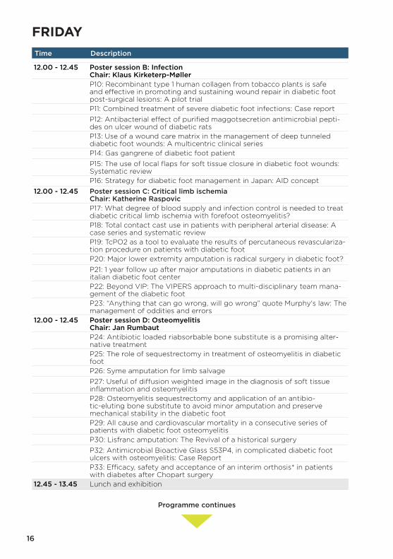
FRIDAYTime Description Speaker Room
Programme continues

Info
rmat
ion
Ora
l Ab
stra
cts
Po
ster
Ab
stra
cts
Spo
nso
r-E
xhib
ito
rP
rog
ram
me
Aut
hors
15
08 30 – 10 30 Revascularization Chair: Mauro Gargiulo Discussants: Roberto Cioni, Alberto Cremonesi
Sala 3 and 4
Lecture: The state of the art about the management of PAD Mauro Gargiulo, ItalyRecorded live cases: The PTAs Roberto Ferraresi, ItalyLecture: Angiosomes and revascularization Maarit Venermo, FinlandRecorded live cases: Concept and illustrative cases of ultra-distal bypas-ses
Florian Dick, Switzerland
Lecture: How to assess cardiac risk factors in DF patients Roberto Ferrari, ItalyRecorded live cases: Creative approaches Daniele Adami, ItalyPanel discussion
10 30 - 11 00 Coffee and exhibition Exhibition area11 00 - 12 00 Organization, setting and education
Chair: Kristien van Acker Discussants: Katherine Raspovic, Klaus Kirketerp-Møller
Sala 3 and 4
Lecture: Is there an ideal setting for diabetic foot surgery? Giacomo Clerici, ItalyLecture: The time-dependent network for DF Alberto Piaggesi, ItalyLecture: A new curriculum; the DF specialist Armin Koller, GermanyPanel discussion
12 00 - 12 45 Parallel poster sessions Poster area12 00 - 12 45 Poster session A: Charcot
Chair: Robert FrykbergPoster area
P1: The Circular Arc hindfoot nail for anatomic tibio-talo-calcaneal fusion Kaj Klaue, SwitzerlandP2: The use of a free vascularised osteocutaneous medial femoral condy-le flap to prevent recurrent neuropathic plantar ulcer
Michael Schintler, Austria
P3: Effectiveness of removable cast walker in the healing of diabetic neu-ropathic foot ulcer in the Fayaha diabetic foot clinic
Abdulhussein Marzoq, Iraq
P4: Modifications of external fixation in podiatric surgery Kamil Navratil, Czech RepublicP5: A clinical and quantitative assessment of the FORS™ insole, a novel shoe-based offloading system
Harry Penny, United States
P6: Clinical observation on the correction of diabetic Charcot foot defor-mities with external fixation
Jiangning Wang, China
P7: The use of total contact cast in diabetic foot ulcer on Charcot neuro-arthropathy: Case report
Ciprian Petrisor Vasiluta, Romania
P8: Classification of neuropathic diabetic foot, revisited Vladimir Obolenskiy, Russian FederationP9: Staged treatment protocol for limb salvage in patients with diabetic foot ulcerations and Charcot deformity
Zachary Flynn, United States
10 NOVEMBERTime Description Speaker Room
16
12 00 - 12 45 Poster session B: Infection Chair: Klaus Kirketerp-Møller
Poster area
P10: Recombinant type 1 human collagen from tobacco plants is safe and effective in promoting and sustaining wound repair in diabetic foot post-surgical lesions: A pilot trial
Elisabetta Iacopi, Italy
P11: Combined treatment of severe diabetic foot infections: Case report Srecko Bosic, SerbiaP12: Antibacterial effect of purified maggotsecretion antimicrobial pepti-des on ulcer wound of diabetic rats
Jiangning Wang, China
P13: Use of a wound care matrix in the management of deep tunneled diabetic foot wounds: A multicentric clinical series
Alessia Scatena, Italy
P14: Gas gangrene of diabetic foot patient Nune Soghomonyan, ArmeniaP15: The use of local flaps for soft tissue closure in diabetic foot wounds: Systematic review
Crystal Ramanujam, United States
P16: Strategy for diabetic foot management in Japan: AID concept Shinobu Ayabe, Japan12 00 - 12 45 Poster session C: Critical limb ischemia
Chair: Katherine RaspovicPoster area
P17: What degree of blood supply and infection control is needed to treat diabetic critical limb ischemia with forefoot osteomyelitis?
Miki Fujii, Japan
P18: Total contact cast use in patients with peripheral arterial disease: A case series and systematic review
Anthony Tickner, USA
P19: TcPO2 as a tool to evaluate the results of percutaneous revasculariza-tion procedure on patients with diabetic foot
Victor Rodriguez Saenz de Buruaga, Spain
P20: Major lower extremity amputation is radical surgery in diabetic foot? Danguole Vaznaisiene, LithuaniaP21: 1 year follow up after major amputations in diabetic patients in an italian diabetic foot center
Roberto De Giglio, Italy
P22: Beyond VIP: The VIPERS approach to multi-disciplinary team mana-gement of the diabetic foot
Valerie Marmolejo, USA
P23: “Anything that can go wrong, will go wrong” quote Murphy's law: The management of oddities and errors
Jan Rumbaut, Belgium
12 00 - 12 45 Poster session D: Osteomyelitis Chair: Jan Rumbaut
Poster area
P24: Antibiotic loaded riabsorbable bone substitute is a promising alter-native treatment
Bernd Gächter, Switzerland
P25: The role of sequestrectomy in treatment of osteomyelitis in diabetic foot
Sokol Hasho, Albania
P26: Syme amputation for limb salvage Robert Frykberg, United StatesP27: Useful of diffusion weighted image in the diagnosis of soft tissue inflammation and osteomyelitis
Yuta Terabe, Japan
P28: Osteomyelitis sequestrectomy and application of an antibio-tic-eluting bone substitute to avoid minor amputation and preserve mechanical stability in the diabetic foot
Cristian Nicoletti, Italy
P29: All cause and cardiovascular mortality in a consecutive series of patients with diabetic foot osteomyelitis
Alessia Scatena, Italy
P30: Lisfranc amputation: The Revival of a historical surgery Estelle How Hong, United KingdomP32: Antimicrobial Bioactive Glass S53P4, in complicated diabetic foot ulcers with osteomyelitis: Case Report
Roberto De Giglio, Italy
P33: Efficacy, safety and acceptance of an interim orthosis* in patients with diabetes after Chopart surgery
Roberto De Giglio, Italy
12 45 - 13 45 Lunch and exhibition Exhibition area
FRIDAYTime Description Speaker Room
Programme continues

Info
rmat
ion
Ora
l Ab
stra
cts
Po
ster
Ab
stra
cts
Spo
nso
r-E
xhib
ito
rP
rog
ram
me
Aut
hors
17
12 00 - 12 45 Poster session B: Infection Chair: Klaus Kirketerp-Møller
Poster area
P10: Recombinant type 1 human collagen from tobacco plants is safe and effective in promoting and sustaining wound repair in diabetic foot post-surgical lesions: A pilot trial
Elisabetta Iacopi, Italy
P11: Combined treatment of severe diabetic foot infections: Case report Srecko Bosic, SerbiaP12: Antibacterial effect of purified maggotsecretion antimicrobial pepti-des on ulcer wound of diabetic rats
Jiangning Wang, China
P13: Use of a wound care matrix in the management of deep tunneled diabetic foot wounds: A multicentric clinical series
Alessia Scatena, Italy
P14: Gas gangrene of diabetic foot patient Nune Soghomonyan, ArmeniaP15: The use of local flaps for soft tissue closure in diabetic foot wounds: Systematic review
Crystal Ramanujam, United States
P16: Strategy for diabetic foot management in Japan: AID concept Shinobu Ayabe, Japan12 00 - 12 45 Poster session C: Critical limb ischemia
Chair: Katherine RaspovicPoster area
P17: What degree of blood supply and infection control is needed to treat diabetic critical limb ischemia with forefoot osteomyelitis?
Miki Fujii, Japan
P18: Total contact cast use in patients with peripheral arterial disease: A case series and systematic review
Anthony Tickner, USA
P19: TcPO2 as a tool to evaluate the results of percutaneous revasculariza-tion procedure on patients with diabetic foot
Victor Rodriguez Saenz de Buruaga, Spain
P20: Major lower extremity amputation is radical surgery in diabetic foot? Danguole Vaznaisiene, LithuaniaP21: 1 year follow up after major amputations in diabetic patients in an italian diabetic foot center
Roberto De Giglio, Italy
P22: Beyond VIP: The VIPERS approach to multi-disciplinary team mana-gement of the diabetic foot
Valerie Marmolejo, USA
P23: “Anything that can go wrong, will go wrong” quote Murphy's law: The management of oddities and errors
Jan Rumbaut, Belgium
12 00 - 12 45 Poster session D: Osteomyelitis Chair: Jan Rumbaut
Poster area
P24: Antibiotic loaded riabsorbable bone substitute is a promising alter-native treatment
Bernd Gächter, Switzerland
P25: The role of sequestrectomy in treatment of osteomyelitis in diabetic foot
Sokol Hasho, Albania
P26: Syme amputation for limb salvage Robert Frykberg, United StatesP27: Useful of diffusion weighted image in the diagnosis of soft tissue inflammation and osteomyelitis
Yuta Terabe, Japan
P28: Osteomyelitis sequestrectomy and application of an antibio-tic-eluting bone substitute to avoid minor amputation and preserve mechanical stability in the diabetic foot
Cristian Nicoletti, Italy
P29: All cause and cardiovascular mortality in a consecutive series of patients with diabetic foot osteomyelitis
Alessia Scatena, Italy
P30: Lisfranc amputation: The Revival of a historical surgery Estelle How Hong, United KingdomP32: Antimicrobial Bioactive Glass S53P4, in complicated diabetic foot ulcers with osteomyelitis: Case Report
Roberto De Giglio, Italy
P33: Efficacy, safety and acceptance of an interim orthosis* in patients with diabetes after Chopart surgery
Roberto De Giglio, Italy
12 45 - 13 45 Lunch and exhibition Exhibition area
10 NOVEMBERTime Description Speaker Room

18
13 45 - 14 45 Bonesupport satellite symposium: Bone loss and biofilm - Overcoming challenges in diabetic foot surgery Chair: Venu Kavarthapu
Sala 3 and 4
Comparison of two techniques for operative treatment of calcaneal osteomyelitis using biodegradable antibiotic carriers Armin Koller, Germany
The role of antibiotic-eluting Cerament™ G bone void filler on the extreme rescue of infected Charcot foot
Enrico Brocco, Italy
Cerament™ allows successful reconstruction of infected Charcot foot Venu Kavarthapu, United Kingdom
Conservative surgery of diabetic foot osteomyelitis with Cerament™ Cristian Nicoletti, Italy14 50 - 17 10 Wound closure strategies
Chair: Alberto Piaggesi Discussants: Dane Wukich, Thomas Zgonis
Sala 3 and 4
Lecture: Local flaps/skin grafts: Does function affect the reconstructive choice?
Christopher Attinger, USA
Lecture: Bioengineered tissues and dermal substitutes: what's new? Franco Bassetto, ItalyRecorded live cases: Application of dermal substitutes Alberto Piaggesi, ItalyLecture: Local muscle and pedicle flaps for wound coverage Thomas Zgonis, USARecorded live cases: Pedicled flaps of the foot and ankle Christopher Attinger, USAPanel discussion
17 10 - 17 40 Coffee and exhibition Exhibition area17 40 - 18 10 Urgo satellite mini symposium:
Revealing new clinical evidence for DFU treatment Chair: Kristien van Acker
Sala 3 and 4
Explorer methodology José Luis Lázaro Martínez, SpainExplorer results Alberto Piaggesi, Italy
18 10 - 19 10 Murphy's law session Chair: Javier Aragon Sanchez Discussants: Sigurd Kessler, Jan Rumbaut
Sala 3 and 4
Mini lecture: "If anything can go wrong, it will". The management of oddi-ties and errors
Jan Rumbaut, Belgium
Three cases badly ended, to be presented and discussed by three diffe-rent KOL
Luca Dalla Paola, Robert Frykberg, John Giurini
19 10 Evening free19 30 - 24 00 Faculty dinner (by invitation only) Venice
FRIDAYTime Description Speaker Room

Info
rmat
ion
Ora
l Ab
stra
cts
Po
ster
Ab
stra
cts
Spo
nso
r-E
xhib
ito
rP
rog
ram
me
Aut
hors
19
13 45 - 14 45 Bonesupport satellite symposium: Bone loss and biofilm - Overcoming challenges in diabetic foot surgery Chair: Venu Kavarthapu
Sala 3 and 4
Comparison of two techniques for operative treatment of calcaneal osteomyelitis using biodegradable antibiotic carriers Armin Koller, Germany
The role of antibiotic-eluting Cerament™ G bone void filler on the extreme rescue of infected Charcot foot
Enrico Brocco, Italy
Cerament™ allows successful reconstruction of infected Charcot foot Venu Kavarthapu, United Kingdom
Conservative surgery of diabetic foot osteomyelitis with Cerament™ Cristian Nicoletti, Italy14 50 - 17 10 Wound closure strategies
Chair: Alberto Piaggesi Discussants: Dane Wukich, Thomas Zgonis
Sala 3 and 4
Lecture: Local flaps/skin grafts: Does function affect the reconstructive choice?
Christopher Attinger, USA
Lecture: Bioengineered tissues and dermal substitutes: what's new? Franco Bassetto, ItalyRecorded live cases: Application of dermal substitutes Alberto Piaggesi, ItalyLecture: Local muscle and pedicle flaps for wound coverage Thomas Zgonis, USARecorded live cases: Pedicled flaps of the foot and ankle Christopher Attinger, USAPanel discussion
17 10 - 17 40 Coffee and exhibition Exhibition area17 40 - 18 10 Urgo satellite mini symposium:
Revealing new clinical evidence for DFU treatment Chair: Kristien van Acker
Sala 3 and 4
Explorer methodology José Luis Lázaro Martínez, SpainExplorer results Alberto Piaggesi, Italy
18 10 - 19 10 Murphy's law session Chair: Javier Aragon Sanchez Discussants: Sigurd Kessler, Jan Rumbaut
Sala 3 and 4
Mini lecture: "If anything can go wrong, it will". The management of oddi-ties and errors
Jan Rumbaut, Belgium
Three cases badly ended, to be presented and discussed by three diffe-rent KOL
Luca Dalla Paola, Robert Frykberg, John Giurini
19 10 Evening free19 30 - 24 00 Faculty dinner (by invitation only) Venice
10 NOVEMBERTime Description Speaker Room

20
08 30 – 9 15 Oral presentations from abstract submissions I Chairs: Alberto Piaggesi, José Lazaro Martinez
Sala 3 and 4
O1: Deep veins arterialization: A limb salvage alternative? The P.I.S.A. Tech-nique (Peripheral Intravascular and Surgical vein Arterialization)
Daniele Adami, Italy
O2: The clinical research of transverse tibial bone flap transport for treat-ment of diabetic foot
Jiangning Wang, China
O3: Autologous peripheral blood mononuclear cells implant in a series of diabetic patients with critical limb ischaemia not eligible for revasculariza-tion
Alessia Scatena, Italy
O4: Keller arthroplasty: A cure for the chronic hallux ulceration Robert Frykberg, USA
9 15 - 10 15 Integra LifeSciences satellite symposium: A complete approach in the treatment of DFU – the key for success Chair: Luca Dalla Paola
Sala 3 and 4
Offloading the DFU in the perioperative period Luca Dalla Paola, ItalyAddressing soft tissue defects in limb salvage Christopher Attinger, USAOrthopedic perspectives in the lower extremity wound Dane Wukich, USA
10 15 - 11 00 Oral presentations from abstract submissions II Chairs: Alberto Piaggesi, José Lazaro Martinez
Sala 3 and 4
O5: The Transmetatarsal amputation: A retrospective analysis of 106 patients
Robert Frykberg, USA
O6: Local antibiotic devices to improve wound healing following surgical management of diabetic foot infection: a systematic review
Ben Marson, United Kingdom
O7: Corrective mini-ivasive osteotomy in treatment of diabetic with the forefoot ulcer
Vladimir Obolenskiy, Russian Federation
O8: Tibiocalcaneal arthrodesis as a surgical option for Charcot ankle deformity
Vladimir Obolenskiy, Russian Federation
O9: The clinical outcomes of Charcot foot surgery Elena Komelyagina, Russian Federation
SATURDAYTime Description Speaker Room
Programme continues

Info
rmat
ion
Ora
l Ab
stra
cts
Po
ster
Ab
stra
cts
Spo
nso
r-E
xhib
ito
rP
rog
ram
me
Aut
hors
21
08 30 – 9 15 Oral presentations from abstract submissions I Chairs: Alberto Piaggesi, José Lazaro Martinez
Sala 3 and 4
O1: Deep veins arterialization: A limb salvage alternative? The P.I.S.A. Tech-nique (Peripheral Intravascular and Surgical vein Arterialization)
Daniele Adami, Italy
O2: The clinical research of transverse tibial bone flap transport for treat-ment of diabetic foot
Jiangning Wang, China
O3: Autologous peripheral blood mononuclear cells implant in a series of diabetic patients with critical limb ischaemia not eligible for revasculariza-tion
Alessia Scatena, Italy
O4: Keller arthroplasty: A cure for the chronic hallux ulceration Robert Frykberg, USA
9 15 - 10 15 Integra LifeSciences satellite symposium: A complete approach in the treatment of DFU – the key for success Chair: Luca Dalla Paola
Sala 3 and 4
Offloading the DFU in the perioperative period Luca Dalla Paola, ItalyAddressing soft tissue defects in limb salvage Christopher Attinger, USAOrthopedic perspectives in the lower extremity wound Dane Wukich, USA
10 15 - 11 00 Oral presentations from abstract submissions II Chairs: Alberto Piaggesi, José Lazaro Martinez
Sala 3 and 4
O5: The Transmetatarsal amputation: A retrospective analysis of 106 patients
Robert Frykberg, USA
O6: Local antibiotic devices to improve wound healing following surgical management of diabetic foot infection: a systematic review
Ben Marson, United Kingdom
O7: Corrective mini-ivasive osteotomy in treatment of diabetic with the forefoot ulcer
Vladimir Obolenskiy, Russian Federation
O8: Tibiocalcaneal arthrodesis as a surgical option for Charcot ankle deformity
Vladimir Obolenskiy, Russian Federation
O9: The clinical outcomes of Charcot foot surgery Elena Komelyagina, Russian Federation
11 NOVEMBERTime Description Speaker Room

22
11 00 - 12 00 Lunch, coffee and exhibition Exhibition area12 00 - 12 30 Lecture: Charcot's foot and surgery: What have we done so far Robert Frykberg, USA Sala 3 and 412 30 - 15 00 Charcot - diagnosis and treatment
Chairs: Robert Frykberg Discussants: Luca Dalla Paola, Dane Wukich
Sala 3 and 4
Lecture: Markers for the early diagnosis of the Charcot's foot Nina Petrova, United KingdomRecorded live cases: Midfoot fusion John Giurini, USALecture: What modern imaging may do for the diagnosis of the Charcot's foot
Virna Zampa, Italy
Lecture: Failed fixation for the Charcot foot/ankle and diabetic foot/ankle trauma
Thomas Zgonis, USA
Lecture: Use of bone substitutes in Charcot foot surgery Dane Wukich, USARecorded live cases: Surgical treatment of Charcot ankle Dane Wukich, USALecture: New technologies for the diagnosis and treatment of Charcot foot
Katherine Raspovic, USA
Recorded live cases : Midfoot Charcot foot complicated by osteomyelitis Luca Dalla Paola, ItalyA duel: Internal vs external fixation Duelists: Armin Koller, Venu Kavarthapu
Referee: Robert Frykberg
Panel discussion15 05 - 15 30 Prizes: Poster presentation prize and oral presentation prize
Chairs: Armin Koller, Luca Dalla PaolaSala 3 and 4
O10 Oral prize: A prompt surgical management of necrotizing fasciitis in diabetic foot patients saves limbs and lives
Chiara Goretti, Italy
P31 Poster prize: Skinstretching device for repair of diabetic foot ulcer Jiangning Wang, China
15 30 - 16 00 Results of the polls - Closing of the Meeting Sala 3 and 416 00 - 16 30 A-DFS Board meeting
SATURDAYTime Description Speaker Room

Info
rmat
ion
Ora
l Ab
stra
cts
Po
ster
Ab
stra
cts
Spo
nso
r-E
xhib
ito
rP
rog
ram
me
Aut
hors
23
11 00 - 12 00 Lunch, coffee and exhibition Exhibition area12 00 - 12 30 Lecture: Charcot's foot and surgery: What have we done so far Robert Frykberg, USA Sala 3 and 412 30 - 15 00 Charcot - diagnosis and treatment
Chairs: Robert Frykberg Discussants: Luca Dalla Paola, Dane Wukich
Sala 3 and 4
Lecture: Markers for the early diagnosis of the Charcot's foot Nina Petrova, United KingdomRecorded live cases: Midfoot fusion John Giurini, USALecture: What modern imaging may do for the diagnosis of the Charcot's foot
Virna Zampa, Italy
Lecture: Failed fixation for the Charcot foot/ankle and diabetic foot/ankle trauma
Thomas Zgonis, USA
Lecture: Use of bone substitutes in Charcot foot surgery Dane Wukich, USARecorded live cases: Surgical treatment of Charcot ankle Dane Wukich, USALecture: New technologies for the diagnosis and treatment of Charcot foot
Katherine Raspovic, USA
Recorded live cases : Midfoot Charcot foot complicated by osteomyelitis Luca Dalla Paola, ItalyA duel: Internal vs external fixation Duelists: Armin Koller, Venu Kavarthapu
Referee: Robert Frykberg
Panel discussion15 05 - 15 30 Prizes: Poster presentation prize and oral presentation prize
Chairs: Armin Koller, Luca Dalla PaolaSala 3 and 4
O10 Oral prize: A prompt surgical management of necrotizing fasciitis in diabetic foot patients saves limbs and lives
Chiara Goretti, Italy
P31 Poster prize: Skinstretching device for repair of diabetic foot ulcer Jiangning Wang, China
15 30 - 16 00 Results of the polls - Closing of the Meeting Sala 3 and 416 00 - 16 30 A-DFS Board meeting
11 NOVEMBERTime Description Speaker Room

Oral Abstracts
24
[O1] DEEP VEINS ARTERIALIZATION: A LIMB SALVAGE ALTERNATIVE? THE P.I.S.A. TECHNIQUE (PERIPHERAL INTRAVASCULAR AND SURGICAL VEIN ARTERIALIZATION)
Daniele Adami1, Marta Mari1, Davide Maria Mocellin1, Francesca Tomei1, Michele Marconi1, Raffaella Berchiolli1, Mauro Ferrari1
1Azienda Ospedaliero Universitaria Pisana, Pisa, Italy
Aim: In the absence of any possible surgical/endovascular revascularization of dorsal and plantar arterial circulation, three patients underwent surgical arterialization of lower limb deep veins as extreme limb salvage attempt. Short and middle term results were evaluated.
Method: Throughout 2016 three diabetic patients (four limbs) underwent failed revascularization of lower limb, due to complete occlusion of foot arterial circulation, both on plantar and dorsal side. Each patient belonged to class 5 of Rutherford classification and required a major amputation. Vascu-lar tree of these patients was characterized by massive calcifications due to chronic metabolic disease (diabetes and chronic kidney disease) and chronic corticosteroid therapy (heart transplant and polymyalgia). As extreme limb salvage attempt, we proposed to create a surgical popliteal artery-pos-terior tibial vein arterovenous fistula by using great saphenous vein as conduit and to “arterialize” posterior tibial vein and distal deep veins through endovascular techniques.
Results/Discussion: Technical success was obtained in each case. In two cases arterialization of deep veins was incomplete due to concomitant plantar vein thrombosis. The postoperative stay was com-plications free. At 1 month follow up the two patients with plantar vein thrombosis required major amputation, despite peripheral gangrene demarcation has occurred. In two patients lower limb was saved.
Conclusion: From our preliminary experience, an attempt of deep veins arterialization could rep-resent an alternative option to major amputation in patients affected by critical limb ischemia at “terminal stage”. Outstanding problems are how to solve venous valves stenosis, timing and ways for eventual conservative foot amputations.

Info
rmat
ion
Ora
l Ab
stra
cts
Po
ster
Ab
stra
cts
Spo
nso
r-E
xhib
ito
rP
rog
ram
me
Aut
hors
25
[O2] THE CLINICAL RESEARCH OF TRANSVERSE TIBIAL BONE FLAP TRANS-PORT FOR TREATMENT OF DIABETIC FOOT
Jiangning Wang1, Lei Gao1, Tiangui Chen1
1Shijitan Hospital Affiliated Capital Medical University, Beijing, China
Aim: To evaluate the clinical outcome of transverse tibial bone flap transport for treatment of diabetic foot infections.
Method: From August 2012 to December 2016, 38 patients with diabetic foot infections were treated with transverse tibial bone flap transport operation, including 22 males and 16 females aged from 43 years to 82 years with an average of 62.5 years. Of them, 18 patients were affected on the left limb, and the remaining 20 patients were on the right sides. According to Wagner’s criteria, 17 patients had Grade II, 15 patients had Grade III, and 4 patients had Grade IV ulcer.
Results/Discussion: All of the 38 patients were available for follow-up ranged from 6 to 21 months with a mean of 13.5 months. Successful limb salvage and complete healing of the ulcer were achieved in all affected limbs with lesions healing time in a mean of 11.8 weeks. Limb pain and numbness were alleviated or even disappeared. The skin temperature of affected foot was significantly improved from (29.8±0.5)oC preoperatively to (31.5±0.9)oC at 1 year after opera-tion (P<0.05). Correspondingly, VAS decreased remarkably from (4.3±0.6) before operation to (0.4±0.1) at 1 year following surgery (P<0.05).
Conclusion: Transverse tibial bone flap transport is an effective way to treat diabetic foot infec-tions and to avoid amputation.

Oral Abstracts
26
[O3] AUTOLOGOUS PERIPHERAL BLOOD MONONUCLEAR CELLS IMPLANT IN A SERIES OF DIABETIC PATIENTS WITH CRITICAL LIMB ISCHAEMIA NOT ELIGIBLE FOR REVASCULARIZATION
Alessia Scatena1, Filippo Maioli2, Pasquale Petruzzi3, Giorgio Ventoruzzo2, Francesco Liistro4, Leonardo Bolognese4, Leonardo Ercolini2, Lucia Ricci5
1Diabetic Foot Care Unit - San Donato Hospital Arezzo, Arezzo, Italy2Vascular Surgery Unit, San Donato Hospital Arezzo, Arezzo, Italy3Interventional Radiology Unit, San Donato Hospital Arezzo, Arezzo, Italy4Cardiovascular and Neurologic Department, San Donato Hospital Arezzo, Arezzo, Italy5Diabetology Unit, San Donato Hospital Arezzo, Arezzo, Italy
Aim: Evaluate the effectiveness of therapeutic angiogenesis using autologous peripheral blood mononuclear cells (A-PBMNC) in diabetic patients with critical limb ischaemia (CLI) not eligible for revascularization.
Method: From September 2016 to January 2017 we collected 6 diabetic patients with CLI and isch-emic not infected wounds. We implanted in the limb 12 mL of A-PBMNC, 0.2–0.3 mL for each bolus, collected by selective filtration from 120mL of peripheral blood. Treatment was repeated three times.
Results/Discussion: We enrolled 5 male and 1 female, with a mean age of 77,4±5,2 years, mean di-abetes duration of 16±7,4 years. Two patients (33,3%) had a bypass occlusion and 4 patients (66,6%) had unsuccessful previous percutaneous transluminal angioplasty because of very distal arterial occlusion and/or severe calcifications. Mean transcutaneous oxygen tension (TcpO2) was 15,8±6,6 mmHg. Rest pain was present in all cases. Wifi Classification System score was W3I3Fi0 in 5 patients (83,3%) and W1I3Fi0 in 1 (16,7%). 5 lesion were in the forefoot (83,3%) and 1 (16,7%) in the malle-olus. After a mean follow-up of 195±41,35 days, mean TcpO2 was 40±14,6 mmHg. Complete wound healing was achieved in 3 patients (50%) with a mean healing time of 120±51,9 days. Improvement of ischemic symptoms was reached in 4 patients (66,7%). 2 patients obtained a 46% reduction of the area (baseline 6,3±7,6 cm2; timeline 2,9±3,4 cm2). 1 patient underwent above the knee amputation.
Conclusion: Implant of A-PBMNC shows to be useful in diabetic patients not eligible for revascular-ization.

Info
rmat
ion
Ora
l Ab
stra
cts
Po
ster
Ab
stra
cts
Spo
nso
r-E
xhib
ito
rP
rog
ram
me
Aut
hors
27
[O4] KELLER ARTHROPLASTY: A CURE FOR THE CHRONIC HALLUX ULCERATION
Robert Frykberg1, Jaminelli Banks1, Dane Wukich2
1Phoenix Va Hospital, Phoenix, United States2University of Pittsburgh Medical Center, Pittsburgh, United States
Aim: Diabetic foot ulcerations (DFU) of the great toe are fairly common complications in patients with neuropathy. Frequently they are associated with restricted motion of the 1st metatarsal-phalangeal (MTP) joint and can be recalcitrant. We herein present the results of our experience with the Keller (1st MTP) arthroplasty to treat such conditions.
Method: We retrospectively evaluated 16 patients undergoing first MTP arthroplasty proce-dures, all of which were performed as a curative measure for plantar ulcers of the hallux.
Results/Discussion: 88 % (14/16) of patients had diabetes. All subjects were males with an average age of 66.9 years. Only 2/16 had an open ulcer at the time of the procedure while all patients had history of hallux ulceration. 14 (88%) procedures healed uneventfully. No patients underwent a hallux amputation during our 112 ± 21 month follow up period. At the time of sur-gery, average HgA1C was 7.8%. 9 (56%) patients developed post-operative surgical site wound dehiscences and/or surgical site infections. The median healing time for the incision was 25 days. Average healing time for the plantar hallux ulcer was 22 days post procedure.
Conclusion: We have shown a high degree of success when performing the Keller arthroplasty, although complications are common. Though many of our patients experienced post–operative wound dehiscences, all eventually healed and no amputations were necessary. Complications can be anticipated in neuropathic patients, but the long term successful outcomes seem to justify this corrective procedure.

Oral Abstracts
28
[O5] THE TRANSMETATARSAL AMPUTATION: A RETROSPECTIVE ANALYSIS OF 106 PATIENTS
Robert Frykberg1, Priyanka Begur2, Jaminelli Banks1
1Phoenix Va Hospital, Phoenix, United States2Phoenix Va Medical Center, Phoenix, United States
Aim: Foot ulcerations are a common cause of morbidity among diabetics, and in many cases, lead to infection and amputations. The transmetatarsal amputation (TMA) is one such amputation that was first described as a limb salvaging technique. In this study, we will review both the outcomes and possible contributing factors.
Method: This is a retrospective, single site, chart review of 106 patients between 2003 to 2017. Patients were included if they had a TMA performed at the Phoenix VA Medical Center.
Results/Discussion: Among 106 patients in this study, the average age was 64 years old, with 105 males and 1 female. About 89 had confirmed diabetes and 88 patients had a history of prior ulcer-ations. 31 individuals had positive MRSA infections and 60 had confirmed osteomyelitis. A statistically significant probability ratio (PR) value of 0.04 showed that wounds positive for MRSA were less likely to heal post-operatively. Patients were followed up for an average of 6 years with 70 TMAs complete-ly healed over an average of 155 days. 36 individuals had palpable pedal pulses preoperatively and 56 individuals had confirmed peripheral arterial disease (PAD). Through Spearman analysis, a PR value of 0.00 showed that the absence of pedal pulses was an accurate indicator of PAD. 30 individuals went on to a more proximal amputation post-operatively.
Conclusion: With a 66% success rate, we conclude that the TMA is a key limb salvaging technique to consider. Though post-operative complications can occur, the high success rate of the TMA outweighs these.

Info
rmat
ion
Ora
l Ab
stra
cts
Po
ster
Ab
stra
cts
Spo
nso
r-E
xhib
ito
rP
rog
ram
me
Aut
hors
29
[O6] LOCAL ANTIBIOTIC DEVICES TO IMPROVE WOUND HEALING FOLLOWING SURGICAL MANAGEMENT OF DIABETIC FOOT INFECTION: A SYSTEMATIC REVIEW
Ben Marson1, Douglas Grindlay1, Ben Ollivere1, Brigitte Scammell1
1Orthopaedics and Trauma Group, University of Nottingham, Nottingham, United Kingdom
Aim: Surgical management of diabetic foot infection is indicated when conservative measures have failed or in the presence of sepsis.
Local antibiotics have been proposed as a method of improving delivery of antibiotic to distal tis-sues without relying on the impaired micro-circulation. This review aims to systematically review the available evidence for such technology as an adjunct to surgical therapy.
Method: OVID Medline, EMBASE PubMed and Cochrane CENTRAL databases were searched to identify eligible trials. Following searching and screening 12 studies were identified for inclusion.
Results/Discussion: Overall study quality was poor. 1 randomised control trial, 2 case control and 9 case series were included. Results from the RCT suggest that wound healing is quicker when a collagen-gentamycin sponge is implanted at time of surgery, but no difference in length of stay or amputation rate was demonstrated. Results from case-control trials with high risk of bias indicated no change in wound healing rate when collagen-gentamycin sponge was implant-ed during transmetatarsal amputation, but a reduction in wound breakdown (8% vs 25%) was identified. A significant cost reduction was identified when using a bespoke antimicrobial gel to deliver antibiotics and other agents.
Analyses of case series identified 473 patients who were treated using local antibiotic delivery devices. Wound healing, reoperation rates and mortality were comparable to previous literature on general treatment of these infection.
Conclusion: There is a lack of good quality evidence to support the use of local antibiotic deliv-ery devices in the treatment of diabetic foot infections.

Oral Abstracts
30
[O7] CORRECTIVE MINI-INVASIVE OSTEOTOMY IN TREATMENT OF DIABETIC WITH THE FOREFOOT ULCER
Vladimir Obolenskiy1, Viktor Protsko2
1City Hospital #13, Rnrmu, Moscow, Russian Federation2Russian University of Friendship of Peoples, City Hospital #79, Moscow, Russian Federation
Aim: To assess the clinical effectiveness of minimally invasive corrective osteotomy (MICO) in treat-ment of diabetic foot (DF) with the forefoot ulcer.
Method: We have analyzed the treatment outcomes of 23 patients suffering from diabetic foot with the ulcerative defect localized at the area of metatarsophalangeal (MTP) joints. Follow up is more than 1 year. In 1 patient the ulcer was classified as Grade 1 (Wagner), 15 patients - Grade 2, and in 7 patients - Grade 3. MICO was performed for all 23 cases. In addition, skin defect reconstruction was done in 4 cases, tenotomy in 2 cases. One month without operated foot weight-bearing on operated foot was recommended for all patients.
Results/Discussion: The average length of hospital stay was 8.5 days. No complications were observed after surgery. In 73.9% of cases, ulcers healed in the first 1-1.5 month after MICO without any intervention on the ulcer site, the osteotomy was healed at the same terms. The recurrence occurred in two patients (8.7%) with neuropathic ulcer after 4 and 7 months (associated with early weight-bearing): in the first case we performed ulcer excision, MTP joint osteotomy; the second patient rejected the further treatment.
MICO is an effective surgical option allowing us to eliminate the pressure area by changing the anato-my of metatarsal bone, thus removing the main cause of the ulcer. Patient with good glucose control mat be treated outpatient.
Conclusion: The successful treatment before development of osteomyelitis prevents patients from the further more traumatic surgeries.

Info
rmat
ion
Ora
l Ab
stra
cts
Po
ster
Ab
stra
cts
Spo
nso
r-E
xhib
ito
rP
rog
ram
me
Aut
hors
31
[O8] TIBIOCALCANEAL ARTHRODESIS AS A SURGICAL OPTION FOR CHARCOT ANKLE DEFORMITY
Vladimir Obolenskiy1, Viktor Protsko2
1City Hospital #13, Rnrmu, Moscow, Russian Federation2Russian University of Friendship of Peoples, City Hospital #79, Moscow, Russian Federation
Aim: To assess the effectivity and safety of different tibiocalcaneal arthrodesis (TCA) types in treatment of patients with Charcot ankle deformity depending on disease severity.
Methods: We have analyzed the outcomes after treatment of 16 patients with diabetic neuro-pathic ankle arthropathy (Charcot ankle) at the stage of septic complications in bones of ankle and subtalar joints. All patients were treated in the Department of Septic Complications in 13th Moscow City Clinical Hospital between 2014 and 2016. The observation period was more than 1 year. The cohort consisted of 14 men and 2 women who had a mean age of 45.3 years (range: 32 to 69 years). Among them 14 patients had Type II and 2 patients had Type I diabetes mellitus. The average duration of diabetes was 8.2 years, Charcot ankle – 1.3 years, septic complications – 24.7 days. Ilizarov fixator for TCA was used in 8 cases; internal fixation with cannulated screws (IFCS) was applied in 6 cases; for one patient we performed supra-ankle wedge osteotomy and used IFCS; in one case only debridement of the infected bone and soft tissues was performed.
Results: Complications developed in 4 patients (3 – IFCS, 1 – Ilizarov frame) in different times: in 1 case after 2 months (screws were removed, however the in 3 months amputation below the knee was performed due to infection spread), in 2 cases – after 9 and 13 months (screws were removed, without stability and correction loss; no recurrences), in 1 case midshafttibial wire-tract osteomyelitis developed (sequestrectomy, no recurrences). Patients were divided into groups according to the proposed combined classification of SERW (anatomy, pathophysiology, deformity and affected tissues depth - Sanders L. & Frykberg R., 1991, Eichenholtz S.N., 1966, Rogers L.C., 2012 иWagner F.W., 1979) and the outcomes were analyzed.
Discussion: The conducted analysis has not revealed any correlation between developed complications in different treatment tactics and spread of pathophysiologic process (S). Disease stage had influence on complications rate: 3 of 4 complications developed in patients who had been treated at fragmentation stage, 1 of 4 – in patient who had surgery at consolidation stage (E). The highest influence on complications rate had R and W components: all complications developed at RD stage (foot deformity in presence of open wound and osteomyelitis) and at W3 stage(phlegmon, abscess or osteomyelitis).
Conclusion: Risk factors assessment should be done in all patients who undergo fixation after TCA. Ilizarov frame is a preferable option for high risk patients, which were defined as those on stages E1, RD and W3. In all other cases internal fixation is treatment of choice at it provides more rapid rehabilitation and higher quality of life.

Oral Abstracts
32
[O9] THE CLINICAL OUTCOMES OF CHARCOT FOOT SURGERY
Elena Komelyagina1, Vladimir Obolenskiy2, Viktor Protsko3, Nuria Sabanchieva4, Mikhail Antsiferov4
1Endocrinological Dispensary, Department of Health of Moscow, Moscow State Out-Patient Endocrine Center, Moscow, Russian Federation2City Hospital #13, Rnrmu, Moscow, Russian Federation3City Hospital #7, Moscow, Russian Federation4Moscow State Out-Patient Endocrine Center, Moscow, Russian Federation
Aim: To assess the outcomes of Charcot foot (CF) surgery.
Method: 22 CF diabetic patients who underwent surgery from 2014 to January of 2017 took part in this study.
Results: Exosectomies were performed in 4 (18%) of the cases, arthrodesis in 9 (41%) patients, external fixation in 3 (14%) of the procedures. Hybrid surgery were made in 5 (23%) of the cases. The complications included infection in 2 (9%) patients, removal of the device in 4 (18%) cases, nonheal-ing postoperative wound in 1 patient. In 12 (55%) of the cases there were no complications. All the ulcers healed in a three months after surgery. In 16 patients who have follow up more than 1 year 6 (27%) have no complications, 2 (9%) developed migration or breakdown of the nail; in 2 cases (9%) there were loss of initial correction; recurrence of the ulcer occurred in 4 patients (18%). In 8 cases (36%) more than one procedure were needed. 2 patients died in a 2 year after surgery due to cardio-vascular events. There were no major or minor amputations. All the patients are weight-bearing on their feet.
Conclusion: Any type of surgical treatment resulted in healing of the ulcer in this group of patients. There were no major or minor amputations in all observed CF patients. In terms of the results ob-tained we can conclude that Charcot foot surgery is a good tool to reach limb salvage in patients with nonhealing ulcers and deformities considered hazardous for amputation.

Info
rmat
ion
Ora
l Ab
stra
cts
Po
ster
Ab
stra
cts
Spo
nso
r-E
xhib
ito
rP
rog
ram
me
Aut
hors
33
[O10] A PROMPT SURGICAL MANAGEMENT OF NECROTIZING FASCIITIS IN DIABETIC FOOT PATIENTS SAVES LIMBS AND LIVES
Chiara Goretti1, Elisabetta Iacopi1, Nicola Riitano1, Alberto Coppelli1, Alberto Piaggesi1
1Pisa University Hospital, Medicine Department, Diabetic Foot Section, Pisa, Italy
Aim: Necrotizing fasciitis (NF) is a life-threatening infection. It requires prompt surgical treat-ment and is associated with a high mortality rate. We evaluated outcomes of surgical manage-ment of NF in diabetic foot (DF) patients in a tertiary referral centre.
Method: We retrospectively searched NF pts in the database of our DF Section from 2012 to 2014. All patients were admitted, began antibiotic therapy and promptly underwent to extensive surgical debridement. We analysed short-term (surgery and major amputation), and long-term outcomes (healing rate).
Results/Discussion: 68 patients was referred to our clinic for a suspicion of NF. Diagnosis was confirmed in 54 pts (79.4%; male/female 40/14; type 1/2 diabetes 6/48; age 62.8±8.1 yrs; dia-betes duration 13.6±10.1 yrs). The cases was classified as Type 1 (33-61.1%), Type 2 (7-13.0%) and Type 3 (14-25.9%). No differences were observed between the groups. Six pts (11.1%) underwent to forefoot amputation, 12 (22.2%) required toe or ray amputation. No primary ma-jor amputation was performed. Of the 54 patients, 23 (42.6%) required a second and 5 (9.2%) a third surgical procedure. Complete healing was achieved in 46 pts (85%). Healing time was 94±11 days. Of the remaining 8 pts: 5 (9.2%) died for other reason, 2 (3.7%) recurred and one (1.9%) required a major amputation.
Conclusion: We observed a higher prevalence of NF in DF, compared to literature. Despite these, when promptly and aggressively treated, NF has a relatively good prognosis and it is not associated with an excess of limb loss and death.

Poster Abstracts
34
[P1] THE CIRCULAR ARC HINDFOOT NAIL FOR ANATOMIC TIBIO-TALO-CALCANEAL FUSION
Kaj Klaue1, Thomas Mittlmeier2
1Clinica Luganese, Moncucco, Switzerland2Unfallchirurgie, Universitätsmedizin Rostock, Rostock, Germany
Aim: Normal anatomy of the hindfoot demonstrates alignment of the heel, the posterior subtalar facet, the talus, the ankle joint and the distal tibia on a circular arc. This arc lies on a vertical plane which is slightly angulated inwards in relation to the sagittal plane. Purpose of the study is to optimize the technique to stabilize the hindfoot in anatomical alignment.
Method: An instrumentation was designed to create a circular arc bore hole crossing the heel, the posterior subtalar facet, the tibio-talar joint and the distal tibia metaphysis. Using an image amplifier the hole is bored using a motor driven end cutting flexible reamer which is seated within a rigid curved hull. The nail has the same shape than the hull and is impacted up to the distal tibia. 18 pa-tients have been treated so far using this technique.
Results/Discussion: The pathology of the operated patients include post-traumatic, congenital and metabolic (diabetes) conditions. We did observe 3 ruptures of the tibial locking screw. All cases went to consolidation without malunion or other complications. One diabetic patient developed a stable pseudarthrosis at the midfoot joints. After 2 weeks our patients did practise partial to full weight bearing using a cam walker for other 6 weeks.
Conclusion: The tibio-talo-calcaneal arthrodesis can be successfully treated using a central circular arc shaped nail allowing for full form fit between implant and bone.

Info
rmat
ion
Ora
l Ab
stra
cts
Po
ster
Ab
stra
cts
Spo
nso
r-E
xhib
ito
rP
rog
ram
me
Aut
hors
35
[P2] THE USE OF A FREE VASCULARISED OSTEOCUTANEOUS MEDIAL FEMO-RAL CONDYLE FLAP TO PREVENT RECURRENT NEUROPATHIC PLANTAR ULCER
Michael Schintler1, Martin Grohmann1, Stetan Benedikt1, Anna Vasilyeva1, Lars-Peter Kamolz2
1Division of Plastic, Aesthetic and Reconstructive Surgery, Department of Surgery, Medical University of Graz, Graz, Austria2Medical University of Graz, Graz, Austria
Aim: A new surgical method to prevent pressure ulcer, using a free vascularized medial femoral condyle bone transplant.
Method: In our article, we report the application of a free vascularized medial femoral condyle (MFC) flap to prevent the recurrence of a pressure ulcer in a patient suffering from diabetic foot syndrome. Our patient had been diagnosed with Type II Diabetes and presented with osteomy-elitis of several metatarsal heads after great toe amputation. The good vascular situation of the larger vessels and the relatively young age of the patient made us think of a new indication for MFC Flap using the vascularized bone graft as a damper in the area of the metatarsal heads by shaping the graft like a ski and thereby prevent perforation of the plantar skin by distributing the pressure. Good results were achieved in terms of wound healing, pain reduction and improve-ment of gait.
Results/Discussion: There was no recurrence of a pressure ulceration after a one year follow up. The versatility of the corticoperiosteal graft from the medial femoral condyle makes it an important reconstructive tool for addressing major surgical problems also for patients suffering from diabetic foot syndrome in the lower extremities. Our case presents the first use of a MFC Flap in the treatment of a pressure ulcer in a diabetic foot.
Conclusion: In selected patients, our method could prevent premature and extended amputa-tions thereby providing a good improvement in the quality of life.

Poster Abstracts
36
[P3] EFFECTIVENESS OF REMOVABLE CAST WALKER IN THE HEALING OF DIABETIC NEUROPATHIC FOOT ULCER IN THE FAYAHA DIABETIC FOOT CLINIC
Abdulhussein Marzoq1
1Al Fayha, Basrah, Iraq
Aim: To evaluate the effectiveness of the use of a removable cast walker (RCW) in the heading of the diabetic neuropathic plantar foot ulceration.
Method: This study was done at Al-Fayha general hospital, Basra, Iraq. Prospective study between August 2014 and September 2015 on 29 adult diabetic patients, 22 males and 7 females with neuro-pathic plantar foot ulcer.
Results/Discussion: Twenty-nine patients were included in our study, twenty-two of them males (75.9%) and seven females (24.1%). The mean age of the patients was (55.79 +/- 7.7) years old and the mean duration of the diabetic mellitus was (11.52 +/- 4.0) years. The mean duration of the neu-ropathic non-ischemic ulcers was (7.1 +/- 4.3) months. The percentage of planter neuropathic ulcers with 1A class according to (UT-Classification) was (44.8%) with a healing rate of (76.9%) whereas the percentage of ulcers with 2A class was (55.2%), with a healing rate of (62.5%) and we found that the total percentage of healing of ulcers with (1A,2A) classes was (69.0%) with a mean duration of (8.45 +/- 2.2 ) weeks.
Conclusion: Removable cast walker (RCW) is an effective method in the treatment of diabetic neuro-pathic non-ischemic foot ulceration.

Info
rmat
ion
Ora
l Ab
stra
cts
Po
ster
Ab
stra
cts
Spo
nso
r-E
xhib
ito
rP
rog
ram
me
Aut
hors
37
[P4] MODIFICATIONS OF EXTERNAL FIXATION IN PODIATRIC SURGERY
Kamil Navratil1, Robert Bem2, Michal Dubský2, Veronika Woskova3, Bedřich Sixta2
1 Transplant Surgery Clinic, Institute for Clinical and Experimental Medicine, Prague, Czech Republic2 Institute for Clinical and Experimental Medicine, Prague, Czech Republic3 Diabetes Center, Institute for Clinical and Experimental Medicine, Prague, Czech Republic
Aim: To discuss modifications of external fixator (EF) used for stabilization and offloading after reconstructive surgery for diabetic foot (DF) and Charcot neuropathy(CN) suitable in case of partial foot ischemia or patient discomfort.
Method: A tube-to-bar EF* was used in three variants: standard Δ-frame (full pins were passed through the metatarsal heads, then into the calcaneus and tibia and stabilized by double framed rods), hybrid frame (same placement but one or two semicircles with tarso-metatarsal K-wire fixation), used in case of partial ischemia and unilateral frame in cases of discomfort from traumatization. 20 patients (13 men, 7 women), average age 57(39-78) were observed prospec-tively within years 2014 – 5/2017 (follow-up 2-41 months), CN in 15 cases, other types 5 cases. Δ-frame was used in 7, hybrid EF in 7 and unilateral EF in 6 cases. Rehospitalisations, major complications – severe pin-tract infection (PTI), osteomyelitis recurrence (OM), non-union rates, hardware failures and EF adjustments episodes were tracked in each group.
Results/Discussion: No major amputations was needed, one rehospitalisation (severe recurrent osteomyelitis). Overall PTI rate was 49%, in one case a premature EF removement was needed. Two non-union cases underwent further fixation. No serious hardware failure occurred, 8 pa-tients needed EF adjustments.
Conclusion: Results showed no differences in complications between EF types. Hybrid EF lowers the risk of blood vessels traumatization due to thinner diameter of the pins. The main advantage of unilateral EF technique is decreased traumatization of the skin, but should not be applied in patients with higher BMI.
*ProSpon, Medin CZ

Poster Abstracts
38
[P5] A CLINICAL AND QUANTITATIVE ASSESSMENT OF THE OFF-LOADING INSOLE DEVICES*, A NOVEL SHOE-BASED OFFLOADING SYSTEM
Harry Penny1, James McGuire2, Payam Rafat3, Regino Flores4, Adam Weaver5, Chad Allender6, Emma Kreuz7
1 Upmc Altoona, Altoona, United States2 Temple University, Philadelphia, United States3 Montefiore Medical Center, New York City, United States4 Gesisinger Commonwealth School of Medicine, Scranton, United States5 Philadelphia College of Osteopathic Medicine, Philadelphia, United States6 Saint Francis University, Loretto, United States7 Junaita College, Huntingdon, United States
Aim: To evaluate the Off-Loading Insole devices* in a patient-based series of diabetic foot ulcers.
Method: Patients were selected based on previous non-compliance, contraindication to TCC, or failure of other off-loading modalities. Also, the Off-Loading Insoles* were implemented in patients transitioning out of TCC until full recovery. The Off-Loading Insoles* were customized by removing plugs from bottom of the insole that correspond to ulcer location, then inserting the insole into a surgical rocker bottom inlay shoe provided to the patient. Wound dimensions were recorded and photographed with each visit to the wound clinic.
Results/Discussion: In three independent trial sites, patients using the Off-Loading Insoles* con-sistently demonstrated a high level of compliance with the device. Patients rated the Off-Loading Insoles* as more comfortable and convenient than other offloading modalities. Features of the Off-Loading Insoles* include a top cover** minimizing shear forces/slippage and absorbing moisture, a polyurethane foam construction providing durable cushioning and shock absorbance, and a fabric mid-layer minimizing collapse and “edge effects”.
Conclusion: The Off-Loading Insole*is an effective shoe-based offloading device when used in conjunction with modern wound care techniques. In addition to being cost-effective, use of the Off-Loading Insoles* improved patient compliance, and reduced healing times, DFU recurrence rates, amputation, and mortality rates. The Off-Loading Insole* is a viable alternative to the TCC, or as a transitional method with TCC. Use of the Off-Loading Insoles* correlated to patient adherence and wound recovery. The insole was highly durable, easy to use, and had no observable contraindications in this study.
*FORSTM-15 Off-Loading Insole **Alcantara®

Info
rmat
ion
Ora
l Ab
stra
cts
Po
ster
Ab
stra
cts
Spo
nso
r-E
xhib
ito
rP
rog
ram
me
Aut
hors
39
[P6] CLINICAL OBSERVATION ON THE CORRECTION OF DIABETIC CHARCOT FOOT DEFORMITIES WITH EXTERNAL FIXATION
Jiangning Wang1, Lei Gao1
¹ Shijitan Hospital Affiliated Capital Medical University, Beijing, China
Aim: To investigate the clinical results of external fixation on the correction of diabetic Charcot foot deformities.
Method: 16 patients (22 feet) with Charcot foot deformities were treated with external fixation from October 2014 to December 2015.All patients were male, and 10 cases were on the unilat-eral side and 3 cases were on the bilateral sides. All patients suffered from ulceration at the bot-tom of the foot. The age of patients ranged from 40 to 56 with an average of 48 years old. The history of diabetics was from10 to 12 years. X ray and CT were used to evaluate bone correction, and AOFAS was applied to estimate recovery of joint function.
Results/Discussion: All patients were followed up from 12 to 24 months with an average of 18 months. The kirschner wire didn’t break. No infection of kirschner wire path, no looseness of external fixation and other complications occurred after operation. According to postoperative X ray and CT results, the time of bone correction ranged from12 to 18 weeks with an average of 15 weeks. 12 feet got excellent results, 3 good and 1 moderate based on AOFAS score.
Conclusion: Using external fixation for the correction of diabetic foot deformity can make the feet receive enough offloading at the same time. It is a perfect method to accelerate the ulcer-ation healing and improve the sole stress points which can avoid ulceration occur again.

Poster Abstracts
40
[P7] THE USE OF TOTAL CONTACT CAST IN DIABETIC FOOT ULCER ON CHARCOT NEUROARTHROPATHY - CASE REPORT
Ciprian Petrisor Vasiluta1, Lidia Iuliana Arhire2, Otilia Nita2, George Nita2, Cosmin Uliniuc1, Bogdan Ciuntu2, Andreea Gherasim2, Laura MIhalache2, Mariana Graur1, Stefan Georgescu1
1 Sf. Spiridon Clinical Emergency Hospital, Iasi, Romania2 Grigore T. Popa University of Medicine and Pharmacy, Iasi, Romania
Aim: Chronic diabetic foot ulcers (DFU) on Charcot neuroarthropathy (CN) are a challenge to the treating team. Efficiency in management requires a specialized multidisciplinary team and the use of all treatment principles. Among these, off-loading is frequently overlooked, especially the “gold” stan-dard technique, the total contact cast (TCC). We present our results in the management of a complex case of DFU on CN, where the application of TCC made the major difference in healing and we aim to draw the necessary conclusions for future implications in Romania.
Method: A 50 year-old diabetic patient presented for a >6 month history of DFU on the left plantar mid-foot. Her wound was 50x100 mm in surface and 8 mm depth, infected, but without osteomy-elites. She had multiple complications of diabetes, including bilateral CN.
Results/Discussion: The initial management included serial surgical debridement, antibiotics, bed rest. She was discharged after 24 days with a 20% decrease in wound surface, but three months later she returned aggravated due to lack of compliance with follow-up and simple standard off-loading. After retaking all the above mentioned measures, we obtained the patient’s consent for applying TCC. We used a fiberglass off-loading cast changed weekly which produced an 80% reduction in wound size and depth after one month and the patient mentioned better quality of life.
Conclusion: Off-loading is essential in healing a chronic DFU and we need to take the necessary steps to support clinicians to develop skills in casting and introduce casting services in Romania.

Info
rmat
ion
Ora
l Ab
stra
cts
Po
ster
Ab
stra
cts
Spo
nso
r-E
xhib
ito
rP
rog
ram
me
Aut
hors
41
[P8] CLASSIFICATION OF NEUROPATHIC DIABETIC FOOT, REVISITED
Vladimir Obolenskiy1, Viktor Protsko2, Elena Komelyagina3
1 City Hospital #13, Rnrmu, Moscow, Russian Federation2 Russian University of Friendship of Peoples, City Hospital #79, Moscow, Russian Federation3 Moscow State Out-Patient Endocrine Center, Endocrinological Dispensary, Department of Health of Moscow, Moscow, Russian Federation
All the existing classifications of the diabetic foot syndrome have their advantages and disadvan-tages, and each of them does not fully reflect the entire scope of possible problems that a pa-tient with diabetic foot may encounter. In the pathogenesis of the complications the leading role belongs to diabetic neuropathy and angiopathy, as well as osteoarthropathy, which is considered a subspecies of the neuropathic form of this syndrome. In our view, osteoarthropathy and foot deformity have nothing to do with having or not having tissue ischemia. In addition, the widely used classification (e.g., Wagner or the IWGDF-recommended PEDIS classification) do not illus-trate the severity of orthopedic problems associated with diabetic foot. Orthopedists may also use other types of classifications – anatomical (Brodsky, Sanders & Frykberg), pathologoanatom-ic/pathophysiological (Eichenholtz, Sella & Barrette), clinical (Rogers, Chantelau & Grutzner) that unfortunately fail to holistically describe the problem.
Let us assume that ischemia, and even more so critical limb ischemia primarily requires re-vascularization in one way or another. Without this intervention any attempts to treat ulcers, deformations and bone-destructive processes in the foot are of little effect and the organ-pre-serving tactics has no chance of success. Therefore vascular pathology needs to be diagnosed, classified and if possible arrested before the treatment of the bone-destructive processes in the foot begins. Let us consider the issue of treatment of complicated neuropathic form of diabetic foot syndrome (and/or its neuro-ischemic form after revascularization).
To describe the entire complex of pathological changes in neuropathic diabetic foot we offer a combination of classifications that describe the foot anatomy, stages of the pathoanatomical/pathophysiological processes in the foot bones, foot deformation degree as well as presence and depth of the infectious process: SERW (Sanders-Eichenholtz-Rogers-Wagner).
This combination allows us to localize the lesions, describe the nature of changes in bones and joints, account for wounds and ulcerations and indicate the severity of infection. Comprehensive understanding of the issue, firstly, facilitates communication between podiatric endocrinologists, surgeons and orthopedists; secondly, it helps to determine the modality of treatment in each case: indications for and the type of surgery, type and duration of limb immobilization and stress relief, indications for and the duration of antibiotic therapy, etc.

Poster Abstracts
42
[P9] STAGED TREATMENT PROTOCOL FOR LIMB SALVAGE IN PATIENTS WITH DIABETIC FOOT ULCERATIONS & CHARCOT DEFORMITY
Zachary Flynn1, Lawrence DiDomenico1
1Ankle & Foot Care Centers, Youngstown, United States
Aim: We present our protocol for a staged reconstruction to achieve wound healing, deformity correction and limb salvage.
Methods: A retrospective chart analysis was performed on patients that underwent a staged Charcot reconstruction and wound healing. Pre-operatively, patients underwent non-invasive vascular testing and referral to vascular surgery if appropriate. Stage 1 consisted of wound debridement with bone culture and biopsy, application of wound vac, reduction of deformity and application of an exter-nal fixator. Once wound healing was achieved, stage 2 consisted of removing the external fixator, aggressive bone resection eliminating the non-viable bone, correction of the deformity via arthrode-sis. Patients were excluded if they had only a single stage reconstruction. 38 patients were identified and 37 charts were available for review. Inclusion criteria included patients that underwent a staged Charcot reconstruction.
Results/ Discussion: Thirteen patients’ (35%) bone biopsies were negative for osteomyelitis. Six (16%) underwent removal of hardware due to infection. Regardless, thirty six (95%) of the thirty eight patients achieved successful limb salvage. Two patients (5%) went on to below knee amputation.
Conclusion: Successful limb salvage was achieved with proper pre-operative vascular evaluation and staged correction of the deformity. We recommend non-invasive vascular testing and referral to vascular surgery prior to reconstruction, along with an initial surgery consisting of bone debridement with biopsy and culture, wound vac application to underlying open ulcerations and application of external fixator device. When appropriate, a referral to infectious disease is made. Once the wound is completely healed, the patient undergoes stage 2 consisting of deformity correction.

Info
rmat
ion
Ora
l Ab
stra
cts
Po
ster
Ab
stra
cts
Spo
nso
r-E
xhib
ito
rP
rog
ram
me
Aut
hors
43
[P10] RECOMBINANT TYPE 1 HUMAN COLLAGEN FROM TOBACCO PLANTS IS SAFE AND EFFECTIVE IN PROMOTING AND SUSTAINING WOUND REPAIR IN DIABETIC FOOT (DF) POST-SURGICAL LESIONS: A PILOT TRIAL
Elisabetta Iacopi1, Elisa Banchellini1, Lorenza Abbruzzese1, Nicola Riitano1, Alberto Cop-pelli1, Chiara Goretti1, Alberto Piaggesi1
1Pisa University Hospital, Medicine Department, Diabetic Foot Section, Pisa, Italy
Aim: A type 1 recombinant human collagen produced by engineered tobacco plants*, registered in Europe for chronic and acute wounds. It provides a bio-degradable scaffold of human colla-gen. We aimed to evaluate the safety and efficacy of the type 1 recombinant human collagen* in the management of diabetic foot post-surgical ulcers left to heal for secondary intent.
Method: We tested the product in a group of consecutive patients surgically treated for DF infection between March and May 2017, applying it directly, in form of a gel, in the operating room after surgical debridement and accurate hemostasis. Polyurethane film was the secondary dressing. After discharge patients were followed weekly in our DF clinic until complete re-epite-lizathion. We analysed healing rate and time and evolution of lesions. The follow up was 45±15 days.
Results/Discussion: We treated 10 type 2 diabetic patients (male/female 8/2; age 57.4±3.1 yrs; duration of diabetes 10.4±6.6 yrs, HbA1c 7.6±1.2), after toe or ray amputation (4), drainage of abscess (2) necrosectomy (2), ulcerectomy (2). 5 patients (50%) healed during the first month and 2 of not healed patients presented a reduction of ulcer size greater than 50%. Mean time of healing was 23.4±4.7 days. No patients presented adverse events or recurrences, neither required further antibiotic treatment after peri-procedural period.
Conclusion: Despite the small size of patients treated and the short observation period, our pilot clinical experience with the type 1 recombinant human collagen* gave positive results and promoted the decision of performing a prospective RCT with the same indications.
*Vergenix FG (Collplant, Ness-Ziona, Israel)

Poster Abstracts
44
[P11] COMBINED TREATMENT OF SEVERE DIABETIC FOOT INFECTIONS - CASE REPORT
Srecko Bosic1
1General Hospital Pozarevac, Pozarevac, Serbia
Aim: The first hospitalization, female, age 49. No information on previous illnesses, diabetes, wounds on the foot. On admission, while on-duty, with high values of glycemia and serious infection of the feet, with phlegmon of soft tissue and areas of necrotic skin of the foot.
Method: Immediately upon admission empirical triple antibiotic therapy/treatment was initiated (Ceftriaxone, Metronydasol, Chinolon all-IV). During first hospitalization three operations made under local anaesthesia, amputation of fingers and foot necrectomy. Two weeks of VAC therapy.
She was given triple antibiotictherapy for 14 days and then she was treated for 10 days with Colistin and Imipenem.
Results/Discussion: First hospitalization was terminated after 36 days on personal request. Four days later she was again hospitalized because of the swelling of the operated foot. Antibiotics treatment and toilet of wound. Second hospitalization lasted for 18 days.
Hospitalized for the third time in order to definitely resolve the wound on her foot. Thiersch’s operati-on performed in order to cover the defect on foot.
Conclusion: Severe foot infection was given hospital and dispensary treatment. 80 days of hospitali-zation, 4 surgeries, 3 in local and one in general anaesthesia. 36 days of parenteral antibiotics therapy and another 20 and some days per os th. The price of total treatment so far according to pricelist of only state insurance company in Serbia – is 5.500 EUR. The price of A_C by ass surgery in private sector in Serbia is around 5.000 EUR. Does it has to be?

Info
rmat
ion
Ora
l Ab
stra
cts
Po
ster
Ab
stra
cts
Spo
nso
r-E
xhib
ito
rP
rog
ram
me
Aut
hors
45
[P12] ANTIBACTERIAL EFFECT OF PURIFIED MAGGOT SECRETION ANTIMICRO-BIAL PEPTIDES ON ULCER WOUND OF DIABETIC RATS
Jiangning Wang1, Lei Gao1
1Shijitan Hospital Affiliated Capital Medical University, Beijing, China
Aim: To investigate the antibacterial effect and wound healing of purified maggot secretion antimicrobial peptides on ulcer wound of diabetic rats.
Method: Twenty 3.5-month-old male SD rats weighing 330-370 g was used to prepare diabetic ulcer wound model.
The rats were randomly divided into two groups (n=10). In the experimental group, the wounds were coated with purified maggot secretion antimicrobial peptide; the control group was not treated.
Results/Discussion: In the experimental group, the wounds were clean and healed well with fresh granulation, no purulent secretions and no staphylococcus aureus infections. In the control group, wound exudates were found with severe erosion, and the wound was enlarged and deepened that healed poor. The staphylococcus aureus infection rate in the control group was 70%. After 7, 14, 21 and 28 days, the ulcer area in the experimental group was significantly smaller than that in the control group (P < 0.05).
Conclusion: It indicates that the purified maggot secretion antimicrobial peptide can promote the ulcers wound healing of diabetic rats and prevent the tissue bacterial infection.

Poster Abstracts
46
[P13] USE OF A WOUND CARE MATRIX IN THE MANAGEMENT OF DEEP TUNNELED DIABETIC FOOT WOUNDS. A MULTICENTRIC CLINICAL SERIES
Alessia Scatena1, Lucia Ricci2, Edoardo Mannucci3, Matteo Monami4
1Diabetic Foot Care Unit, San Donato Hospital Arezzo, Arezzo, Italy2Diabetology Unit, San Donato Hospital Arezzo, Arezzo, Italy3Diabetology Agency, University of Florence, Florence, Italy4Diabetic Foot Unit, University of Florence, Florence, Italy
Aim: Evaluate the effectiveness of a wound care matrix in the management of deep diabetic foot wounds after drainage for acute infection.
Method: 6 diabetic patients with deep non-infected tunneled foot wounds were enrolled. After evaluation and surgical debridement we administered an advance wound care matrix comprised of granulated cross-linked bovine tendon collagen and glycosamminoglycan. Dimensions were collected at the baseline and at each weekly visit for 4 weeks.
Results/Discussion: From March 2017 to May 2017 we enrolled 5 male and 1 female. Peripheral neuropathy and arterial disease were present in all the patients (100%). After revascularization mean transcutaneous oxygen tension (TcpO2) was 46,7±7,4 mmHg. Texas University Classification (TUC) grade was 3A in 4 patients (66,7%) and 3C in 2 (33,3%); Wifi Classification System score was W3I1Fi0 in 4 patients (66,7%) and W3I2Fi0 in 2 (33,3%). Baseline area was 3,97±2,3 cm2, depth 0,75±0,22 cm, volume 3,73±1,75 cm3. At the end of the follow-up one ulcer was completely heal. The others obtained 100% coverage with granulating tissue (depth 0,12±0,11 cm with 16% reduction), area was 2,52±0,4 cm2 (63,5% reduction) and volume 0,37±0,25 cm3 (90% reduction).
Conclusion: The use of advanced wound care matrix for deep tunneled diabetic foot wounds could represent an option to reach the complete coverage.

Info
rmat
ion
Ora
l Ab
stra
cts
Po
ster
Ab
stra
cts
Spo
nso
r-E
xhib
ito
rP
rog
ram
me
Aut
hors
47
[P14] GAS GANGRENE OF DIABETIC FOOT PATIENT
Nune Soghomonyan1, Hamazasp Khachatryan2
1Armenian Association of Diabetic Foot, Chair, Head of Diabetic Foot Department, MC Kanaker Zeytoun, Yerevan, Armenia2Armenian Association of Diabetic Foot, Yerevan, Armenia
Aim: Severe progressive anaerobic infection of foot and leg may appear a major challenge for diabetic foot diagnostic algorithm and standard treatment.
Method: A 51 y. o. male patient with prandial and uncontrolled serum glucose presented severe diabetic polyneuropathy, developed progressive edema of I, II and III toes on the left foot, ankle and lower 1/3 of leg after prolonged wear of unfitting shoes a day before. Lab tests revealed leucocytes 22.800, peripheral blood pressure 80/40 mmHg, t0 38,50C, oliguria 700ml per 24 hours, serum creatinine 180 mcmol/ l. Skin of the toes was black with liquid leaking smelling specifically with mixture of gas. Crepitations were positive on the anterior surface of the leg, ankle and foot. ABI bilaterally estimated 1, 0, transcutaneous PSO2 95%. X-ray revealed no bone abnormalities. Amputation of I, II, III toes and corresponding metatarsal heads, subfascial surgi-cal decompensation was performed. The procedure accomplished with continuous s/c 3% hy-drogenium peroxide irrigation of the wound. Bacteriological presence of anaerobic Clostridium perfringens and Enterobacter was estimated. Intra- and postoperative treatment was supported by i/v administration of Penicillini 5000000 IU, Moxifloxacini 400mg for 14 days followed by Me-tronidazoli 500mg x3 i /v and Clarythromycini 500mg x 2. Additionally HBO intermittent courses were organized. Patient was discharged from the hospital after 4 weeks with weight bearing foot and well granulating wound with constant normoglycemia.
Results/Discussion: Multidisciplinary management of diabetic foot complicated with gas gangrene may succeed with partial foot resection.
Conclusion: Diabetic foot treatment must include colonic bacterial superinfection.
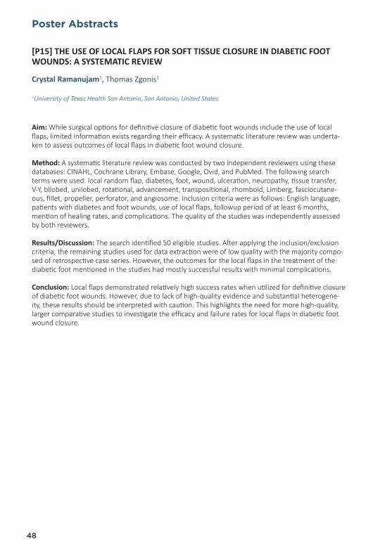
Poster Abstracts
48
[P15] THE USE OF LOCAL FLAPS FOR SOFT TISSUE CLOSURE IN DIABETIC FOOT WOUNDS: A SYSTEMATIC REVIEW
Crystal Ramanujam1, Thomas Zgonis1
1University of Texas Health San Antonio, San Antonio, United States
Aim: While surgical options for definitive closure of diabetic foot wounds include the use of local flaps, limited information exists regarding their efficacy. A systematic literature review was underta-ken to assess outcomes of local flaps in diabetic foot wound closure.
Method: A systematic literature review was conducted by two independent reviewers using these databases: CINAHL, Cochrane Library, Embase, Google, Ovid, and PubMed. The following search terms were used: local random flap, diabetes, foot, wound, ulceration, neuropathy, tissue transfer, V-Y, bilobed, unilobed, rotational, advancement, transpositional, rhomboid, Limberg, fasciocutane-ous, fillet, propeller, perforator, and angiosome. Inclusion criteria were as follows: English language, patients with diabetes and foot wounds, use of local flaps, followup period of at least 6 months, mention of healing rates, and complications. The quality of the studies was independently assessed by both reviewers.
Results/Discussion: The search identified 50 eligible studies. After applying the inclusion/exclusion criteria, the remaining studies used for data extraction were of low quality with the majority compo-sed of retrospective case series. However, the outcomes for the local flaps in the treatment of the diabetic foot mentioned in the studies had mostly successful results with minimal complications.
Conclusion: Local flaps demonstrated relatively high success rates when utilized for definitive closure of diabetic foot wounds. However, due to lack of high-quality evidence and substantial heterogene-ity, these results should be interpreted with caution. This highlights the need for more high-quality, larger comparative studies to investigate the efficacy and failure rates for local flaps in diabetic foot wound closure.

Info
rmat
ion
Ora
l Ab
stra
cts
Po
ster
Ab
stra
cts
Spo
nso
r-E
xhib
ito
rP
rog
ram
me
Aut
hors
49
[P16] STRATEGY FOR DIABETIC FOOT MANAGEMENT IN JAPAN: AID CONCEPT
Shinobu Ayabe1
1Yao Tokushukai General Hospital, Osaka, Japan
Diabetic ulcers are a major risk factor for lower extremity amputations. Diabetic foot problem are a common cause of morbidity. The aim of treatment for diabetic foot wound must be not only limb salvage but also gait salvage. Improving the management of diabetic foot disease is a mission for plastic surgeon in Japan because there are no specialist podiatrists.
Diabetic ischemic foot patients with infection are the most difficult to treat.
In patient with arterial insufficiency ulcers, restoration of blood flow by revascularization is the intervention that will most likely lead to healing. Infected feet require surgical debridement to achieve healing. The incision planning for debridement was designed based on the assumption that the defect would be closed by fillet flaps. The wounds were left open, and treated with NPWT. Fillet flaps were gradually advanced by NPWT, and complete wound closure was achie-ved.
But in ischemic foot, early debridement could result in worsened necrosis by increasing metabo-lic demand. So we performed marginal debridement without bleeding, not to worsen ischemia. And if necessary, we performed longitudinal incision and drainage to disturb the skin blood flow.
Foot deformities are a cause of pressure concentrations and create biomechanical stresses that cause ulcerations. The mainstay of treatment for foot deformities is offloading using proper footwear.
Here we propose a new concept of diabetic foot wound management, which we have termed AID (acronym for arterial insufficiency, infection and deformity) concept.
We reports strategy for diabetic foot wound management in Japan.

Poster Abstracts
50
[P17] WHAT DEGREE OF BLOOD SUPPLY AND INFECTION CONTROL IS NEEDED TO TREAT DIABETIC CRITICAL LIMB ISCHEMIA WITH FOREFOOT OSTEOMYELITIS?
Miki Fujii1, Hiroto Terashi2, Koichi Yokono3, David G. Armstrong4
1Department of Plastic and Reconstructive Surgery, Critical Limb Ischemia Center, Kitaharima Medical Center, Ono, Japan2Kobe University Hospital, Kobe, Japan3Kitaharima Medical Center, Ono, Japan4Southern Arizona Limb Salvage Alliance, University of Arizona College of Medicine, Tuscon, United States
Aim: Diabetic foot ulcer combined with ischemia and infection can be difficult to treat. However, no studies have shown the level of blood supply and infection control required to treat such complex ulcers. We aimed to develop an actual index for ischemia and infection control in the treatment of diabetic CLI with osteomyelitis that would increase the likelihood of limb salvage.
Method: We retrospectively studied the records of 30 patients with diabetic CLI combined with forefoot osteomyelitis (26 men, 4 women; mean age 68.4±11 years) treated surgically from 2009 to 2016. After 44 surgeries, we compared the differences between the healing group and the non-he-aling group in terms of patient background (age, sex, hemodialysis), infection status (pre-, post-, 1-week, 2-week postoperative C-reactive protein level (mg/l)), surgical bone margin (with or without osteomyelitis), vascular supply (skin perfusion pressure (mmHg)), ulcer size (Rutherford 5 or 6), and time to wound healing.
Results: Preoperative C-reactive protein levels (healing: 15±17 vs. non-healing: 43±32; p<0.05) and the ratio of patients with a Rutherford 6 (p<0.05) in the healing group were significantly lower than that in the non-healing group. The skin perfusion pressure level in the healing group was significantly higher than that in the non-healing group (54.2±14.1 mmHg vs. 36.4±14.8 mmHg; p<0.05).
Conclusion: Our study demonstrated that if the preoperative C-reactive protein level is greater than 40, debridement should be performed first to control infection. A 55 mmHg skin perfusion pressure value is necessary to successfully treat diabetic critical limb ischemia with forefoot osteomyelitis.

Info
rmat
ion
Ora
l Ab
stra
cts
Po
ster
Ab
stra
cts
Spo
nso
r-E
xhib
ito
rP
rog
ram
me
Aut
hors
51
[P18] TOTAL CONTACT CAST USE IN PATIENTS WITH PERIPHERAL ARTERIAL DISEASE: A CASE SERIES AND SYSTEMATIC REVIEW
Anthony Tickner1, Cheri Klinghard1, Jonathan2, Valerie Marmolejo3
1Restorix Health Wound Healing Center of Saint Vincent Hospital, Worcester, United States2Great River Medical Center, West Burlington, United States3Scriptum Medica, United States
Aim: The gold standard treatment to offload ulcerations due to excessive pressure is the use of a total contact cast (TCC). Yet, use of a TCC is contraindicated in patients with ischemia. Lower extremity ischemia typically presents in the more severe stages of peripheral arterial disease (PAD). As PAD exists on a spectrum of severity from mild to severe, designation of a clear cutoff where use of a TCC is an absolute contraindication would assist those who treat DFUs on a daily basis.
Method: This retrospective review of patient with mild to moderate PAD treated with a TCC and systematic review of the literature was performed to determine a potential cutoff value where TCC use would be an absolute contraindication.
Results/Discussion: TCC use appears to be a viable treatment option for pressure related DFUs in patients with an ankle pressure ≥70mmHG, a toe pressure ≥ 50mmHg, an ABI ≥0.45 or a TBI ≥0.4.
Conclusion: Risk/benefit analyze of each patient by the treating provider, close follow up, and patient education are essential components of TCC use in these patients. Formal vascular evalu-ation is recommended if the wound fails to progress towards resolution with TCC use.

Poster Abstracts
52
[P19] TCPO2 AS A TOOL TO EVALUATE THE RESULTS OF PERCUTANEOUS REVASCU-LARIZATION PROCEDURE ON PATIENTS WITH DIABETIC FOOT
Victor Rodriguez Saenz de Buruaga1, Gabriel Rivera1, Cristina Alzate1, Maider Mendia1, Jose Luis Higuera1, Jose Maria Egaña1
1San Sebastian University Hospital, Spain
Aim: Patients having an ischaemic or neuroischaemic diabetic foot (DF) are treated with endovascular techniques. There are different tools for testing the results of this treatment but amongst them our proposal is the Transcutaneous Oxygen Pressure (TcPO2). This indicator provides objectively and non-aggressively information about patient’s metabolic situati-on as well as the ability to healing lesions.
Method: Evaluation and following up of 14 patients diagnosed with ischemic or neuroischemic DF by monitoring TcPO2 before and after the endovascular treatment. First TcPO2 measurement M1 is taken 24 hours before and arteriography, secondly M2 the 9th day after the arteriography and lastly M3 after 15 days. The anatomic references for taking the samples are the thorax and forefoot.
Results/Discussion: We compared the foot TcPO2 M1, M2 and M3 before and after revascularization and the difference between direct and indirect revascularization according to angiosome concept. The results are: Foot TcPO2 M1 – M2 p of 0,001, Foot TcPO2 M1 – M3 p of 0,002, TcPO2 M2-3 p of 0,9. Foot TcPO2 M1 direct vs indirect p of 0,1, foot TcPO2 M2 direct vs indirect p < 0,05, foot TcPO2 M3 direct vs indirect p < 0,05.
Conclusion: These results reveal that there has been an increase of oxygen pressure comparing pre and post revascularization treatment and the direct vs indirect revascularization groups. Measuring the TCPO2 becomes a useful tool to evaluate the impact in tissues of the revascularization treatment as well as a guide to monitoring all the cases to be studied.

Info
rmat
ion
Ora
l Ab
stra
cts
Po
ster
Ab
stra
cts
Spo
nso
r-E
xhib
ito
rP
rog
ram
me
Aut
hors
53
[P20] MAJOR LOWER EXTREMITY AMPUTATION IS RADICAL SURGERY IN DIABETIC FOOT?
Danguole Vaznaisiene1, Rita Sulcaite2, Beltrand Eric3, Arturas Spucis4, Anatolijus Reingar-das4, Daiva Jomantiene5, Vytautas Kymantas5, Aukse Mickiene1, Eric Senneville6
1Infectious Diseases Department, Lithuanian University of Health Sciences, Kaunas Clinical Hospital, Kau-nas, Lithuania2 Department of Endocrinology, Lithuanian University of Health, Kaunas, Lithuania3Orthopedic Surgery, Tourcoing Hospital, Tourcoing, France4Lithuanian University of Health Sciences, Republican Hospital of Kaunas, Kaunas, Lithuania5Lithuanian University of Health Sciences, Kaunas, Lithuania6Service Universitaire des Maladies Infectieuses et du Voyageur, Centre Hospitalier Gustave Dron, Tourco-ing, France
Aim: To assess the spread of foot infection and its impact on the outcomes of major amputati-ons of lower extremities in diabetic patients.
Method: In a multicentre retrospective and prospective cohort study, we included adult diabetic patients (≥ 18 years) who underwent a major amputation of a lower limb in 5 hospitals between 2000 and 2009, 2012 and 2014. A total of 51 patients were included (of which 27 (52.94%) were men and 24 (47.06%) were women) with the mean age of 65.51 years (SD=16.99). Concomitant section’s osseous slice biopsy (BA) and percutaneous bone biopsy of the distal site (BD) were performed during limb amputation. The patients were followed-up for 1 year. Stump outcomes were assessed on the delay of complete healing, equipment, need of re-intervention and anti-biotics.
Results: In total, 51 BA were performed during major lower limb amputations (17 above the knee and 34 below the knee) in diabetic patients. Nine (17.65%) bacterial culture results from BA specimens were positive, 7 (13.73%) doubtful and 35 (68.63%) sterile. Microorganisms identified in BA were also cultured from the distal site in 33.33% of the cases. Positive BA was associated with prolonged complete stump healing, delay of complete healing (more than 6 months), re-amputation and the need of antibiotics
Conclusions: The microorganisms identified from BA play a role in stump healing in diabetic pa-tients. BA is useful during major limb amputation due to infectious complications and antibiotic therapy could be corrected on the basis of the BA culture results.

Poster Abstracts
54
[P21] 1 YEAR FOLLOW UP AFTER MAJOR AMPUTATIONS IN DIABETIC PATIENTS IN AN ITALIAN DIABETIC FOOT CENTER
Benedetta Masserini1, Fabrizio Signorelli2, Giacoma Di Vieste1, Ilaria Formenti1, Sara Lodi-giani1, Teresa Mondello1, Paola Cavaiani1, Gianmario Balduzzi1, Simone Introini3, Roberto De Giglio1
1 Internal Medicine, Diabetic Foot Center, Ospedale Cantù, Abbiategrasso, Italy2General Surgery, Ospedale Cantù, Abbiategrasso, Italy3Anesthesiology, Ospedale Cantù, Abbiategrasso, Italy
Aim: Despite the improved treatment of diabetic foot a persistent cohort of patients succumb to major amputation. Diabetic patients are usually older and with more severe comorbidities than other groups of patients who undergo major amputation and the outcome can be worse due to these associated conditions. The aim of the study was to evaluate the mortality and functional outcomes in a diabetic population who underwent major amputation (above knee - AKA and below knee - BKA) one year after surgery.
Method: 28 patients (5 AKA, 1 female, 4 male; 23 BKA 4 female, 19 male) were consecutively recru-ited in 2015 in the Diabetic Foot Center of Abbiategrasso Hospital. Evidence of adeguate perfusion after distal endorevascularization was obtained. They were evaluated one year after surgery in terms of mortality, wound healing and ambulation.
Results/Discussion: As AKA patients were compared with BKA subjects no difference in age (73.0±12.3 yrs vs 69.5±8.6 yrs, P=NS), glucose control (HbA1c 60±6 mmol/mol vs 64±16 mmol/mol P=NS) diabetes duration (18.2±8.6 yrs vs 13.8±7.2 yrs P=NS) and time required to heal (4.7±3.2 months vs 3.8±2.0 months P=NS) was observed. After one year one AKA patient ambulated (20%), while 13 subjects of BKA group (56.5%) had a functional recovery. One AKA patient (20%) and one BKA subject (4.3 %) died during follow-up.
Conclusion: These preliminary data suggest that in diabetic patients referred to a diabetic foot center BKA is associated with better functional and mortality outcomes and therefore should be preferred to AKA.

Info
rmat
ion
Ora
l Ab
stra
cts
Po
ster
Ab
stra
cts
Spo
nso
r-E
xhib
ito
rP
rog
ram
me
Aut
hors
55
[P22] BEYOND VIP: THE VIPERS APPROACH TO MULTI-DISCIPLINARY TEAM MANAGEMENT OF THE DIABETIC FOOT
Valerie Marmolejo1, Anthony Tickner2, Cheri Klinghard2
1Scriptum Medica, United States2Restorix Health Wound Healing Center of Saint Vincent Hospital, Worcester, United States
Aim: To provide an evidence base for the necessity for multidisciplinary teams treating the diabetic foot to shift beyond focus on the VIPs to VIPERS.
Method: The limb salvage team at Restorix Health Wound Healing Center of Saint Vincent Hospital has gone beyond the VIPs focusing on VIPERS: Vascular compromise, Infection mana-gement, Pressure reduction, Endocrine/glycemic control, Rehabilitation, and Surgical Interven-tion and Social/support network to provide evidence-based care of diabetic patients with foot ulceration.
Results/Discussion: With the worldwide prevalence of diabetes expected to be 550 million pe-ople by 2030, the concept of limb salvage through development of multidisciplinary teams has become an international and national trend. These teams have been shown to reduce major lower extremity amputation rates by 50%. Multi-disciplinary limb salvage teams have focused on the management of the VIPs of that delay wound healing, vascular compromise, infection and pressure. However, focus on the VIPs only does not optimize management for these patients on a whole as each of them present with a myriad of comorbidities and psychosocial attributes affecting treatment.
Conclusion: A dynamic shift from focusing on the VIPs to VIPERS provides optimal evidence-ba-sed, cost-effective care for patients afflicted with diabetic foot ulceration, which in turn will reduce morbidity and mortality and healthcare costs and increase patient quality of life.

Poster Abstracts
56
[P23] “ANYTHING THAT CAN GO WRONG, WILL GO WRONG” QUOTE MURPHY’S LAW THE MANAGEMENT OF ODDITIES AND ERRORS
Jan Rumbaut1
1Olv Ziekenhuis Aalst Belgium, Denderhoutem, Belgium
When dealing with diabetic foot problems, even in optimal conditions, it is well known that healing means extending the relapse free interval. The natural history of the illness is a balance between the new symptom and the reaction time of the health care team.
On the other hand, in recent times, we see a change in clinical presentations towards more complex problems, due to limb and patient related factors, as well as socio-economic situations.
Unexpected events, post-operative complications and dramatic evolution as a result of a wrong decision are truly feared.
The shift from a controlled to a dangerous situation usually starts with one or several of the following problems: acute vascular deterioration, rapid onset or progression of infection, tissue necrosis, skele-tal instability and non-union.
Acute vascular trombosis or embolism demands urgent revascularisation, but ischemia due to swelling and pus collection needs evacuation and decompression. Hyperbaric oxigen can be helpful in bridging before vascular procedure. Non reconstructable dry gangrene of toes is better lead to auto-amputation.
Infectious complications are the main indications for amputation; although immediate and repeated debridement respecting compartments, angiosomes and pathways of progression, can achieve more tissue preservation. Continuous or vacuum assisted intermediate irrigation prevents from drying. Spreading along tendon sheets and the dorsal subcutaneum cannot be underestimated. The need for resecting infected bone should be evaluated individually, potentially following MRI scan.
Major instability mainly arises from Charcot, sometimes due to fracture non-union or resection of in-fected bone. It can lead to complete loss of function, as a risk factor for pressure ulcer and infection, it even leads to amputation. As a consequence the decision to stabilize, internally or externally, has to be made urgently, before developing more bone loss.
The management of complications in diabetic foot is a dynamic process. Sometimes we face an acute life threatening situation. A well-structured team with therapeutic schedules and permanent access to operation time and vascular surgery is essential. By repeated evaluation the therapy should be planned step-by-step, always considering the functional outcome and the limits of footwear and ort-hosis. Partial amputation or even below knee amputation is considered as a reconstructive, functional procedure.
At the first presentation of an ambulatory patient, the chymaera of consecutive complications is already present. Integrating the patient and his family in each decision making process, is crucial for both the patient and the medical team in difficult and challenging situations.

Info
rmat
ion
Ora
l Ab
stra
cts
Po
ster
Ab
stra
cts
Spo
nso
r-E
xhib
ito
rP
rog
ram
me
Aut
hors
57
[P24] ANTIBIOTIC LOADED RIABSORBABLE BONE SUBSTITUTE IS A PROMISING ALTERNATIVE TREATMENT
Bernd Gächter1, Stephan Schlunke2
1Surgery & Wound-Care-Center in Southern Switzerland, Surgery, Minusio (Locarno, Bellinzona and Luga-no), Switzerland2Clinica Luganese Moncucco, Lugano, Switzerland
Aim: This case report demonstrates an until now little known possibility to treat in a very effective way patients affected by diabetic foot syndrome and infected malum perforans with osteomyelitis.
Method: A 67-year-old patient presented during a time lapse of one year a double relapse of a classical malum perforans with osteomyelitis of a metatarsal (MT) bone on the same foot but not the same MT. We found satisfactory arterial perfusion, the diabetes was well under control and radiologic evident osteolysis on both occasions.
We treated him for the first episode with limited resection of 4th MT head by means of a dorsal surgical access, microbiologic sample, leaving the plantar ulcer of malum perforans open, trea-ted with modern wound care dressings according to it’s evolution; an orthesis avoiding fore-foot weight bearing allowed him to walk normally. Antibiotic treatment was given according to the antibiogram for three months (picture series B).
The second episode of osteomyelitis involved the 1rst MT head: we treated it again by rese-ction of MT head, drilling open longitudinally the remaining MT and filling the canal with an absorbable antibiotic loaded bone graft substitute (Gentamycin), packing the void space of the MT head and direct skin closure. The patient dismissed autonomously the long term antibiotic therapy because of it’s side effects (picture series A).
Results/Discussion/Conclusion: Helped by orthopedic foam discharging soles, both plantar ulcers healed within 6 weeks. All biopsies showed Gentamicin sensible germs. Radiologic follow up at 2 month for 4th MT and 12 month for 1rst MT showed no recurrence of osteomyelitis. The material used as substitute in the 1rst MT produced a bone growth induction with complete resorption of the bone cement at one year. Using this later antibiotic loaded bone substitute, the total cost could be diminished by the price of the intravenous and oral long term antibiotic therapy, and also without any side effect for the patient.

Poster Abstracts
58
[P25] THE ROLE OF SEQUESTRECTOMY IN TREATMENT OF OSTEOMYELITIS IN DIABETIC FOOT
Sokol Hasho1, Eni Celo2
1Shefqet Ndroqi University Hospital, Tirana, Albania2Endocrinology, Mother Theresa Universitary Hospital, Tirana, Albania
Aim: To evaluate the role of sequestrectomy as a better solution compared to long term conservative treatment in diabetic patients.
Method: We performed 52 consecutive sequestrectomies in 51 diabetic patients with bone involve-ment demonstrate by a positive probe to bone and a positive rx. For each patient the part of bone resected resulted with abnormal consistence and was collected in a sample. In our study there were 80% males and 20% females. Mean age was 70±10 years old. Mean HbA1c values 8.2±2.4%.
Results/Discussion: In our study 37 patients (72%) resulted with peripheral vascular disease, while 15 patients (28%) had no arterial problems. In the hystological examination 48/52 patients (92%) resulted positive for osteomyelitis (acute inflammation, micro-abscesses, necrosis of trabecolae). In 4/52 patients (8%) resulted absence of osteomyelitis (presence of fibro productive process without infection). There were isolated a total of 54 strains. Among them 10 alert pathogens were identified (1 MRSA, 2 MRSCN, 1 Escherichia coli ESBL, 1 Klebsiella pneumoniae ESBL, 1 Pseudomonas aerugi-nosa ESBL, 2 VRE, 1 Acinetobacter lwofii MDR and 1 Acinetobacter calcoaceticus-baumanii complex MDR). 22 patients presented complete healing with a mean healing time of 85 ± 48 days. Antibiotic therapy was given orally for a mean duration of 22±9 days. No relapse of wounds or osteomyelitis was observed at the site of previous lesions in the follow up of 6 months.
Conclusion: Limited removal of infected bone is associated with a high percentage of success in healing osteomielitis with a very low relapse rate.

Info
rmat
ion
Ora
l Ab
stra
cts
Po
ster
Ab
stra
cts
Spo
nso
r-E
xhib
ito
rP
rog
ram
me
Aut
hors
59
[P26] SYME AMPUTATION FOR LIMB SALVAGE
Robert Frykberg1, Jaminelli Banks1, Edward Tierney1, Jaclyn Marino1, Priyanka Begur1
1Phoenix Va Hospital, Phoenix, United States
Aim: The objective of our study is to determine the criteria that can assist the surgeon in predic-ting successful outcomes when performing Syme amputations for limb salvage.
Method: We performed a single-center, retrospective cohort study of 76 patients (80 amputati-ons). All patients underwent a Syme amputation. Pre-operative data, short-term and long-term surgical outcomes were collected.
Results/Discussion: The mean age of our study population was 65 years (43–91 years), all males. 71/76 (93.4%) had DM2. 48/76 had PAD with 17 patients had ipsilateral lower extremity bypass surgery. 74/80 limbs had peripheral neuropathy. 39/76 had renal impairment (creatinine >1.3), and 8 patients were on hemodialysis.
43/80 (53%) healed their Syme amputation without any long-term complications at an average of 17.1 weeks. Only 54/80 limbs had a skin perfusion pressure test performed, 15/18 (83.3%) ca-ses with skin perfusion pressures below 30 mmHg did fail to heal. Forty three of the 80 (53.7%) surgical sites in this study initially healed the surgical incision at a mean time of 17.1 weeks. Fifty seven (71.3%) patients developed postoperative complications. Twenty three persons progres-sed to BKA. Fifty four patients died during the follow-up period for a death rate of 71.0%. The 3 year survival rate was 64.5% (49 out of 76 patients) and the 5 year survival rate was 51.3% (39 out of 76 patients). Of those that died, 14 (18.4%) required a proximal amputation.
Conclusion: The Syme amputation is an important limb salvaging procedure in diabetic patients with forefoot/midfoot necrosis, osteomyelitis, and/or ischemia.

Poster Abstracts
60
[P27] USEFUL OF DIFFUSION WEIGHTED IMAGE IN THE DIAGNOSIS OF SOFT TIS-SUE INFLAMMATION AND OSTEOMYELITIS
Yuta Terabe1
1Plastic and Reconstructive Surgery, Tokyo Nishi Tokushukai Hospital, Akishima, Japan
Aim: One of the most important factors in foot ulcer is to determine the extent of inflammatory and infection before debridement. Although Magnetic resonance imaging (MRI) of T1 and STIR sequence are useful for diagnosing osteomyelitis in recent years, bone marrow edema also appears in the same way, so it is difficult to differentiate at the boundary between reversible (acute inflammation) and irreversible inflammation tissue (chronic inflammation). MRI by diffusion weighted image (DWI) and apparent diffusion coefficient (ADC) is differentiate at the boundary between these tissues.
MRI by DWI and ADC is useful for determination of resected range of soft tissue and osteomyelitis.
Method: The case was 5 cases (M/F: 4/1, mean age 66±24.2) of critical limb ischemia (Rutherford 5, 6) treated at the Kasukabe Chuo General Hospital, department of Cardiovascular. MRI (STIR, T1, DWI and ADC) was taken after the revascularization. After confirming the inflammation range, acute inflammation (DWI and ADC are high signals) was preserved as much as possible, and all parts of chronic inflammation (DWI and ADC are low signals) were resected.
Results/Discussion: All cases didn’t lead to reoperative surgery, but maintenance debridement was necessary by bedside. Three cases were able to treat within a range smaller than the inflammation range found at STIR and T1, and two cases were in the same range.
Conclusion: DWI and ADC in MRI may be a new contributing factor in determining the scope of debridement in foot ulcers.

Info
rmat
ion
Ora
l Ab
stra
cts
Po
ster
Ab
stra
cts
Spo
nso
r-E
xhib
ito
rP
rog
ram
me
Aut
hors
61
[P28] OSTEOMYELITIS SEQUESTRECTOMY AND APPLICATION OF AN ANTI-BIOTIC-ELUTING BONE SUBSTITUTE TO AVOID MINOR AMPUTATION AND PRESERVE MECHANICAL STABILITY IN THE DIABETIC FOOT
Cristian Nicoletti1, Emanuele Nasole2, Elena Solagna2, Luca Spazzapan2
1Diabetic Foot Unit, Pederzoli Hospital, Peschiera del Garda, Italy2Pederzoli Hospital, Peschiera del Garda, Italy
Aim: To evaluate the effectiveness of an antibiotic eluting injectable synthetic bone graft substi-tute in the treatment of diabetic foot osteomyelitis (OM).
Method: Consecutive type 2-diabetic patients affected by OM who presented from October 2016 to March 2017 were included. All patients underwent standard care, bone biopsy, systemic antibiotic therapy according to antibiogram, OM sequestrectomy and implantation of a synthetic bone graft eluting either gentamicin or vancomycin, according to antibiogram*. All patients had negative vascular assessment for critical limb ischemia (CLI) and were assigned our standard offloading protocol. Patients were considered as “healed” if they had first-intention closure and/or did not undergo any other surgical procedures to heal.
Results/Discussion: A total of 12 patients were included: 8 with 1st-metatarsal head OM, 2 with cuboid and 2 with heel OM. Nine patients received the gentamicin-eluting bone substitute and 3 the vancomycin-eluting product. Six patients (50%) healed after the treatment. Three patients (25%) failed to heal due to CLI recurrence. The remaining 3 patients (25%) failed to heal pro-bably due to low compliance to the offloading protocol. One patient (with heel OM and ESRD) underwent Lower Extremity Amputation (LEA).
Conclusion: OM sequestrectomy with the use of the antibiotic-eluting synthetic bone graft substitutes*, was effective in healing diabetic patients with OM of the foot, thereby avoiding minor amputations, with consequences for biomechanical stability, and reducing the need for long-term antibiotic therapy.
*CERAMENT G or CERAMENT V, BONESUPPORT AB

Poster Abstracts
62
[P29] ALL CAUSE AND CARDIOVASCULAR MORTALITY IN A CONSECUTIVE SERIES OF PATIENTS WITH DIABETIC FOOT OSTEOMYELITIS
Alessia Scatena1, Lucia Ricci2, Danilo Tacconi3, Giorgio Ventoruzzo4, Francesco Liistro5, Leonardo Bolognese5, Matteo Monami6, Edoardo Mannucci7
1Diabetic Foot Care Unit, San Donato Hospital Arezzo, Arezzo, Italy2Diabetology Unit, San Donato Hospital Arezzo, Arezzo, Italy3Infective Disease Unit, San Donato Hospital Arezzo, Arezzo, Italy4Vascular Surgery Unit, San Donato Hospital Arezzo, Arezzo, Italy5Cardiovascular and Neurologic Department, San Donato Hospital Arezzo, Arezzo, Italy6Diabetic Foot Unit, University of Florence, Florence, Italy7Diabetology Agency, University of Florence, Florence, Italy
Aim: Mortality in patients with type 2 diabetes and diabetic foot osteomyelitis (DFO) have been explored in few small studies with a short follow-up. Aim of the present study is to assess all-cause and cardiovascular mortality and predictors of mortality in a consecutive series of patients with DFO.
Method: Patients with a diagnosis of DFO, attending the Diabetic Foot Unit of San Donato Hospital in Arezzo between January 1st, 2012 and December 31st, 2013, were included in this retrospective study. Information on all-cause mortality up to December 1st, 2016, was obtained from the registry of the Local Health Unit of Arezzo, which contains updated records of all persons living in Tuscany.
Results/Discussion: One hundred ninety four patients were included in the study. During a mean period of observation of 2.8±1.4 years, 73 (37.6%) died, with a yearly rate of 13.2%. Of the 73 deaths, 59 were attributable to cardiovascular causes. After adjusting for possible confounders in a Cox analysis, site of osteomyelitis (hindfoot vs mid/forefoot) was associated with a higher mortality, and surgical treatment with a lower mortality.
Conclusion: Mortality in patients with DFO appears to be much higher than that reported in clinical series of patients with diabetic foot ulcers, particularly when hindfoot is affected.

Info
rmat
ion
Ora
l Ab
stra
cts
Po
ster
Ab
stra
cts
Spo
nso
r-E
xhib
ito
rP
rog
ram
me
Aut
hors
63
[P30] LISFRANC AMPUTATION, THE REVIVAL OF A HISTORICAL SURGERY
Estelle How Hong1, Raeesa Patel1, Jennifer Buxton1
1Department of Vascular Surgery, Royal Blackburn Hospital, Blackburn, Lancashire, United Kingdom
Aim: The use of Lisfranc amputation for limb salvage, named after French surgeon Jaques Lisfranc de St Martin, has previously faced scepticism due to its poor prosthesis suitability and abnormal loading. With a rising frequency of diabetic foot infections, the rate of amputation is rapidly climbing. We propose that Lisfranc amputation may be used in selected cases for limb salvage and hinder the progression to major amputation especially when infection/osteomyelitis and vascular compromise are involved. We describe a recent case of a successful limb salvage after distal angioplasty and Lisfranc amputation with a review of the literature.
Method: Patient’s permission was gained to review all notes pertaining to surgery. A Pubmed search was carried out using the keywords: ‘Lisfranc’, ‘amputation’ and ‘diabetes’.
Results/Discussion: A total of 14 articles were reviewed (2 excluded, 1 systematic review, 1 retrospective study, 5 prospective cohort study, 2 case series, 1 review article, 1 case report and 1 expert opinion) which mainly focussed on evaluating midfoot amputations and the factors im-proving its post surgical outcomes. Two articles were rejected as they were mainly focussed on Charcot arthrodesis. Midfoot amputation, when performed under the hands of an experienced surgeon, with early revascularisation intervention, adequate skin flaps and good prothesis, can achieve excellent functional outcome, shorter hospital stay and reduced morbidity and mortality rate associated with major amputation.
Conclusion: In diabetic foot disease, aggressive limb salvage with midfoot amputation should consider first as an alternative to higher level amputations which are associated with high mor-tality and morbidity rate.

Poster Abstracts
64
[P31] SKIN STRETCHING DEVICE SSD FOR REPAIR OF DIABETIC FOOT ULCER
Jiangning Wang1, Lei Gao1
1Shijitan Hospital Affiliated Capital Medical University, Beijing, China
Aim: To investigate the effect of the skin stretching device (SSD) in repair of the diabetic foot ulcer.
Method: 32 patients with diabetic foot ulcer were randomly divided into two groups after debride-ment (Soft tissue defects formed in the ulcer area, the area of defect is between 20cm2 and 30cm2),
one group treated with vacuum sealing drainage (VSD), combined with skin graft; the other group treated with skin stretching device (SSD) only. The wound infection rate, wound healing time and the curative effect were evaluated.
Results/Discussion: All the patients were followed up for 1 to 4 months. There were 1 cases with wound infection in VSD group, and 0 case in SSD group, there was no difference between two groups (P =0.500). Healing time in the SSD group was significantly shorter than that in the VSD group (P =0.010.
Conclusion: Compared to the VSD, the SSD in Soft tissue defect healing is easy and convenient, the wound healing time is shorter, with small scars, and the curative effect is obvious, especially, the skin in the reparation area is adjacent to the surrounding skin, and the skin histology is similar after healing.

Info
rmat
ion
Ora
l Ab
stra
cts
Po
ster
Ab
stra
cts
Spo
nso
r-E
xhib
ito
rP
rog
ram
me
Aut
hors
65
[P32] ANTIMICROBIAL BIOACTIVE GLASS S53P4, IN COMPLICATED DIABETIC FOOT ULCERS WITH OSTEOMYELITIS: CASE REPORT
De Giglio R1, Masserini B1, Mondello T1, Di Vieste G1, Lodigiani S1, Formenti I1, Cavaiani P1, Signorelli F1, Balduzzi G1, Mazzone A.1
1Medical Department, Diabetic foot unit, Abbiategrasso Hospital, Asst Ovest Milanese, Italy
The aim of the study is to evaluate the efficacy of BioActive Glass (BAG-S53P4) in the treatment of complex diabetic foot ulcers with bone exposure and osteomyelitis, in the attempt to limb salvage. BAG-S53P4 is a bone substitute with proven antibacterial and bone bonding properties commonly used in orthopedic surgery.
Two patients with complicated insulin treated diabetes aged 54 and 62 years were treated. In the first patient who was in hemodialysis and had undergone transmetatarsal amputation, a non-healing plantar ulcer after one year of antibiotic treatment, off-loading and local surgical debridement was described.
In the second patient who had undergone Chopart amputation and revascularization a complete wound healing was not reached 6 months after surgery.
The outcome of both patients with clinically and radiologically verified chronic osteomyelitis, requiring surgical debridement and bone void filling using the antibacterial BAG-S53P4 as bone graft substitute, is reported.
Both patients showed complete wound healing after 3 and 2 months respectively and post-ope-rative X-ray at three and six months follow up showed no radiological sign of infection.
The results suggest that use of BAG-S53P4 may be effective in improving the healing of complex diabetic foot ulcers that present with osteomyelitis. Prospective, randomised controlled trials are warranted to confirm the BAG-S53P4 efficacy as well as its possible application in other chronic wounds.

Poster Abstracts
66
[P33] EFFICACY, SAFETY AND ACCEPTANCE OF AN INTERIM ORTHOSIS* IN PATIENTS WITH DIABETES AFTER CHOPART SURGERY
Di Vieste G1, Balduzzi G1, De Giglio R1, Masserini B1, Mondello T1, Lodigiani S1, Formenti I1, Cavaiani P1, Signorelli F1, Mazzone A.1
1Medical Department, Diabetic foot unit, Abbiategrasso Hospital, Asst Ovest Milanese, Italy
Introduction Chopart amputation is the consequence of severe diabetes-related foot complications. Current clinical management after surgery consists in promoting healing until wearing a cus-tom-made prosthesis.
The interim orthosis* is a new device that can be used after Chopart surgery. It potentially could allow the patient greater degree of motility than the currently used systems.
Aim of our study was to evaluate efficacy, safety and acceptance of this new orthosis.
Methods We have performed a prospective observational study involving people with diabetes who have undergone Chopart surgery from January 2016 until February 2017 at the Cantù Hospital, Abbi-ategrasso, Italy. After surgery patients were followed up until prosthesis application.
Only patients without complications requiring re-amputation in the immediate post-operative period were selected. Main study outcomes were major amputation occurrence, ulcer recurrence, healing time and patients’ acceptance of the orthosis. Foot was considered healed when complete re-epithe-lization of the surgical wound had occurred. Patients’ acceptance was evaluated by using the Italian validated version of the Orthotic Prosthetic User’s Survey (OPUS) questionnaire.
Results Overall, 14 subjects with diabetes (mean age 67.3±9.2 years, 57.1% males, mean diabetes duration 27±15.3 years, 64.2% insulin treated) were enrolled. Mean follow-up was of 12.5±4.1 months. All the patients were alive at the last follow-up visit. None of the patients had major amputa-tion or ulcer recurrence. All the patients had their wound healed. Mean time of wound healing was of 113.6±75 days. No adverse events were recorded. Patients’ acceptance of the new orthosis was high.
Conclusions The interim orthosis* is an efficient, safe and well accepted orthosis for patients with diabetes who have undergone Chopart surgery, therefore it use could be always suggested after Chopart surgery.
* the Body Armor®Pro Term orthosis

Info
rmat
ion
Ora
l Ab
stra
cts
Po
ster
Ab
stra
cts
Spo
nso
r-E
xhib
ito
rP
rog
ram
me
Aut
hors
67

68
Company Description
AcelityTel. +1 800 275-4524 ext. [email protected]
Acelity L.P. Inc. and its subsidiaries are a global ad-vanced wound care company that leverages the strengths of Kinetic Concepts, Inc. and Systagenix Wound Management, Limited. Available in more than 90 countries, the innovative and complementary ACELITY™ product portfolio delivers value through solutions that speed healing and lead the industry in quality, safety and customer experience. At Acelity, our shared values drive our company’s culture and priorities by providing a framework for making decisions. At the forefront of our values is Customer First – the idea that we provide the best solutions for our customers and patients. We work with integrity, and are accountable for our actions. We have a will to win, and enjoy being the best at what we do. And we work as One Acelity, executing as one team with a global mindset.
BONESUPPORT ABTel. +44 7568 337 984 Rich [email protected] [email protected]
BONESUPPORT™ is an orthobiologic company specializing in the development of innovative injectable bone graft substitutes that remodel into bone within 6 to 12 months. Used in more than 30,000 patients, and includes the only CE marked injectable antibiotic eluting bone graft substitutes; CERAMENT|G with gentamicin, and CERAMENT|V with vancomycin.
DARCO (Europe) GmbHTel.: +49 887 922 [email protected]
DARCO is dedicated to being one of the leading providers of post op, trauma and wound care solutions to the global foot and ankle community.
DEKATel.:+390558826807 www.dekalaser.com [email protected]
A spin-off of the El.En. Group, DEKA is a world leader in the design and manufacture of lasers and light sources for medical applications in more than 80 countries. Excellence is the hallmark of the DEKA’s experience and recognition garnered in the sphere of R&D in over 30 years of activity. Quality, innovation and technological excellence place DEKA and its products in a unique and distinguished position in the global arena.
DM Systems’ HeeliftTel.: +1 847 328 [email protected]
Avoid amputation, heal chronic wounds & prevent heel pressure ulcers. See published clinical evidence, request a sample. The Heelift® Suspension Boot.
SPONSOR AND EXHIBITOR INFORMATION

Info
rmat
ion
Ora
l Ab
stra
cts
Po
ster
Ab
stra
cts
Spo
nso
r-E
xhib
ito
rP
rog
ram
me
Aut
hors
69
Company Description
Genadyne BiotechnologiesTel.: +1 732 672 7056 [email protected] www.genadyne.com
Genadyne specializes in Negative Pressure Wound Therapy (NPWT) systems, including a variety of single patient use systems. Advanced wound dressings with Nanotechnology to address non healing chronic wounds. And the most advanced colloidal silver spray solution for infected wounds.Through continuous research and development, Genadyne has been able to provide a new dimension to wound care and has been able to create some of the most advanced, cost-effective products for the healthcare community.
Integra LifeSciencesRoberto Andreose Tel.: +39 366 7753414 roberto.andreose@ integralife.comwww.integralife.eu
Integra is a world leader in medical technology, dedicated to limiting uncertainty for clinicians, so they can concentrate on providing the best treatment options for their patients. Integra offers innovative regenerative collagen based solutions for nerve repair, dermal repair and soft tissue reconstruction. The Integra collagen has been used successfully in more than 10 million procedures worldwide.With the completion of the acquisition of Dermas-ciences, a tissue regeneration company focused on advanced wound and burn care solutions featuring a wide portfolio including the TCC-EZ offloading total contact cast for the management of DFU plantar ulcers, Integra positions itself as a leader in the treatment of diabetic foot, and in Wound Reconstruction and Care.
MimedexTel.:+43 664 142 5224 Milena Ridl [email protected] www.mimedx.com
MiMedx is the leading regenerative medicine company utilizing human placental tissue and patent-protected processes to develop and market advanced products and therapies for the Wound Care, Surgical, Orthopedic, Spine, Sports Medicine, Ophthalmic, and Dental sectors of healthcare.
Orthofix Tel.: +39 045 [email protected]
Orthofix International N.V. is a diversified, global medical device company focused on improving patients’ lives by providing superior reconstructive and regenerative orthopedic and spine solutions to physicians worldwide. The company has four strategic business units that include BioStim, Biologics, Extremity Fixation and Spine Fixation.
Ottobock Soluzioni OrtopedicheTel.: +39051 [email protected]
We’re ottobock’s orthopaedic workshop network: we provide technological devices (prosthetics, orthotics and mobility solutions) in order to help patients improving their quality of life. We’re special-ized in the application of high-tec prosthtetics and made-to measure orthopaedic devices.

70
Company Description
Perimed ABTel.: +46 8 580 119 90 [email protected] www.perimed-instruments.com
PERIMED is a global provider of diagnostic solutions for patients with peripheral vascular diseases and complex diabetic foot ulcers. Our new PeriFlux 6000 offers a unique combination of tests: ABI, toe pressure and transcutaneous oximetry (tcpO2).
SALUBER SRLTel. +39 0424 570321 Mob.+39 349 [email protected]
Saluber srl, based in Veneto region Italy, is the man-ufacturer of thr FORS™ offloading insole for diabetic foot ulcers which has recently been evaluated at multiple academic institutions with excellent results. Additionally we wiil be exhibiting our non-custom or-thotics products, which are an effective an luxurious alternative to other OTS orthotics.
Salvatelli S r L Tel.: +39 0733 801 060 [email protected] www.molliter.com
Salvatelli S.r.L. is a company that produces orthopaedic shoes (Molliter) and dynamic Walkers for the management of the Diabetic foot ulceration or post-surgery/trauma (Optima).
Urgo MedicalTel. +33 3 80 54 50 00www.urgomedical.com
URGO Medical is the Healing Company committed each day to improve wound care treatments for both patients and health care professionals by offering highly innovative solutions.
Woundcare-CircleTel.: +49 880 792 [email protected]
The 3 Woundcare-Circle founders, OPTIMA, Heelift and DARCO are international market leaders providing innovative product solutions. The group permanently supports research & science as well as the advancement of foot disease management
Wright Medical Tel.: +44 (0)845 833 4435 Inter-national Headquarters www.wright.com/contact-uswww.wright.com
Wright medical focus on Extremities and Biologics through our team of passionate and dedicated people, we deliver innovative, value-added solutions improving quality of life for patients worldwide. We are committed to compliance and the highest standards of ethical conduct.
SPONSOR AND EXHIBITOR INFORMATION

Info
rmat
ion
Ora
l Ab
stra
cts
Po
ster
Ab
stra
cts
Spo
nso
r-E
xhib
ito
rP
rog
ram
me
Aut
hors
71

72
Abbruzzese, Lorenza P10
Adami, Daniele O1
Allender, Chad P5
Alzate, Cristina P19
Antsiferov, Mikhail O9
Arhire, Lidia Iuliana P7
Armstrong, David P17
Ayabe, Shinobu P16
Balduzzi, Gianmario P21, P32, P33
Banchellini, Elisa P10
Banks, Jaminelli P26, O4, O5
Begur, Priyanka O5, P26
Bem, Robert P4
Benedikt, Stetan P2
Berchiolli, Raffaella O1
Bolognese, Leonardo O3, P29
Bosic, Srecko P11
Buxton, Jennifer P30
Cavaiani, Paola P21, P32, P33
Celo, Eni P25
Chen, Tiangui O2
Ciuntu, Bogdan P7
Coppelli, Alberto O10, P10
De Giglio, Roberto P21, P32, P33
Di Vieste, Giacoma P21, P32, P33
Didomenico , Lawrence P9
Dubský, Michal P4
Egaña, Jose Maria P19
Ercolini, Leonardo O3
Eric, Beltrand P20
Ferrari, Mauro O1
Flores, Regino P5
Flynn, Zachary P9
Formenti, Ilaria P21, P32, P33
Frykberg, Robert P26, O4, O5
Fujii, Miki P17
Gächter, Bernd P24
Gao, Lei P12, O2, P31, P6
Georgescu, Stefan P7
Gherasim, Andreea P7
Goretti, Chiara O10, P10
Graur, Mariana P7
Grindlay, Douglas O6
Grohmann, Martin P2
Hasho, Sokol P25
Higuera, Jose Luis P19
How Hong, Estelle P30
Iacopi, Elisabetta O10, P10
Introini, Simone P21
Jomantiene, Daiva P20
Kamolz, Lars-Peter P2
Khachatryan, Hamazasp P14
Klaue, Kaj P1
Klinghard, Cheri P18, P22
Komelyagina, Elena P8, O9
Kreuz, Emma P5
Kymantas, Vytautas P20
Liistro, Francesco O3, P29
Lodigiani, Sara P21, P32, P33
Maioli, Filippo O3
Mannucci, Edoardo P13, P29
Marconi, Michele O1
Mari, Marta O1
Marino, Jaclyn P26
Marmolejo, Valerie P18, P22
Name Abstract Bold = Presenting author
AUTHORS

Info
rmat
ion
Ora
l Ab
stra
cts
Po
ster
Ab
stra
cts
Spo
nso
r-E
xhib
ito
rP
rog
ram
me
Aut
hors
73
Marson, Ben O6
Marzoq, Abdulhussein P3
Masserini, Benedetta P21, P32, P33
Mazzone, A. P32, P33
Mcguire, James P5
Jonathan P18
Mendia, Maider P19
Mickiene, Aukse P20
Mihalache, Laura P7
Mittlmeier, Thomas P1
Mocellin, Davide Maria O1
Monami, Matteo P13, P29
Mondello, Teresa P21, P32, P33
Nasole, Emanuele P28
Navratil, Kamil P4
Nicoletti, Cristian P28
Nita, George P7
Nita, Otilia P7
Obolenskiy, Vladimir O7, O8, P8, O9
Ollivere, Ben O6
Patel, Raeesa P30
Penny, Harry P5
Petruzzi, Pasquale O3
Piaggesi, Alberto O10, P10
Protsko, Viktor O7, O8, O9, P8
Rafat, Payam P5
Ramanujam, Crystal P15
Reingardas, Anatolijus P20
Ricci, Lucia O3, P13, P29
Riitano, Nicola O10, P10
Rivera, Gabriel P19
Rodriguez Saenz de Buruaga, Victor
P19
Rumbaut, Jan P23
Sabanchieva, Nuria O9
Scammell, Brigitte O6
Scatena, Alessia O3, P13, P29
Schintler, Michael P2
Schlunke, Stephan P24
Senneville, Eric P20
Signorelli, Fabrizio P21, P32, P33
Sixta, Bedřich P4
Soghomonyan, Nune P14
Solagna, Elena P28
Spazzapan, Luca P28
Spucis, Arturas P20
Sulcaite, Rita P20
Tacconi, Danilo P29
Terabe, Yuta P27
Terashi, Hiroto P17
Tickner, Anthony P18, P22
Tierney, Edward P26
Tomei, Francesca O1
Uliniuc, Cosmin P7
Vasiluta, Ciprian Petrisor P7
Vasilyeva, Anna P2
Vaznaisiene, Danguole P20
Ventoruzzo, Giorgio O3, P29
Wang, Jiangning P12, O2, P31, P6
Weaver, Adam P5
Woskova, Veronika P4
Wukich, Dane O4
Yokono, Koichi P17
Zgonis, Thomas P15
Name Abstract Bold = Presenting author

NOTES


The A-DFS 2017 is organised with the highly appreciated support of:
EXHIBITORS
BRONZE SPONSORS
SILVER SPONSORS
GOLD SPONSORS