Embed Size (px)
Citation preview

BearWorks BearWorks
MSU Graduate Theses
Fall 2017
Assessment Of Reliability And Stability Of Various Visual Search Assessment Of Reliability And Stability Of Various Visual Search
Parameters Parameters
Michael Don Mizer Missouri State University, [email protected]
As with any intellectual project, the content and views expressed in this thesis may be
considered objectionable by some readers. However, this student-scholar’s work has been
judged to have academic value by the student’s thesis committee members trained in the
discipline. The content and views expressed in this thesis are those of the student-scholar and
are not endorsed by Missouri State University, its Graduate College, or its employees.
Follow this and additional works at: https://bearworks.missouristate.edu/theses
Part of the Cognition and Perception Commons
Recommended Citation Recommended Citation Mizer, Michael Don, "Assessment Of Reliability And Stability Of Various Visual Search Parameters" (2017). MSU Graduate Theses. 3204. https://bearworks.missouristate.edu/theses/3204
This article or document was made available through BearWorks, the institutional repository of Missouri State University. The work contained in it may be protected by copyright and require permission of the copyright holder for reuse or redistribution. For more information, please contact [email protected].

ASSESSMENT OF RELIABILITY AND STABILITY OF VARIOUS
VISUAL SEARCH PARAMETERS
A Masters Thesis
Presented to
The Graduate College of
Missouri State University
TEMPLATE
In Partial Fulfillment
Of the Requirements for the Degree
Master of Science, Psychology
By
Michael Don Mizer
December 2017

ii
Copyright 2017 by Michael Don Mizer
iii
ASSESSMENT OF RELIABILITY AND STABILITY OF VARIOUS VISUAL
SEARCH PARAMETERS
Psychology
Missouri State University, December 2017
Masters of Science
Michael Don Mizer
ABSTRACT
Research in social science has been on a continuous self-correcting path as scientists find
new ways to look at old problems. Recent technology has given us the ability to perform
compounded calculations in a fraction of previous times while recording complex
measurements with greater degrees of precision. While this is helpful regarding corporeal
measures, quantifying cognition is still a difficult task. Recently, many computer-aided
eye tracking devices have been developed and used to validate visual search theories.
However, few inquiries have been made assessing the reliability and stability of these
methods. This study assessed the reliability and stability of visual attention tasks using
the Gazepoint eye-tracker. Visual scanning behaviors of 46 participants were recorded to
provide evidence of reliability and stability of four measurement outcomes: (1) total
number of fixations, (2) latency to first fixation, (3) total time attending, and (4) total
number of switches between areas of interest. All visual scanning measures were found to
be stable across stimuli and trials with total number of fixations and total fixation time
being the most reliable visual scanning measure. These findings can afford better visual
theory development and predictions of subsequent development outcomes.
KEYWORDS: reliability, stability, visual scanning, saliency, faces, individual
differences,
This abstract is approved as to form and content
_______________________________
D. Wayne Mitchell, PhD
Chairperson, Advisory Committee
Missouri State University
iv
ASSESSMENT OF RELIABILITY AND STABILITY OF VARIOUS VISUAL
SEARCH PARAMETERS
By
Michael Don Mizer
A Masters Thesis
Submitted to the Graduate College
Of Missouri State University
In Partial Fulfillment of the Requirements
For the Degree of Master of Science, Psychology
December 2017
Approved:
_______________________________________
D. Wayne Mitchell, PhD
_______________________________________
Erin M. Buchanan, PhD
_______________________________________
Melissa D. Fallone, PhD
_______________________________________
Julie Masterson, PhD: Dean, Graduate College
In the interest of academic freedom and the principle of free speech, approval of this
thesis indicates the format is acceptable and meets the academic criteria for the discipline
as determined by the faculty that constitute the thesis committee. The content and views
expressed in this thesis are those of the student-scholar and are not endorsed by Missouri
State University, its Graduate College, or its employees.

v
ACKNOWLEDGEMENTS
Firstly, I would like to thank my wife and family for their unconditional love and
support throughout my studies. To my thesis committee, Dr. Wayne Mitchell, Dr. Erin
Buchanan, and Dr. Melissa Fallone, I am grateful for the knowledge gained and the
positive Missouri State experience brought about through your unwavering commitment
and support to the students and psychology program. To the “labbies” (Buddy, Christina,
Derby, and Stacy) for keeping me in check and ensuring everything ran smoothly. Last
and certainly not least, to the “nerd herd,” your constant support and encouragement over
the past two years made my graduate experience not only manageable, but memorable.
vi
TABLE OF CONTENTS
Introduction ..........................................................................................................................1
Attention ..................................................................................................................2
Visual Attention .......................................................................................................3
Visual Scanning Theoretical Perspectives ...............................................................6
Visual Perception Measurements .............................................................................7
Purpose of This Study ............................................................................................10
Primary Hypotheses ...............................................................................................12
Methods..............................................................................................................................14
Sample....................................................................................................................14
Materials ................................................................................................................15
Procedures ..............................................................................................................15
Debriefing ..............................................................................................................18
Results ...............................................................................................................................19
Preliminary Analyses .............................................................................................19
Primary Analyses ...................................................................................................19
Total Number of Fixations .....................................................................................21
Latency to First Fixation ........................................................................................22
Total Time Attending .............................................................................................23
Total Number of Shifts Between AOI’s ................................................................25
Discussion ..........................................................................................................................27
Total Number of Fixations .....................................................................................27
Latency to First Fixation ........................................................................................28
Total Time Attending .............................................................................................28
Total Number of Shifts ..........................................................................................29
Two-Trial Consistency Combinations ...................................................................30
Limitations .............................................................................................................30
Conclusion .............................................................................................................31
References ..........................................................................................................................32
Appendices ........................................................................................................................35
Appendix A. Consent Form ...................................................................................35
Appendix B. Demographic Information Form.......................................................36
Appendix C. WAIS Participant Response Form....................................................37
vii
LIST OF TABLES
Table 1. Presentation Effects – Order and Gender. ...........................................................38
Table 2. Primary Analysis .................................................................................................39
Table 3-6. Two-Trial Consistencies for Face and Object. .................................................40
Table 7-10. Two-Trial Consistencies for Face and Object Matched Pairs ........................44
Table 11-14. Two-Trial Consistencies for Faces ...............................................................48
Table 15-18. Two-Trial Consistencies for Objects ............................................................52
Table 19-22. Two-Trial Consistencies for Face Matched Pairs ........................................56
Table 23-26. Two-Trial Consistencies for Object Matched Pairs .....................................60
viii
LIST OF FIGURES
Graph 1. Group differences: Total number of fixations ....................................................64
Graph 2. Group differences: Latency to first fixation ........................................................65
Graph 3. Groups differences: Total fixation time ..............................................................66
Graph 4. Groups differences: Total number of shifts ........................................................67
1
INTRODUCTION
In the advent of technology, computer aided measurement has become a preferred
method of investigation by many scientists in social science research. In the field of
cognitive science, it has “provided that much needed assurance that cognitive processes
were real; that they could be studied and perhaps understood” (Neisser, 1976). While
technology supports the scientific process, researchers in the field of cognition and
perception continue to face the challenge of identifying “physiological correlates of
cognitive processes…[and] have typically been motivated by two primary goals: (1) to
discover the mechanisms underlying these processes and (2) to develop empirical indices
that will mark the occurrences of a cognitive event, thereby validating the process”
(Cohen & O’Donnell, 1993).
Regarding visual search processes, many models have been proposed; however,
while these models may share theoretical overlap of causal agents and relationships,
differences among measurements are impossible to discern if we cannot obtain repeatedly
the same results by using the same criteria over repeated trials. Researchers have
continued to invent unique methods of measuring visual attention and subsequent
underlying cognitive processes. Specifically, computer-aided eye tracking devices have
been developed (i.e. brands: Gazepoint GP3, Tobii, Pupil Labs, Eye Tribe, etc.) and used
to validate visual search theories. For instance, numerous theories have embraced the
phenomena that visual attention is directed to the items in the visual field in the order of
decreasing saliency irrespective of the task at hand (Theeuwes, 1992; Wykowska &
Schubo, 2009). This phenomena relates to research concerning automaticity (Schneider
2
& Shiffrin, 1977; Shiffrin & Schneider, 1977), pre-attentive processes (Neisser, 1967),
pop-out effect (Treisman & Gelade, 1980) , and other similar events that occur without
attentional effort. To date these phenomena have not been tested using visual scanning
techniques which afford an active (moment to moment) measurement (milliseconds) of
visual attention.
Attention
Cognition can be described as the way in “which sensory input is transformed,
reduced, elaborated, stored, recovered, and used. It is concerned with these processes
even when they operate in the absence of relevant stimulation, as in images and
hallucinations” (Neisser, 1967). Attention is the salient feature of cognition; a conscious
manifestation of experience. It allows “high-level processing of information in a capacity
limited manner” (Kalivas & Petralia, 2012). The study of attention has had an irregular,
often seemingly absent, path of existence during the dawning years of psychological
theory (Kahneman, 1973; Neisser, 1976). While theories of attention have been sources
of debate throughout time, they began to consistently gain credence toward the late 19th
and early 20th century. However, with the onset Gestalt psychology and behaviorism, it
was the efforts of philosophers/researchers such as William James (1890), Edward
Titchener (1908), Wilhelm Wundt (1910), and others who kept the concept of attention
from fading completely. Early on, attention was most aptly defined as “the taking
possession by the mind, in clear and vivid form, of one out of what seem several
simultaneously possible objects or trains of thought. Focalization and concentration, of
consciousness are of its essence” (James, 1890). James theorized an analogous

3
“spotlight” model that likened attention to having a focus, a margin, and a fringe, much
like a beam of light with higher-resolution toward the center (focus) and subsequently
deteriorating as it moves outward (fringe). What James offered was a framework for a
definition that continues to exist today. Half of a century later, Donald Broadbent (1958)
introduced a filter model of selective attention followed by Charles Eriksen’s (Eriksen &
James, 1986) zoom lens model of attention, both theories further defined and supported
James’ earlier reasoning. Broadbent’s Filter Theory (1958, p. 43) further supposed a type
of “bottleneck” theory—based on the work of Kenneth Craik and his single-channel
theory (Craik, 1947)—which allows one bundle of relevant information to pass while
rejecting another. The bottleneck theory surmises a problem of attention in that,
regardless of conscious effort, an individual can only attend to a limited number of things
at any given time (Deutsch & Deutsch, 1963; Kahneman, 1973).
Visual Attention
The human optic nerve is said to be able to transmit 107-108 bits (1 gigabyte =
134,217,728 bits) of information each second (Itti & Koch, 2001). However, visual
attention, like attention in general, is capacity limited, except it is based solely on data
collected from visual input. Visual attention is a function of person-centric biological
mediators—i.e. anatomical integrity, biochemical makeup, physiological processes, etc.
(Campbell & Green, 1965) and external stimulus attributes (size, color, form, etc.).
When an individual visually scans an area of interest (AOI), only a limited
amount of information is consciously attended. The rest is either filtered and allocated to
the subconscious mind or due to functional design, it is never realized in the first place.

4
The latter deals with one way a person scans his or her environment; a phenomenon
known as saccadic eye movement.
Saccadic Movement. A saccade is a rapid movement of the eye between two
fixation points, approximately 30/ms in duration, with each fixation (still period between
saccades) lasting on average 30/ ms (Irwin, 1991). Saccadic eye movement allows an
individual to take in target information through simultaneous movements of both eyes,
however it is not a fluid movement that allows for encoding of all available stimuli. As
the eyes shift, higher level images may be processed while lower level images are
attenuated, a phenomenon known as saccadic masking. As the eyes move from one AOI
to another (approximately three times per second), minimal information is processed due
to selective blocking by the brain. It is supposed that selective blocking during transient
looks allows the brain time to encode visual information from both eyes and interpret
what the eyes are essentially “seeing.” If continuous information were to be collected
from both eyes, which offer two different vantage points, without blocking, the result
would literally be a blur of information. This is a concept that can be thought of
analogous to a film projector. When a movie is shown at a theatre, film runs between a
light source and a lens, subsequently “projecting” it onto a screen. Movie film is a series
of still images, each slightly different than the next, continuously displayed giving the
effect of simultaneous fluid motion. However, if each frame were to pass the lens
without interruption, the output cast onto the screen would be nothing but a blur. To
remedy this, a shutter is used. A shutter opens for a fraction of a second, when the full
frame appears between the light source and the lens, and closes as the film transitions to
the next frame. This happens very quickly, up to 24 times per second, and gives the

5
illusion of a smooth continuous event. The brain has evolved to take in all incoming
stimuli, analyze it, and to either encode or ignore information. When watching a film, the
brain ignores the microsecond delays, gathers the information and produces conscious
experience.
Transsaccadic Memory. When visually scanning a scene, the brain collects
information from saccadic eye shifts, ignores any delays and differences, and in a
piecemeal fashion, manifests a stable and continuous conscious experience, a
phenomenon called transsaccadic memory (Irwin, 1991). While transsaccadic memory
can be defined as a process that allows information collected from fixation points to be
combined “in such a way that a percept of a stable and continuous world is produced”
(Irwin, 1991), it is unclear how the underlying sub-process(es) work. Irwin offers a
perspective of how a person perceives a stable environment that may seem
counterintuitive. Cognitive processes may not facilitate a detailed memory of successive
fixations, but rather the brain may be parsimonious with detailed memory and very little
may be remembered from one fixation to the next.
While this is an effective way to encode visual stimuli, it is not without its
drawbacks. Rapid shifts between AOI’s create blind spots in our visual field called
transsaccadic change blindness (Henderson and Hollingworth, 2003). This creates a
temporal problem of experience between real and perceived events since, theoretically
speaking, change is constant and our experience is intermittent.

6
Visual Scanning Theoretical Perspectives.
Over the past few decades, a vast amount of interest has been garnered in the field
of visual search research. Numerous theories/paradigms have been proposed to explain
visual phenomena to include preattentive processing versus attentional focus, integral
versus separable perceptions, global versus local processing, and many others.
Preattentive Processing. Asking what captures attention before attention is
captured seems like a paradoxical question. In a way, it is, but more so it depends on
how we define attention. Earlier attention was described as a conscious manifestation of
experience. Visually speaking, it is when we become consciously aware of something
within our visual field. But what directs our eyes to different target areas in our
environment to facilitate our “awareness?”
Attentional Capture. At any given time, senses of the human body are
monitoring and encoding an insurmountable amount of exogenous data—while
concurrently reconciling endogenous feedback, only retaining a fraction of what is
potentially available. As theorists often find, dichotomous trends present ; (a) attention is
automatic or purposeful (Treisman & Glade, 1980; Treisman & Schmidt, 1982), (b)
attention is divided or selective (Kahneman & Treisman, 1984), (c) input is processes
consecutively (single-channel theory) or through multiple processes at once. About the
latter, multiple processing theories are further broken down; two or more stages
processed simultaneously, additivity (Sternberg, 2010), or two signals processed in one
stage, parallel processing (Sternberg, 2010; Treisman & Gelade, 1980; Wickens &
McCarley, 2008). Once again, these theories call upon the work of James, “[H]ow many
7
ideas or things can we attend to at once, …the answer is, not easily more than one, unless
the processes are very habitual; but then two, or three…” (James, 1890, p. 409).
A popular, and often misunderstood, concept used to explain the lack of encoding
of all information suggests that a person filters out irrelevant information and stores only
pertinent information (Broadbent, 1958); Neisser states that is a false assumption. He
believes, “Perceivers pick up only what they have schemata for, and willy-nilly ignore
the rest” (Neisser, 1976). While a willy-nilly ignoring of information may help explain
the lack of attending to one’s environment, there have been other theories posited that
have garnered more empirical support. One such theory is Anne Treisman’s selective
attention model based on attenuation (1964). In lieu of discarding information altogether,
Treisman proposed a weakening of information in which irrelevant input is diminished
and does not enter the conscious mind. Unattended items are hierarchically processed
and acquire different thresholds depending on personal significance and relevance.
Simply speaking, when attending to multiple stimuli, an individual focuses attention on
one stimulus, the other stimuli may not completely evade attention, but rather decrease in
intensity. Any one of the stimuli can be called to/back to focal attention and processing
intensity will increase.
Visual Perception Measurements
A few common measurements used to assess visual perception are evoked
potentials, reaction time, and response latency. Computer assisted measurement has
allowed our knowledge base to grow exponentially by quantifying that which we cannot
see with the naked eye (Evans & Abarbanel, 1999).
8
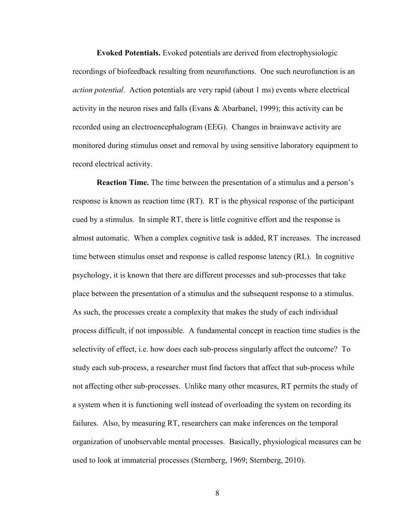
Evoked Potentials. Evoked potentials are derived from electrophysiologic
recordings of biofeedback resulting from neurofunctions. One such neurofunction is an
action potential. Action potentials are very rapid (about 1 ms) events where electrical
activity in the neuron rises and falls (Evans & Abarbanel, 1999); this activity can be
recorded using an electroencephalogram (EEG). Changes in brainwave activity are
monitored during stimulus onset and removal by using sensitive laboratory equipment to
record electrical activity.
Reaction Time. The time between the presentation of a stimulus and a person’s
response is known as reaction time (RT). RT is the physical response of the participant
cued by a stimulus. In simple RT, there is little cognitive effort and the response is
almost automatic. When a complex cognitive task is added, RT increases. The increased
time between stimulus onset and response is called response latency (RL). In cognitive
psychology, it is known that there are different processes and sub-processes that take
place between the presentation of a stimulus and the subsequent response to a stimulus.
As such, the processes create a complexity that makes the study of each individual
process difficult, if not impossible. A fundamental concept in reaction time studies is the
selectivity of effect, i.e. how does each sub-process singularly affect the outcome? To
study each sub-process, a researcher must find factors that affect that sub-process while
not affecting other sub-processes. Unlike many other measures, RT permits the study of
a system when it is functioning well instead of overloading the system on recording its
failures. Also, by measuring RT, researchers can make inferences on the temporal
organization of unobservable mental processes. Basically, physiological measures can be
used to look at immaterial processes (Sternberg, 1969; Sternberg, 2010).

9
Because cognitive processes cannot be directly observed, we are forced to find
alternate ways to look at and subsequently measure a response(s), RT allows this
advantage. For example, during visual search tasks we know that there are two different
processes taking place, pre-attentive and attentional. During pre-attentive processes,
visual stimuli is encoded in parallel and rapidly. Searches are susceptible to distinct
differences in luminance, color, orientation, motion direction, form, and velocity. During
a target detection task, when a stimulus is introduced, the participant scans the stimulus
searching for the target. By manipulating the variables, RT can be increased or decreased
(pop-out affect) depending on the complexity and ambiguousness of the visual task. The
RT measure can give us insight to underlying processes at work during changes to our
environment. Another advantage to RT, it offers a way to study cognitive processes by
looking at temporal organization. As mentioned above, many of these processes can be
extremely complex and difficult to delineate. Input maybe processed sequentially or at
the same time. If multiple factors are processed at the same time, sub-process factors can
be manipulated and processes timed and analyzed through mean comparisons. If
information is believed to be processed serially, then differences between self-
terminating and exhausting processes can be analyzed. Distinct changes in time between
stimulus onset and response can be used to make inferences about the overall cognitive
processes regardless of how it is processed (Muller & Krummenacher, 2006).
While RT allows for an alternative method of measurement, it is not without its
drawbacks. One issue that arises when studying RT of any cognitive process is the trade-
off that occurs between speed and accuracy. It stands to reason that preattentive
processing is much faster than attentional effort, but when we look at each process
10
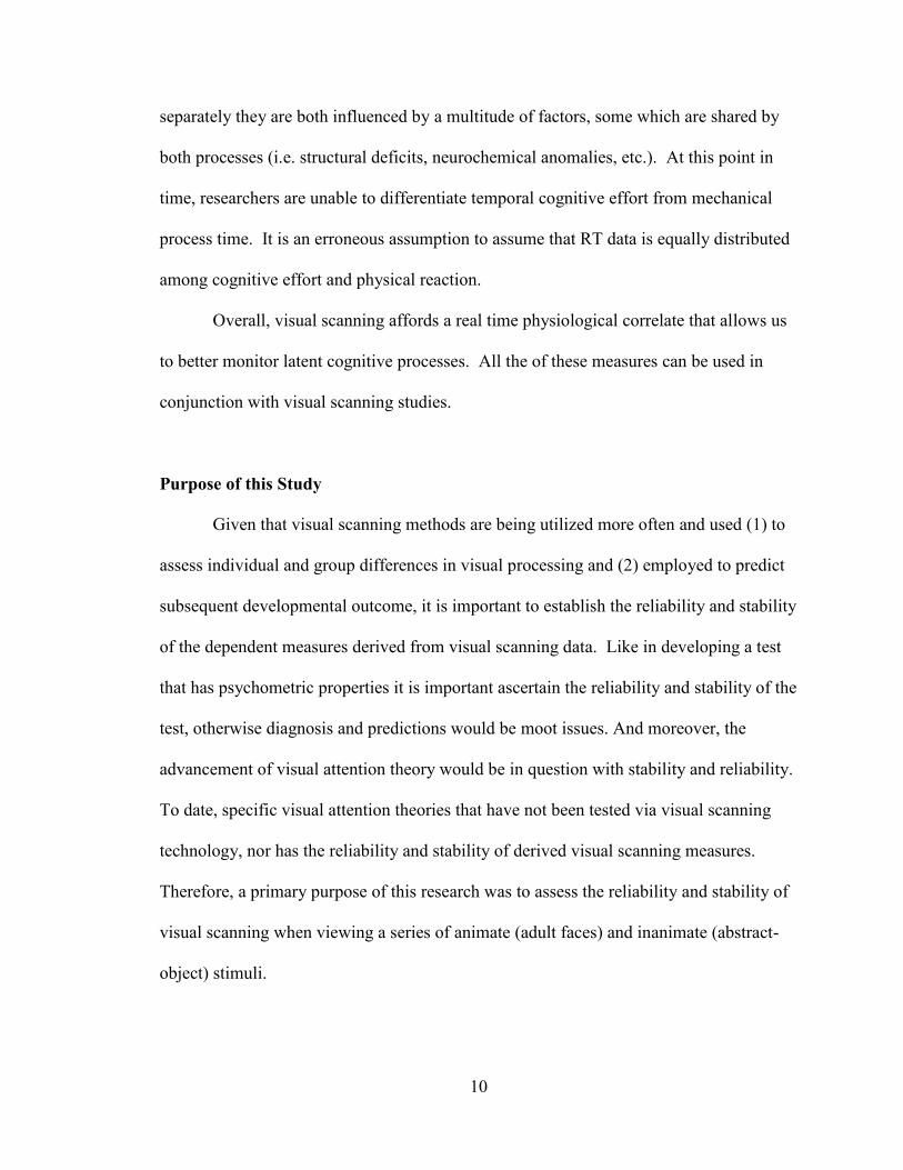
separately they are both influenced by a multitude of factors, some which are shared by
both processes (i.e. structural deficits, neurochemical anomalies, etc.). At this point in
time, researchers are unable to differentiate temporal cognitive effort from mechanical
process time. It is an erroneous assumption to assume that RT data is equally distributed
among cognitive effort and physical reaction.
Overall, visual scanning affords a real time physiological correlate that allows us
to better monitor latent cognitive processes. All the of these measures can be used in
conjunction with visual scanning studies.
Purpose of this Study
Given that visual scanning methods are being utilized more often and used (1) to
assess individual and group differences in visual processing and (2) employed to predict
subsequent developmental outcome, it is important to establish the reliability and stability
of the dependent measures derived from visual scanning data. Like in developing a test
that has psychometric properties it is important ascertain the reliability and stability of the
test, otherwise diagnosis and predictions would be moot issues. And moreover, the
advancement of visual attention theory would be in question with stability and reliability.
To date, specific visual attention theories that have not been tested via visual scanning
technology, nor has the reliability and stability of derived visual scanning measures.
Therefore, a primary purpose of this research was to assess the reliability and stability of
visual scanning when viewing a series of animate (adult faces) and inanimate (abstract-
object) stimuli.

11
The reliability of the following derived measures of visual scanning to pre-
defined Areas of Interest (AOI) within each of the stimuli: (1) the total number of
fixations during stimulus-interval; (2) latency to first fixation; (3) the total time attending;
(4) and the number of shifts between stimulus pairs were examined in this study via a
series of Pearson correlations (two-trial consistencies) across pairs of facial and object
stimuli.
To assess the stability of the visual scanning, the means and standard deviations
for the derived measures (e.g., total number of fixations, latency to first fixation, total
time attending to target, and number of shifts between stimulus pairs) within participants
will be assessed across a series of facial and object stimuli.
Although face stimuli and abstract stimuli have been employed in a variety of
studies with infants, children and adults; and the stimulus features attended to have been
well documented, no visual scanning norms have been developed. There are
standardized sets of visual stimuli (e.g., Brodeur, Dionne-Dostie, Montreuil, & Lepage,
2010); however, the researchers employed a less than optimal method of equating the
stimuli; subjective participant personal ratings using Likert-like rating scales were used.
It has become evident that stimulus characteristics (e.g., size, contrast, familiarity,
novelty, and linguistically based – stimuli that can be named) can impact significantly
visual scanning and therefore can produce confounds in the interpretation of individual
differences on subsequent recognition memory tasks. Hence, there is need to develop
visual stimuli that have been normed in concordance with current visual scanning
technology so to (1) better advance our work in recognition memory and attention, and

12
(2) to develop appropriate diagnostic visual tests that could be used to predict
development outcome or detect cognitive anomalies.
Primary Hypotheses
Hypotheses 1 thru 4 relate to assessing the stability of the visual scanning
measures of number fixations, latency to first fixation, total fixation time, and the number
of visual shifts. Although the null hypothesis is predicted, in tests development theory
stability of a given measure(s) is expected.
HYPOTHESIS 1. The mean and standard deviation of the number of fixations
across trials should remain consistent with no significant differences.
HYPOTHESIS 2. The mean and standard deviation of the latency to first fixation
across trials should remain consistent with no significant differences.
HYPOTHESIS 3. The mean and standard deviation of the total time attending
across trials should remain consistent with no significant differences.
HYPOTHESIS 4. The mean and standard deviation of the number of shifts
between stimulus pairs across trials should remain consistent with no significant
differences.
Hypotheses 5 thru 8 relate to assessing the reliability of the visual scanning
measures of number fixations, latency to first fixation, total fixation time, and the number
of visual shifts. As in test development theory reliability of a given measure(s) are
expected. Within groups two-trial consistencies (reliability) are hypothesized to be
significant statistically. Given these are laboratory measures, the normal test-retest
reliability of r = 0.80 required for standardized tests will not be expected, however
significant two-trial consistencies are predicted.
HYPOTHESIS 5. Within groups, significant two-trial consistencies for number of
fixations are predicted.

13
HYPOTHESIS 6. Within groups, significant two-trial consistencies for the
latency to first fixation are predicted.
HYPOTHESIS 7. Within groups, significant two-trial consistencies for the total
time attending are expected.
HYPOTHESIS 8. Within groups, significant two-trial consistencies for the
number of shifts are expected.

14
METHODS
Sample
Eighty participants were recruited from a pool of Missouri State University’s
(MSU) students through use of SONA, consisting primarily of students currently enrolled
in PSY 121 classes. Participation in a research project is part of the PSY 121 students
required course work. Missouri State University’s Institutional Review Board (IRB) has
reviewed and approved this research project (January 22, 2017; approval #2017-422).
Prior to data analysis, a total of thirty-four participants were removed from the study due
to equipment malfunction, system lag, task misunderstanding and/or lack of scanning.
Multiple participants commented during the debrief that they were unsure if they were
supposed to look at the stimulus presentations or remain focused where the “X” appeared
and reappeared or that during the trials, they believed their task may have been different
than what they were doing.
Data were further screened for assumptions and outliers. Mean replacement was
used within participant trials for scores that deviated from the participant trend. In
Latency to First Response columns, mean replacement was used within participant trials
(n = 20) if first response was greater than or equal to 1000/ms in two or less trials. In
Total Number of Switches Between AOI’s, mean replacement was used on participant
data (n = 2) if total number of switches deviated more than 75% of the within participant
trend. The final sample size was n = 46 participants.

15
Materials
Subtest. A Wechsler Adult Intelligence Scale-Revised—WAIS-R (1981) picture
arrangement subtest was administered to each participant. The subtest was used to
validate the normality of participants regarding attention to detail and to assess the
concurrent relationship between this subscale and the derived visual scanning measures.
Stimuli. Three sets of visual stimuli (faces, abstract-object, and manipulation
check) were created. Each set of the first two sets consisted of three stimuli. The face
stimuli (Lundqvist, Flykt, & Öhman, 1998) differed in emotion; neutral, happy, and
anger. For the abstract-object, three stimuli were created with differing levels of
complexity (saliency map); low, medium, and high. For the manipulation check, two
new stimuli were used; a novel face pair and an abstract-object image.
Apparatus. The stimulus images were displayed on a 60 cm color monitor. Eye
tracking was recorded using GazePoint GP3 Eye Tracker sensor and data collected using
GazePoint Analysis and Control software.
Procedure
The design of the study was a 2 (Stimulus: Face vs. Object) X 2 (Gender: Male
vs. Female) X 2 (Presentation Order: Forward vs. Reverse) X 9 (Trials) factorial design
with a repeated measure on the last factor. Each stimulus set was followed by a
manipulation check (Trial 10) to assess whether participants were exerting cognitive
effort when scanning. Data from the manipulation check (Trial 10) was used to validate
the link between visual scanning and cognitive performance.
16
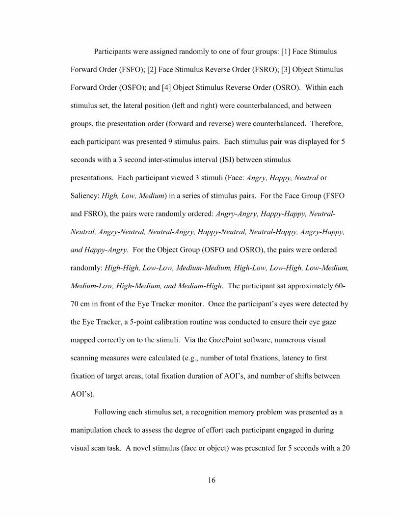
Participants were assigned randomly to one of four groups: [1] Face Stimulus
Forward Order (FSFO); [2] Face Stimulus Reverse Order (FSRO); [3] Object Stimulus
Forward Order (OSFO); and [4] Object Stimulus Reverse Order (OSRO). Within each
stimulus set, the lateral position (left and right) were counterbalanced, and between
groups, the presentation order (forward and reverse) were counterbalanced. Therefore,
each participant was presented 9 stimulus pairs. Each stimulus pair was displayed for 5
seconds with a 3 second inter-stimulus interval (ISI) between stimulus
presentations. Each participant viewed 3 stimuli (Face: Angry, Happy, Neutral or
Saliency: High, Low, Medium) in a series of stimulus pairs. For the Face Group (FSFO
and FSRO), the pairs were randomly ordered: Angry-Angry, Happy-Happy, Neutral-
Neutral, Angry-Neutral, Neutral-Angry, Happy-Neutral, Neutral-Happy, Angry-Happy,
and Happy-Angry. For the Object Group (OSFO and OSRO), the pairs were ordered
randomly: High-High, Low-Low, Medium-Medium, High-Low, Low-High, Low-Medium,
Medium-Low, High-Medium, and Medium-High. The participant sat approximately 60-
70 cm in front of the Eye Tracker monitor. Once the participant’s eyes were detected by
the Eye Tracker, a 5-point calibration routine was conducted to ensure their eye gaze
mapped correctly on to the stimuli. Via the GazePoint software, numerous visual
scanning measures were calculated (e.g., number of total fixations, latency to first
fixation of target areas, total fixation duration of AOI’s, and number of shifts between
AOI’s).
Following each stimulus set, a recognition memory problem was presented as a
manipulation check to assess the degree of effort each participant engaged in during
visual scan task. A novel stimulus (face or object) was presented for 5 seconds with a 20

17
second ISI, following which, the participant was asked to recall image features that were
present (a recall memory assessment).
Pretest Process. Upon arrival, participants were greeted and escorted to a
designated assessment room. They were sat at a table and asked to please read and sign
the informed consent form and fill out the demographics sheet. They were asked if they
have any questions regarding the forms. Next, participants were given summaries of the
tasks, stated as follows:
“There will be two tasks. The first task will be a WAIS-R picture arrangement
subtest; it is simply a working and spatial memory assessment. Secondly, we will
perform a visual assessment. The computer is equipped to perform an analysis of
your visual field. We will briefly calibrate the system to your eyes and then
display a series of photos. The entire process should take no longer than 20-25
minutes.”
Phase 1. The researcher began the WAIS-R Picture Arrangement task per the
WAIS-R Manual (1981). Scores were recorded.
Phase 2. After the WAIR-R subtest was completed, participants were moved to a
workstation setup with the GazePoint program. The participant was instructed to sit
squarely in front of the monitor and to rest his/her chin comfortably on the stabilization
bar in front of him/her. The system was calibrated to each participant after the following
statement was read to him/her,
“You will see a white dot with a red center enter the screen from the upper left
hand corner of the monitor and it will quickly move to the center. It will slowly
shrink before moving to the upper right hand corner. Do your best to focus on the
center of the dot at all times without looking at the area surrounding the dot or its
perimeter. Try to keep your head in a fixed position and only follow the dot with
your eyes. This ensures the most accurate calibration. The dot will make its way
around the screen to five locations (the researcher will motion with his/her hands
the calibration path).”

18
Once calibration was complete, the researcher began the primary visual task. Before
clicking “Record,” the task was explained. “You will see the word ‘Start’ followed by an
‘X’ in the center of the screen. While the ‘X’ is present, please focus your attention to
the center of it. When the ‘X’ disappears, you are free to view anywhere on the screen.
When the ‘X’ reappears, please focus on the center of it once again. Are you ready to
begin?”
Once the final stimulus was presented, the participant was asked to recall details
of the previous image while his/her responses were recorded.
Debriefing
As soon as each participant completed all assessments, he or she was debriefed
and allowed to ask questions. Participants were informed upon exiting that results would
be provided to participants upon request once the study was complete.
19
RESULTS
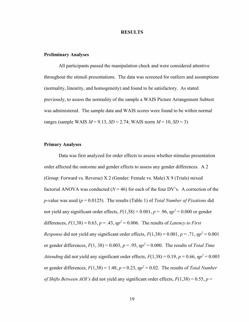
Preliminary Analyses
All participants passed the manipulation check and were considered attentive
throughout the stimuli presentations. The data was screened for outliers and assumptions
(normality, linearity, and homogeneity) and found to be satisfactory. As stated
previously, to assess the normality of the sample a WAIS Picture Arrangement Subtest
was administered. The sample data and WAIS scores were found to be within normal
ranges (sample WAIS M = 9.13, SD = 2.74; WAIS norm M = 10, SD = 3)
Primary Analyses
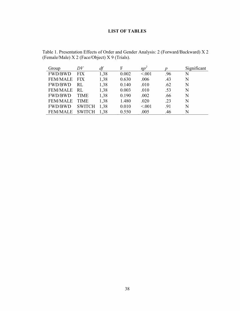
Data was first analyzed for order effects to assess whether stimulus presentation
order affected the outcome and gender effects to assess any gender differences. A 2
(Group: Forward vs. Reverse) X 2 (Gender: Female vs. Male) X 9 (Trials) mixed
factorial ANOVA was conducted (N = 46) for each of the four DV’s. A correction of the
p-value was used (p = 0.0125). The results (Table 1) of Total Number of Fixations did
not yield any significant order effects, F(1,38) = 0.001, p = .96, ηp2 = 0.000 or gender
differences, F(1,38) = 0.63, p = .43, ηp2 = 0.006. The results of Latency to First
Response did not yield any significant order effects, F(1,38) = 0.001, p = .71, ηp2 = 0.001
or gender differences, F(1, 38) = 0.003, p = .95, ηp2 = 0.000. The results of Total Time
Attending did not yield any significant order effects, F(1,38) = 0.19, p = 0.66, ηp2 = 0.003
or gender differences, F(1,38) = 1.48, p = 0.23, ηp2 = 0.02. The results of Total Number
of Shifts Between AOI’s did not yield any significant order effects, F(1,38) = 0.55, p =

20
.46, ηp2 = .005 or gender differences, F(1,38) = 0.01, p = 0.91, ηp2 = 0.000. For all
subsequent analyses comparing means, gender and test order was collapsed within
Groups, hence the primary statistical analyses approach was a 2 (Group: Face vs. Object)
X 9 (Trials) mixed ANOVA with a repeated measure on the last factor.
After groups were collapsed, stability was assessed through used of a series of 2
X 9 ANOVAs calculating means and standard deviations using two one-sided tests
(TOST) of equivalence (Lakens, 2016); reliability was assessed looking at two-trial
correlations. Two-trial consistencies were analyzed several ways: (1) Face and Object
groups combined with correlations ran in sequential order (Trial 1/Trial 2, Trial 2/Trial 3,
etc.); (2) Face and Object groups combined with correlations ran with respective matched
pair trials (Happy Angry/Angry Happy; High Low/Low High); (3) Face and Object
groups separate with correlations ran in sequential order; (4) Face and Object groups
combined with correlations ran with respective matched pairs. Note: in the second group,
Happy was combined with High Saliency, Neutral was combined with Medium Saliency,
and Angry was combined with Low Saliency. These iterations provided for multifarious
organized comparisons of the correlations and in the following sections the stability and
reliability analyses of each of the derived scanning measures will be discussed in turn.
Means and standard deviations were analyzed for measurement reliability through
use of TOST. A test of equivalence was used to assess whether the groups means differ
“too much” for setting up two one-sided t-tests. If significant, the means will be less than
the upper limit and greater than the lower limit. Finally, two-trial consistency (Pearson’s
Correlation) was used to determine the reliability of various visual scanning measures
over trials. Given the magnitude of two-trial consistency correlations only the range and
21
magnitude will be presented and discussed, however, all two-trial consistency and
summary statistics are tabled in the appendices.
Total Number of Fixations
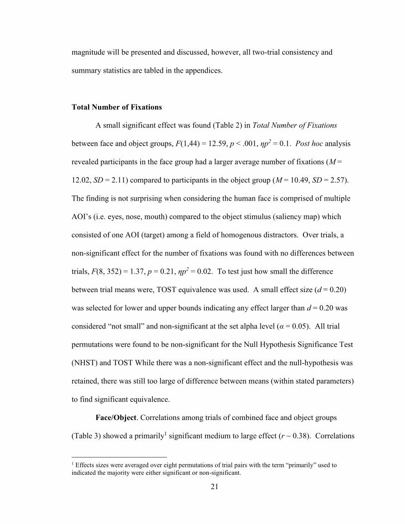
A small significant effect was found (Table 2) in Total Number of Fixations
between face and object groups, F(1,44) = 12.59, p < .001, ηp2 = 0.1. Post hoc analysis
revealed participants in the face group had a larger average number of fixations (M =
12.02, SD = 2.11) compared to participants in the object group (M = 10.49, SD = 2.57).
The finding is not surprising when considering the human face is comprised of multiple
AOI’s (i.e. eyes, nose, mouth) compared to the object stimulus (saliency map) which
consisted of one AOI (target) among a field of homogenous distractors. Over trials, a
non-significant effect for the number of fixations was found with no differences between
trials, F(8, 352) = 1.37, p = 0.21, ηp2 = 0.02. To test just how small the difference
between trial means were, TOST equivalence was used. A small effect size (d = 0.20)
was selected for lower and upper bounds indicating any effect larger than d = 0.20 was
considered “not small” and non-significant at the set alpha level (α = 0.05). All trial
permutations were found to be non-significant for the Null Hypothesis Significance Test
(NHST) and TOST While there was a non-significant effect and the null-hypothesis was
retained, there was still too large of difference between means (within stated parameters)
to find significant equivalence.
Face/Object. Correlations among trials of combined face and object groups
(Table 3) showed a primarily1 significant medium to large effect (r ~ 0.38). Correlations
1 Effects sizes were averaged over eight permutations of trial pairs with the term “primarily” used to
indicated the majority were either significant or non-significant.

22
among matched pair trials (Table 4) showed a significant averaged large effect for all
permutations (r ~ 0.46)
Faces. Correlations among face trials (Trial 1-2, Trial 2-3, Trial 3-4, etc.) returned
mixed reliability results (Table 9); an average correlation of nine repeated trials yielded a
primarily non-significant small-medium effect sizes (r ~ 0.24). Correlations among face
matched pair trials (Table 19-22) showed primarily non-significant averaged medium
effects (r ~ 0.27).
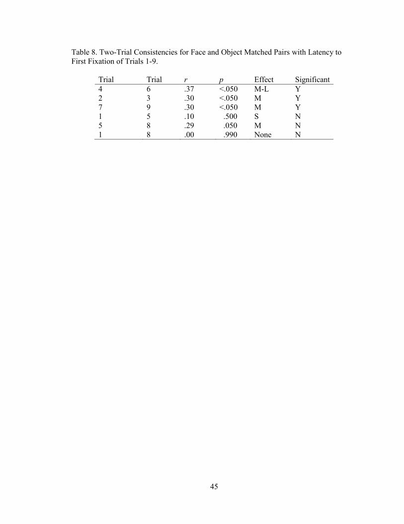
Objects. Correlations among object trials (Table 15) returned mixed reliability
results; an average correlation of nine repeated trials showed a primarily non-significant
medium to large effect (r ~ 0.39). Correlations among object matched pair trials (Table
8) yielded a significant averaged large effect (r ~ 0.56).
Latency to First Fixation
A non-significant effect was found in the analysis of Latency to First Fixation
between face and object groups, F(1,44) = 0.28, p = .60, η2 = 0.002. Over trials, a non-
significant effect for the number of fixations was found, F(8, 352) = 0.99, p = 0.44, η2 =
0.01. Once again, to test how small the difference between trial means were, the TOST
equivalence was used. A small effect size (d = 0.20) was selected for lower and upper
bounds indicating any effect larger than d = 0.20 was considered “not small” and non-
significant at the set alpha level (α = 0.05). All trial permutations were found to be non-
significant for NHST and TOST. While there is a non-significant effect and the null-
hypothesis is retained, there is still too large of difference between means (within stated
parameters) to find significant equivalence.
23
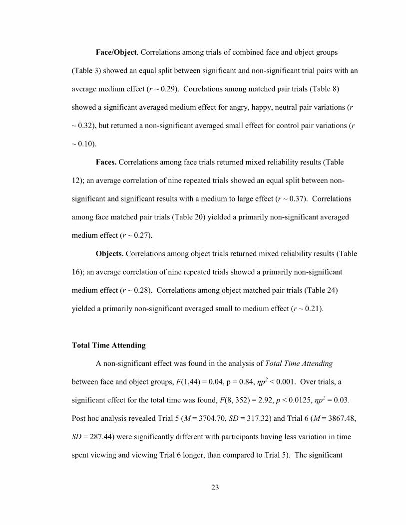
Face/Object. Correlations among trials of combined face and object groups
(Table 3) showed an equal split between significant and non-significant trial pairs with an
average medium effect (r ~ 0.29). Correlations among matched pair trials (Table 8)
showed a significant averaged medium effect for angry, happy, neutral pair variations (r
~ 0.32), but returned a non-significant averaged small effect for control pair variations (r
~ 0.10).
Faces. Correlations among face trials returned mixed reliability results (Table
12); an average correlation of nine repeated trials showed an equal split between non-
significant and significant results with a medium to large effect (r ~ 0.37). Correlations
among face matched pair trials (Table 20) yielded a primarily non-significant averaged
medium effect (r ~ 0.27).
Objects. Correlations among object trials returned mixed reliability results (Table
16); an average correlation of nine repeated trials showed a primarily non-significant
medium effect (r ~ 0.28). Correlations among object matched pair trials (Table 24)
yielded a primarily non-significant averaged small to medium effect (r ~ 0.21).
Total Time Attending
A non-significant effect was found in the analysis of Total Time Attending
between face and object groups, F(1,44) = 0.04, p = 0.84, ηp2 < 0.001. Over trials, a
significant effect for the total time was found, F(8, 352) = 2.92, p < 0.0125, ηp2 = 0.03.
Post hoc analysis revealed Trial 5 (M = 3704.70, SD = 317.32) and Trial 6 (M = 3867.48,
SD = 287.44) were significantly different with participants having less variation in time
spent viewing and viewing Trial 6 longer, than compared to Trial 5). The significant

24
effect was the only one out of 36 permutations and the researcher determined the result is
not meaningful and likely an artifact of the analysis. The differences were tested using
TOST equivalence. A small effect size (d = 0.20) was selected for lower and upper
bounds indicating any effect larger than d = 0.20 was considered “not small” and non-
significant at the set alpha level (α = 0.05). All trial permutations were found to be non-
significant for NHST and TOST. While there is a non-significant effect and the null-
hypothesis is retained, there is still too large of difference between means (within stated
parameters) to find significant equivalence.
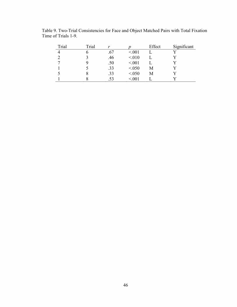
Face/Object. Correlations among trials of combined face and object groups
(Table 5) showed a primarily significant effect for trial pairs with an average large effect
(r ~ 0.46). Average correlations among matched pair trials (Table 9) showed a
significant large effect for all pair variations (r ~ 0.47).
Faces. Correlations among face trials returned significant reliability results (Table
13); an average correlation of nine repeated trials showed significant results with a
medium to large effect (r ~ 0.44). Correlations among face matched pair trials (Table 21)
showed a significant averaged large for angry, happy, neutral pair variations (r ~ 0.56),
but returned a primarily non-significant averaged large effect for control pair variations (r
~ 0.10).
Objects. Correlations among object trials returned mixed reliability results (Table
17); an average correlation of nine repeated trials showed a primarily significant large
effect (r ~ 0.53). Correlations among object matched pair trials (Table 25) yielded a
primarily non-significant averaged medium to large effect (r ~ 0.44).
25
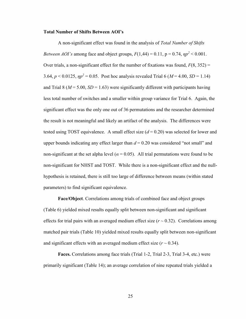
Total Number of Shifts Between AOI’s
A non-significant effect was found in the analysis of Total Number of Shifts
Between AOI’s among face and object groups, F(1,44) = 0.11, p = 0.74, ηp2 < 0.001.
Over trials, a non-significant effect for the number of fixations was found, F(8, 352) =
3.64, p < 0.0125, ηp2 = 0.05. Post hoc analysis revealed Trial 6 (M = 4.00, SD = 1.14)
and Trial 8 (M = 5.00, SD = 1.63) were significantly different with participants having
less total number of switches and a smaller within group variance for Trial 6. Again, the
significant effect was the only one out of 36 permutations and the researcher determined
the result is not meaningful and likely an artifact of the analysis. The differences were
tested using TOST equivalence. A small effect size (d = 0.20) was selected for lower and
upper bounds indicating any effect larger than d = 0.20 was considered “not small” and
non-significant at the set alpha level (α = 0.05). All trial permutations were found to be
non-significant for NHST and TOST. While there is a non-significant effect and the null-
hypothesis is retained, there is still too large of difference between means (within stated
parameters) to find significant equivalence.
Face/Object. Correlations among trials of combined face and object groups
(Table 6) yielded mixed results equally split between non-significant and significant
effects for trial pairs with an averaged medium effect size (r ~ 0.32). Correlations among
matched pair trials (Table 10) yielded mixed results equally split between non-significant
and significant effects with an averaged medium effect size (r ~ 0.34).
Faces. Correlations among face trials (Trial 1-2, Trial 2-3, Trial 3-4, etc.) were
primarily significant (Table 14); an average correlation of nine repeated trials yielded a
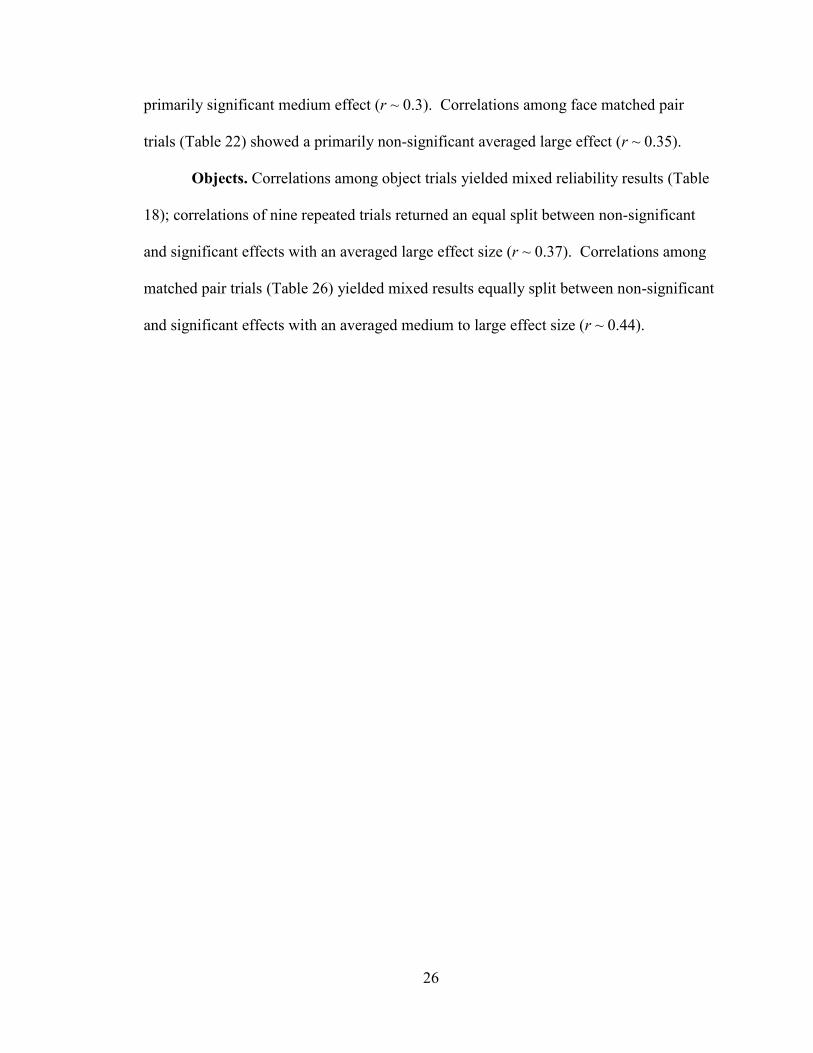
26
primarily significant medium effect (r ~ 0.3). Correlations among face matched pair
trials (Table 22) showed a primarily non-significant averaged large effect (r ~ 0.35).
Objects. Correlations among object trials yielded mixed reliability results (Table
18); correlations of nine repeated trials returned an equal split between non-significant
and significant effects with an averaged large effect size (r ~ 0.37). Correlations among
matched pair trials (Table 26) yielded mixed results equally split between non-significant
and significant effects with an averaged medium to large effect size (r ~ 0.44).

27
DISCUSSION
Total Number of Fixations
A main effect was found between face and object groups indicating participants in
the face group had a larger number of fixations as compared to the object group. The
finding is not surprising when considering the human face is comprised of multiple
AOI’s (i.e. eyes, nose, mouth) compared to the object stimulus (saliency map) which
consisted of one AOI (target) among a field of homogenous distractors. In terms of
stability over trials, no significant difference was found between groups; the mean
number of fixations from one trial to the next did not vary significantly. However, after
analyzing how similar the groups are using a small effect, a non-significant effect was
found. Groups still had too large of a difference between means to be practically
equivalent. Further research should be conducted to determine outcome modulation and
why groups are not significantly different, but are not significantly the same. In terms of
reliability, the average number of group fixations between trial pairs was found to have a
medium to large effect, but only significant in half of the analyses. Previous research
would suggest that reliability of the total number of fixations is expected to higher than
other indices. A participant’s task understanding, search method, encoding speed, etc.
should not vary greatly between trials. Any variation is likely due to novelty effects of
the stimuli or environmental distractions.
28
Latency to First Fixation
A non-significant effect was found between face and object groups; there were no
apparent differences between groups in how long it took participants to record their first
fixation once the stimulus presented. This finding is not surprising in that both stimulus
sets consisted of a pair of stimuli that appeared in the same location over trials; each
having salient features that garner attention. Future research may want to look at
differences between groups and latency to first fixation regarding salient AOI’s. In terms
of stability over trials, no significant difference was found between groups; the mean
number of fixations from one trial to the next did not vary significantly. After analyzing
how similar the groups are using a small effect, a non-significant effect was found.
Groups still had too large of a difference between means to be practically equivalent.
Further research should be conducted to determine outcome modulation and why groups
are not significantly different, but are not significantly the same. In terms of reliability,
the average number of group fixations between trial pairs was found to have a small to
medium effect, but only significant in less than half of the analyses.
Total Time Attending
A non-significant effect was found between face and object groups; there were no
apparent differences between groups in how long participants fixated on the viewing area
once the stimulus presented. This finding is not surprising in that saccadic movement,
blink rate, and focus should primarily be a function of individual differences with each
participant behaving the same over trial periods over a short time frame. Future research
may want to look at differences between groups and changes individual behavior over
29
time. In terms of stability over trials, no significant difference was found between trial
pairs (only 1 of 36 found significant). After analyzing how similar the groups are using a
small effect, a non-significant effect was found. Groups still had too large of a difference
between means to be practically equivalent. Further research should be conducted to
determine outcome modulation and why groups are not significantly different, but are not
significantly the same. In terms of reliability, total time attending was found to be the
most reliable. When looking at trial pairs, the average time attending was found to have a
primarily significant medium to large effect. This measure should return the highest
reliability which suggests that each participant is cognitively engaged and visually
attending approximately the same amount of time per each five-second presentation.
Total Number of Shifts
A non-significant effect was found between face and object groups; there were no
apparent differences between groups in how many times participants shifted from one
AOI to another once the stimulus presented. This finding is a little surprising in that, like
the total number of fixations, face stimuli has more detail to consider between stimulus
pairs while object stimuli only has one target area of interest. This could be a function of
the ambiguous task. Participants, knowing it was a psychological study, may have been
come up with their own search paradigms that they used to help determine the purpose of
the study. In other words, scanning patterns may have had less to do with the stimulus
sets and more to do with mental tasks arising from not knowing what they should be
looking at. Future research may want to look at differences between groups with detailed
task descriptions compared to ambiguous task descriptions. In terms of stability over

30
trials, no significant difference was found between trial pairs (only 1 of 36 found
significant). After analyzing how similar the groups are using a small effect, a non-
significant effect was found. Groups still had too large of a difference between means to
be practically equivalent. Further research should be conducted to determine outcome
modulation and why groups are not significantly different, but are not significantly the
same. In terms of reliability, the average number of switches between stimulus pairs was
found to be split equally between non-significant and significant with a medium to large
effect over trials.
Two-Trial Consistency Combinations
Reliability data should be carefully interpreted with consideration given to
changing rank order between trial pairs. Novelty of one trial over another may influence
outcomes and change the rank order due to a function of individual differences; i.e. a
happy face may garner one person’s attention longer in a trial as compared to another
emotion.
Limitations
The ambiguous nature of the task should be reconsidered in future studies.
During the debrief, multiple participants suggested they changed their scanning behavior
during one or more trials due to uncertainty of task requirements. Participants were not
given a goal, but they assumed that there was a goal they should be trying to achieve; i.e.
“Should I have kept my focus on the spot where the “X” was?”, “Was I looking for
differences between the pictures?”, “After the first couple of pictures, they looked like
31
the same things.” A second limitation to the study is an afterimage effect. Two
participants commented after the study that when the “X” disappeared, they could still
see it in their field of vision. This effect was undoubtedly intensified due to the white
“X” on a black background.
Conclusion
The primary purpose of this study was to assess the reliability and stability of
visual scanning when viewing a series of animate (adult faces) and inanimate (abstract-
object) stimuli. While no significant differences were found between test measurements,
there were no significant equivalencies between groups either. A measure of the total
number of fixations and the total time attending during visual scanning tasks appear to be
the most reliable measures. Because of this, future studies may utilize the reliability of
these measures to be more confident about the consistency of their task measures and
variable influences. Future research should also explore factors contributing to group
differences and limited equivalency. Overall, an average medium correlation was found
between trial pairs, but with mixed results of statistical significance. With a larger
sample size, statistical significance would likely be achieved in further studies. Likewise,
as technology improves and we learn more about visual attention, more reliable results
will likely be obtained.
32
REFERENCES
Broadbent, D. (1958). Perception and communication. London: Pergamon Press.
Brodeur M., Dionne-Dostie E., Montreuil T., Lepage M. (2010). The bank of
standardized stimuli (BOSS), a new set of 480 normative photos of objects to be
used as visual stimuli in cognitive research. PLoS ONE 5(5): e10773.
https://doi.org/10.1371/journal.pone.0010773
Campbell, F., & Green, D. (1965). Optical and retinal factors affecting visual resolution.
The Journal of Physiology, 181(3), 576–593.
Cohen, R. & O’Donnell, B. (1993). Physiological substrates of attention. The
Neuropsychology of Attention. (p. 115-144). New York: Springer U.S.
Craik, K. (1947). Theory of the human operator in control systems. British Journal of
Psychology. General Section, 38, 56-61. doi: 10.1111/j.2044-
8295.1947.tb01141.x
Deutsch, J., & Deutsch, D. (1963). Attention: Some theoretical considerations.
Psychological Review, 70(1), 80.
Eriksen, C. & St. James, J. (1986). Visual attention within and around the field of focal
attention: A zoom lens model. Perception Psychophysics, 40(2), 225-240.
Evans, J. & Abarbanel, A. (1999). Introduction to quantitative EEG and neurofeedback.
San Diego, Calif: Academic Press.
Henderson, J. & Hollingworth, A. (2003). Global transsaccadic change blindness during
scene perception. Psychological Science, 14, (5), 493-497.
Irwin, D. (1991). Integrating information across saccadic eye movements. Cognitive
Psychology, 23(3), 420-456.
Itti, L. & Koch, C. (2001). Computational modelling of visual attention. Nature Reviews
Neuroscience, 2(3), 194-203.
James, W. (1890). The principles of psychology. New York: Henry, Holt, and Company.
Jennings, J. (1986). Bodily changes during attending. In M. G. H. Coles, E. Donchin, &
S. W. Porges (Eds.), Psychophysiology: Systems, processes and application (p.
268–289). New York: Guilford Press.

33
Kahneman, D. (1973). Attention and effort. Englewood Cliffs, New Jersey: Prentice-Hall,
Inc.
Kahneman, D. & Treisman, A. (1984). Changing views of attention and automaticity. In
R. Parasuraman & D.R. Davis (Eds.), Varieties of attention (p. 29-61). Orlando:
Academic Press.
Kalivas, G. & Petralia, S. (2012). Short-term memory: New Research. New York: Nova
Science Publishers, Inc.
Lakens, D. (2016, December 9). TOST equivalence testing R package (TOSTER) and
spreadsheet. Retrieved April 15, 2017, from
http://daniellakens.blogspot.com/2016/12/tost-equivalence-testing-r-package.html
Lundqvist, D., Flykt, A., & Öhman, A. (1998). The karolinska directed emotional faces -
KDEF, CD ROM from department of clinical neuroscience, psychology section,
karolinska institutet, ISBN 91-630-7164-9.
Mackay, D. (1972). Visual stability. Investigative ophthalmology, 11(6), 518-524.
Müller, H., & Krummenacher, J. (2006). Visual search and selective attention. Visual
cognition, 14(4-8), 389-410.
Neisser, U. (1967). Cognitive psychology. New Jersey: Prentice-Hall, Inc.
Neisser, U. (1976). Cognition and reality. San Francisco: W. H. Freeman and Company.
Schneider, W., & Shiffrin, R. (1977). Controlled and automatic human information
processing: I. Detection, search, and attention. Psychological review, 84(1), 1.
Shiffrin, R., & Schneider, W. (1977). Controlled and automatic human information
processing: II. Perceptual learning, automatic attending and a general theory.
Psychological review, 84(2), 127.
Sternberg, S. (1969). Memory-scanning: Mental processes revealed by reaction-time
experiments. American scientist, 57(4), 421-457.
Sternberg, S. (2010). Reaction-time experimentation. Proseminar in psychological
methods. Retrieved from
http://www.psych.upenn.edu/~saul/rt.experimentation.pdf
Theeuwes, J. (1992). Perceptual selectivity for color and form. Perceptions and
psychometrics, 51(6), 599-606.
Titchener, B. (1908). Lectures on the elementary psychology of feeling and attention.
New York: The Macmillan Company

34
Treisman, A. (1964). Verbal cues, language, and meaning in selective attention. The
american journal of psychology, 77(2), 206-219. doi:1. Retrieved from
http://www.jstor.org/stable/1420127 doi:1
Treisman, A. & Gelade, G. (1980). A feature-integration theory of attention. Cognitive
psychology, 12, 97-136.
Treisman, A. & Schmidt, H. (1982). Illusory conjunctions in the perception of objects.
Cognitive psychology, 14(1), 107-141.
Wickens, C. & McCarley, J. S. (2008). Attention theory. Boca Raton: CRC Press.
Wright, R. (1998). Visual Attention. New York: Oxford University Press.
Wundt, W. (1910). Principles of physiological psychology, Volume 1. (E. B. Titchener,
Trans.) New York: The Macmillan Company. (Original work published 1902)
Wykowska, A. & Schubo, A. (2009). Irrelevant singletons in visual search do not capture
attention but can produce nonspatial filtering costs. Journal of Cognitive
Neuroscience, 23(3), 645-660.

35
APPENDICIES
Appendix A. Consent Form.
Missouri State University Consent of Participation
Infant Perception and Learning Laboratory
This study is part of the Missouri State University Psychology Graduate Program
designed to give us more information and to fulfill a thesis requirement for Michael
Mizer. The following information is provided so that you can decide whether you wish to
participate in this study. If you agree to participate, we will administer an intelligence
subtest and observe your visual responses to a series of slides of human faces and abstract
shapes. One of the members of the research lab should have explained the purposes and
procedures of the study to you, and will answer any questions you might have. Please be
assured that if you agree to participate, you are free to withdraw from the study even after
you have signed this consent form. If you wish to withdraw, simply stop any on-going
task and tell the research staff you wish not to continue. Should you decide to terminate
the research session; all data pertaining to you that have been collected will be destroyed.
Since it is our policy to protect the confidentiality of all our participants, your name
will not be included in any data analyses, subsequent publication or presentations related
to this research study. All raw data collected during this study will be identified only by
code-number to insure confidentiality of the information collected.
If questions arise after you have left the research laboratory, feel free to give D.
Wayne Mitchell, Ph.D. a call at 417-836-6941 or at
[email protected] do not anticipate any risk to you as a result of
participating in this study, but it is unlikely that this study will provide you with any
direct benefits. Your participation will, however, make an important contribution to our
scientific knowledge, and we very much appreciate your cooperation.
In addition, we would appreciate your filling out the attached demographic sheet so
we can document the characteristics of our participants. Any of the questions you feel
uncomfortable about answering, please feel free to leave blank. As with the raw data
collected, this information will be entered into our computer system and only identified
by code-number to insure confidentiality.
I have read the above description of the study and I agree to participate.
Participant's Name (please print):___________________________________________
Participant’s Signature: __________________________________________________
Witness’s Signature: _____________________________________________________
Date: ____/____/_______

36
Appendix B. Demographics Information Form.
Participant's Name: _______________________________________________________
1. Date of Birth __________________________
2. Gender ______________________________
3. Major _______________________________
4. Do you wear any prescription eye wear? Yes ______ No ______
If yes, Glasses? ______ Contacts ______
5. Are you aware of any other vision problems you may have? Yes _______ No ______
If yes, please explain ________________________________________________
_________________________________________________________________
_________________________________________________________________

37
Appendix C. WAIS Participant Response Form.
Item Time (sec) Letters (L to R) Points
1.
House
2.
Flirt
3.
Romeo
4.
Louie
5.
Enter
6.
Escape
7.
Hill
8.
Fish
9.
Robber
10.
Taxi
Face Manipulation Check
What differences did you see?
Smiley face
Vampire teeth
Red eyes
Missing nostril
Missing eyebrows
Object Manipulation Check
How many blue squares did you see? ______________
What color was the roof? _______________
What color was the circle? _______________
38
LIST OF TABLES
Table 1. Presentation Effects of Order and Gender Analysis: 2 (Forward/Backward) X 2
(Female/Male) X 2 (Face/Object) X 9 (Trials).
Group DV df F ηp2 p Significant
FWD/BWD FIX 1,38 0.002 <.001 .96 N
FEM/MALE FIX 1,38 0.630 .006 .43 N
FWD/BWD RL 1,38 0.140 .010 .62 N
FEM/MALE RL 1,38 0.003 .010 .53 N
FWD/BWD TIME 1,38 0.190 .002 .66 N
FEM/MALE TIME 1,38 1.480 .020 .23 N
FWD/BWD SWITCH 1,38 0.010 <.001 .91 N
FEM/MALE SWITCH 1,38 0.550 .005 .46 N

39
Table 2. Primary Analysis: 2 (Face/Object) X 9 (Trials).
Group DV df F η2 p Significant
FACE/OBJ FIX 1,44 12.590 .100 .013 Y
TRIAL FIX 8,352 1.370 .020 .020 N
FACE/OBJ RL 1,38 0.280 .002 .002 N
TRIAL RL 8,352 0.990 .010 .010 N
FACE/OBJ TIME 1,38 0.040 .000 .000 N
TRIAL TIME 8,352 2.920 .030 .030 N
FACE/OBJ SWITCH 1,38 0.110 .000 .000 Y
TRIAL SWITCH 8,352 3.640 .050 .050 N

40
Table 3. Two-Trial Consistencies for Face and Object with Total Number of Fixations of
Trials 1-9.
Trial Trial r p Effect Significant
1
2
3
4
5
6
7
8
2
3
4
5
6
7
8
9
.40
.51
.37
.49
.28
.34
.37
.29
<.010
<.001
<.050
<.001
.060
<.050
<.050
.050
M-L
L
M
L
M
M
M
M
YES
YES
YES
YES
NO
YES
YES
NO

41
Table 4. Two-Trial Consistencies for Face and Object with Latency to First Fixation of
Trials 1-9.
Trial Trial r p Effect Significant
1
2
3
4
5
6
7
8
2
3
4
5
6
7
8
9
.40
.51
.37
.49
.28
.34
.37
.29
.570
<.050
.080
<.050
<.001
.250
.420
<.001
S
M
M
M
L
S-M
S
L
NO
YES
NO
YES
YES
NO
NO
YES

42
Table 5. Two-Trial Consistencies for Face and Object with Total Fixation Time of Trials
1-9.
Trial Trial r p Effect Significant
1
2
3
4
5
6
7
8
2
3
4
5
6
7
8
9
.48
.50
.32
.42
.46
.57
.64
.31
<.050
<.010
.120
<.050
<.050
<.010
<.001
.120
L
L
M
M-L
L
L
L
M
N
Y
N
Y
Y
N
N
Y

43
Table 6. Two-Trial Consistencies for Face and Object with Total Number of Shifts
Between AOI’s of Trials 1-9.
Trial Trial r p Effect Significant
1
2
3
4
5
6
7
8
2
3
4
5
6
7
8
9
.37
.51
.58
.26
.16
.08
.11
.48
.060
<.010
<.010
.200
.430
.710
.590
<.050
M-L
L
L
S-M
S-M
S
S
L
N
Y
Y
N
N
N
N
Y

44
Table 7. Two-Trial Consistencies for Face and Object Matched Pairs with Total Number
of Fixations of Trials 1-9.
Trial Trial r p Effect Significant
4
2
7
1
5
1
6
3
9
5
8
8
.40
.39
.33
.46
.51
.67
<.010
<.010
<.050
<.010
<.001
<.001
M-L
M-L
M
L
L
L
Y
Y
Y
Y
Y
Y
45
Table 8. Two-Trial Consistencies for Face and Object Matched Pairs with Latency to
First Fixation of Trials 1-9.
Trial Trial r p Effect Significant
4
2
7
1
5
1
6
3
9
5
8
8
.37
.30
.30
.10
.29
.00
<.050
<.050
<.050
.500
.050
.990
M-L
M
M
S
M
None
Y
Y
Y
N
N
N
46
Table 9. Two-Trial Consistencies for Face and Object Matched Pairs with Total Fixation
Time of Trials 1-9.
Trial Trial r p Effect Significant
4
2
7
1
5
1
6
3
9
5
8
8
.67
.46
.50
.33
.33
.53
<.001
<.010
<.001
<.050
<.050
<.001
L
L
L
M
M
L
Y
Y
Y
Y
Y
Y

47
Table 10. Two-Trial Consistencies for Face and Object Matched Pairs with Total Number
of Shifts Between AOI’s of Trials 1-9.
Trial Trial r p Effect Significant
4
2
7
1
5
1
6
3
9
5
8
8
.34
.47
.28
.21
.52
.19
<.050
<.001
.060
.150
<.001
.200
M
L
M
L
L
L
Y
Y
N
N
Y
N

48
Table 11. Two-Trial Consistencies for Faces with Total Number of Fixations of Trials
1-9.
Trial Stimulus Trial Stimulus r p Effect Significant
1 ANG-ANG 2 NEU-HAP .04 .830 None N
2 NEU-HAP 3 HAP-NEU .25 .210 M N
3 HAP-NEU 4 ANG-HAP .18 .370 S-M N
4 ANG-HAP 5 NEU-NEU .32 .120 M N
5 NEU-NEU 6 HAP-HAP .20 .320 S-M N
6 HAP-ANG 7 ANG-ANG .10 .640 S N
7 ANG-NEU 8 HAP-HAP .56 <.050 L Y
8 HAP-HAP 9 NEU-NEU .29 .150 M N

49
Table 12. Two-Trial Consistencies for Faces with Latency to First Fixation of Trials 1-9.
Trial Stimulus Trial Stimulus r p Effect Significant
1 ANG-ANG 2 NEU-HAP .03 .880 None N
2 NEU-HAP 3 HAP-NEU .42 <.050 M-L Y
3 HAP-NEU 4 ANG-HAP .34 .090 M N
4 ANG-HAP 5 NEU-NEU .46 <.020 L Y
5 NEU-NEU 6 HAP-HAP .69 <.001 L Y
6 HAP-ANG 7 ANG-ANG .23 .260 S-M N
7 ANG-NEU 8 HAP-HAP .24 .240 S-M N
8 HAP-HAP 9 NEU-NEU .56 <.010 L Y
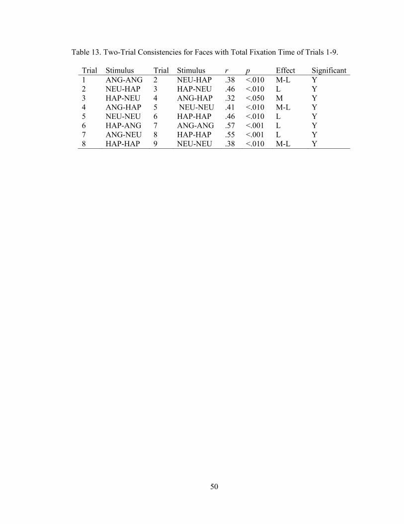
50
Table 13. Two-Trial Consistencies for Faces with Total Fixation Time of Trials 1-9.
Trial Stimulus Trial Stimulus r p Effect Significant
1 ANG-ANG 2 NEU-HAP .38 <.010 M-L Y
2 NEU-HAP 3 HAP-NEU .46 <.010 L Y
3 HAP-NEU 4 ANG-HAP .32 <.050 M Y
4 ANG-HAP 5 NEU-NEU .41 <.010 M-L Y
5 NEU-NEU 6 HAP-HAP .46 <.010 L Y
6 HAP-ANG 7 ANG-ANG .57 <.001 L Y
7 ANG-NEU 8 HAP-HAP .55 <.001 L Y
8 HAP-HAP 9 NEU-NEU .38 <.010 M-L Y
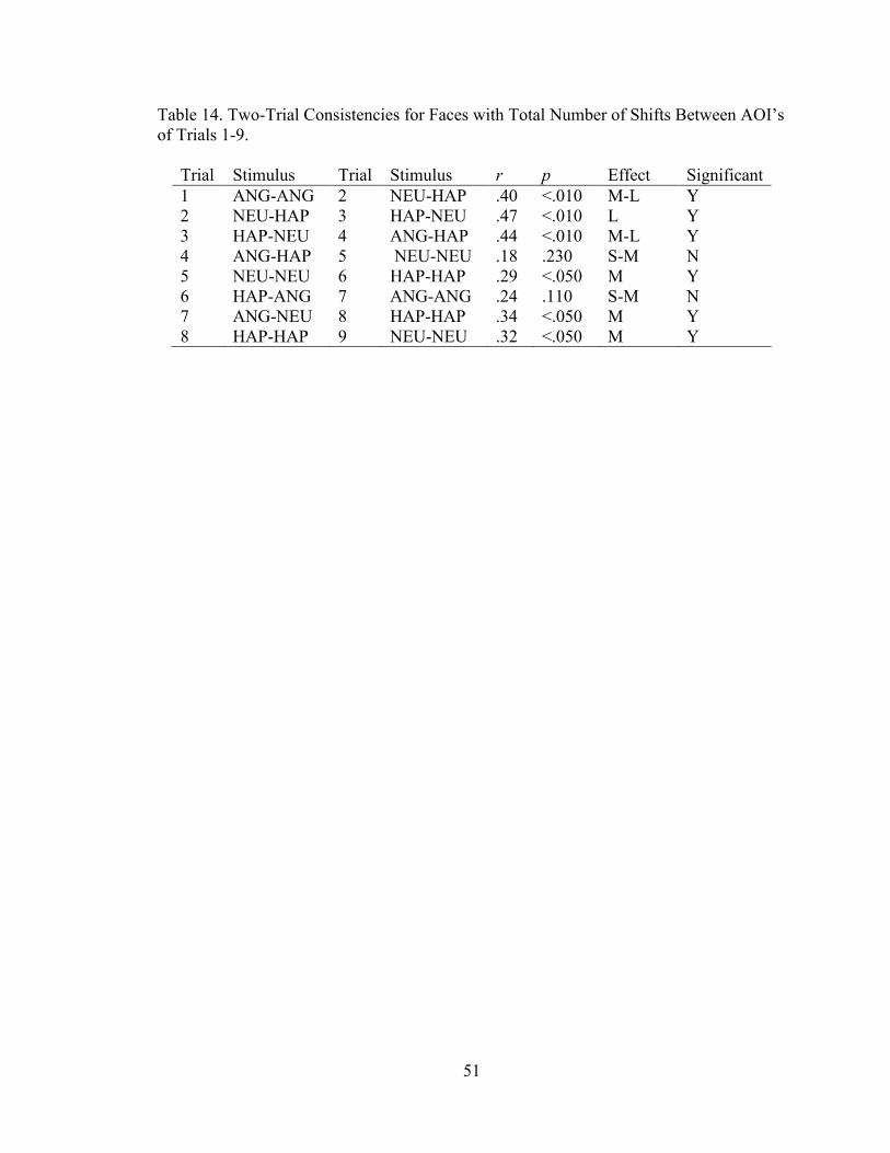
51
Table 14. Two-Trial Consistencies for Faces with Total Number of Shifts Between AOI’s
of Trials 1-9.
Trial Stimulus Trial Stimulus r p Effect Significant
1 ANG-ANG 2 NEU-HAP .40 <.010 M-L Y
2 NEU-HAP 3 HAP-NEU .47 <.010 L Y
3 HAP-NEU 4 ANG-HAP .44 <.010 M-L Y
4 ANG-HAP 5 NEU-NEU .18 .230 S-M N
5 NEU-NEU 6 HAP-HAP .29 <.050 M Y
6 HAP-ANG 7 ANG-ANG .24 .110 S-M N
7 ANG-NEU 8 HAP-HAP .34 <.050 M Y
8 HAP-HAP 9 NEU-NEU .32 <.050 M Y

52
Table 15. Two-Trial Consistencies for Objects with Total Number of Fixations of Trials
1-9.
Trial Stimulus Trial Stimulus r p Effect Significant
1 LOW-LOW 2 MED-HGH .56 <.050 L Y
2 MED-HGH 3 HGH-MED .65 <.010 L Y
3 HGH-MED 4 LOW-HGH .21 .370 S-M N
4 LOW-HGH 5 MED-MED .60 <.010 L Y
5 MED-MED 6 HGH-LOW .17 .490 S-M N
6 HGH-LOW 7 HGH-LOW .40 .080 M-L N
7 LOW-MED 8 LOW-MED .21 .380 S-M N
8 HGH-HGH 9 HGH-HGH .31 .180 M N

53
Table 16. Two-Trial Consistencies for Objects with Latency to First Fixation of Trials 1-
9.
Trial Stimulus Trial Stimulus r p Effect Significant
1 LOW-LOW 2 MED-HGH .32 .170 M N
2 MED-HGH 3 HGH-MED .09 .700 S N
3 HGH-MED 4 LOW-HGH .17 .480 S-M N
4 LOW-HGH 5 MED-MED .15 .540 S N
5 MED-MED 6 HGH-LOW .24 .310 S-M N
6 HGH-LOW 7 HGH-LOW .09 .690 S N
7 LOW-MED 8 LOW-MED .01 .970 None N
8 HGH-HGH 9 HGH-HGH .52 <.050 L Y
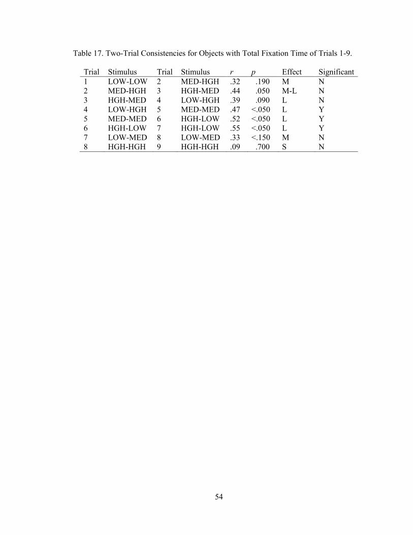
54
Table 17. Two-Trial Consistencies for Objects with Total Fixation Time of Trials 1-9.
Trial Stimulus Trial Stimulus r p Effect Significant
1 LOW-LOW 2 MED-HGH .32 .190 M N
2 MED-HGH 3 HGH-MED .44 .050 M-L N
3 HGH-MED 4 LOW-HGH .39 .090 L N
4 LOW-HGH 5 MED-MED .47 <.050 L Y
5 MED-MED 6 HGH-LOW .52 <.050 L Y
6 HGH-LOW 7 HGH-LOW .55 <.050 L Y
7 LOW-MED 8 LOW-MED .33 <.150 M N
8 HGH-HGH 9 HGH-HGH .09 .700 S N

55
Table 18. Two-Trial Consistencies for Objects with Total Number of Shifts Between
AOI’s of Trials 1-9.
Trial Stimulus Trial Stimulus r p Effect Significant
1 LOW-LOW 2 MED-HGH .42 .060 M-L N
2 MED-HGH 3 HGH-MED .47 <.050 L Y
3 HGH-MED 4 LOW-HGH .21 .370 S-M N
4 LOW-HGH 5 MED-MED .08 .720 S N
5 MED-MED 6 HGH-LOW .45 <.050 L Y
6 HGH-LOW 7 HGH-LOW .50 <.050 L Y
7 LOW-MED 8 LOW-MED .58 <.010 L Y
8 HGH-HGH 9 HGH-HGH .26 .260 M N

56
Table 19. Two-Trial Consistencies for Face Matched Pairs with Total Number of
Fixations of Trials 1-9.
Trial Stimulus Trial Stimulus r p Effect Significant
4 ANG-HAP 6 HAP-ANG .17 .390 S-M N
2 NEU-ANG 3 HAP-NEU .25 .210 M N
7 ANG-NEU 9 NEU-ANG .48 <.050 L Y
1 ANG-ANG 5 NEU-NEU .27 .190 M N
5 NEU-NEU 8 HAP-HAP .23 .250 S-M N
1 ANG-ANG 8 HAP-HAP .20 .330 S-M N
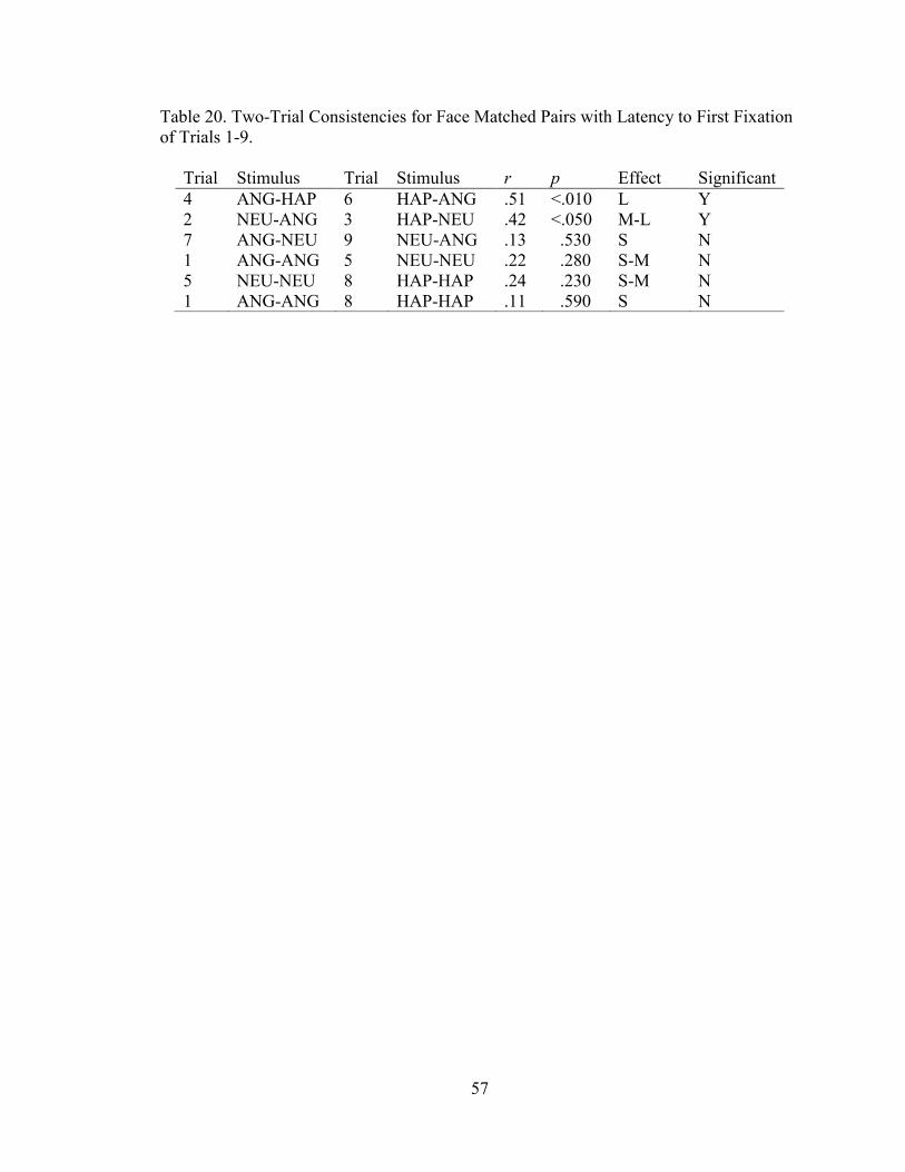
57
Table 20. Two-Trial Consistencies for Face Matched Pairs with Latency to First Fixation
of Trials 1-9.
Trial Stimulus Trial Stimulus r p Effect Significant
4 ANG-HAP 6 HAP-ANG .51 <.010 L Y
2 NEU-ANG 3 HAP-NEU .42 <.050 M-L Y
7 ANG-NEU 9 NEU-ANG .13 .530 S N
1 ANG-ANG 5 NEU-NEU .22 .280 S-M N
5 NEU-NEU 8 HAP-HAP .24 .230 S-M N
1 ANG-ANG 8 HAP-HAP .11 .590 S N

58
Table 21. Two-Trial Consistencies for Face Matched Pairs with Total Fixation Time of
Trials 1-9.
Trial Stimulus Trial Stimulus r p Effect Significant
4 ANG-HAP 6 HAP-ANG .68 <.001 L Y
2 NEU-ANG 3 HAP-NEU .50 <.010 L Y
7 ANG-NEU 9 NEU-ANG .51 <.010 L Y
1 ANG-ANG 5 NEU-NEU .37 .060 M-L N
5 NEU-NEU 8 HAP-HAP .32 .110 M N
1 ANG-ANG 8 HAP-HAP .70 <.001 L Y
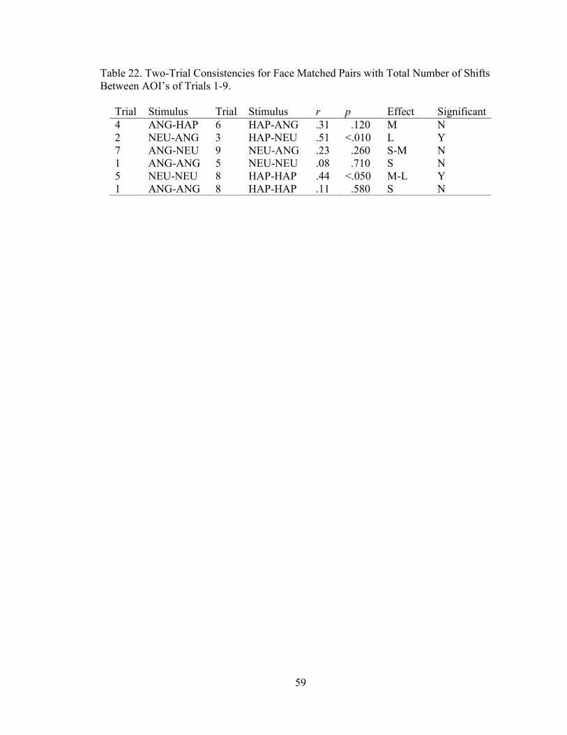
59
Table 22. Two-Trial Consistencies for Face Matched Pairs with Total Number of Shifts
Between AOI’s of Trials 1-9.
Trial Stimulus Trial Stimulus r p Effect Significant
4 ANG-HAP 6 HAP-ANG .31 .120 M N
2 NEU-ANG 3 HAP-NEU .51 <.010 L Y
7 ANG-NEU 9 NEU-ANG .23 .260 S-M N
1 ANG-ANG 5 NEU-NEU .08 .710 S N
5 NEU-NEU 8 HAP-HAP .44 <.050 M-L Y
1 ANG-ANG 8 HAP-HAP .11 .580 S N

60
Table 23. Two-Trial Consistencies for Face Matched Pairs with Total Number of
Fixations of Trials 1-9.
Trial Stimulus Trial Stimulus r p Effect Significant
4 LOW-HGH 6 HGH-LOW .46 <.050 L Y
2 MED-HGH 3 HGH-MED .65 <.010 L Y
7 LOW-MED 9 MED-LOW .80 <.001 L Y
1 LOW-LOW 5 MED-MED .45 <.050 L Y
5 MED-MED 8 HGH-HGH .54 <.050 L Y
1 LOW-LOW 8 HGH-HGH .45 <.050 L Y

61
Table 24. Two-Trial Consistencies for Face Matched Pairs with Latency to First Fixation
of Trials 1-9.
Trial Stimulus Trial Stimulus r p Effect Significant
4 LOW-HGH 6 HGH-LOW .18 .460 S-M N
2 MED-HGH 3 HGH-MED .09 .700 S N
7 LOW-MED 9 MED-LOW .58 <.010 L Y
1 LOW-LOW 5 MED-MED .01 .950 None N
5 MED-MED 8 HGH-HGH .35 .130 M-L N
1 LOW-LOW 8 HGH-HGH .07 .760 S N

62
Table 25. Two-Trial Consistencies for Face Matched Pairs with Total Fixation Time of
Trials 1-9.
Trial Stimulus Trial Stimulus r p Effect Significant
4 LOW-HGH 6 HGH-LOW .67 <.010 L Y
2 MED-HGH 3 HGH-MED .44 .050 M-L N
7 LOW-MED 9 MED-LOW .49 <.050 L Y
1 LOW-LOW 5 MED-MED .40 .080 M-L N
5 MED-MED 8 HGH-HGH .32 .170 M N
1 LOW-LOW 8 HGH-HGH .29 .210 M N

63
Table 26. Two-Trial Consistencies for Face Matched Pairs with Total Number of Shifts
Between AOI’s of Trials 1-9.
Trial Stimulus Trial Stimulus r p Effect Significant
4 LOW-HGH 6 HGH-LOW .35 .130 M-L N
2 MED-HGH 3 HGH-MED .47 <.050 L Y
7 LOW-MED 9 MED-LOW .36 .120 M-L N
1 LOW-LOW 5 MED-MED .54 <.050 L Y
5 MED-MED 8 HGH-HGH .66 <.010 L Y
1 LOW-LOW 8 HGH-HGH .28 .220 M N

64
LIST OF FIGURES
Group Mean Differences
Figure 1. Group Differences: Total Number of Fixations
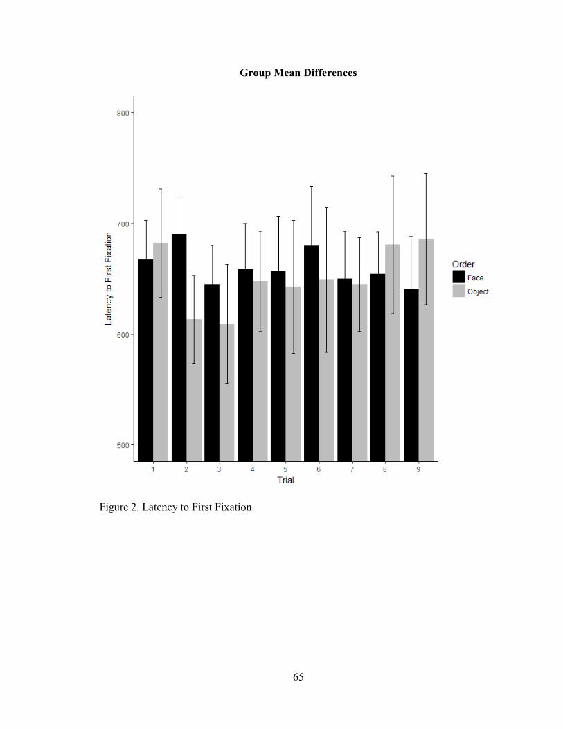
65
Group Mean Differences
Figure 2. Latency to First Fixation

66
Group Mean Differences
Figure 3. Total Fixation Time.

67
Group Mean Differences
Figure 4. Total Number of Shifts.