Embed Size (px)
Citation preview

Research ArticleAssessment of Osteoporosis in Injured Older Women Admittedto a Safety-Net Level One Trauma Center: A Unique Opportunityto Fulfill an Unmet Need
Elisabeth S. Young,1,2 May J. Reed,2 TamN. Pham,3 Joel A. Gross,4
Lisa A. Taitsman,5 and Stephen J. Kaplan2,6
1University of Hawaii College of Medicine, Honolulu, HI, USA2Division of Geriatrics and Gerontology, Department of Medicine, Harborview Medical Center, University of Washington,Seattle, WA, USA3Department of Surgery, Harborview Medical Center, University of Washington, Seattle, WA, USA4Department of Radiology, Harborview Medical Center, University of Washington, Seattle, WA, USA5Department of Orthopedics and Sports Medicine, Harborview Medical Center, University of Washington, Seattle, WA, USA6Section of General, Thoracic, and Vascular Surgery, Department of Surgery, Virginia Mason Medical Center, Seattle, WA, USA
Correspondence should be addressed to Stephen J. Kaplan; [email protected]
Received 10 July 2017; Revised 28 September 2017; Accepted 10 October 2017; Published 6 November 2017
Academic Editor: Francesc Formiga
Copyright © 2017 Elisabeth S. Young et al. This is an open access article distributed under the Creative Commons AttributionLicense, which permits unrestricted use, distribution, and reproduction in any medium, provided the original work is properlycited.
Background. Older trauma patients often undergo computed tomography (CT) as part of the initial work-up. CT imaging can also beused opportunistically to measure bone density and assess osteoporosis.Methods. In this retrospective cohort study, osteoporosiswas ascertained from admission CT scans in women aged ≥65 admitted to the ICU for traumatic injury during a 3-year periodat a single, safety-net, level 1 trauma center. Osteoporosis was defined by established CT-based criteria of average L1 vertebralbody Hounsfield units <110. Evidence of diagnosis and/or treatment of osteoporosis was the primary outcome. Results. The studycohort consisted of 215 women over a 3-year study period, of which 101 (47%) had evidence of osteoporosis by CT scan criteria.There were no differences in injury severity score, hospital length of stay, cost, or discharge disposition between groups with andwithout evidence of osteoporosis. Only 55 (59%) of the 94 patients with osteoporosis who survived to discharge had a documentedosteoporosis diagnosis and/or corresponding evaluation/treatment plan. Conclusion. Nearly half of older women admitted withtraumatic injuries had underlying osteoporosis, but 41% had neither clinical recognition of this finding nor a treatment planfor osteoporosis. Admission for traumatic injury is an opportunity to assess osteoporosis, initiate appropriate intervention, andcoordinate follow-up care. Trauma and acute care teams should consider assessment of osteoporosis in women who undergo CTimaging and provide a bridge to outpatient services.
1. Introduction
Adults aged 65 and older constitute over 25% of traumarelated admissions and are the fastest growing trauma patientpopulation, with a significant proportion sustaining fractures[1]. Osteoporotic fractures, particularly hip fracture, are asignificant cause of morbidity and mortality in these patients[2]. Older women are particularly vulnerable to osteoporosisas declining estrogen contributes to an increased rate of bone
loss and thus reduced bone mineral density (BMD) that con-tributes to fracture risk. As the population ages, osteoporosisis a growing individual and public health concern with morethan 40 million Americans at risk for this diagnosis [2–4].The lifetime risk of developing a fracture in patients withunderlying osteoporosis is estimated to be up to 50% inwomen and 20% in men [2].
Over 80 million CT scans are performed annually in theUnited States andmany of these scans are performed on older
HindawiCurrent Gerontology and Geriatrics ResearchVolume 2017, Article ID 4658050, 6 pageshttps://doi.org/10.1155/2017/4658050

2 Current Gerontology and Geriatrics Research
patients who have undiagnosed chronic conditions. In theroutine evaluation of patientswith traumatic injury, thoroughimaging via computed tomography (CT) is often obtained[5]. Nevertheless, the implications of routine opportunisticutilization of CT scans for the assessment of osteoporosis inacute care settings have not been previously evaluated. Asidefrom the immediate needs of addressing traumatic injury,harnessing the diagnostic power of routine evaluations inacute care settings may provide significant health and costbenefits.
This repurposing of diagnostic imaging is not a newidea. CT-derived BMD is an established method to identifychronic bone loss, diagnose vertebral fractures, and improvereliability of BMD estimates in patients with aortic calcifi-cation [6]. Others have reported diagnosing osteopenia andosteoporosis with CT scans ordered for other reasons andnoted substantial opportunities for savings with respect toobviating the expense of additional imaging and the cost ofpreventable fractures [7, 8].
The use of computed tomography to evaluate bone min-eral density has been broadly described, although it has notbeenwidely accepted as a gold standard for routine outpatientscreening. Specifically, Pickhardt et al. compared CT-derivedBMD toDual EnergyX-rayAbsorptiometry (DXA)measuresin over 2000 paired comparisons and found highly predictivevalues for osteoporosis diagnosis (area under the curve[AUC]=0.83, 95%CI 0.81–0.85) [8]. Subsequent authors haveindependently demonstrated significant predictive valuesand correlations between DXA and CT [7, 9–11].
The US Preventive Service Task Force (USPSTF) recom-mends that all women over 65 years of age should be screenedfor osteoporosis and that women below 65 years of age shouldbe tested in the presence of additional risk fractures, such as afragility fracture [2, 3]. Despite these guidelines, many olderwomen do not undergo formal BMD evaluation, even in thecontext of falls or known fall risk [1, 2].
The purpose of this study was to assess the prevalenceof osteoporosis by opportunistic CT imaging in older adultwomen admitted for trauma, a high-risk population, andmeasure recognition of this chronic disease by acute careproviders in a safety-net hospital.
2. Methods
2.1. Study Design and Participants. This retrospective cohortstudy included all women of age ≥ 65 who were in-stateresidents, sustained traumatic injury without serious headinjury (maximum head abbreviated injury severity [AIS]score < 3), had CT imaging of L1 within 7 days of admission,and were admitted to the intensive care unit (ICU) at a safety-net, level one trauma center from January 2011 to February2014. Our target population was chosen because women area traditionally at-risk population, with female to male 4 : 1prevalence of osteoporosis in the US [2]. We also restrictedour analysis to patients admitted to the ICU because severelyinjured patients were more likely to undergo truncal CTevaluation on admission. Inclusion of only in-state residentsallowed for readmissions and mortality linkage using stateregistries as described below. Women who died within 24
Trauma ICU admissions
CT scan available
(i) Fracture, 21(ii) Implant, 3
(iii) Other abnormality, 8(iv) Death within 1 day of
admission, 5
No osteoporosis Osteoporosis
Study cohort
Female, age ≥ 65,Head AIS ≤2 , in-state,
L1 HU ≥ 110 L1 HU < 110
1/1/2011–2/28/2014
n =
n = (53%) n = (%)
n =
n = Exclusions,
Figure 1: Flow diagram of study patients, showing inclusion andexclusion criteria, grouping of study cohort.
hours of admission or whose L1 imaging was inadequate foranalysis were excluded (Figure 1).
2.2. Study Setting. This study was conducted at a level onetrauma center, which serves the surrounding metropolitanarea, state, and surrounding four-state region. In additionto being the only level one adult trauma, pediatric trauma,and burn center in the state, the facility also serves as thestate’s main safety-net hospital. Fifteen percent of the statepopulation is ≥65 years old; 12% of the population is livingin poverty (income below 100% of the federal poverty line).Sixty-two percent of patients visiting this hospital qualifyfor Medicaid or premium subsidies under the state HealthInsurance Marketplace. Nearly 50% of the service populationaremembers of racial and/or ethnicminorities;more than 8%are non-English speaking; and more than 2% are indigentswithout third-party coverage.
2.3. Definition of Osteoporosis by CT. Patients were dividedinto groups by presence or absence of osteoporosis, whichwasdefined as average vertebral bodyHounsfield units (HU)< 110(90% specificity) as described by Pickard et al., 2013 [7, 8, 10,11].
2.4. Covariates. Patient data, injury details, and clinicalmeasures were queried through the state trauma registry.Ground-level falls were determined according to ICD-9E codes (E880.1, E884.2, E884.3, E884.4, E884.6, E885.9,E888.1, and E888.8). To determine outcomes after trauma,the registry was linked to the Comprehensive HospitalAbstract Reporting System (CHARS), a statewide databasethat contains hospital admission information. Only the firstnonelective readmission after the index trauma hospitaliza-tion was included (identified by categorization in CHARS).

Current Gerontology and Geriatrics Research 3
Trauma Registry and CHARS datasets were further linked tothe Washington State Death Registry to assess 30-day and 1-year mortality.
2.5. Evidence of Osteoporosis Recognition and Treatment byInpatient Providers. Recognition of osteoporosis was ascer-tained from problem list; patient education materials; dis-charge summary; or treatment of osteoporosis (calcium,vitamin D, bisphosphonate, teriparatide, or denosumab) thatwas abstracted from the discharge medication list.
2.6. Image Analysis Protocol. In the sagittal midline plane,the L1 vertebral body was identified by locating the superioraspect of the sacrum and labeling the immediately superiorvertebral body as L5. Identification of L1 was confirmed byabsence of ribs at that level. T12was utilized if L1 was excludeddue to Genant Grade II or III compression fracture, neoplas-tic lesion, hemangioma, or any compromising abnormalitythat resulted in nonhomogenous bone. L2 was utilized ifT12 required exclusion. The most superior axial plane of thechosen vertebral body, which minimized presence of corticalbone and excluded comprising abnormalities, was chosen.Vertebral BMD was assessed by placing a single ellipticalregion of interest (ROI) 100–120mm2 on the central part ofthe vertebral body excluding cortical bone, sclerotic bone,or fracture lines. Average HU measurement and SD fromthe selected vertebra was recorded. Intra- and Interraterreliability of HU measurements was confirmed using intr-aclass correlation coefficients utilizing the first 32 patientsincluded in the study.The images were analyzed separately bya trauma radiologist (J. A. G.), a research scholar and surgicalresident (S. J. K.), and a medical student (E. S. Y.). Intraclasscorrelation coefficients were 0.98 [95% Cl 0.96–0.99] forHU calculation and 0.99 [95% Cl 0.993–0.999] for axialimage selection. All three evaluators excluded the same threepatients due to a compromising abnormality.
2.7. Statistical Analysis. Data normality was evaluated withthe Shapiro-Wilk test and histogram visualization. Contin-uous, normally distributed data are reported as mean ±standard deviation (SD) and compared between groups usingthe t-test. Discrete and skewed continuous data are reportedas median (interquartile range [IQR]) and compared usingthe Mann–Whitney 𝑈 test. Categorical data are reportedas count (proportion) and compared using Pearson’s 𝜒2 orFisher’s exact test, as appropriate. Confidence intervals forrelative proportions of CT-identified osteoporosis and thesubset of patients who did not have a diagnosis ormedicationlisted in discharge data were calculated using the thresholdsdescribed by Pickhardt et al. [8]. All statistical calculationswere performed with Stata/SE 14.1 (StataCorp LP, CollegeStation, TX) using an a priori two-sided significance level of0.05.
3. Results
Of the 252 women ≥ 65 years old who met inclusioncriteria, 37 patients were excluded for death within 24 hours
of admission (𝑛 = 5) or inadequate imaging/unusablevertebral bodies at or adjacent to the L1 level (𝑛 = 32).The remaining 215 comprised the study cohort. Using thethreshold described above, 101 women (47%) retrospectivelyhad evidence of osteoporosis by CT scan, leaving 114 (53%)without osteoporosis.
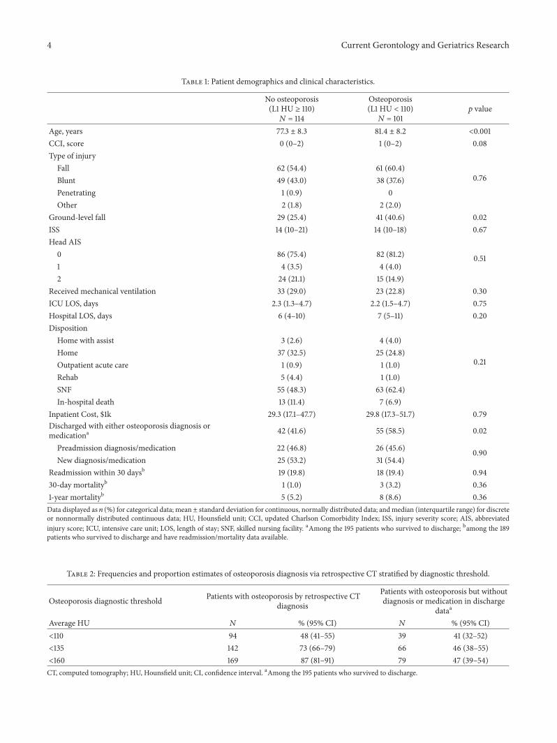
Women with osteoporosis were older (81.4 ± 8.2 versus77.3 ± 8.3, 𝑝 < 0.001) and more likely to have sustained aground-level fall (41 [40.6%] versus 29 [25.4%], relative risk[RR] 1.59 [95% CI 1.08–2.36], 𝑝 = 0.02). Demographics andclinical characteristics between osteoporotic and nonosteo-porotic groups were otherwise relatively similar (Table 1).Twenty patients died in hospital. There were no differencesbetween groups with regard to any of the following outcomes:hospital length of stay, discharge disposition, inpatient cost,30-day readmission, 30-day mortality, and 1-year mortality.
Among survivors to discharge, 63 (67.0%) of osteoporoticpatients were discharged to a skilled nursing facility (SNF)compared to 55 (54.5%) of nonosteoporotic patients. Thisdifference approached significance on multivariate analysis(RR 1.23 [95% CI 0.98–1.55], 𝑝 = 0.07).
Only 55 (59%) of the 94 patients withCT-identified osteo-porosis who survived to discharge had a listed osteoporosisdiagnosis and/or corresponding evaluation/treatment plan:24 had amedication prescribed before their traumatic injury;the other 31 of 55 had a new medication prescribed at timeof discharge. The remaining thirty-nine (41%) patients withretrospectively identified osteoporosis did not have a markerfor the recognition of osteoporosis by the acute care team.Undiagnosed and untreated osteoporosis proportions did notdiffer markedly using more sensitive, less specific criteria(Table 2) [8]. Among women with retrospectively identifiedosteoporosis, the proportion of osteoporosis recognition didnot differ between women who sustained a ground-level falland those with other injury mechanisms (17 [45%] versus 22[39%], 𝑝 = 0.60).
4. Discussion
In this retrospective study, we utilized routine admissionCT in an opportunistic fashion to evaluate older women forlow L1 BMD. We found that nearly half of those admittedfor traumatic injuries had underlying osteoporosis using themost specific of criteria of <110 HU. However, 41% of thosewomen did not have documentation conveying this finding,either in the discharge summary or problem list, or thedocumentation of medications used to treat osteoporosis. Ofthe women with evidence of osteoporosis by CT, we foundthat just 12% were deemed osteoporotic in the problem list ordischarge summary.
DXA remains the objective gold standard in BMD assess-ment (osteoporosis defined as a 𝑇-score of <−2.5) duringroutine outpatient care. Despite increased fracture risk andincreased mortality in the population with falls, screeningfor chronic bone loss remains underutilized even in thispatient population [1, 12]. It is worth noting that DXA is notreimbursed in the inpatient setting and is largely delegatedto outpatient providers. As a result, appropriate follow-upfor this chronic disease is susceptible to the communication
4 Current Gerontology and Geriatrics Research
Table 1: Patient demographics and clinical characteristics.
No osteoporosis(L1 HU ≥ 110)𝑁 = 114
Osteoporosis(L1 HU < 110)𝑁 = 101
𝑝 value
Age, years 77.3 ± 8.3 81.4 ± 8.2 <0.001CCI, score 0 (0–2) 1 (0–2) 0.08Type of injury
0.76Fall 62 (54.4) 61 (60.4)Blunt 49 (43.0) 38 (37.6)Penetrating 1 (0.9) 0Other 2 (1.8) 2 (2.0)
Ground-level fall 29 (25.4) 41 (40.6) 0.02ISS 14 (10–21) 14 (10–18) 0.67Head AIS
0.510 86 (75.4) 82 (81.2)1 4 (3.5) 4 (4.0)2 24 (21.1) 15 (14.9)
Received mechanical ventilation 33 (29.0) 23 (22.8) 0.30ICU LOS, days 2.3 (1.3–4.7) 2.2 (1.5–4.7) 0.75Hospital LOS, days 6 (4–10) 7 (5–11) 0.20Disposition
0.21
Home with assist 3 (2.6) 4 (4.0)Home 37 (32.5) 25 (24.8)Outpatient acute care 1 (0.9) 1 (1.0)Rehab 5 (4.4) 1 (1.0)SNF 55 (48.3) 63 (62.4)In-hospital death 13 (11.4) 7 (6.9)
Inpatient Cost, $1k 29.3 (17.1–47.7) 29.8 (17.3–51.7) 0.79Discharged with either osteoporosis diagnosis ormedicationa 42 (41.6) 55 (58.5) 0.02
Preadmission diagnosis/medication 22 (46.8) 26 (45.6) 0.90New diagnosis/medication 25 (53.2) 31 (54.4)
Readmission within 30 daysb 19 (19.8) 18 (19.4) 0.9430-day mortalityb 1 (1.0) 3 (3.2) 0.361-year mortalityb 5 (5.2) 8 (8.6) 0.36Data displayed as 𝑛 (%) for categorical data; mean ± standard deviation for continuous, normally distributed data; andmedian (interquartile range) for discreteor nonnormally distributed continuous data; HU, Hounsfield unit; CCI, updated Charlson Comorbidity Index; ISS, injury severity score; AIS, abbreviatedinjury score; ICU, intensive care unit; LOS, length of stay; SNF, skilled nursing facility. aAmong the 195 patients who survived to discharge; bamong the 189patients who survived to discharge and have readmission/mortality data available.
Table 2: Frequencies and proportion estimates of osteoporosis diagnosis via retrospective CT stratified by diagnostic threshold.
Osteoporosis diagnostic threshold Patients with osteoporosis by retrospective CTdiagnosis
Patients with osteoporosis but withoutdiagnosis or medication in discharge
dataa
Average HU 𝑁 % (95% CI) 𝑁 % (95% CI)<110 94 48 (41–55) 39 41 (32–52)<135 142 73 (66–79) 66 46 (38–55)<160 169 87 (81–91) 79 47 (39–54)CT, computed tomography; HU, Hounsfield unit; CI, confidence interval. aAmong the 195 patients who survived to discharge.

Current Gerontology and Geriatrics Research 5
breakdowns that are common when transitioning from inpa-tient to outpatient settings [13].
Of the 94 women with evidence of osteoporosis by CTwho survived to discharge, only 31 (31%) were prescribednew medications during admission and only 26 (25%) wereon previously prescribed medications that could benefitosteoporosis. It is possible that some acute care providersdeferred initiation of bone modifying medications, such asbisphosphonates, because of the theoretical concern thatthese medications impair fracture healing [14, 15]. In orderto improve sensitivity, we also included vitamin D and/orcalcium as additional surrogates for initiation of osteoporosistreatment. Still, a substantial number of older women withosteoporosis did not receive any medications to promotebone health.
Of note, there were also a number of patients who didnot meet the CT-based threshold for osteoporosis but yethad evidence of osteoporosis recognition based on theirmedication list or diagnosis list (42 [41%] among survivorsto discharge). This is likely an effect of the highly specific,but poorly sensitive HU-based threshold of 110. When moresensitive thresholds are considered, more patients are consid-ered osteoporotic (Table 2). However, regardless of thresholdused, the estimated proportion of patients with osteoporosisby CT criteria who are discharged without medications orformal diagnoses remains between 41 and 47% in this studycohort.
Osteoporosis evaluation and treatment is largely con-sidered within the purview of primary care: a perspectivethat may explain the limited evaluation and treatment ini-tiation in an at-risk population during an admission fortrauma. Multiple investigations have focused on improvingthe transition to outpatient care and referral for evaluationof osteoporosis after discharge [16, 17]. These studies havedemonstrated improved treatment and evaluation with suchmethods as a dedicated osteoporosis health professional,fracture liaison nurse, or a letter to the patient’s primarycare provider. However, diagnosis by CT-derived BMD couldstreamline initiation of interventions, reduce risk of missedcommunication, and provide considerable cost savings.
Fragility fractures are a significant public health issue andtreatment of osteoporosis has been found to be effective inreducing morbidities, such as secondary fracture prevention[18, 19]. Many of the organizations and countries that havefinancial responsibility for covered lives have instituted for-mal protocols for identifying and treating fragility fracturesand osteoporosis, ultimately to the benefit of the patient[20, 21]. The ability to utilize existing CT scans to assessosteoporosis could be beneficial for patients and the healthcare system. Simply providing patients with informationregarding their diagnosis of osteoporosis improves the like-lihood that a patient will have their osteoporosis addressedby their primary care provider [22].
Opportunistic diagnosis of osteoporosis using CT scanscould also serve an unmet need in hospitals that serveas a safety-net, such as ours [23]. By definition, safety-nethospitals serve low income,medically, and socially vulnerablepatients regardless of their ability to pay. Economicallydisadvantaged individuals with chronic conditions have high
rates of readmission and emergency department usage fol-lowing initial hospitalization. Additionally, this populationfaces greater challenges in receiving pre- and postinjurycare [24]. Point-of-care diagnosis could be valuable in theacute care setting, as hospitalization is an opportunity forthe patient to be assessed for osteoporosis by CT BMD in acost- and time-effective manner. Recognition of low BMDas part of trauma care may improve care transitions andlead to efficient arrangement of subsequent interventionsand appointments. In a safety-net hospital, CT could alsoprovide an early diagnosis of bone loss in the late-middle agepopulation (55–64 years of age), who do not typically qualifyfor insurance coverage of outpatient DXA [2, 25].
The present study has several limitations. It is retro-spective and excludes patients without imaging, which con-tributed to a smaller sample size. Participants are exclusivelyfrom an ICU population, so severity of injuries is greater thanthat of a typical population of older adults admitted withtrauma.We note that patients admitted to general orthopedicservices, especially those with medicine comanagement, aremore likely to receive a diagnosis and subsequent plan of carefor osteoporosis.
5. Conclusion
Trauma patients often undergo routine CT imaging, whichprovides a unique opportunity to diagnose older womenwithosteoporosis. Osteoporosis poses a significant risk factor forfractures, future falls, and death. Trauma and other acute careteams should consider using opportunistic imaging to assessolder women for osteoporosis, especially those in safety-netsettings, and provide a bridge to outpatient services.
Disclosure
Thecontent is solely the responsibility of the authors and doesnot necessarily represent the official views of the sponsoringinstitutions or supporting agencies.
Conflicts of Interest
The authors declare that they have no conflicts of interest.
Acknowledgments
The authors wish to acknowledge Mamatha Damodarasamyfor assistance with the manuscript and Zeyno Shorter, Ph.D.,MPH, for data acquisition. Additionally, they thank ItayBentov, MD, Ph.D., Steven H. Mitchell, MD, and SamanArbabi, MD, MPH, and the Geriatric Injury Workgroupat Harborview Medical Center, for their assistance on thisproject. This work was supported in part by the JohnA. Hartford Foundation Center of Excellence in GeriatricMedicine and Training at the University of Washington, theMedical Student Training in Aging Research Program atthe University of California, Los Angeles, and the PattersonSurgery Research Endowment at Benaroya Research Insti-tute/Virginia Mason.

6 Current Gerontology and Geriatrics Research
References
[1] P. Ayoung-Chee, L. McIntyre, B. E. Ebel, C. D. MacK, W.McCormick, and R. V. Maier, “Long-term outcomes of ground-level falls in the elderly,” Journal of Trauma and Acute CareSurgery, vol. 76, no. 2, pp. 498–503, 2014.
[2] K. E. Ensrud and C. J. Crandall, “Osteoporosis,” Annals ofInternal Medicine, vol. 167, no. 3, pp. ITC17–ITC32, 2017.
[3] U.S. Preventive Services Task Force, “Screening for osteo-porosis: U.S. Preventive Services Task Force recommendationstatement,” Annals of Internal Medicine, vol. 154, no. 5, pp. 356–364, 2011.
[4] N. C. Wright, A. C. Looker, K. G. Saag et al., “The recentprevalence of osteoporosis and low bone mass in the UnitedStates based on bone mineral density at the femoral neck orlumbar,” Journal of Bone and Mineral Research, vol. 29, no. 11,pp. 2520–2526, 2014.
[5] S. J. Kaplan, T. N. Pham, S. Arbabi et al., “Association ofradiologic indicators of frailty with 1-year mortality in oldertrauma patients: Opportunistic screening for sarcopenia andosteopenia,” JAMA Surgery, vol. 152, no. 2, article e164604, 2017.
[6] S. Kinsella, K. Murphy, M. Breen et al., “Comparison ofsingle CT scan assessment of bone mineral density, vascularcalcification and fat mass with standard clinical measurementsin renal transplant subjects: The ABC HeART study,” BMCNephrology, vol. 16, no. 1, article 188, 2015.
[7] C. F. Buckens, G. Dijkhuis, B. de Keizer, H. J. Verhaar, and P. A.de Jong, “Opportunistic screening for osteoporosis on routinecomputed tomography?An external validation study,”EuropeanRadiology, vol. 25, no. 7, pp. 2074–2079, 2015.
[8] P. J. Pickhardt, B. D. Pooler, T. Lauder, A. M. del Rio, R. J. Bruce,andN. Binkley, “Opportunistic screening for osteoporosis usingabdominal computed tomography scans obtained for otherindications,”Annals of InternalMedicine, vol. 158, no. 8, pp. 588–595, 2013.
[9] O. Emohare, M. Wiggin, P. Hemmati, and J. Switzer, “Assessingbone mineral density following acute hip fractures: the roleof computed tomography attenuation,” Geriatric OrthopaedicSurgery & Rehabilitation, vol. 6, no. 1, pp. 16–21, 2015.
[10] M. K. Choi, S. M. Kim, and J. K. Lim, “Diagnostic efficacyof Hounsfield units in spine CT for the assessment of realbone mineral density of degenerative spine: correlation studybetween T-scores determined by DEXA scan and Hounsfieldunits from CT,” Acta Neurochirurgica, vol. 158, no. 7, pp. 1421–1427, 2016.
[11] S. Y. Lee, S.-S. Kwon, H. S. Kim et al., “Reliability and validityof lower extremity computed tomography as a screening toolfor osteoporosis,” Osteoporosis International, vol. 26, no. 4, pp.1387–1394, 2015.
[12] D. Bliuc, D. Alarkawi, T. V. Nguyen, J. A. Eisman, and J. R.Center, “Risk of subsequent fractures and mortality in elderlywomen andmenwith fragility fractureswith andwithout osteo-porotic bone density: The dubbo osteoporosis epidemiologystudy,” Journal of Bone and Mineral Research, vol. 30, no. 4, pp.637–646, 2015.
[13] C. J. Yates, M.-A. Chauchard, D. Liew, A. Bucknill, and J. D.Wark, “Bridging the osteoporosis treatment gap: Performanceand cost-effectiveness of a fracture liaison service,” Journal ofClinical Densitometry, vol. 18, no. 2, pp. 150–156, 2015.
[14] S. Larsson and N. L. Fazzalari, “Anti-osteoporosis therapy andfracture healing,” Archives of Orthopaedic and Trauma Surgery,vol. 134, no. 2, pp. 291–297, 2014.
[15] V. Hegde, J. E. Jo, P. Andreopoulou, and J. M. Lane, “Effectof osteoporosis medications on fracture healing,” OsteoporosisInternational, vol. 27, no. 3, pp. 861–871, 2016.
[16] K. Bell, H. Strand, and W. J. Inder, “Effect of a dedicatedosteoporosis health professional on screening and treatment inoutpatients presenting with acute low trauma non-hip fracture:a systematic review,” Archives of Osteoporosis, vol. 9, article 167,2014.
[17] W. W. Hung, K. A. Egol, J. D. Zuckerman, and A. L. Siu,“Hip fracturemanagement: Tailoring care for the older patient,”Journal of the AmericanMedical Association, vol. 307, no. 20, pp.2185–2194, 2012.
[18] H. S. Bawa, J. Weick, and D. R. Dirschl, “Anti-osteoporotictherapy after fragility fracture lowers rate of subsequent frac-ture: Analysis of a large population sample: Analysis of alarge population sample,” Journal of Bone and Joint Surgery -American Volume, vol. 97, no. 19, pp. 1555–1562, 2014.
[19] E. F. Ekman, “The role of the orthopaedic surgeon in minimiz-ing mortality and morbidity associated with fragility fractures,”American Academy of Orthopaedic Surgeon, vol. 18, no. 5, pp.278–285, 2010.
[20] S. Drew, S. Sheard, J. Chana, C. Cooper, M. K. Javaid, andA. Judge, “Describing variation in the delivery of secondaryfracture prevention after hip fracture: an overviewof 11 hospitalswithin one regional area in England,”Osteoporosis International,vol. 25, no. 10, pp. 2427–2433, 2014.
[21] K. Ganda, M. Puech, J. S. Chen et al., “Models of care for thesecondary prevention of osteoporotic fractures: a systematicreview and meta-analysis,” Osteoporosis International, vol. 24,no. 2, pp. 393–406, 2013.
[22] M. J. Gardner, R. H. Brophy, D. Demetrakopoulos et al.,“Interventions to improve osteoporosis treatment following hipfracture: a prospective, randomized trial,” The Journal of Bone& Joint Surgery—American Volume, vol. 87, no. 1, pp. 3–7, 2005.
[23] R. V. Maier, “Seattle’s Harborview Medical Center, 1877–2003,”Archives of Surgery, vol. 139, no. 1, pp. 14-15, 2004.
[24] S. Hewner, S. Casucci, and J. Castner, “The roles of chronicdisease complexity, health system integration, and caremanage-ment in post-discharge healthcare utilization in a low-incomepopulation,” Research in Nursing & Health, vol. 39, no. 4, pp.215–228, 2016.
[25] S. Nayak, M. S. Roberts, and S. L. Greenspan, “Cost-effectiveness of different screening strategies for osteoporosis inpostmenopausal women,” Annals of Internal Medicine, vol. 155,no. 11, pp. 751–761, 2011.

Submit your manuscripts athttps://www.hindawi.com
Stem CellsInternational
Hindawi Publishing Corporationhttp://www.hindawi.com Volume 2014
Hindawi Publishing Corporationhttp://www.hindawi.com Volume 2014
MEDIATORSINFLAMMATION
of
Hindawi Publishing Corporationhttp://www.hindawi.com Volume 2014
Behavioural Neurology
EndocrinologyInternational Journal of
Hindawi Publishing Corporationhttp://www.hindawi.com Volume 2014
Hindawi Publishing Corporationhttp://www.hindawi.com Volume 2014
Disease Markers
Hindawi Publishing Corporationhttp://www.hindawi.com Volume 2014
BioMed Research International
OncologyJournal of
Hindawi Publishing Corporationhttp://www.hindawi.com Volume 2014
Hindawi Publishing Corporationhttp://www.hindawi.com Volume 2014
Oxidative Medicine and Cellular Longevity
Hindawi Publishing Corporationhttp://www.hindawi.com Volume 2014
PPAR Research
The Scientific World JournalHindawi Publishing Corporation http://www.hindawi.com Volume 2014
Immunology ResearchHindawi Publishing Corporationhttp://www.hindawi.com Volume 2014
Journal of
ObesityJournal of
Hindawi Publishing Corporationhttp://www.hindawi.com Volume 2014
Hindawi Publishing Corporationhttp://www.hindawi.com Volume 2014
Computational and Mathematical Methods in Medicine
OphthalmologyJournal of
Hindawi Publishing Corporationhttp://www.hindawi.com Volume 2014
Diabetes ResearchJournal of
Hindawi Publishing Corporationhttp://www.hindawi.com Volume 2014
Hindawi Publishing Corporationhttp://www.hindawi.com Volume 2014
Research and TreatmentAIDS
Hindawi Publishing Corporationhttp://www.hindawi.com Volume 2014
Gastroenterology Research and Practice
Hindawi Publishing Corporationhttp://www.hindawi.com Volume 2014
Parkinson’s Disease
Evidence-Based Complementary and Alternative Medicine
Volume 2014Hindawi Publishing Corporationhttp://www.hindawi.com