Embed Size (px)
Citation preview

University of Birmingham
Assessment of Alveolar Macrophage DysfunctionUsing an in vitro Model of Acute RespiratoryDistress SyndromeMahida, Rahul Y.; Scott, Aaron; Parekh, Dhruv; Lugg, Sebastian T.; Belchamber, Kylie B. R.;Hardy, Rowan S.; Matthay, Michael A.; Naidu, Babu; Thickett, David R.DOI:10.3389/fmed.2021.737859
License:Creative Commons: Attribution (CC BY)
Document VersionPublisher's PDF, also known as Version of record
Citation for published version (Harvard):Mahida, RY, Scott, A, Parekh, D, Lugg, ST, Belchamber, KBR, Hardy, RS, Matthay, MA, Naidu, B & Thickett,DR 2021, 'Assessment of Alveolar Macrophage Dysfunction Using an in vitro Model of Acute RespiratoryDistress Syndrome', Frontiers in Medicine, vol. 8, 737859. https://doi.org/10.3389/fmed.2021.737859
Link to publication on Research at Birmingham portal
General rightsUnless a licence is specified above, all rights (including copyright and moral rights) in this document are retained by the authors and/or thecopyright holders. The express permission of the copyright holder must be obtained for any use of this material other than for purposespermitted by law.
•Users may freely distribute the URL that is used to identify this publication.•Users may download and/or print one copy of the publication from the University of Birmingham research portal for the purpose of privatestudy or non-commercial research.•User may use extracts from the document in line with the concept of ‘fair dealing’ under the Copyright, Designs and Patents Act 1988 (?)•Users may not further distribute the material nor use it for the purposes of commercial gain.
Where a licence is displayed above, please note the terms and conditions of the licence govern your use of this document.
When citing, please reference the published version.
Take down policyWhile the University of Birmingham exercises care and attention in making items available there are rare occasions when an item has beenuploaded in error or has been deemed to be commercially or otherwise sensitive.
If you believe that this is the case for this document, please contact [email protected] providing details and we will remove access tothe work immediately and investigate.
Download date: 03. Apr. 2022

ORIGINAL RESEARCHpublished: 29 September 2021
doi: 10.3389/fmed.2021.737859
Frontiers in Medicine | www.frontiersin.org 1 September 2021 | Volume 8 | Article 737859
Edited by:
Gary Frank Nieman,
SUNY Upstate Medical University,
United States
Reviewed by:
Francesco Poti,
University of Parma, Italy
Anna Dmitriyevna Krasnodembskaya,
Queen’s University Belfast,
United Kingdom
*Correspondence:
Rahul Y. Mahida
Specialty section:
This article was submitted to
Pulmonary Medicine,
a section of the journal
Frontiers in Medicine
Received: 07 July 2021
Accepted: 30 August 2021
Published: 29 September 2021
Citation:
Mahida RY, Scott A, Parekh D,
Lugg ST, Belchamber KBR, Hardy RS,
Matthay MA, Naidu B and Thickett DR
(2021) Assessment of Alveolar
Macrophage Dysfunction Using an in
vitro Model of Acute Respiratory
Distress Syndrome.
Front. Med. 8:737859.
doi: 10.3389/fmed.2021.737859
Assessment of Alveolar MacrophageDysfunction Using an in vitro Modelof Acute Respiratory DistressSyndromeRahul Y. Mahida 1*, Aaron Scott 1, Dhruv Parekh 1, Sebastian T. Lugg 1,
Kylie B. R. Belchamber 1, Rowan S. Hardy 2, Michael A. Matthay 3, Babu Naidu 1 and
David R. Thickett 1
1 Birmingham Acute Care Research Group, Institute of Inflammation and Ageing, University of Birmingham, Birmingham,
United Kingdom, 2 Institute of Metabolism and Systems Research, University of Birmingham, Birmingham, United Kingdom,3Departments of Medicine and of Anaesthesia, Cardiovascular Research Institute, University of California, San Francisco,
San Francisco, CA, United States
Background: Impaired alveolar macrophage (AM) efferocytosis may contribute to
acute respiratory distress syndrome (ARDS) pathogenesis; however, studies are
limited by the difficulty in obtaining primary AMs from patients with ARDS. Our
objective was to determine whether an in vitro model of ARDS can recapitulate
the same AM functional defect observed in vivo and be used to further investigate
pathophysiological mechanisms.
Methods: AMs were isolated from the lung tissue of patients undergoing lobectomy
and then treated with pooled bronchoalveolar lavage (BAL) fluid previously collected
from patients with ARDS. AM phenotype and effector functions (efferocytosis and
phagocytosis) were assessed by flow cytometry. Rac1 gene expression was assessed
using quantitative real-time PCR.
Results: ARDS BAL treatment of AMs decreased efferocytosis (p = 0.0006) and
Rac1 gene expression (p = 0.016); however, bacterial phagocytosis was preserved.
Expression of AM efferocytosis receptors MerTK (p = 0.015) and CD206 (p = 0.006)
increased, whereas expression of the antiefferocytosis receptor SIRPα decreased
following ARDS BAL treatment (p = 0.036). Rho-associated kinase (ROCK) inhibition
partially restored AM efferocytosis in an in vitro model of ARDS (p = 0.009).
Conclusions: Treatment of lung resection tissue AMs with ARDS BAL fluid induces
impairment in efferocytosis similar to that observed in patients with ARDS. However, AM
phagocytosis is preserved following ARDS BAL treatment. This specific impairment in
AM efferocytosis can be partially restored by inhibition of ROCK. This in vitro model of
ARDS is a useful tool to investigate the mechanisms by which the inflammatory alveolar
microenvironment of ARDS induces AM dysfunction.
Keywords: ARDS (acute respiratory disease syndrome), alveolar macrophage (AM), efferocytosis, BAL
(bronchoalveolar lavage), Rac1, Rho-associated kinase (ROCK) inhibitor

Mahida et al. Modeling Alveolar Macrophage Dysfunction in ARDS
INTRODUCTION
Acute respiratory distress syndrome (ARDS) is an inflammatorypulmonary disorder, which results in hypoxemic respiratoryfailure. ARDS may develop in response to various insults,with sepsis being the underlying etiology in > 75% ofcases (1). Since December 2019, the emergence of severeacute respiratory syndrome coronavirus-2 (SARS-CoV-2) andthe ensuing pandemic has vastly increased the incidence ofARDS; initial studies showed that 41.8% of adult patientsadmitted with SARS-CoV-2 pneumonia developed ARDS (2, 3).Notwithstanding advances in supportive care and ventilationstrategies, mortality for moderate to severe ARDS remainsat 40–46%, and ARDS-specific treatment options are limited(1). Pharmacological therapies such as dexamethasone andtocilizumab have only been shown to be efficacious inSARS-CoV-2 ARDS (4, 5). We now understand more abouthow ARDS develops: It requires damage to the alveolarepithelium and endothelium (6), leading to reduced alveolar fluidclearance (7), increased permeability, exaggerated inflammation,and neutrophilic alveolar edema (8). However, the role ofalveolar macrophages (AMs) in ARDS pathogenesis is notfully understood.
We have previously shown that AM efferocytosis is impaired
in patients with sepsis-related ARDS, compared to a control
group of ventilated sepsis patients without ARDS (9). ImpairedAM efferocytosis is associated with increased alveolar neutrophilapoptosis and worse clinical outcomes (increased duration ofmechanical ventilation and mortality), indicating this defect inefferocytosis plays a key role in the pathogenesis of ARDS (9).Further studies are required to investigate the pathophysiologicalrole of AM dysfunction in ARDS; however, the difficulty inobtaining relevant cells from these patients is a major barrier toundertaking this work. Safety concerns preclude bronchoscopyin many patients with ARDS due to their ventilation status (9).For those patients in whom bronchoscopy can be performed,the bronchoalveolar lavage (BAL) fluid is highly neutrophilic,resulting in a relatively low AM yield, which is often insufficientto undertake all necessary experiments in every patient (9).This significantly limits the effectiveness of ARDS-relatedAM research.
Owing to these difficulties with isolating AMs from patientswith ARDS, we sought to develop an in vitro model ofARDS. A previous study has shown that the treatment ofmonocyte-derived macrophages (MDMs) with ARDS patientBAL impairs macrophage efferocytosis (10). However, AMsare distinct from MDMs in terms of origin, function, andphenotype. Resident AMs develop from yolk sac progenitorsat the embryonic stage (11) and can self-renew throughoutlife (12, 13), independently from monocytes and hematopoieticstem cells. AMs are crucial for maintaining alveolar immunehomeostasis; exposure to the external environment requires AMsto finely balance inflammatory responses to infection againstresolving functions to prevent immune-mediated tissue damage(14). The intrinsic protolerogenic characteristic of AMs haslikely evolved to prevent excessive inflammation in the face ofcontinuous low-level stimulation from a diverse range of foreign
particles (15). Therefore, while previous studies utilizing MDMsare useful, they do not constitute the most representative modelof ARDS and, therefore, would not be as appropriate to informa mechanistic investigation. We postulated that by using AMsfrom lung resection tissue and treating with pooled ARDS patientBAL, we could develop a more accurate model of the AM defectin ARDS. Since ARDS patient BAL contains high concentrationsof proinflammatory cytokines, we hypothesized that followingtreatment AMs will be driven away from a proresolvingphenotype and toward a proinflammatory phenotype, which isassociated with reduced efferocytosis capacity (16, 17).
Alveolar macrophage expression of the Mer tyrosine kinase(MerTK) receptor may be critical for efferocytosis (16, 18).
MerTK signaling via phosphatidylinositol 3′-OH kinase
(PI3K) results in activation of Rac1, which causes cytoskeletalrearrangement and engulfment of the apoptotic cell (19, 20). AMsurface receptors, namely, CD206 and CD163, are also thoughtto mediate efferocytosis (14). AMs also express signal regulatoryprotein-α (SIRPα) on their surface, which binds surfactantproteins or CD47 on healthy cells (21). SIRPα signaling activatesRho-associated kinase (ROCK), and phosphatase and tensinhomolog (PTEN), which oppose PI3K signaling, resulting inRac1 inhibition and suppression of efferocytosis (22). The statusof these important efferocytosis-related receptors in ARDS AMsremains unknown.
We hypothesized that treatment of lung resection tissue AMswith pooled ARDS patient BAL will recapitulate the defect inefferocytosis we observed in vivo and allow us to determinethe mechanism by which this defect occurs. Our study had thefollowing aims:
(1) To determine whether treatment of lung resection tissueAMs with ARDS patient BAL can replicate the impairedefferocytosis observed in patients with ARDS.
(2) To determine whether treatment of lung resection tissueAMswith ARDS patient BAL decreases AM expression ofMerTKand increases expression of SIRPα.
(3) To determine whether inhibition of ROCK-PTENsignaling can increase AM efferocytosis in this in vitro modelof ARDS.
MATERIALS AND METHODS
Ethical ApprovalEthical approval was obtained to recruit ventilated sepsis patientswith and without ARDS (REC 16/WA/0169) and for theuse of lung tissue samples from patients undergoing routinethoracic surgery (REC 17/WM/0272). For patients who lackedcapacity, permission to enroll was sought from a personal legalrepresentative following the UKMental Capacity Act (2005). Forpatients with capacity, written informed consent was obtainedfrom the patient.
Patient RecruitmentInvasively ventilated adult patients with ARDS and sepsis wererecruited from the intensive care unit of the Queen ElizabethHospital, Birmingham, UK, from December 2016 to February2019, and BAL was collected as previously described (9).
Frontiers in Medicine | www.frontiersin.org 2 September 2021 | Volume 8 | Article 737859

Mahida et al. Modeling Alveolar Macrophage Dysfunction in ARDS
Demographic and physiological details of the patients can also befound in this prior publication. BAL fluid was rendered acellularby centrifuging at 500 g for 5min. Acellular supernatant BAL wasthen pooled and stored at−80◦C before use in this study.
Adult patients who underwent lung lobectomy as part oftheir clinical treatment plan for malignancy at BirminghamHeartlands Hospital from September 2017 to July 2019 were alsorecruited. Recruited patients were never-smokers or long-termex-smokers (quit> 5 years), with normal spirometry and withoutairways disease. No patient received chemotherapy beforesurgery. Following lobectomy, lung tissue resection samplessurplus to histopathological requirements were collected.
Alveolar Macrophage IsolationMacroscopically normal lung tissue samples were perfused with0.15M saline via pressure bag by inserting a needle (21-gauge)in bronchioles. When saturated, the tissue was gently massagedto facilitate emptying lavage fluid from the tissue, ready for thenext instillation. This process was repeated until the lavage fluidcontained <1× 10⁴ cells/ml (23).
Cells were pelleted from the lavage fluid by centrifugationat 500 g for 5min. Mononuclear cells were then separatedby gradient centrifugation using Lymphoprep (StemCellTechnologies, Vancouver, BC, Canada), according to theinstructions of the manufacturer. Mononuclear cells were thencultured in RPMI-1640 media supplemented with 10% fetal calfserum (FCS), 100 U/ml penicillin, 100µg/ml streptomycin, and2mM L-glutamine (Sigma-Aldrich, Darmstadt, Germany) at37◦C and 5% CO2 for 24 h to allow adherence. After 24-h culture,the wells were washed and media changed, thereby removingnon-adherent mononuclear cells (24, 25). AMs were assessed forpurity by cytospin (23); AM purity was consistently >95% acrossall samples.
Alveolar Macrophage Efferocytosis AssayThe efferocytosis assay was modified from published protocols(26–29). Neutrophils were isolated from the blood of healthyvolunteers using Percoll density centrifugation (30) as previouslydescribed by our group (31). Neutrophil purity was >96% asassessed by cytospin and viability >97% as assessed by trypanblue exclusion. Neutrophils were suspended in a 5µM solutionof CellTrackerTM Deep Red fluorescent dye (ThermoFisher,Waltham, MA, USA) in 10% FCS/RPMI at 4 × 10⁶/ml,then incubated for 30min at 37◦C. Stained neutrophils werecentrifuged at 1,500 g for 5min then resuspended at 2 × 10⁶/mlin serum-free RPMI and incubated at 37◦C and 5% CO2 for24 h to allow apoptosis. Flow cytometric assessment of neutrophilapoptosis was performed using a fluorescein isothiocyanate(FITC)-conjugated Annexin V and 7-aminoactinomycin D (7-AAD) apoptosis detection kit (BioLegend, San Diego, CA,USA): mean neutrophil apoptosis of 93% with necrosis of <2%was observed.
Alveolar macrophages were cultured at 2.5 × 10⁵/well in24-well plates. As a negative control, 5µg/ml Cytochalasin D(CytoD, Sigma-Aldrich, Darmstadt, Germany) was added for30min to inhibit actin filament polymerization required forefferocytosis. Stained apoptotic neutrophils (ANs) were added toAMs at a 4:1 ratio before incubation for 2 h at 37◦C. The optimal
assay duration of 2 h had previously been determined by time-course experiments. Media was removed and wells washed twotimes with ice-cold phosphate-buffered saline (PBS) to removenon-adherent/engulfed neutrophils. Cells were harvested using a5-min TrypLETM express (ThermoFisher, Waltham, MA, USA)incubation at 37◦C, before acquisition using an Accuri C6 flowcytometer and software (BD Biosciences, Franklin Lakes, NJ,USA). AMs and ANs alone were used to set gates for theirrespective populations on forward and side-scatter plots. ANsalone were used to set a positive gate on the allophycocyanin(APC) plot, which was subsequently used to identify AMs whichhad engulfed ANs. Minimum 5,000 events gated as AMs werecounted for each experimental condition and the percentage ofAPC+ AMs calculated. CytoD-treated AMs (negative control)determined the background fluorescence present due to ANsadhering to the surface of AMs but not being engulfed. Thisbackground fluorescence was subtracted from the percentageof APC+ AMs in other experimental conditions to give acorrected net efferocytosis index representative of neutrophilengulfment (Supplementary Figure 1). Steps were taken to avoidbias, including drawing gates based on single-cell populations(ANs and AMs) before assessing efferocytosis.
Alveolar Macrophage Phagocytosis AssayAlveolar macrophage phagocytosis assays were performed usingpHrodoTM red Escherichia coli and Staphylococcus aureusBioParticle R© conjugates (ThermoFisher, Waltham, MA, USA) ina 96-well plate according to the instruction of the manufacturerand as previously described (23). The pHrodoTM beads wereprepared according to the instructions of the manufacturer ata final concentration of 1 mg/ml. AMs were seeded at 50,000cells per well in black well, clear bottomed 96-well plates,and cultured overnight. For negative control wells, 5µg/mlCytoD (Sigma-Aldrich, Darmstadt, Germany) was added for30min. A total of 50 µl of pHrodo bead suspension was addedper well and incubated for 6 h at 37◦C. After 6 h, cells werewashed three times with PBS before adding 100 µl fresh PBS.Fluorescence was measured using a microplate reader (Synergy2, Bio-Tek, Winooski, VT, USA) set at the excitation/emissionspectra of pHrodoTM red dye: 560/585 nm. The negative control(cytochalasin D treated) AMs were used to determine thebackground fluorescence present due to stained pHrodoTM
red BioParticles R© adhering to the outside of macrophagesbut not being engulfed. This background fluorescence valuewas subtracted from fluorescence values of other experimentalconditions, to give the corrected net fluorescence valuethe representative of phagocytosis. Phagocytosis results wereexpressed as fold change in relative fluorescence unit fromuntreated AMs.
Use of Alveolar Macrophages in an in vitro
Model of ARDSBronchoalveolar lavage from 14 recruited patients with sepsis-related ARDS was rendered acellular by centrifugation andthen pooled. The acellular pooled BAL was mixed in a1:1 ratio with 10% FCS/RPMI. To elicit functional changesassociated with ARDS, AMs were treated with this 50% ARDSBAL mixture. AMs were also treated with a 1:1 mixture of
Frontiers in Medicine | www.frontiersin.org 3 September 2021 | Volume 8 | Article 737859

Mahida et al. Modeling Alveolar Macrophage Dysfunction in ARDS
0.9% saline and 10% FCS/RPMI, as vehicle control (VC).Other treatments given in conjunction with 50% ARDS BALor saline included 200 nM Y-27632 dihydrochloride (Rho-associated protein kinase inhibitor, Apexbio, Houston, TX,USA), 2µM SF1670 (phosphatase and tensin homolog inhibitor,Selleckchem, Houston, TX, USA), and dimethyl sulfoxide (VCfor Y-27632 and SF1670, Sigma-Aldrich, Darmstadt, Germany) ata 1:50,000 dilution. ROCK and PTEN inhibitor treatment doseswere determined by dose response on untreated AM efferocytosis(Supplementary Figure 2). Other treatments not combined with50% ARDS BAL or saline included 50 ng/ml interferon-γ (IFN-γ, Peprotech, UK), 1µg/ml ultrapure lipopolysaccharide (LPS,Invitrogen, Waltham, MA, USA), 40 ng/ml interleukin-4 (IL-4,Peprotech, UK), and 40 ng/ml IL-13, (Peprotech, UK). Previousstudies have shown that macrophage treatment with IFN-γ andLPS can induce a proinflammatory phenotype, whereas IL-4and IL-13 treatment can induce a proresolving phenotype (17).Cytokine concentrations were based on published methods (32).The 1µg/ml dose of LPS was based on the lowest dose requiredto elicit tumor necrosis factor-α (TNFα) production fromAMs (Supplementary Figure 3). Efferocytosis, phagocytosis,apoptosis/viability, and RNA extraction for gene expressionwere performed 24 h after treatment with 50% ARDS BAL.Phenotyping was performed 48 h after treatment. AM apoptosisand viability were assessed using a flow cytometric apoptosisdetection kit (BioLegend, San Diego, CA, USA).
Flow Cytometric Assessment of AMSurface MarkersAlveolar macrophages were labeled with the followingantihuman antibodies or their isotype controls: CD206-APC, CD80-PE, CD163-FITC, Mer-APC, and SIRPα-FITC(see Supplementary Table 1). Surface marker expression wasassessed by an Accuri C6 flow cytometer and software (BDBiosciences, Franklin Lakes, NJ, USA). AM population wasgated on forward and side-scatter plot. The median fluorescenceintensity (MFI) in relevant channels from isotype control AMswas subtracted from the MFIs of stained AMs, to give the netMFI for each antibody fluorophore. Results presented as foldchange-corrected MFI, as a measure of change in cell surfaceexpression, compared to VC (50% saline).
Assessment of AM Gene ExpressionRNA was isolated from AMs using Nucleospin RNA kits(Machery-Nagel, Düren, Germany) as per the instructions of themanufacturer. RNA quantity was assessed with the NanoDrop2000 UV-Vis Spectrophotometer (ThermoFisher, Waltham, MA,USA). One-Step Quantifast Probe RT-PCR Kits (Qiagen, Hilden,Germany) were used to assess gene expression with a CFX384Touch Real-Time PCR Detection System (BioRad, Hercules,CA, USA). Taqman R© gene expression assays (ThermoFisher,Waltham, MA, USA) were purchased for 18S on VIC-MGB(ref 4318839) and RAC1 on FAM-MGB (Hs01025984_m1).PCR conditions were used as per the recommendation of themanufacturer. Triplicate data were analyzed using CFX Maestrosoftware (BioRad, Hercules, CA, USA). Relative quantification oftarget gene mRNA was calculated relative to expression of 18sendogenous control gene.
TABLE 1 | Characterization of pooled ARDS patient BAL.
Characterization of pooled ARDS patient BAL
IL-6 453 pg/ml
IL-8 4,268 pg/ml
IL-1β 98 pg/ml
IL-1ra 3,023 pg/ml
IL-10 6 pg/ml
TNF-α 3 pg/ml
VEGF 209 pg/ml
MCP-1 1,045 pg/ml
IFN-γ 0 pg/ml
LPS 57 pg/ml
Total protein 2.89 mg/ml
IFN-γ, interferon-γ; IL, interleukin; TNF-α, tumor necrosis factor-α; MCP-1, macrophage
chemoattractant protein-1; LPS, lipopolysaccharide; VEGF, vascular endothelial
growth factor.
Bronchoalveolar Lavage Cytokine andProtein QuantificationInflammatory cytokine (IL-6, IL-8, TNF-α, IL-1β, macrophagechemoattractant protein-1, IL-10, IL-1ra, and vascularendothelial growth factor) content of pooled patient BALfluid was measured by a commercially available customMagneticLuminex R© Performance Assay (R&D Systems, UK) as per theinstructions of the manufacturer. Protein concentration inpooled patient BAL fluid was measured using the PierceTM BCA(Bicinchoninic Acid) Protein Assay Kit (ThermoFisher Scientific,Waltham, MA, USA) as per the instructions of the manufacturer.
Statistical AnalysisData were analyzed using Prism 8 software (GraphPad, SanDiego, CA, USA). Parametric data are shown as mean and SD.Non-parametric data are shown as the median and interquartilerange (IQR). Differences between continuously distributed datawere assessed using Welch’s t-tests for parametric data or Mann–Whitney tests for non-parametric data. Differences between non-parametric paired data were assessed using Wilcoxon matched-pairs signed-rank test. Differences between three or moreunpaired parametric data sets were assessed using ANOVAfollowed by Dunn’s multiple comparison tests. Differencesbetween three or more paired parametric data sets assessedusing the repeated measures ANOVA followed by Tukey’smultiple comparison tests. Two-tailed p-values of ≤ 0.05 wereconsidered significant.
RESULTS
Patient CharacteristicsSamples of BAL from the first 14 patients with sepsis-relatedARDS recruited to a previous study (9) were pooled and usedto treat AMs isolated from lung resections. This pooled ARDSpatient BAL was characterized with regards to inflammatorycytokine and LPS content (Table 1). AMs were isolated fromthe lung tissue of 16 patients who underwent lobectomy (meanyield of 9 million AMs per patient). The mean age of lobectomy
Frontiers in Medicine | www.frontiersin.org 4 September 2021 | Volume 8 | Article 737859

Mahida et al. Modeling Alveolar Macrophage Dysfunction in ARDS
FIGURE 1 | Effect of pooled ARDS BAL treatment on lobectomy patient alveolar macrophage efferocytosis. (A) Alveolar macrophages (AMs) from sepsis patients with
ARDS have significantly reduced efferocytosis index compared to AMs from lobectomy patients (means 7.6 vs. 32.2%, p < 0.0001). Statistical analysis by Welch’s
t-test, n = 11–12. (B) UTC, Untreated control (cultured in RPMI + 10% FBS); VC, vehicle control (50% saline). Effect of ARDS BAL treatment on lobectomy patient
AM efferocytosis. Treatment with 50% ARDS BAL significantly reduced lobectomy patient AM efferocytosis compared to VC treatment (mean of differences 13.0%,
p = 0.0006) and UTC (mean of differences 20.6%, p = 0.0009). Treatment of AMs with VC did not affect efferocytosis compared to UTC (mean of differences 7.6%, p
= 0.053). Statistical analysis by the repeated measures ANOVA and Tukey’s multiple comparisons tests, n = 11 for all groups. (C) Effect of ARDS BAL treatment on
Rac1 gene transcription in lobectomy patient AMs. Data are shown as fold change in AM Rac1 mRNA expression from 50% saline treatment. Statistical analysis by
the Wilcoxon matched-pairs signed-rank test, n = 8. VC, vehicle control (50% saline). Treatment with 50% ARDS BAL significantly reduced Rac1 mRNA expression in
lobectomy patient AMs, compared to VC treatment (median of differences 0.48, p = 0.016). Error bars are shown as mean and SD. ARDS, acute respiratory distress
syndrome; BAL, bronchoalveolar lavage; FBS, fetal bovine serum.
patients was 70 years (SD = 6.9 years). The male:female split forlobectomy patients was 9:7.
ARDS BAL Treatment of AlveolarMacrophages Impairs Efferocytosis andPreserves Bacterial PhagocytosisAlveolar macrophage efferocytosis was impaired in sepsispatients with ARDS compared to lobectomy patients(Figure 1A, mean 7.6% [SD = 5.1] vs. 32.2% [SD = 9.4],p < 0.0001). We established an in vitro model of ARDS,by treating lung resection tissue AMs with pooled ARDSBAL to induce AM dysfunction. ARDS BAL or saline VCtreatment did not affect AM apoptosis or viability comparedto standard culture (Supplementary Figure 4). Treatment
of AMs with ARDS BAL reduced efferocytosis comparedto VC treatment (Figure 1B, mean 11.7% [SD = 6.4] vs.24.7% [SD = 7.6], p = 0.0006). Treatment of AMs withARDS BAL reduced Rac1 mRNA expression compared toVC treatment (Figure 1C, median of differences 0.48, p =
0.016), which supports our finding that ARDS BAL inhibitsAM efferocytosis.
Treatment of AMs with VC reduced phagocytosis of both S.aureus and E. coli pHrodo R© bioparticles compared to untreatedcontrols (Figures 2A,B, p = 0.031). Treatment of AMs withARDS BAL increased phagocytosis of S. aureus pHrodo R©
bioparticles compared to VC treatment (Figure 2A, median ofdifferences 0.32, p = 0.031). There was no difference in AMphagocytosis of E. coli pHrodo R© bioparticles following ARDSBAL treatment compared to VC treatment (Figure 3B, p =
Frontiers in Medicine | www.frontiersin.org 5 September 2021 | Volume 8 | Article 737859

Mahida et al. Modeling Alveolar Macrophage Dysfunction in ARDS
FIGURE 2 | Effect of pooled ARDS BAL treatment on lobectomy patient alveolar macrophage phagocytosis. Alveolar macrophage (AM) phagocytic index in
lobectomy AMs treated with 50% ARDS BAL. Data corrected to fold change in the phagocytic index from untreated control (UTC—cultured in RPMI + 10% FBS).
Statistical analysis by Wilcoxon matched-pairs signed-rank test, n = 6 for all groups. VC, vehicle control (50% saline). VC values are non-identical in graphs C and D.
Data are shown as median and interquartile range. ARDS BAL or saline VC-treated AMs receive half the volume of culture media and FCS compared to UTC AMs. (A)
Treatment of AMs with 50% saline VC reduced phagocytosis of S. aureus bioparticles (median of differences −0.38, p = 0.031). Treatment with 50% ARDS BAL
caused a significant increase in AM phagocytosis of S. aureus bioparticles compared to VC (median of differences 0.32, p = 0.031). (B) Treatment of AMs with 50%
saline VC reduced phagocytosis of E. coli bioparticles (median of differences −0.43, p = 0.031). No significant difference in AM phagocytosis of E. coli bioparticles
was observed following treatment with 50% ARDS BAL compared to VC (median of differences 0.59, p = 0.063). ARDS, acute respiratory distress syndrome; BAL,
bronchoalveolar lavage; UTC, untreated control; FCS, fetal calf serum.
0.063). Thus, ARDS BAL treatment had divergent effects on AMefferocytosis and phagocytosis.
ARDS BAL Treatment of AlveolarMacrophages Alters Surface-ReceptorExpressionSince ARDS BAL treatment of AMs had divergent effects onefferocytosis and phagocytosis, we used this in vitro model toinvestigate the association between AM phenotype and function.Treatment of AMs with ARDS BAL increased expression ofCD206 (Figure 3A, mean fold change 0.39, p = 0.006) andMerTK (Figure 3B, mean fold change 0.3, p = 0.028) comparedto VC treatment. Treatment of AMs with ARDS BAL decreasedSIRPα expression compared to VC treatment (Figure 3C, meanfold change −0.26, p = 0.006). Treatment of AMs with ARDSBAL did not change the expression of CD163 or CD80 comparedto VC treatment (Figures 3D,E, p > 0.05 for both). Thesechanges in AM surface-receptor expression were incongruentwith the observed defect in AM efferocytosis following ARDSBAL treatment.
Treatment of AMs with proinflammatory mediators IFN-γand LPS also impaired efferocytosis (Figure 4A, mean difference16.5%, p = 0.008). However, in contrast to ARDS BAL,proinflammatory mediator treatment decreased expression ofboth MerTK (Figure 4B, mean fold change −0.58, p = 0.015)and CD163 (Figure 4C, mean fold change −0.58, mean foldchange −0.55, p = 0.005) while increasing expression of SIRPα
(Figure 4D, mean fold change 2.48, p= 0.036). Proinflammatorymediator treatment had no significant effect on the expression ofAM surface markers CD206 or CD80 (Figures 4E,F). Treatmentwith proresolving mediators IL-4 and IL-13 did not affect
AM efferocytosis; however, their effect on AM surface-receptorexpression was also assessed: expression of MerTK, CD163, andSIRPα was decreased while expression of CD206 was increased(Figures 4A–E). These findings indicate that the effect of ARDSBAL treatment on AMs cannot solely be explained by changes insurface-receptor expression.
Rho-Associated Kinase Inhibition PartiallyRestores Alveolar MacrophageEfferocytosis in an in vitro Model of ARDSSince this model effectively replicated in vitro the efferocytosisdefect in ARDS AMs evident in vivo, we next used this modelto explore the mechanism driving this defect. Rac1 intracellularsignaling pathways are summarized in Figure 5. From thispathway, we identified ROCK and PTEN as potential targets tomodify activity. The addition of ROCK-inhibitor to ARDS BALtreatment increased AM efferocytosis compared to treatmentwith ARDS BAL plus VC (Figure 6A, mean fold change 0.17,p = 0.009). The addition of PTEN inhibitor to ARDS BALtreatment did not affect efferocytosis compared to treatmentwith ARDS BAL plus VC (Figure 6B). ROCK inhibition didnot affect AM phagocytosis of E. coli or S. aureus bioparticles(Supplementary Figure 5). ROCK inhibition also did not affectAM surface marker expression of CD206, CD163, CD80, SIRPα,and MerTK (Supplementary Figure 6).
DISCUSSION
Herein, we have established a phenotypically and functionallyaccurate in vitro model through which we can model theeffects of ARDS on AM function. In this model, ARDS BAL
Frontiers in Medicine | www.frontiersin.org 6 September 2021 | Volume 8 | Article 737859

Mahida et al. Modeling Alveolar Macrophage Dysfunction in ARDS
FIGURE 3 | Effect of ARDS BAL treatment on lobectomy patient alveolar macrophage surface-receptor expression. UTC, untreated control (RPMI + 10% FBS); VC,
vehicle control (50% saline). Statistical analysis by paired t-test, n ≥ 8 for all groups. (A) Treatment with 50% ARDS BAL significantly increased CD206 expression on
AMs compared to VC treatment (mean of differences 0.39, p = 0.006), (B) 50% ARDS BAL treatment significantly increased MerTK expression on AMs compared to
VC treatment (mean of differences 0.30, p = 0.028), (C) 50% ARDS BAL treatment significantly decreased SIRPα expression on AMs compared to VC treatment
(mean of differences −0.26, p = 0.006), (D) 50% ARDS BAL treatment increased CD163 expression on AMs compared to VC treatment; AM, alveolar macrophage;
however, this difference did not reach statistical significance (mean of differences 0.44, p = 0.061), and (E) 50% ARDS BAL treatment did not significantly change
CD80 expression on AMs compared to VC treatment (mean of differences 0.19, p = 0.122). ARDS, acute respiratory distress syndrome; BAL, bronchoalveolar lavage;
FCS, fetal calf serum; MerTK, Mer receptor tyrosine kinase; SIRPα, signal regulatory protein alpha; AMs, alveolar macrophage.
treatment of lung resection tissue AMs induced an impairmentin efferocytosis observed in ARDS patients, but preservedAM bacterial phagocytosis. Thus, the inflammatory contentsof ARDS BAL do not induce a global impairment in AMfunction but rather a specific impairment in efferocytosis.The impairment in AM efferocytosis caused by ARDSBAL treatment is not mediated by changes in surface-receptor expression. ROCK inhibition partially restoresAM efferocytosis in an in vitro model of ARDS. Modulationof the ROCK-PI3K-Rac1 intracellular signaling pathway mayoffer a therapeutic strategy to upregulate AM efferocytosisin ARDS.
In early ARDS, proinflammatory monocytes migrate to thealveoli and then differentiate into “recruited” AMs (33). A directcorrelation was observed between alveolar monocyte influx, theseverity of the respiratory failure, and mortality in ARDS (33).Murine models showed that following the initiation of lunginjury, the majority of inflammatory cytokines (namely, tumornecrosis factor-alpha, IL-6, and IL-1β) were released by recruitedAMs (34). Our study assessed total AM efferocytosis, and did notdistinguish between resident and recruited AMs. Therefore, we
initially postulated that the decreased AM efferocytosis in ARDSmay be due to the polarization of AMs to a proinflammatoryphenotype, which is associated with reduced efferocytosis (17).The concentrations of inflammatory cytokines within our pooledARDS BAL are in keeping with those reported in previous studies(35, 36).We then undertook experiments using the in vitromodelof ARDS to investigate the association between AM phenotypeand function.
Intriguingly, ARDS BAL treatment of AMs increasedexpression of efferocytosis receptors (CD206 and MerTK)and decreased expression of the antiefferocytosis receptorSIRPα. These phenotypic changes were incongruent with thefunctional defect in AM efferocytosis induced by ARDS BAL. Incomparison, treatment with proinflammatory mediators (IFN-γand LPS) also decreased AM efferocytosis, but induced SIRPα
expression and decreased MerTK expression. Although bothARDS BAL and proinflammatory mediator treatments impairedAM efferocytosis, they had opposite effects on efferocytosisreceptor expression. A similar association was observed incigarette smokers, between decreased AM efferocytosis (37),increased MerTK expression (38), and increased transcription
Frontiers in Medicine | www.frontiersin.org 7 September 2021 | Volume 8 | Article 737859

Mahida et al. Modeling Alveolar Macrophage Dysfunction in ARDS
FIGURE 4 | Effect of pro- and anti-inflammatory mediators on alveolar macrophage efferocytosis and surface-receptor expression. VC, vehicle control (distilled water
added at 1:500 to RPMI + 10% FBS). IL-4 + IL-13, interleukins 4 and 13 used at 40 ng/ml each; IFN-γ, 50 ng/ml interferon-γ; LPS, 1µg/ml lipopolysaccharide. (A)
Data are shown as mean and SD, n = 7 for all groups. Cytokine treatment significantly affected alveolar macrophage (AM) efferocytosis (repeated measures ANOVA
p = 0.004). IFN-γ and LPS treatment significantly reduced AM efferocytosis compared to VC (Dunnett’s multiple comparisons test mean difference 16.5%, p =
0.008). IL-4 and IL-13 had no significant effect on AM efferocytosis compared to VC (Dunnett’s multiple comparisons test mean difference −1.8%, p = 0.897). (B–F)
Statistical analysis by the repeated measures ANOVA with Dunnett’s multiple comparison test, n = 6–8. Linear y-axis was used for all graphs, except SIRPα (D) for
which a log scale y-axis was used. (B) Cytokine treatments significantly affected AM surface expression of MerTK (repeated measures ANOVA p = 0.003). Compared
to treatment with VC, AM surface expression of MerTK was significantly decreased following treatment with IFN-γ + LPS (mean of differences −0.58, p = 0.015), and
IL-4 + IL-13 (mean of differences −0.47, p = 0.004). (C) Cytokine treatments significantly affected AM surface expression of CD163 (repeated measures ANOVA p =
0.002). Compared to treatment with VC, AM surface expression of CD163 was significantly decreased following treatment with IFN-γ + LPS (mean of differences
−0.55, p = 0.005), and IL-4 + IL-13 (mean of differences −0.45, p = 0.002). (D) Cytokine treatments significantly affected AM surface expression of SIRPα (repeated
measures ANOVA p = 0.012). Compared to treatment with VC, AM surface expression of SIRPα was significantly increased following treatment with IFN-γ + LPS
(median of differences 2.48, p = 0.036). Compared to treatment with VC, AM surface expression of SIRPα was significantly decreased following treatment with IL-4 +
IL-13 (median of differences −0.59, p < 0.0001). (E) Cytokine treatments significantly affected AM surface expression of CD206 (repeated measures ANOVA p =
0.007). Compared to treatment with VC, AM surface expression of CD206 was significantly increased following treatment with IL-4 + IL-13 (mean of differences 0.92,
p = 0.013). There were no significant changes in CD206 expression following treatment with IFN-γ + LPS (mean of differences −0.13, p = 0.777). (F) Cytokine
treatments did not significantly affect AM surface expression of CD80 (repeated measures ANOVA p = 0.098). MerTK, Mer tyrosine kinase receptor; SIRPα, signal
regulatory protein-α.
of genes associated with a proresolving phenotype (39).Patients with chronic obstructive pulmonary disease (COPD)also have impaired AM efferocytosis (40) with overexpressionof efferocytosis receptors CD206 and CD163 (41). Ourdata, therefore, suggest that the AM efferocytosis defectinduced by ARDS BAL treatment is not mediated by surface-receptor changes.
Strategies to upregulate AM efferocytosis may reducesecondary necrosis of alveolar neutrophils, thereby attenuatinginflammation in ARDS. Since in vitro ARDS BAL treatmentdownregulated AM Rac1 gene expression, we sought toupregulate Rac1 expression and restore efferocytosis by
inhibiting ROCK and PTEN. The addition of ROCK inhibitorto ARDS BAL treatment partially restored AM efferocytosisfunction and did not affect bacterial phagocytosis. However, theaddition of PTEN inhibitor had no significant effect; this may bebecause the role of PTEN is less important in antagonizing thePI3K pathway. ROCK inhibitors have been shown to increaseefferocytosis in MDMs and AMs from patients with COPD (42).Further studies to investigate the role of the ROCK-PTEN-Rac1pathway in ARDS AM dysfunction are required. Measurementsof Rac1 and PI3K proteins expression in our model, withand without ROCK inhibition, are required to support thehypothesis that Rac1 inhibition is partially responsible for
Frontiers in Medicine | www.frontiersin.org 8 September 2021 | Volume 8 | Article 737859
Mahida et al. Modeling Alveolar Macrophage Dysfunction in ARDS
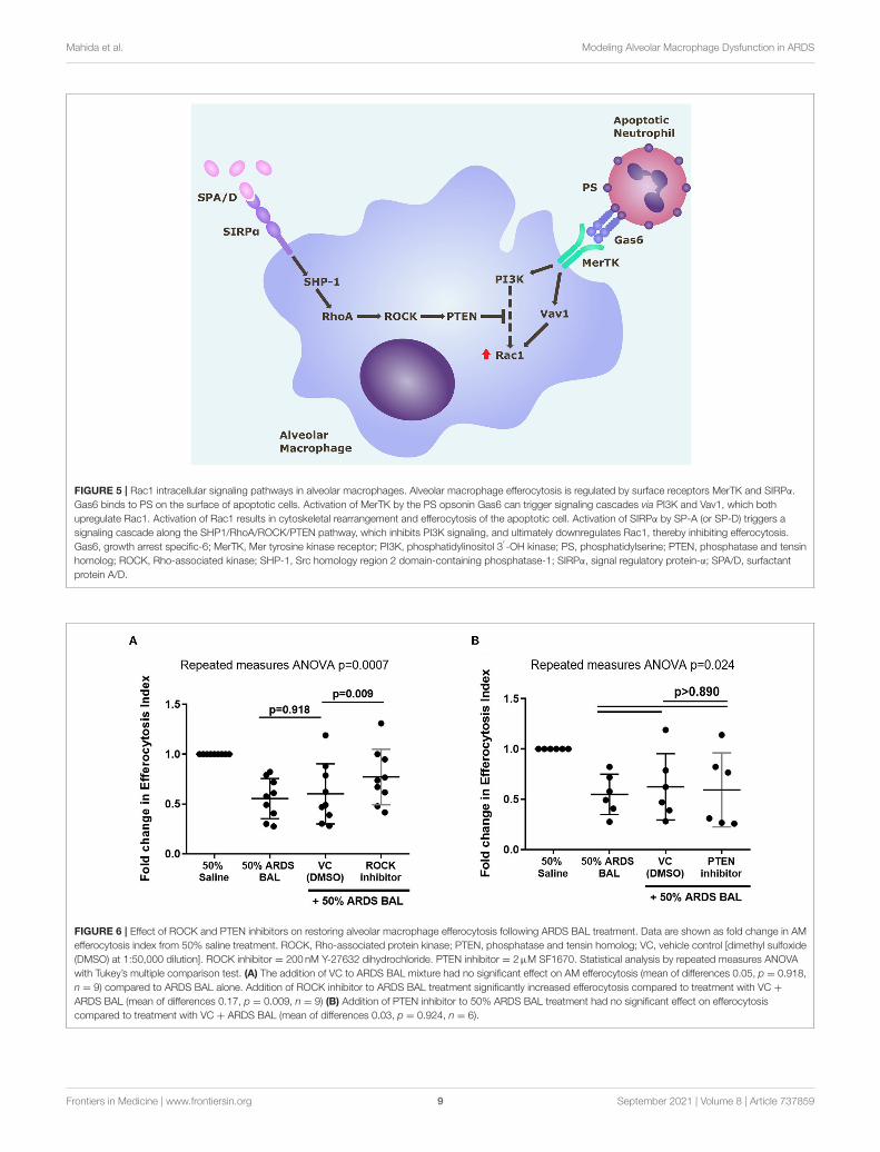
FIGURE 5 | Rac1 intracellular signaling pathways in alveolar macrophages. Alveolar macrophage efferocytosis is regulated by surface receptors MerTK and SIRPα.
Gas6 binds to PS on the surface of apoptotic cells. Activation of MerTK by the PS opsonin Gas6 can trigger signaling cascades via PI3K and Vav1, which both
upregulate Rac1. Activation of Rac1 results in cytoskeletal rearrangement and efferocytosis of the apoptotic cell. Activation of SIRPα by SP-A (or SP-D) triggers a
signaling cascade along the SHP1/RhoA/ROCK/PTEN pathway, which inhibits PI3K signaling, and ultimately downregulates Rac1, thereby inhibiting efferocytosis.
Gas6, growth arrest specific-6; MerTK, Mer tyrosine kinase receptor; PI3K, phosphatidylinositol 3′-OH kinase; PS, phosphatidylserine; PTEN, phosphatase and tensin
homolog; ROCK, Rho-associated kinase; SHP-1, Src homology region 2 domain-containing phosphatase-1; SIRPα, signal regulatory protein-α; SPA/D, surfactant
protein A/D.
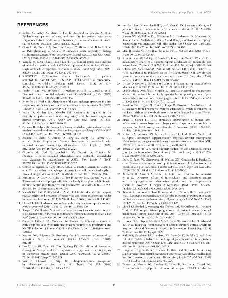
FIGURE 6 | Effect of ROCK and PTEN inhibitors on restoring alveolar macrophage efferocytosis following ARDS BAL treatment. Data are shown as fold change in AM
efferocytosis index from 50% saline treatment. ROCK, Rho-associated protein kinase; PTEN, phosphatase and tensin homolog; VC, vehicle control [dimethyl sulfoxide
(DMSO) at 1:50,000 dilution]. ROCK inhibitor = 200 nM Y-27632 dihydrochloride. PTEN inhibitor = 2µM SF1670. Statistical analysis by repeated measures ANOVA
with Tukey’s multiple comparison test. (A) The addition of VC to ARDS BAL mixture had no significant effect on AM efferocytosis (mean of differences 0.05, p = 0.918,
n = 9) compared to ARDS BAL alone. Addition of ROCK inhibitor to ARDS BAL treatment significantly increased efferocytosis compared to treatment with VC +
ARDS BAL (mean of differences 0.17, p = 0.009, n = 9) (B) Addition of PTEN inhibitor to 50% ARDS BAL treatment had no significant effect on efferocytosis
compared to treatment with VC + ARDS BAL (mean of differences 0.03, p = 0.924, n = 6).
Frontiers in Medicine | www.frontiersin.org 9 September 2021 | Volume 8 | Article 737859

Mahida et al. Modeling Alveolar Macrophage Dysfunction in ARDS
impaired AM efferocytosis in ARDS. ROCK inhibition promotesPI3K signaling, which has multiple effects on a cellular functionbeyond upregulation of Rac1, namely, proliferation, chemotaxis,andmigration (43). ROCK inhibition would havemany off-targeteffects, thereby limiting its therapeutic potential as a strategy toupregulate AM function in ARDS. Existing medications could betested using the in vitromodel of ARDS, to determine if they canrestore AM efferocytosis, e.g., N-acetylcysteine (44), macrolideantibiotics (45), statins (46), and glucocorticoids (47).
Studies utilizing an ex-vivo perfused human lung model ofARDS have shown that extracellular vesicles (EVs) are releasedfollowing lung injury with E. coli; these isolated EVs subsequentlymediated inflammatory lung injury when administered touninjured lungs (48). Murine models of LPS lung injury haveshown that EV transfer of microRNA cargo to AMs can increaseinflammatory cytokine release (49). Further analysis of ARDSBAL and studies utilizing our in vitro model of ARDS arerequired to determine whether EV transfer of microRNA to AMsmay affect intracellular pathways regulating efferocytosis (50).
Our study had some limitations. Due to logistical constraints,efferocytosis assays were undertaken with heterologousneutrophils, as opposed to autologous neutrophils, whichwould have more accurately reflected the environment in vivo.Although unaffected lung tissue was processed, we cannotrule out contamination with tumor-associated macrophageswhich are characterized by an immunosuppressive phenotypeand may exhibit increased efferocytosis (51), which couldaccount for some of the divergent effects observed. Expression ofintracellular signaling mediators (e.g., Rac1) was only measuredat the mRNA level. To draw definitive conclusions regardingthe mechanism of impaired efferocytosis in ARDS, data on theprotein expression of these mediators will be required. Ideally,the use of healthy human BAL would be a more appropriate VCinstead of saline in this model; however, healthy BAL is a highlylimited resource.
Another limitation to our study is that when assessing AMexpression of TAM receptors (key mediators of macrophageefferocytosis), only MerTK was investigated (52). We hadpredominantly focused on MerTK, as this efferocytosis receptorwas best characterized in the context of ARDS within theliterature (18, 53–55). However, we omitted to investigate otherimportant TAM receptors: Axl and Tyro3 (52). Impairment ofthe Axl signaling pathway has been associated with decreased AMefferocytosis in asthma (56). We report a contradictory increasein MerTK expression associated with decreased efferocytosisin AMs treated with ARDS BAL; however, this may, in part,be explained if the expression of TAM receptors Axl and/orTyro3 were decreased. Further studies will be required toinvestigate this.
Studies have previously shown that the microenvironmentcan influence AM metabolism, inflammatory response, andgene expression (57, 58). In vitro culture of AMs canalter efferocytosis receptor expression profiles (56), thereforeundertaking efferocytosis assays directly in situ on lungtissue may provide a more accurate representation of invivo AM function (59). For future studies, precision-cutlung slices could be incubated with ARDS BAL before theassessment of efferocytosis directly on lung tissue (60); terminal
deoxynucleotidyl transferase dUTP nick end labeling couldbe used to identify ANs in a double immunofluorescencemethod (59).
In conclusion, in vitro treatment of lung resection tissueAMs with pooled ARDS patient BAL can recapitulate the samefunctional defect observed in vivo. This dysfunction can bepartially restored by ROCK inhibition. The in vitro model ofARDS is a useful tool to investigate the mechanisms by whichthe inflammatory alveolar microenvironment of ARDS inducesAM dysfunction.
DATA AVAILABILITY STATEMENT
The original contributions presented in the study are includedin the article/Supplementary Material, further inquiries can bedirected to the corresponding author/s.
ETHICS STATEMENT
The studies involving human participants were reviewedand approved by Wales Research Ethics Committee 1 (REC16/WA/0169) and West Midlands - Solihull Research EthicsCommittee (REC 17/WM/0272). The patients/participantsprovided their written informed consent to participate inthis study.
AUTHOR CONTRIBUTIONS
RM, AS, MM, and DT contributed to the study conception anddesign. RM, AS, DP, and SL contributed to data acquisition. RM,AS, and DT drafted the manuscript. All the authors contributedto the data analysis and interpretation, critically revised themanuscript for intellectual content, and approved the finalversion before submission.
FUNDING
This work was funded by Medical Research Council grantsMR/N021185/1 (RM) and MR/L002736/1 (DT/AS).
ACKNOWLEDGMENTS
We would thank the nurses and thoracic surgeons of theThoracic Research Team, University Hospitals BirminghamNHSFoundation Trust, for their help consenting patients undergoingthoracic surgery. We also thank Dr. Gerald Langman and Dr.Andrew Robinson, Department of Cellular Pathology, UniversityHospitals Birmingham NHS Foundation Trust, for their helpin obtaining lung tissue samples from patients undergoinglobectomy. We thank Dr. Hui Li for her assistance in processinghuman lung tissue samples.
SUPPLEMENTARY MATERIAL
The Supplementary Material for this article can be foundonline at: https://www.frontiersin.org/articles/10.3389/fmed.2021.737859/full#supplementary-material
Frontiers in Medicine | www.frontiersin.org 10 September 2021 | Volume 8 | Article 737859
Mahida et al. Modeling Alveolar Macrophage Dysfunction in ARDS
REFERENCES
1. Bellani G, Laffey JG, Pham T, Fan E, Brochard L, Esteban A, et al.
Epidemiology, patterns of care, and mortality for patients with acute
respiratory distress syndrome in intensive care units in 50 countries. JAMA.
(2016) 315:788–800. doi: 10.1001/jama.2016.0291
2. Grasselli G, Tonetti T, Protti A, Langer T, Girardis M, Bellani G, et
al. Pathophysiology of COVID-19-associated acute respiratory distress
syndrome: a multicentre prospective observational study. Lancet Respir Med.
(2020) 8:1201–1208. doi: 10.1016/S2213-2600(20)30370-2
3. Yang X, Yu Y, Xu J, Shu H, Xia J, Liu H, et al. Clinical course and outcomes
of critically ill patients with SARS-CoV-2 pneumonia in Wuhan, China: a
single-centered, retrospective, observational study. Lancet Respir Med. (2020)
8:475–81. doi: 10.1016/S2213-2600(20)30079-5
4. RECOVERY Collaborative Group. Tocilizumab in patients
admitted to hospital with COVID-19 (RECOVERY): a randomised,
controlled, open-label, platform trial. Lancet. (2021) 397:1637–
45. doi: 10.1016/S0140-6736(21)00676-0
5. Horby P, Lim WS, Emberson JR, Mafham M, Bell JL, Linsell L, et al.
Dexamethasone in hospitalized patients with Covid-19. N Engl J Med. (2021)
384:693–704. doi: 10.1056/NEJMoa2021436
6. Bachofen M, Weibel ER. Alterations of the gas exchange apparatus in adult
respiratory insufficiency associated with septicemia.AmRev Respir Dis. (1977)
116:589–615. doi: 10.1164/arrd.1977.116.4.589
7. Ware LB, Matthay MA. Alveolar fluid clearance is impaired in the
majority of patients with acute lung injury and the acute respiratory
distress syndrome. Am J Respir Crit Care Med. (2001) 163:1376–
83. doi: 10.1164/ajrccm.163.6.2004035
8. Zemans RL, Colgan SP, Downey GP. Transepithelial migration of neutrophils:
mechanisms and implications for acute lung injury. Am J Respir Cell Mol Biol.
(2009) 40:519–35. doi: 10.1165/rcmb.2008-0348TR
9. Mahida RY, Scott A, Parekh D, Lugg ST, Hardy RS, Lavery GG,
et al. Acute Respiratory Distress Syndrome is associated with
impaired alveolar macrophage efferocytosis. Euro Respir J. (2021)
58:2100829. doi: 10.1183/13993003.00829-2021
10. Gregoire M, Uhel F, Lesouhaitier M, Gacouin A, Guirriec M,
Mourcin F, et al. Impaired efferocytosis and neutrophil extracellular
trap clearance by macrophages in ARDS. Euro Respir J. (2018)
52:1702590. doi: 10.1183/13993003.02590-2017
11. Gomez Perdiguero E, Klapproth K, Schulz C, Busch K, Azzoni E, Crozet L,
et al. Tissue-resident macrophages originate from yolk-sac-derived erythro-
myeloid progenitors. Nature. (2015) 518:547–51. doi: 10.1038/nature13989
12. Hashimoto D, Chow A, Noizat C, Teo P, Beasley MB, Leboeuf M, et al.
Tissue-resident macrophages self-maintain locally throughout adult life with
minimal contribution from circulating monocytes. Immunity. (2013) 38:792–
804. doi: 10.1016/j.immuni.2013.04.004
13. Yona S, Kim KW, Wolf Y, Mildner A, Varol D, Breker M, et al. Fate mapping
reveals origins and dynamics of monocytes and tissue macrophages under
homeostasis. Immunity. (2013) 38:79–91. doi: 10.1016/j.immuni.2012.12.001
14. Hussell T, Bell TJ. Alveolar macrophages: plasticity in a tissue-specific context.
Nat Rev Immunol. (2014) 14:81–93. doi: 10.1038/nri3600
15. Thepen T, Van Rooijen N, Kraal G. Alveolar macrophage elimination in vivo
is associated with an increase in pulmonary immune response in mice. J Exp
Med. (1989) 170:499–509. doi: 10.1084/jem.170.2.499
16. Zizzo G, Hilliard BA, Monestier M, Cohen PL. Efficient clearance of
early apoptotic cells by human macrophages requires M2c polarization and
MerTK induction. J Immunol. (2012) 189:3508–20. doi: 10.4049/jimmunol.
1200662
17. Mosser DM, Edwards JP. Exploring the full spectrum of macrophage
activation. Nat Rev Immunol. (2008) 8:958–69. doi: 10.1038/
nri2448
18. Lee YJ, Lee SH, Youn YS, Choi JY, Song KS, Cho MS, et al. Preventing
cleavage of Mer promotes efferocytosis and suppresses acute lung injury
in bleomycin treated mice. Toxicol Appl Pharmacol. (2012) 263:61–
72. doi: 10.1016/j.taap.2012.05.024
19. Wu Y, Tibrewal N, Birge RB. Phosphatidylserine recognition
by phagocytes: a view to a kill. Trends Cell Biol. (2006)
16:189–97. doi: 10.1016/j.tcb.2006.02.003
20. van der Meer JH, van der Poll T, van’t Veer C. TAM receptors, Gas6, and
protein S: roles in inflammation and hemostasis. Blood. (2014) 123:2460–
9. doi: 10.1182/blood-2013-09-528752
21. Janssen WJ, McPhillips KA, Dickinson MG, Linderman DJ, Morimoto K,
Xiao YQ, et al. Surfactant proteins A and D suppress alveolar macrophage
phagocytosis via interaction with SIRP alpha. Am J Respir Crit Care Med.
(2008) 178:158–67. doi: 10.1164/rccm.200711-1661OC
22. Meili R, Sasaki AT, Firtel RA. Rho rocks PTEN. Nat Cell Biol. (2005) 7:334–
5. doi: 10.1038/ncb0405-334
23. Scott A, Lugg ST, Aldridge K, Lewis KE, Bowden A, Mahida RY, et al. Pro-
inflammatory effects of e-cigarette vapour condensate on human alveolar
macrophages. Thorax. (2018) 73:1161–9. doi: 10.1136/thoraxjnl-2018-211663
24. O’Kane CM, McKeown SW, Perkins GD, Bassford CR, Gao F, Thickett DR,
et al. Salbutamol up-regulates matrix metalloproteinase-9 in the alveolar
space in the acute respiratory distress syndrome. Crit Care Med. (2009)
37:2242–9. doi: 10.1097/CCM.0b013e3181a5506c
25. Davies JQ, Gordon S. Isolation and culture of human macrophages. Methods
Mol Biol. (2005) 290:105–16. doi: 10.1385/1-59259-838-2:105
26. Michlewska S, Dransfield I, Megson IL, Rossi AG. Macrophage phagocytosis
of apoptotic neutrophils is critically regulated by the opposing actions of pro-
inflammatory and anti-inflammatory agents: key role for TNF-alpha. FASEB
J. (2009) 23:844–54. doi: 10.1096/fj.08-121228
27. Wootton DG, Diggle PJ, Court J, Eneje O, Keogan L, Macfarlane L, et
al. Recovery from pneumonia requires efferocytosis which is impaired in
smokers and those with low bodymass index and enhanced by statins. Thorax.
(2016) 71:1052–4. doi: 10.1136/thoraxjnl-2016-208505
28. Zizzo G, Cohen PL. IL-17 stimulates differentiation of human anti-
inflammatory macrophages and phagocytosis of apoptotic neutrophils in
response to IL-10 and glucocorticoids. J Immunol. (2013) 190:5237–
46. doi: 10.4049/jimmunol.1203017
29. Serban KA, Petrusca DN, Mikosz A, Poirier C, Lockett AD, Saint L, et
al. Alpha-1 antitrypsin supplementation improves alveolar macrophages
efferocytosis and phagocytosis following cigarette smoke exposure. PLoSONE.
(2017) 12:e0176073. doi: 10.1371/journal.pone.0176073
30. Jepsen LV, Skottun T. A rapid one-step method for the isolation of human
granulocytes from whole blood. Scand J Clin Lab Investig. (1982) 42:235–
8. doi: 10.3109/00365518209168079
31. Sapey E, Patel JM, Greenwood H, Walton GM, Grudzinska F, Parekh D,
et al. Simvastatin improves neutrophil function and clinical outcomes in
pneumonia: a pilot randomised controlled trial. Am J Respir Crit Care Med.
(2019) 200:1282–93. doi: 10.1164/rccm.201812-2328OC
32. Bonecchi R, Sozzani S, Stine JT, Luini W, D’Amico G, Allavena
P, et al. Divergent effects of interleukin-4 and interferon-gamma
on macrophage-derived chemokine production: an amplification
circuit of polarized T helper 2 responses. Blood. (1998) 92:2668–
71. doi: 10.1182/blood.V92.8.2668.420k39_2668_2671
33. Rosseau S, Hammerl P, Maus U, Walmrath HD, Schutte H, Grimminger F,
et al. Phenotypic characterization of alveolar monocyte recruitment in acute
respiratory distress syndrome. Am J Physiol Lung Cell Mol Physiol. (2000)
279:L25–35. doi: 10.1152/ajplung.2000.279.1.L25
34. Mould KJ, Barthel L, Mohning MP, Thomas SM, McCubbrey AL, Danhorn
T, et al. Cell origin dictates programming of resident versus recruited
macrophages during acute lung injury. Am J Respir Cell Mol Biol. (2017)
57:294–306. doi: 10.1165/rcmb.2017-0061OC
35. Heijnen NFL, Hagens LA, Smit MR, Schultz MJ, van der Poll T, Schnabel
RM, et al. Biological subphenotypes of acute respiratory distress syndrome
may not reflect differences in alveolar inflammation. Physiol Rep. (2021)
9:e14693. doi: 10.14814/phy2.14693
36. Park WY, Goodman RB, Steinberg KP, Ruzinski JT, Radella F, 2nd, Park
DR, et al. Cytokine balance in the lungs of patients with acute respiratory
distress syndrome. Am J Respir Crit Care Med. (2001) 164(10 Pt 1):1896–
903. doi: 10.1164/ajrccm.164.10.2104013
37. Hodge S, Hodge G, Ahern J, Jersmann H, Holmes M, Reynolds PN. Smoking
alters alveolar macrophage recognition and phagocytic ability: implications
in chronic obstructive pulmonary disease. Am J Respir Cell Mol Biol. (2007)
37:748–55. doi: 10.1165/rcmb.2007-0025OC
38. Kazeros A, Harvey BG, Carolan BJ, Vanni H, Krause A, Crystal RG.
Overexpression of apoptotic cell removal receptor MERTK in alveolar
Frontiers in Medicine | www.frontiersin.org 11 September 2021 | Volume 8 | Article 737859
Mahida et al. Modeling Alveolar Macrophage Dysfunction in ARDS
macrophages of cigarette smokers. Am J Respir Cell Mol Biol. (2008) 39:747–
57. doi: 10.1165/rcmb.2007-0306OC
39. Shaykhiev R, Krause A, Salit J, Strulovici-Barel Y, Harvey BG, O’Connor TP, et
al. Smoking-dependent reprogramming of alveolar macrophage polarization:
implication for pathogenesis of chronic obstructive pulmonary disease. J
Immunol. (2009) 183:2867–83. doi: 10.4049/jimmunol.0900473
40. Hodge S, Hodge G, Scicchitano R, Reynolds PN, Holmes M.
Alveolar macrophages from subjects with chronic obstructive
pulmonary disease are deficient in their ability to phagocytose
apoptotic airway epithelial cells. Immunol cell Biol. (2003)
81:289–96. doi: 10.1046/j.1440-1711.2003.t01-1-01170.x
41. Kaku Y, Imaoka H, Morimatsu Y, Komohara Y, Ohnishi K, Oda H, et al.
Overexpression of CD163, CD204 and CD206 on alveolar macrophages in
the lungs of patients with severe chronic obstructive pulmonary disease. PLoS
ONE. (2014) 9:e87400. doi: 10.1371/journal.pone.0087400
42. Bewley MA, Belchamber KB, Chana KK, Budd RC, Donaldson G, Wedzicha
JA, et al. Differential effects of p38, MAPK, PI3K or Rho kinase inhibitors on
bacterial phagocytosis and efferocytosis bymacrophages in COPD. PLoS ONE.
(2016) 11:e0163139. doi: 10.1371/journal.pone.0163139
43. Vanhaesebroeck B, Stephens L, Hawkins P. PI3K signalling: the
path to discovery and understanding. Nat Rev Mol Cell Biol. (2012)
13:195. doi: 10.1038/nrm3290
44. Moon C, Lee YJ, Park HJ, Chong YH, Kang JL. N-acetylcysteine inhibits RhoA
and promotes apoptotic cell clearance during intense lung inflammation. Am
J Respir Crit CareMed. (2010) 181:374–87. doi: 10.1164/rccm.200907-1061OC
45. Hodge S, Hodge G, Jersmann H, Matthews G, Ahern J, Holmes M, et al.
Azithromycin improves macrophage phagocytic function and expression of
mannose receptor in chronic obstructive pulmonary disease. Am J Respir Crit
Care Med. (2008) 178:139–48. doi: 10.1164/rccm.200711-1666OC
46. Morimoto K, Janssen WJ, Fessler MB, McPhillips KA, Borges VM, Bowler
RP, et al. Lovastatin enhances clearance of apoptotic cells (efferocytosis) with
implications for chronic obstructive pulmonary disease. J Immunol. (2006)
176:7657–65. doi: 10.4049/jimmunol.176.12.7657
47. McColl A, Bournazos S, Franz S, Perretti M, Morgan BP, Haslett
C, et al. Glucocorticoids induce protein S-dependent phagocytosis of
apoptotic neutrophils by human macrophages. J Immunol. (2009) 183:2167–
75. doi: 10.4049/jimmunol.0803503
48. Liu A, Park JH, Zhang X, Sugita S, Naito Y, Lee JH, et al. Therapeutic effects of
hyaluronic acid in bacterial pneumonia in ex vivo perfused human lungs.Am J
Respir Crit CareMed. (2019) 200:1234–45. doi: 10.1164/rccm.201812-2296OC
49. Shikano S, Gon Y, Maruoka S, Shimizu T, Kozu Y, Iida Y,
et al. Increased extracellular vesicle miRNA-466 family in the
bronchoalveolar lavage fluid as a precipitating factor of ARDS.
BMC Pulmonary Med. (2019) 19:110. doi: 10.1186/s12890-019-
0876-9
50. Mahida RY, Matsumoto S, Matthay MA. Extracellular vesicles: a new frontier
for research in acute respiratory distress syndrome. Am J Respir Cell Mol Biol.
(2020) 63:15–24. doi: 10.1165/rcmb.2019-0447TR
51. Zhou J, Tang Z, Gao S, Li C, Feng Y, Zhou X. Tumor-associated
macrophages: recent insights and therapies. Front Oncol. (2020)
10:188. doi: 10.3389/fonc.2020.00188
52. Lemke G, Rothlin CV. Immunobiology of the TAM receptors. Nat Rev
Immunol. (2008) 8:327–36. doi: 10.1038/nri2303
53. Choi JY, Park HJ, Lee YJ, Byun J, Youn YS, Choi JH, et al. Upregulation
of Mer receptor tyrosine kinase signaling attenuated lipopolysaccharide-
induced lung inflammation. J Pharmacol Exp Ther. (2013) 344:447–
58. doi: 10.1124/jpet.112.199778
54. Lee YJ, Han JY, Byun J, Park HJ, Park EM, Chong YH, et al. Inhibiting Mer
receptor tyrosine kinase suppresses STAT1, SOCS1/3, and NF-κB activation
and enhances inflammatory responses in lipopolysaccharide-induced acute
lung injury. J Leukocyte Biol. (2012) 91:921–32. doi: 10.1189/jlb.0611289
55. Mohning MP, Thomas SM, Barthel L, Mould KJ, McCubbrey AL, Frasch SC,
et al. Phagocytosis of microparticles by alveolar macrophages during acute
lung injury requires MerTK. Am J Physiol Lung Cell Mol Physiol. (2018)
314:L69–82. doi: 10.1152/ajplung.00058.2017
56. Grabiec AM, Denny N, Doherty JA, Happonen KE, Hankinson J, Connolly E,
et al. Diminished airway macrophage expression of the Axl receptor tyrosine
kinase is associated with defective efferocytosis in asthma. J Allergy Clin
Immunol. (2017) 140:1144–6.e4. doi: 10.1016/j.jaci.2017.03.024
57. Svedberg FR, Brown SL, Krauss MZ, Campbell L, Sharpe C, Clausen M,
et al. The lung environment controls alveolar macrophage metabolism
and responsiveness in type 2 inflammation. Nat Immunol. (2019) 20:571–
80. doi: 10.1038/s41590-019-0352-y
58. Lavin Y, Winter D, Blecher-Gonen R, David E, Keren-Shaul H, Merad M, et
al. Tissue-resident macrophage enhancer landscapes are shaped by the local
microenvironment. Cell. (2014) 159:1312–26. doi: 10.1016/j.cell.2014.11.018
59. Proto JD, Doran AC, Gusarova G, Yurdagul A, Jr., Sozen E, et al. Regulatory
T Cells Promote Macrophage Efferocytosis during Inflammation Resolution.
Immunity. (2018) 49:666–77.e6. doi: 10.1016/j.immuni.2018.07.015
60. Dutra Silva J, Su Y, Calfee CS, Delucchi KL, Weiss D, McAuley DF, et
al. Mesenchymal stromal cell extracellular vesicles rescue mitochondrial
dysfunction and improve barrier integrity in clinically relevant models of
ARDS. Euro Respir J. (2021) 58:2002978. doi: 10.1183/13993003.02978-2020
Conflict of Interest: The authors declare that the research was conducted in the
absence of any commercial or financial relationships that could be construed as a
potential conflict of interest.
Publisher’s Note: All claims expressed in this article are solely those of the authors
and do not necessarily represent those of their affiliated organizations, or those of
the publisher, the editors and the reviewers. Any product that may be evaluated in
this article, or claim that may be made by its manufacturer, is not guaranteed or
endorsed by the publisher.
Copyright © 2021 Mahida, Scott, Parekh, Lugg, Belchamber, Hardy, Matthay,
Naidu and Thickett. This is an open-access article distributed under the terms
of the Creative Commons Attribution License (CC BY). The use, distribution or
reproduction in other forums is permitted, provided the original author(s) and the
copyright owner(s) are credited and that the original publication in this journal
is cited, in accordance with accepted academic practice. No use, distribution or
reproduction is permitted which does not comply with these terms.
Frontiers in Medicine | www.frontiersin.org 12 September 2021 | Volume 8 | Article 737859


![Original Article Amelioration of meconium-induced acute ...inflammation, alveolar exudation, surfactant dysfunction, airway hyperreactivity, and other conditions [2, 3]. Meconium can](https://img.dokumen.tips/doc/110x75/601f8d151da783327f1ffce1/original-article-amelioration-of-meconium-induced-acute-inflammation-alveolar.jpg)


![Activation of Alveolar Macrophage Tumoricidal …...[14C]DSPC. All counts were quench-corrected before calculation of in corporation or recovery. Freeze-dried liposomes were prepared](https://img.dokumen.tips/doc/110x75/5f22ac6bc2a49c46796e8cca/activation-of-alveolar-macrophage-tumoricidal-14cdspc-all-counts-were-quench-corrected.jpg)