Embed Size (px)
Citation preview

Original Investigation | Dermatology
Assessment of Accuracy of an Artificial Intelligence Algorithmto Detect Melanoma in Images of Skin LesionsMichael Phillips, MMedSci; Helen Marsden, PhD; Wayne Jaffe, MB, ChB, FRCS; Rubeta N. Matin, PhD, MBBS, MRCP; Gorav N. Wali, MA, BMBCh, MRCP;Jack Greenhalgh, PhD; Emily McGrath, BMBS, MRCP; Rob James, BSc, RGN; Evmorfia Ladoyanni, PGCME, DTM&H, FRCP; Anthony Bewley, MB ChB, FRCP;Giuseppe Argenziano, MD, PhD; Ioulios Palamaras, MD, PhD
Abstract
IMPORTANCE A high proportion of suspicious pigmented skin lesions referred for investigation arebenign. Techniques to improve the accuracy of melanoma diagnoses throughout the patientpathway are needed to reduce the pressure on secondary care and pathology services.
OBJECTIVE To determine the accuracy of an artificial intelligence algorithm in identifying melanomain dermoscopic images of lesions taken with smartphone and digital single-lens reflex(DSLR) cameras.
DESIGN, SETTING, AND PARTICIPANTS This prospective, multicenter, single-arm, maskeddiagnostic trial took place in dermatology and plastic surgery clinics in 7 UK hospitals. Dermoscopicimages of suspicious and control skin lesions from 514 patients with at least 1 suspicious pigmentedskin lesion scheduled for biopsy were captured on 3 different cameras. Data were collected fromJanuary 2017 to July 2018. Clinicians and the Deep Ensemble for Recognition of Malignancy, adeterministic artificial intelligence algorithm trained to identify melanoma in dermoscopic images ofpigmented skin lesions using deep learning techniques, assessed the likelihood of melanoma. Initialdata analysis was conducted in September 2018; further analysis was conducted from February 2019to August 2019.
INTERVENTIONS Clinician and algorithmic assessment of melanoma.
MAIN OUTCOMES AND MEASURES Area under the receiver operating characteristic curve(AUROC), sensitivity, and specificity of the algorithmic and specialist assessment, determined usinghistopathology diagnosis as the criterion standard.
RESULTS The study population of 514 patients included 279 women (55.7%) and 484 white patients(96.8%), with a mean (SD) age of 52.1 (18.6) years. A total of 1550 images of skin lesions wereincluded in the analysis (551 [35.6%] biopsied lesions; 999 [64.4%] control lesions); 286 images(18.6%) were used to train the algorithm, and a further 849 (54.8%) images were missing orunsuitable for analysis. Of the biopsied lesions that were assessed by the algorithm and specialists,125 (22.7%) were diagnosed as melanoma. Of these, 77 (16.7%) were used for the primary analysis.The algorithm achieved an AUROC of 90.1% (95% CI, 86.3%-94.0%) for biopsied lesions and 95.8%(95% CI, 94.1%-97.6%) for all lesions using iPhone 6s images; an AUROC of 85.8% (95% CI, 81.0%-90.7%) for biopsied lesions and 93.8% (95% CI, 91.4%-96.2%) for all lesions using Galaxy S6 images;and an AUROC of 86.9% (95% CI, 80.8%-93.0%) for biopsied lesions and 91.8% (95% CI,87.5%-96.1%) for all lesions using DSLR camera images. At 100% sensitivity, the algorithm achieved
(continued)
Key PointsQuestion How accurate is an artificial
intelligence–based melanoma detection
algorithm, which analyzes dermoscopic
images taken by smartphone and digital
single-lens reflex cameras, compared
with clinical assessment and
histopathological diagnosis?
Findings In this diagnostic study, 1550
images of suspicious and benign skin
lesions were analyzed by an artificial
intelligence algorithm. When compared
with histopathological diagnosis, the
algorithm achieved an area under the
receiver operator characteristic curve of
95.8%. At 100% sensitivity, the
algorithm achieved a specificity of
64.8%, while clinicians achieved a
specificity of 69.9%.
Meaning As the burden of skin cancer
increases, artificial intelligence
technology could play a role in
identifying lesions with a high likelihood
of melanoma.
+ Supplemental content
Author affiliations and article information arelisted at the end of this article.
Open Access. This is an open access article distributed under the terms of the CC-BY-NC-ND License.
JAMA Network Open. 2019;2(10):e1913436. doi:10.1001/jamanetworkopen.2019.13436 (Reprinted) October 16, 2019 1/12
Downloaded From: https://jamanetwork.com/ on 02/28/2022

Abstract (continued)
a specificity of 64.8% with iPhone 6s images. Specialists achieved an AUROC of 77.8% (95% CI,72.5%-81.9%) and a specificity of 69.9%.
CONCLUSIONS AND RELEVANCE In this study, the algorithm demonstrated an ability to identifymelanoma from dermoscopic images of selected lesions with an accuracy similar to that ofspecialists.
JAMA Network Open. 2019;2(10):e1913436.
Corrected on November 6, 2019. doi:10.1001/jamanetworkopen.2019.13436
Introduction
When compared with other forms of skin cancer, malignant melanoma is relatively uncommon;however, the incidence of melanoma is increasing faster than any other form of cancer, and it isresponsible for the majority of skin cancer deaths.1 Patients in whom melanoma is diagnosed at stageI have more than a 95% 5-year relative survival rate compared with 8% to 25% when the disease isdiagnosed at stage IV.1,2
A person with a suspicious pigmented skin lesion will go through several steps before adefinitive diagnosis of melanoma: self-evaluation, evaluation by a primary care physician, assessmentby a specialist, and excision and assessment by histopathology. Current practice guidelinesrecommend appropriately trained health care professionals assess all suspicious skin lesions usingdermoscopy.3 Techniques to improve diagnostic accuracy can be used at each step to improvedifferentiation between harmless and potentially harmful lesions, thus reducing the pressure on laterservices from lesions that were unnecessarily referred further.4-6 The Cochrane Skin CancerDiagnostic Test Accuracy Group recently published a comprehensive series of reviews on theaccuracy of different diagnostic techniques, including visual assessment with or withoutdermoscopy,7 reflectance confocal microscopy,8 teledermatology,9 computer-aided diagnostictechniques,10 and smartphone applications.11 Dermoscopy was found to improve diagnostic accuracyover visual inspection alone, and reflectance confocal microscopy was more accurate thandermoscopy alone, but the data supporting the widespread adoption of teledermatology, computer-assisted diagnostic techniques, or smartphone applications are limited and/or of poor quality. Clinicaldiagnostic accuracy also depends on the experience of the examiners,12 and the equipment requiredfor reflectance confocal microscopy is expensive.
A large number of smartphone applications for melanoma detection have been released,although there is little evidence of clinical validation.11,13 Of 39 skin cancer applications assessed, 19involved smartphone photography and 4 provided an estimate of the probability of malignancy, butnone were assessed for diagnostic accuracy.14 Poorly designed, inaccurate, and/or misleadingconsumer applications may cause harm to patients.15-18
However, with appropriate development and suitable evaluation, modern electronictechnology could improve diagnostic accuracy. Indeed, artificial intelligence (AI) algorithmscategorizing photographs of lesions have recently been shown to be capable of classifying melanomawith a level of competence comparable with dermatologists.19,20 Deep Ensemble for Recognition ofMalignancy, developed by Skin Analytics Limited, is an AI algorithm that is designed to be used as adecision support tool for health care professionals by determining the likelihood of skin cancer fromdermoscopic images of pigmented skin lesions. It was developed using deep learning techniquesthat identify and assess features of lesions that are associated with melanoma, using more than7000 archived dermoscopic images, and it has been shown to identify melanoma with accuracysimilar to that of specialist physicians.21
The aim of this study was to evaluate the ability of the Deep Ensemble for Recognition ofMalignancy algorithm to detect melanoma from images of both biopsied and nonbiopsied pigmented
JAMA Network Open | Dermatology Accuracy of an Artificial Intelligence Algorithm to Detect Melanoma in Images of Skin Lesions
JAMA Network Open. 2019;2(10):e1913436. doi:10.1001/jamanetworkopen.2019.13436 (Reprinted) October 16, 2019 2/12
Downloaded From: https://jamanetwork.com/ on 02/28/2022

skin lesions, prospectively captured in dermatology and plastic surgery clinics, and to compare thiswith clinical diagnoses made by specialists.
Methods
We conducted a prospective, multicenter, single-arm, masked diagnostic trial. Ethical approval forthe study was granted by the Leicester South National Research Ethics Service committee. This studyfollowed the Standards for Reporting of Diagnostic Accuracy (STARD) reporting guideline.
Patients attending dermatology or plastic surgery clinics (on 2-week wait and general referralpathways) with at least 1 skin lesion referred for histological evaluation were eligible for the study. Atotal of 514 patients provided written informed consent for the study, which was conducted betweenJanuary 2017 and July 2018. Recruitment was on a consecutive, competitive recruitment basis in 7participating hospitals, resulting in 551 suspicious lesions scheduled for biopsy and 999 controllesions. Overall, 13 patients were ineligible or withdrawn, primarily because no skin lesion wasbiopsied (eFigure 1 in the Supplement). One lesion was not biopsied because of specialist decision.Race was defined by the participant.
Lesions not previously biopsied or excised and 2 control lesions (ie, lesions believed to bebenign) were selected if they were less than 15 mm in diameter and not located in an anatomical siteunsuitable for photographing or in an area of visible scarring. These were photographed by 3different cameras: iPhone 6s (Apple, Inc), Galaxy S6 (Samsung), and digital single-lens reflex (DSLR)camera D5500 (Nikon). Operating systems were kept up to date during the study. The dermoscopiclens attachments were DermLite DL1 for the iPhone 6s and Galaxy S6 and DermLite Foto II Pro for theDSLR (DermLite). Clinicians assessed the lesions for likelihood of melanoma on an ordinal scale(range, 1-4; 1 indicates unlikely and 4, highly likely).
Histopathology results were collected on biopsied excised lesions and categorized asmelanoma, dysplastic nevi, or other. Melanoma in situ was classified as melanoma. When there washistopathological uncertainty in the diagnosis, the most likely diagnosis was reported. Clinicians andpatients were not informed of the outcome of the algorithmic assessment, and patients weremanaged in accordance with the usual standard of care. Images were stored electronically untilcompletion of recruitment.
Prior to the study, the algorithm had been trained with published dermoscopic images. A subsetof 289 images from this study were extracted from the data set and used to further train thealgorithm (36 [12.5%] randomly selected confirmed melanoma lesions; 67 [23.2%] randomlyselected nonmelanoma lesions; and 186 [64.4%] control lesions). No single patient had lesions thatappeared in both the training and test split to ensure that overfitting did not occur. Images from eachcamera were used to train a version of the algorithm, which was then used to assess the remainingimages from that camera type. Images were screened for quality, and images that were blurred orotherwise flawed were removed from the data set. The remaining 1550 images were assessed by thealgorithm between September 2018 and February 2019.
The diagnostic performance of the algorithm was reassessed in August 2019 to optimize thealgorithm to better generalize for different cameras. To ensure that any difference in performancewas owing to algorithmic improvements rather than the quantity of data, the image data set waslimited to only those lesions for which images came from all 3 cameras. In total, images of 731 lesionswere used, including 260 (35.6%) biopsied lesions, of which 51 (7.0%) were diagnosed as melanoma.In addition, healthy skin images were included in the training set, but low-quality images remainedexcluded. This data set was combined with a historical data set21 and shuffled. The algorithm wasretrained and validated using 10-fold cross-validation. Lesions from each patient were kept togetherin the same fold to avoid overfitting to a single patient. The model was trained on images taken bythe DSLR and Galaxy S6, then tested using images taken by the iPhone 6s as the naive device as wellas images taken by the DSLR and Galaxy S6 cameras.
JAMA Network Open | Dermatology Accuracy of an Artificial Intelligence Algorithm to Detect Melanoma in Images of Skin Lesions
JAMA Network Open. 2019;2(10):e1913436. doi:10.1001/jamanetworkopen.2019.13436 (Reprinted) October 16, 2019 3/12
Downloaded From: https://jamanetwork.com/ on 02/28/2022

The algorithm generated a numerical response to an image, from 0 to 1, which reflects itsconfidence that the lesion is melanoma (0 indicates certainly benign and 1, certainly malignantmelanoma). A decision threshold defined the point above which a lesion is classed as melanoma.
Statistical AnalysisReceiver operator curves using a nonparametric method with bootstrapped estimation ofuncertainty22 were used to examine the association of the algorithm’s confidence scale with thecriterion-standard, histopathology-confirmed diagnosis for each camera and for the clinicalassessment. The assessment of the clinical accuracy of likelihood of melanoma was estimated on allbiopsied lesions (including the lesions whose images were missing and those used for training thealgorithm). In the direct comparison, only those lesions with images from every camera wereincluded in the analysis. Area under the receiver operating characteristic curve (AUROC) and otherdiagnostic accuracy indices (specificity, predictive values, false-positive rates, and false-negativerates) at the decision thresholds that generated a sensitivity of 100% and 95% were compared withthe clinical assessment of the likelihood of melanoma. The 100% sensitivity threshold was usedbecause all lesions referred for biopsy were assessed by histopathology. A χ2 test of the equality ofthe AUROC between assessments was conducted.
We examined the extent of influence of covariates on the accuracy of each method ofmelanoma detection. The following patient-related covariates were examined: patients’ level ofconcern, age, sex, race (ie, white vs nonwhite), Fitzpatrick skin type, hair color (ie, blond or red vsnot), patient or family history of melanoma and other skin cancer, and lesion-specific body location.
The potential consequences of missing values (from missing or poor-quality images) wereassessed using the Little missing completely at random test.23 We corrected receiver operatingcharacteristic curve estimates using the selection model, as developed by Cook and Rajbhandari.24 AP value of less than .05 was regarded as statistically significant, and all tests were 2-tailed. Statisticalestimates of accuracy are reported with 95% CIs. Statistical analysis was conducted using Statastatistical software version 15 (StataCorp).
Results
The study population of 514 patients included 279 women (55.7%) and 484 white patients (96.8%),with a mean (SD) age of 52.1 (18.6) years (Table 1). Most participants had Fitzpatrick skin sensitivitycategories I to III (417 [81.1%]) and had no family or personal history of any form of skin cancer(352 [71.3%]).
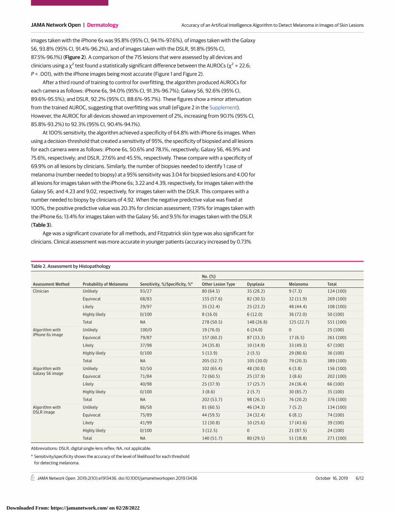
A total of 1550 images of skin lesions were included in the analysis (551 [35.6%] biopsied lesions;999 [64.4%] control lesions). Of the 551 lesions biopsied, 125 (22.7%) were identified as melanomaby histopathology, 148 (26.8%) were dysplastic nevi, and 278 (50.5%) received other diagnoses(eTable in the Supplement). To understand the types of lesions categorized other, a poststudy auditof 119 biopsied lesions from 1 hospital found that, of 40 lesions categorized as other, 3 (7.5%) werediagnosed as basal cell carcinoma and the rest were benign. The most frequent melanoma subtypeswere superficial spreading (67 [53.6%]) and melanoma in situ or lentigo maligna (39 [31.2%]). A totalof 32 melanoma cases (25.6%) had a Breslow thickness greater than 1.0 mm. Of the 50 biopsiedlesions clinicians identified as highly likely to be melanoma, 36 (72.0%) were histologically confirmedas melanoma and 6 (12.0%) were diagnosed as dysplastic nevi. Of the 124 biopsied lesions cliniciansidentified as unlikely to be melanoma, 9 (7.3%) were diagnosed as melanoma and 35 (28.2%) weredysplastic nevi (Table 2). Table 2 shows the similarity between the algorithmic and clinicalassessment at approximately similar levels of sensitivity and specificity. No adverse events wererecorded in the study.
The algorithm training data set included 858 images of 286 lesions from 92 patients. A further627 images were missing owing to technical issues with the camera equipment, data extraction, or aninability to link images to clinical information, and 222 images failed an image quality check (eFigure 1
JAMA Network Open | Dermatology Accuracy of an Artificial Intelligence Algorithm to Detect Melanoma in Images of Skin Lesions
JAMA Network Open. 2019;2(10):e1913436. doi:10.1001/jamanetworkopen.2019.13436 (Reprinted) October 16, 2019 4/12
Downloaded From: https://jamanetwork.com/ on 02/28/2022

in the Supplement). Of the remaining lesions used for the analysis, 77 (16.7%) were identified asmelanoma.
The analysis of images of biopsied lesions using the algorithm trained on published images oflesions produced AUROCs for each camera as follows: iPhone 6s, 87.9% (82.8%-89.9%); Galaxy S6,82.3% (76.9%-87.2%); DSLR, 85.0% (77.3%-90.9%).21 Using the training clinical images from thisstudy, the retrained algorithm produced AUROCs for each camera as follows: iPhone 6s, 90.1% (95%CI, 86.3%-94.0%); Galaxy S6, 85.8% (95% CI, 81.0%-90.7%); and DSLR, 86.9% (95% CI, 80.8%-93.0%) (Figure 1). The AUROC for clinician assessment of melanoma likelihood for biopsied lesionswas 77.8% (95% CI, 72.5%-81.9%).
Assuming none of the control lesions were melanoma, all methods show an improvement inaccuracy when assessing all lesions compared with biopsied lesions alone. The AUROC for clinicalassessment was 90.8% (95% CI, 88.0%-93.6%). The AUROC for the algorithmic assessment of
Table 1. Characteristics of 501 Patients
Characteristic No. (%)Age, mean (SD), y 52.1 (18.6)
Sex
Male 222 (44.3)
Female 279 (55.7)
White race
No 16 (3.20)
Yes 484 (96.8)
Missing 1 (0.1)
No. of nevi
≤10 173 (35.4)
11-50 232 (47.4)
>50 84 (17.2)
Missing 8 (1.6)
Fitzpatrick skin type
Type I, highly sensitive 61 (12.4)
Type II, very sun sensitive 172 (34.9)
Type III, sun-sensitive skin 184 (37.3)
Type IV, minimally sun sensitive 62 (12.6)
Type V, sun-insensitive skin 10 (2.0)
Type VI, sun insensitive, never burns 4 (0.8)
Missing 8 (1.6)
Hair color
Blond 110 (22.8)
Red 39 (8.1)
Brown 298 (61.8)
Black 35 (7.3)
Missing 19 (3.8)
Freckles
No 214 (43.4)
Yes 279 (56.6)
Missing 8 (1.6)
History of skin cancer
No history 352 (71.3)
Family nonmelanoma 30 (6.1)
Family melanoma 38 (7.7)
Patient nonmelanoma 34 (6.9)
Patient melanoma 40 (8.1)
Missing 7 (1.4)
JAMA Network Open | Dermatology Accuracy of an Artificial Intelligence Algorithm to Detect Melanoma in Images of Skin Lesions
JAMA Network Open. 2019;2(10):e1913436. doi:10.1001/jamanetworkopen.2019.13436 (Reprinted) October 16, 2019 5/12
Downloaded From: https://jamanetwork.com/ on 02/28/2022
images taken with the iPhone 6s was 95.8% (95% CI, 94.1%-97.6%), of images taken with the GalaxyS6, 93.8% (95% CI, 91.4%-96.2%), and of images taken with the DSLR, 91.8% (95% CI,87.5%-96.1%) (Figure 2). A comparison of the 715 lesions that were assessed by all devices andclinicians using a χ2 test found a statistically significant difference between the AUROCs (χ2 = 22.6;P < .001), with the iPhone images being most accurate (Figure 1 and Figure 2).
After a third round of training to control for overfitting, the algorithm produced AUROCs foreach camera as follows: iPhone 6s, 94.0% (95% CI, 91.3%-96.7%); Galaxy S6, 92.6% (95% CI,89.6%-95.5%); and DSLR, 92.2% (95% CI, 88.6%-95.7%). These figures show a minor attenuationfrom the trained AUROC, suggesting that overfitting was small (eFigure 2 in the Supplement).However, the AUROC for all devices showed an improvement of 2%, increasing from 90.1% (95% CI,85.8%-93.2%) to 92.3% (95% CI, 90.4%-94.1%).
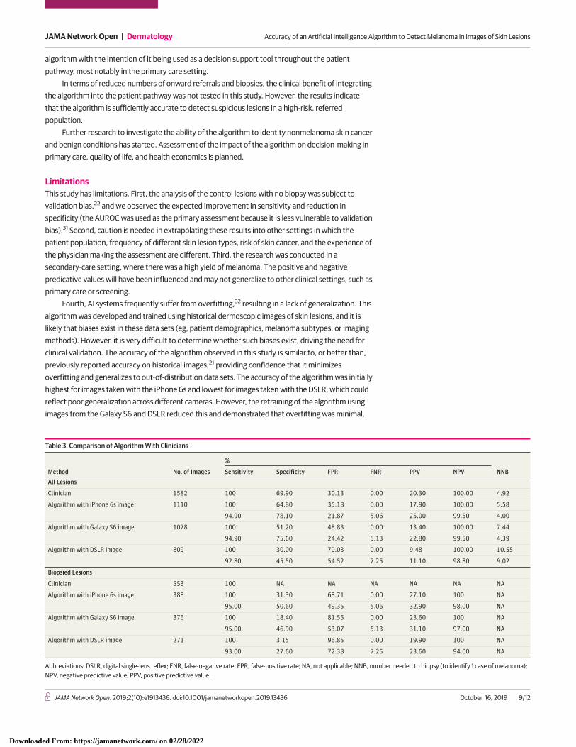
At 100% sensitivity, the algorithm achieved a specificity of 64.8% with iPhone 6s images. Whenusing a decision-threshold that created a sensitivity of 95%, the specificity of biopsied and all lesionsfor each camera were as follows: iPhone 6s, 50.6% and 78.1%, respectively; Galaxy S6, 46.9% and75.6%, respectively; and DSLR, 27.6% and 45.5%, respectively. These compare with a specificity of69.9% on all lesions by clinicians. Similarly, the number of biopsies needed to identify 1 case ofmelanoma (number needed to biopsy) at a 95% sensitivity was 3.04 for biopsied lesions and 4.00 forall lesions for images taken with the iPhone 6s; 3.22 and 4.39, respectively, for images taken with theGalaxy S6; and 4.23 and 9.02, respectively, for images taken with the DSLR. This compares with anumber needed to biopsy by clinicians of 4.92. When the negative predictive value was fixed at100%, the positive predictive value was 20.3% for clinician assessment; 17.9% for images taken withthe iPhone 6s; 13.4% for images taken with the Galaxy S6; and 9.5% for images taken with the DSLR(Table 3).
Age was a significant covariate for all methods, and Fitzpatrick skin type was also significant forclinicians. Clinical assessment was more accurate in younger patients (accuracy increased by 0.73%
Table 2. Assessment by Histopathology
Assessment Method Probability of Melanoma Sensitivity, %/Specificity, %a
No. (%)
Other Lesion Type Dysplasia Melanoma TotalClinician Unlikely 93/27 80 (64.5) 35 (28.2) 9 (7.3) 124 (100)
Equivocal 68/83 155 (57.6) 82 (30.5) 32 (11.9) 269 (100)
Likely 29/97 35 (32.4) 25 (23.2) 48 (44.4) 108 (100)
Highly likely 0/100 8 (16.0) 6 (12.0) 36 (72.0) 50 (100)
Total NA 278 (50.5) 148 (26.8) 125 (22.7) 551 (100)
Algorithm withiPhone 6s image
Unlikely 100/0 19 (76.0) 6 (24.0) 0 25 (100)
Equivocal 79/87 157 (60.2) 87 (33.3) 17 (6.5) 261 (100)
Likely 37/98 24 (35.8) 10 (14.9) 33 (49.3) 67 (100)
Highly likely 0/100 5 (13.9) 2 (5.5) 29 (80.6) 36 (100)
Total NA 205 (52.7) 105 (30.0) 79 (20.3) 389 (100)
Algorithm withGalaxy S6 image
Unlikely 92/50 102 (65.4) 48 (30.8) 6 (3.8) 156 (100)
Equivocal 71/84 72 (60.5) 25 (37.9) 3 (8.6) 202 (100)
Likely 40/98 25 (37.9) 17 (25.7) 24 (36.4) 66 (100)
Highly likely 0/100 3 (8.6) 2 (5.7) 30 (85.7) 35 (100)
Total NA 202 (53.7) 98 (26.1) 76 (20.2) 376 (100)
Algorithm withDSLR image
Unlikely 86/58 81 (60.5) 46 (34.3) 7 (5.2) 134 (100)
Equivocal 75/89 44 (59.5) 24 (32.4) 6 (8.1) 74 (100)
Likely 41/99 12 (30.8) 10 (25.6) 17 (43.6) 39 (100)
Highly likely 0/100 3 (12.5) 0 21 (87.5) 24 (100)
Total NA 140 (51.7) 80 (29.5) 51 (18.8) 271 (100)
Abbreviations: DSLR, digital single-lens reflex; NA, not applicable.a Sensitivity/specificity shows the accuracy of the level of likelihood for each threshold
for detecting melanoma.
JAMA Network Open | Dermatology Accuracy of an Artificial Intelligence Algorithm to Detect Melanoma in Images of Skin Lesions
JAMA Network Open. 2019;2(10):e1913436. doi:10.1001/jamanetworkopen.2019.13436 (Reprinted) October 16, 2019 6/12
Downloaded From: https://jamanetwork.com/ on 02/28/2022

for each decreasing year), whereas the algorithm’s assessment was more accurate in older patients(for each increasing year, iPhone 6s AUROC increased by 0.76%, Galaxy S6 AUROC increased by1.9%, and DSLR AUROC increased by 4.3%). Because clinician accuracy is also influenced byFitzpatrick skin type (ie, AUROC increased by 10% for each level of skin sensitivity above Fitzpatricktype IV), it is possible that clinicians overweighted these risk factors in their determination of thelikelihood of melanoma.
Missing images were independent of patient-related or lesion-related characteristics (missingcompletely at random test results: clinical likelihood: P = .26; n = 1581; iPhone 6s image algorithmicassessment: P = .56; n = 1110; Galaxy S6 image algorithmic assessment: P = .17; n = 1078; and D5500image algorithmic assessment: P = .26; n = 809). However, a degree of bias did appear to beintroduced by the missing image–based assessments, as the AUROC based on the Heckman analysiswas generally lower for every method of assessment of all lesions than the empirical AUROC.Nevertheless, there is very little difference between the empirical and the Heckman AUROC for allassessment methods.
Figure 1. Receiver Operating Characteristic Curves for Clinical and Trained Algorithm Assessment of Biopsied Lesions
1.0
0.8
0.6
0.4
0.2
0
0 1.00.8
Sens
itivi
ty
1 – Specificity0.60.40.2
Clinician assessmentA
1.0
0.8
0.6
0.4
0.2
0
0 1.00.8
Sens
itivi
ty
1 – Specificity0.60.40.2
iPhone 6s imagesB
1.0
0.8
0.6
0.4
0.2
0
0 1.00.8
Sens
itivi
ty
1 – Specificity0.60.40.2
Galaxy S6 imagesC
1.0
0.8
0.6
0.4
0.2
0
0 1.00.8
Sens
itivi
ty
1 – Specificity0.60.40.2
DSLR imagesD
A, Area under receiver operator characteristic curve, 0.779. B, Area under receiver operator characteristic curve, 0.902. C, Area under receiver operator characteristic curve, 0.858.D, Area under receiver operator characteristic curve, 0.869. DSLR indicates digital single-lens reflex.
JAMA Network Open | Dermatology Accuracy of an Artificial Intelligence Algorithm to Detect Melanoma in Images of Skin Lesions
JAMA Network Open. 2019;2(10):e1913436. doi:10.1001/jamanetworkopen.2019.13436 (Reprinted) October 16, 2019 7/12
Downloaded From: https://jamanetwork.com/ on 02/28/2022

Discussion
The results of the study showed that the algorithm and specialists identified melanoma in selectedsuspicious pigmented skin lesions at a similar level of accuracy. More than half of the melanomadiagnoses were either in situ or less than 1 mm deep, indicating that the algorithm could play a role indetecting thin or early-stage lesions. The inclusion of control (ie, believed to be benign) lesions in theanalysis enabled us to show that the algorithm maintains a high specificity when control lesions areassessed.
Primary prevention of melanoma based on so-called sun-safe behaviors has not proved to besuccessful,25 and a US government review of secondary prevention concluded that there isinsufficient evidence to recommend skin cancer screening.2 While this opinion has been challenged26
and guidance in other countries recommends screening in high-risk populations,27-29 the cost ofscreening in populations at lower risk of developing melanoma means it is not cost-effective todeploy widespread screening programs.30 To address this issue, Skin Analytics developed an AI
Figure 2. Receiver Operating Characteristic Curves for Clinical and Trained Algorithm Assessment of All Lesions
1.0
0.8
0.6
0.4
0.2
0
0 1.00.8
Sens
itivi
ty
1 – Specificity0.60.40.2
Clinician assessmentA
1.0
0.8
0.6
0.4
0.2
0
0 1.00.8
Sens
itivi
ty
1 – Specificity0.60.40.2
iPhone 6s imagesB
1.0
0.8
0.6
0.4
0.2
0
0 1.00.8
Sens
itivi
ty
1 – Specificity0.60.40.2
Galaxy S6 imagesC
1.0
0.8
0.6
0.4
0.2
0
0 1.00.8
Sens
itivi
ty
1 – Specificity0.60.40.2
DSLR imagesD
A, Area under receiver operator characteristic curve, 0.909. B, Area under receiver operator characteristic curve, 0.959. C, Area under receiver operator characteristic curve, 0.938.D, Area under receiver operator characteristic curve, 0.918. DSLR indicates digital single-lens reflex.
JAMA Network Open | Dermatology Accuracy of an Artificial Intelligence Algorithm to Detect Melanoma in Images of Skin Lesions
JAMA Network Open. 2019;2(10):e1913436. doi:10.1001/jamanetworkopen.2019.13436 (Reprinted) October 16, 2019 8/12
Downloaded From: https://jamanetwork.com/ on 02/28/2022
algorithm with the intention of it being used as a decision support tool throughout the patientpathway, most notably in the primary care setting.
In terms of reduced numbers of onward referrals and biopsies, the clinical benefit of integratingthe algorithm into the patient pathway was not tested in this study. However, the results indicatethat the algorithm is sufficiently accurate to detect suspicious lesions in a high-risk, referredpopulation.
Further research to investigate the ability of the algorithm to identity nonmelanoma skin cancerand benign conditions has started. Assessment of the impact of the algorithm on decision-making inprimary care, quality of life, and health economics is planned.
LimitationsThis study has limitations. First, the analysis of the control lesions with no biopsy was subject tovalidation bias,22 and we observed the expected improvement in sensitivity and reduction inspecificity (the AUROC was used as the primary assessment because it is less vulnerable to validationbias).31 Second, caution is needed in extrapolating these results into other settings in which thepatient population, frequency of different skin lesion types, risk of skin cancer, and the experience ofthe physician making the assessment are different. Third, the research was conducted in asecondary-care setting, where there was a high yield of melanoma. The positive and negativepredicative values will have been influenced and may not generalize to other clinical settings, such asprimary care or screening.
Fourth, AI systems frequently suffer from overfitting,32 resulting in a lack of generalization. Thisalgorithm was developed and trained using historical dermoscopic images of skin lesions, and it islikely that biases exist in these data sets (eg, patient demographics, melanoma subtypes, or imagingmethods). However, it is very difficult to determine whether such biases exist, driving the need forclinical validation. The accuracy of the algorithm observed in this study is similar to, or better than,previously reported accuracy on historical images,21 providing confidence that it minimizesoverfitting and generalizes to out-of-distribution data sets. The accuracy of the algorithm was initiallyhighest for images taken with the iPhone 6s and lowest for images taken with the DSLR, which couldreflect poor generalization across different cameras. However, the retraining of the algorithm usingimages from the Galaxy S6 and DSLR reduced this and demonstrated that overfitting was minimal.
Table 3. Comparison of Algorithm With Clinicians
Method No. of Images
%
NNBSensitivity Specificity FPR FNR PPV NPVAll Lesions
Clinician 1582 100 69.90 30.13 0.00 20.30 100.00 4.92
Algorithm with iPhone 6s image 1110 100 64.80 35.18 0.00 17.90 100.00 5.58
94.90 78.10 21.87 5.06 25.00 99.50 4.00
Algorithm with Galaxy S6 image 1078 100 51.20 48.83 0.00 13.40 100.00 7.44
94.90 75.60 24.42 5.13 22.80 99.50 4.39
Algorithm with DSLR image 809 100 30.00 70.03 0.00 9.48 100.00 10.55
92.80 45.50 54.52 7.25 11.10 98.80 9.02
Biopsied Lesions
Clinician 553 100 NA NA NA NA NA NA
Algorithm with iPhone 6s image 388 100 31.30 68.71 0.00 27.10 100 NA
95.00 50.60 49.35 5.06 32.90 98.00 NA
Algorithm with Galaxy S6 image 376 100 18.40 81.55 0.00 23.60 100 NA
95.00 46.90 53.07 5.13 31.10 97.00 NA
Algorithm with DSLR image 271 100 3.15 96.85 0.00 19.90 100 NA
93.00 27.60 72.38 7.25 23.60 94.00 NA
Abbreviations: DSLR, digital single-lens reflex; FNR, false-negative rate; FPR, false-positive rate; NA, not applicable; NNB, number needed to biopsy (to identify 1 case of melanoma);NPV, negative predictive value; PPV, positive predictive value.
JAMA Network Open | Dermatology Accuracy of an Artificial Intelligence Algorithm to Detect Melanoma in Images of Skin Lesions
JAMA Network Open. 2019;2(10):e1913436. doi:10.1001/jamanetworkopen.2019.13436 (Reprinted) October 16, 2019 9/12
Downloaded From: https://jamanetwork.com/ on 02/28/2022

Fifth, there were technical issues with the equipment used for the study, leading to a highproportion of lesions that could not be photographed by at least 1 of the 3 cameras or images thatcould not be analyzed by the algorithm. These issues show the influence that the usability ofequipment can have on the effectiveness of a technology, which needs to be reliable for health careprofessionals to use it and have confidence in it. Reasons for the missing images included technologyfailures, such as camera malfunction, and poor optimization of the equipment for use in a clinicalsetting (particularly the DSLR, which was awkward for the study staff to use). Despite this, statisticalanalysis of the influence of missing data suggests that there was little selection bias generated bythe missing data. This provides evidence for the robustness of the algorithm in accurately detectingmelanoma.
Sixth, there were only 3 types of cameras used in this trial. It remains to be tested whether theresults apply to other smartphones or other cameras.
Conclusions
The findings of this diagnostic trial demonstrated that an AI algorithm, using different camera types,can detect melanoma with a similar level of accuracy as specialists. The development of low-costscreening methods, such as AI-based services, could transform patient diagnosis pathways, enablinggreater efficiencies throughout the health care service.
ARTICLE INFORMATIONAccepted for Publication: August 27, 2019.
Published: October 16, 2019. doi:10.1001/jamanetworkopen.2019.13436
Correction: This article was corrected on November 6, 2019, to fix an error in the Key Points.
Open Access: This is an open access article distributed under the terms of the CC-BY-NC-ND License. © 2019 PhillipsM et al. JAMA Network Open.
Corresponding Author: Michael Phillips, MMedSci, Centre for Medical Research, University of Western Australia,Perth, WA 6008, Australia ([email protected]); Helen Marsden, PhD, Skin Analytics Limited,One Phipp Street, The Frames, London EC2A 4PS, United Kingdom ([email protected]).
Author Affiliations: Harry Perkins Institute of Medical Research, Perth, Western Australia, Australia (Phillips);Centre for Medical Research, University of Western Australia, Perth, Western Australia, Australia (Phillips); SkinAnalytics Limited, London, United Kingdom (Marsden, Greenhalgh); Royal Stoke University Hospital, UniversityHospital North Midlands, Stoke, United Kingdom (Jaffe); Oxford University Hospitals NHS Foundation Trust,Oxford, United Kingdom (Matin, Wali); Royal Devon and Exeter NHS Foundation Trust, Exeter, United Kingdom(McGrath, James); Dudley Group NHS Foundation Trust, Corbett Hospital, Stourbridge, United Kingdom(Ladoyanni); Barts Health, London, United Kingdom (Bewley); Queen Mary School of Medicine, University ofLondon, London, United Kingdom (Bewley); Dermatology Unit, University of Campania, Naples, Italy(Argenziano); Barnet and Chase Farm Hospitals, Royal Free NHS Foundation Trust, London, United Kingdom(Palamaras).
Author Contributions: Mr Phillips had full access to all of the data in the study and takes responsibility for theintegrity of the data and the accuracy of the data analysis.
Concept and design: Phillips, Marsden, Greenhalgh, Argenziano, Palamaras.
Acquisition, analysis, or interpretation of data: Phillips, Marsden, Jaffe, Matin, Wali, Greenhalgh, McGrath, James,Ladoyanni, Bewley, Palamaras.
Drafting of the manuscript: Phillips, Marsden, Greenhalgh, McGrath, Palamaras.
Critical revision of the manuscript for important intellectual content: Jaffe, Matin, Wali, Greenhalgh, McGrath,James, Ladoyanni, Bewley, Argenziano, Palamaras.
Statistical analysis: Phillips, Greenhalgh.
Administrative, technical, or material support: Marsden, Jaffe, McGrath, James, Bewley, Palamaras.
Supervision: Phillips, Jaffe, Wali, Ladoyanni, Palamaras.
JAMA Network Open | Dermatology Accuracy of an Artificial Intelligence Algorithm to Detect Melanoma in Images of Skin Lesions
JAMA Network Open. 2019;2(10):e1913436. doi:10.1001/jamanetworkopen.2019.13436 (Reprinted) October 16, 2019 10/12
Downloaded From: https://jamanetwork.com/ on 02/28/2022

Conflict of Interest Disclosures: Mr Phillips reported having a familial relationship with Skin Analytics Limited. DrMarsden reported working for Skin Analytics Limited and receiving share options during the conduct of the study.Dr Matin reported receiving grants from Barco outside the submitted work and being coauthor of a suite ofCochrane diagnostic test accuracy systematic reviews, including the diagnosis of melanoma. Dr Greenhalghreported working for Skin Analytics Limited during the conduct of the study and holding patent US 20150254851A1. Dr Bewley reported working as an ad hoc consultant for Almirall, AbbVie, Galderma, LEO Pharma, Eli Lilly andCo, Sanofi, Novartis, and Janssen Pharmaceuticals. No other disclosures were reported.
Funding/Support: This study was funded by Skin Analytics Limited, which developed and owns Deep Ensemblefor Recognition of Malignancy. The Royal Perth Hospital Medical Research Fund supported the analysis andinterpretation of the data and the preparation of the manuscript.
Role of the Funder/Sponsor: Employees of Skin Analytics Limited designed and conducted the study; collected,managed, and analyzed the images; contributed to the interpretation of the data; and prepared, reviewed, andapproved the decision to submit the manuscript for publication. Royal Perth Hospital Medical Research Fund hadno involvement with the data analysis and interpretation of the data, nor the preparation of the manuscript.
Additional Contributions: We thank all the patients who kindly consented to participate in the study.
REFERENCES1. Cancer Research UK. Melanoma skin cancer survival statistics. https://www.cancerresearchuk.org/health-professional/cancer-statistics/statistics-by-cancer-type/melanoma-skin-cancer/survival#heading-Three. AccessedMarch 21, 2019.
2. Wernli KJ, Henrikson NB, Morrison CC, Nguyen M, Pocobelli G, Whitlock EP; US Preventive Services Task ForceEvidence Syntheses formerly Systematic Evidence Reviews. Screening for skin cancer in adults: an updatedsystematic evidence review for the US Preventive Services Task Force. JAMA. 2016;316(4):436-447.
3. National Institute for Health and Care Excellence. Melanoma: assessment and management. https://www.nice.org.uk/guidance/ng14. Accessed September 5, 2019.
4. Welch HG, Woloshin S, Schwartz LM. Skin biopsy rates and incidence of melanoma: population based ecologicalstudy. BMJ. 2005;331(7515):481. doi:10.1136/bmj.38516.649537.E0
5. Chen SC, Pennie ML, Kolm P, et al. Diagnosing and managing cutaneous pigmented lesions: primary carephysicians versus dermatologists. J Gen Intern Med. 2006;21(7):678-682. doi:10.1111/j.1525-1497.2006.00462.x
6. Bainbridge S, Cake R, Meredith M, Furness P, Gordon B. Testing times to come: an evaluation of pathologycapacity across the UK. https://www.cancerresearchuk.org/sites/default/files/testing_times_to_come_nov_16_cruk.pdf.Accessed September 5, 2019.
7. Dinnes J, Deeks JJ, Chuchu N, et al; Cochrane Skin Cancer Diagnostic Test Accuracy Group. Dermoscopy, withand without visual inspection, for diagnosing melanoma in adults. Cochrane Database Syst Rev. 2018;12(12):CD011902. doi:10.1002/14651858.cd011902.pub2
8. Dinnes J, Deeks JJ, Saleh D, et al; Cochrane Skin Cancer Diagnostic Test Accuracy Group. Reflectance confocalmicroscopy for diagnosing cutaneous melanoma in adults. Cochrane Database Syst Rev. 2018;12(12):CD013190.doi:10.1002/14651858.cd013190
9. Chuchu N, Dinnes J, Takwoingi Y, et al; Cochrane Skin Cancer Diagnostic Test Accuracy Group. Teledermatologyfor diagnosing skin cancer in adults. Cochrane Database Syst Rev. 2018;12(12):CD013193. doi:10.1002/14651858.cd013193
10. Ferrante di Ruffano L, Takwoingi Y, Dinnes J, et al; Cochrane Skin Cancer Diagnostic Test Accuracy Group.Computer-assisted diagnosis techniques (dermoscopy and spectroscopy-based) for diagnosing skin cancer inadults. Cochrane Database Syst Rev. 2018;12(12):CD013186. doi:10.1002/14651858.cd013186
11. Chuchu N, Takwoingi Y, Dinnes J, et al; Cochrane Skin Cancer Diagnostic Test Accuracy Group. Smartphoneapplications for triaging adults with skin lesions that are suspicious for melanoma. Cochrane Database Syst Rev.2018;12(12):CD013192. doi:10.1002/14651858.cd013192
12. Kittler H, Pehamberger H, Wolff K, Binder M. Diagnostic accuracy of dermoscopy. Lancet Oncol. 2002;3(3):159-165. doi:10.1016/S1470-2045(02)00679-4
13. Brewer AC, Endly DC, Henley J, et al. Mobile applications in dermatology. JAMA Dermatol. 2013;149(11):1300-1304. doi:10.1001/jamadermatol.2013.5517
14. Kassianos AP, Emery JD, Murchie P, Walter FM. Smartphone applications for melanoma detection bycommunity, patient and generalist clinician users: a review. Br J Dermatol. 2015;172(6):1507-1518. doi:10.1111/bjd.13665
15. Ferrero NA, Morrell DS, Burkhart CN. Skin scan: a demonstration of the need for FDA regulation of medicalapps on iPhone. J Am Acad Dermatol. 2013;68(3):515-516. doi:10.1016/j.jaad.2012.10.045
JAMA Network Open | Dermatology Accuracy of an Artificial Intelligence Algorithm to Detect Melanoma in Images of Skin Lesions
JAMA Network Open. 2019;2(10):e1913436. doi:10.1001/jamanetworkopen.2019.13436 (Reprinted) October 16, 2019 11/12
Downloaded From: https://jamanetwork.com/ on 02/28/2022

16. Wolf JA, Moreau JF, Akilov O, et al. Diagnostic inaccuracy of smartphone applications for melanoma detection.JAMA Dermatol. 2013;149(4):422-426. doi:10.1001/jamadermatol.2013.2382
17. Stoecker WV, Rader RK, Halpern A. Diagnostic inaccuracy of smartphone applications for melanoma detection:representative lesion sets and the role for adjunctive technologies. JAMA Dermatol. 2013;149(7):884. doi:10.1001/jamadermatol.2013.4334
18. Wolf JA, Ferris LK. Diagnostic inaccuracy of smartphone applications for melanoma detection: reply. JAMADermatol. 2013;149(7):885. doi:10.1001/jamadermatol.2013.4337
19. Esteva A, Kuprel B, Novoa RA, et al. Dermatologist-level classification of skin cancer with deep neuralnetworks. Nature. 2017;542(7639):115-118. doi:10.1038/nature21056
20. Haenssle HA, Fink C, Schneiderbauer R, et al; Reader study level-I and level-II Groups. Man against machine:diagnostic performance of a deep learning convolutional neural network for dermoscopic melanoma recognition incomparison to 58 dermatologists. Ann Oncol. 2018;29(8):1836-1842. doi:10.1093/annonc/mdy166
21. Phillips M, Greenhalgh J, Marsden H, Palamaras I. Detection of malignant melanoma using artificial intelligence:an observational study. Dermatol Pract Concept. In press.
22. Pepe MS. The Statistical Evaluation of Medical Tests for Classification and Prediction. Oxford, UK: OxfordUniversity Press; 2003.
23. Little RJA. A test of missing completely at random for multivariate data with missing values. J Am Stat Assoc.1988;83(404):1198-1202. doi:10.1080/01621459.1988.10478722
24. Cook JA, Rajbhandari A. Heckroccurve: ROC curves for selected samples. Stata J. 2018;18(1):174-183. doi:10.1177/1536867X1801800110
25. Greinert R, Breitbart E, Mohr P, Volkmer B. Health initiatives for the prevention of skin cancer. In: Reichrath J,ed. Sunlight, Vitamin D and Skin Cancer. New York, NY: Springer; 2008:485-499.
26. Johnson MM, Leachman SA, Aspinwall LG, et al. Skin cancer screening: recommendations for data-drivenscreening guidelines and a review of the US Preventive Services Task Force controversy. Melanoma Manag. 2017;4(1):13-37. doi:10.2217/mmt-2016-0022
27. Cancer Council Australia. Position statement: screening and early detection of skin cancer. https://dermcoll.edu.au/wp-content/uploads/2014/05/PosStatEarlyDetectSkinCa.pdf. Accessed September 5, 2019.
28. Royal Australian College of General Practitioners. Guidelines for preventive activities in general practice, 9thedition. https://www.racgp.org.au/download/Documents/Guidelines/Redbook9/17048-Red-Book-9th-Edition.pdf.Accessed September 5, 2019.
29. Marsden JR, Newton-Bishop JA, Burrows L, et al; British Association of Dermatologists (BAD) ClinicalStandards Unit. Revised UK guidelines for the management of cutaneous melanoma 2010. J Plast Reconstr AesthetSurg. 2010;63(9):1401-1419. doi:10.1016/j.bjps.2010.07.006
30. Robinson JK, Halpern AC. Cost-effective melanoma screening. JAMA Dermatol. 2016;152(1):19-21. doi:10.1001/jamadermatol.2015.2681
31. Diamond GA. The wizard of odds: Bayes theorem and diagnostic testing. Mayo Clin Proc. 1999;74(11):1179-1182. doi:10.4065/74.11.1179
32. Obermeyer Z, Emanuel EJ. Predicting the future: big data, machine learning, and clinical medicine. N Engl JMed. 2016;375(13):1216-1219. doi:10.1056/NEJMp1606181
SUPPLEMENT.eTable. Biopsied Lesion CharacteristicseFigure 1. STARD Flow CharteFigure 2. ROC Curves of Algorithm Performance Following Retraining
JAMA Network Open | Dermatology Accuracy of an Artificial Intelligence Algorithm to Detect Melanoma in Images of Skin Lesions
JAMA Network Open. 2019;2(10):e1913436. doi:10.1001/jamanetworkopen.2019.13436 (Reprinted) October 16, 2019 12/12
Downloaded From: https://jamanetwork.com/ on 02/28/2022


![Artificial Intelligence · Artificial Intelligence 2016-2017 Introduction [5] Artificial Brain: can machines think? Artificial Intelligence 2016-2017 Introduction [6] ... Deep Blue](https://img.dokumen.tips/doc/110x75/5f0538917e708231d411e192/artificial-intelligence-artificial-intelligence-2016-2017-introduction-5-artificial.jpg)