Embed Size (px)
Citation preview
Assessment and Treatment of Low Back Pain
Steven Stanos, DO
Medical DirectorCenter for Pain Management
Rehabilitation Institute of ChicagoAsst. Professor, Dept. PM&R
Northwestern University Medical School Feinberg School Of Medicine
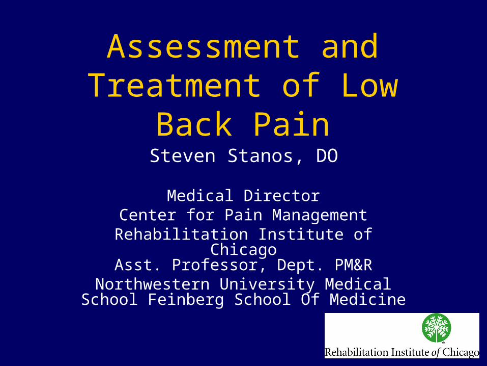
Goals• Individualized yet
comprehensive• Efficient• Comfortable for patient• Comfortable for clinician• Build rapport• Educate and prepare patient for
treatment• Monitor for inconsistencies
Physical Exam Overview
– Pain behavior– Gait– Motor strength– Muscle stretch reflexes– Dural tension testing– Sacral iliac joint testing– Myofascial assessment– Kinetic Chain considerations

Anatomy of LumboSacral Spine
Bogduk N, Clinical Anatomy Lumbar Spine and Sacrum, 3rd. Ed. Churchill Livingstone, 1999.

Bogduk N, Clinical Anatomy Lumbar Spine and Sacrum, 3rd. Ed. Churchill Livingstone, 1999.
Annulus Fibrosis
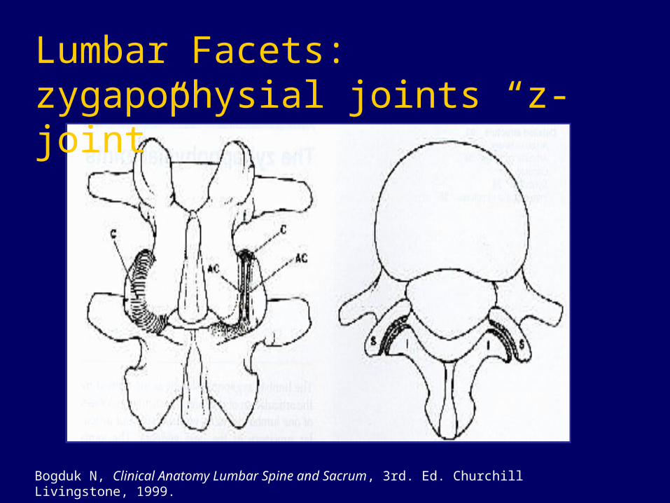
Lumbar Facets: zygapophysial joints “z-joint”
Bogduk N, Clinical Anatomy Lumbar Spine and Sacrum, 3rd. Ed. Churchill Livingstone, 1999.
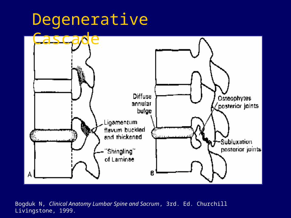
Bogduk N, Clinical Anatomy Lumbar Spine and Sacrum, 3rd. Ed. Churchill Livingstone, 1999.
Degenerative Cascade
Definitions• Somatome: field of somatic and
autonomic innervation based on embryologic segmental origin of somatic tissues
three basic elements:1. Dermatome: cutaneous structures
2. Myotome: skeletal musculature
3. Sclerotome: bones, joints, and ligaments8
Inman VT, Saunders J. J Nerv Ment Dis 1944;99:660-67.

Spinal “stability”
Neural
Control Unit
Spinal Column Spinal Muscles
Vertebral Position
Spinal Loads
Spinal Motions
Muscle
Activation Patterns
Panjabi MM. J Electromyography Kinesiology 2003:12:371-9

“Core” muscle groups
– Abdominals (Front)– Paraspinals and gluteals (Back)– Diaphragm (Roof)– Pelvic floor and hip muscles (Bottom)
Richardson C, et al .Therapeutic exercise for spinal stabilization and low back pain. Edinburgh (Scotland): Churchill Livigstone1999.
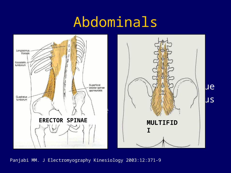
Abdominals
Local muscles
(Slow twitch)• Transversus
abdominus• Multifidi• Internal oblique • Pelvic floor
Global Muscles
(Fast-twitch)• Erector spinae• External oblique• Rectus abdominus
MULTIFIDIERECTOR SPINAE
Panjabi MM. J Electromyography Kinesiology 2003:12:371-9

the “15 minute rotisserie special”
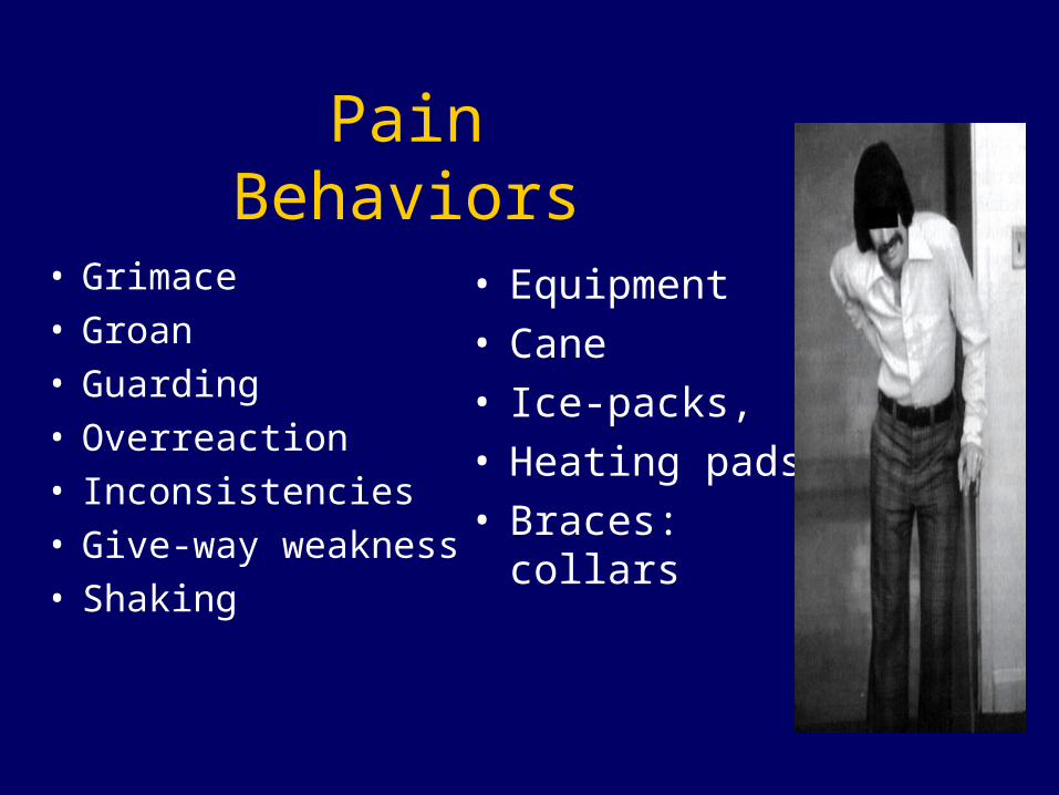
Pain Behaviors
• Grimace• Groan• Guarding• Overreaction• Inconsistencies• Give-way weakness• Shaking
• Equipment• Cane• Ice-packs, • Heating pads• Braces: collars
Gait
• Balance
• Base of support
• Arm swing/ trunk and shoulder rotation
• Cadence
• Leg: cicumduction, stance time, position
• Pain behavior
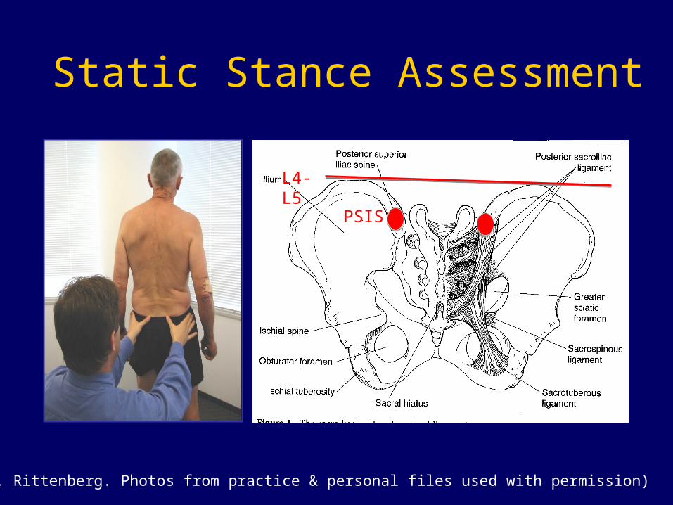
Static Stance Assessment
(J. Rittenberg. Photos from practice & personal files used with permission)
L4-L5
PSIS
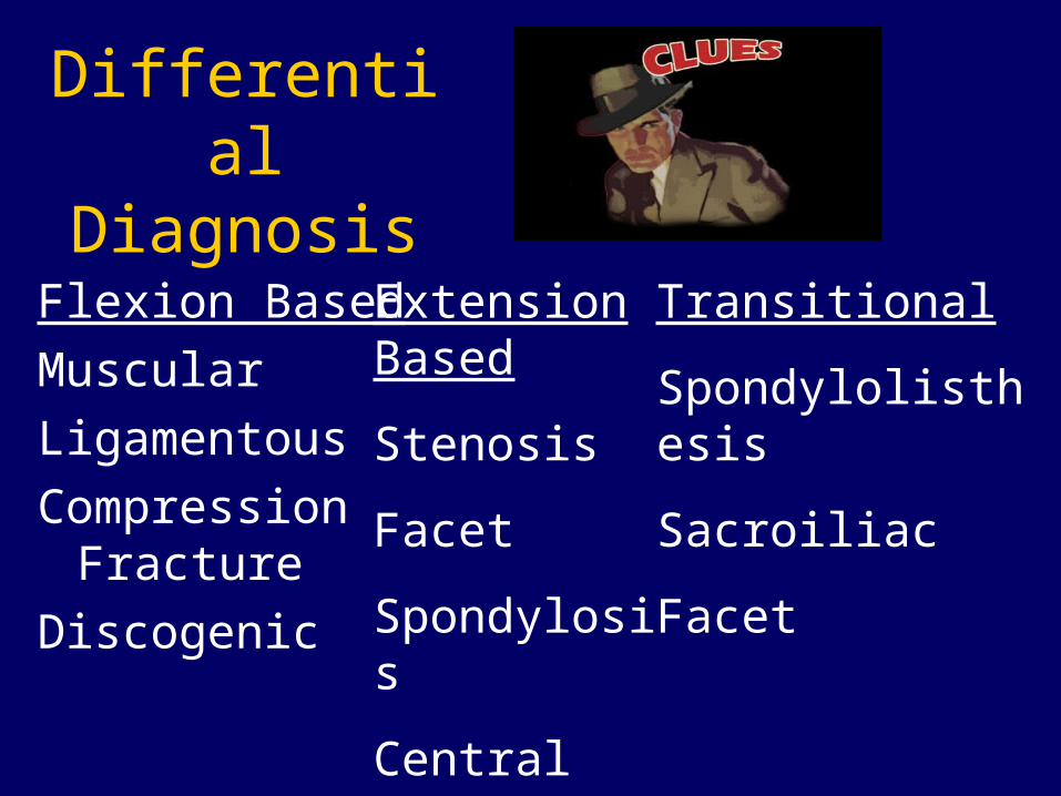
Flexion Based
Muscular
Ligamentous
Compression Fracture
Discogenic
Extension Based
Stenosis
Facet
Spondylosis
Central Disc
Transitional
Spondylolisthesis
Sacroiliac
Facet
Differential Diagnosis

Facet Arthropathy
• Zygapophyseal (z-joint)
• Poor correlation with history and exam1
• Commonly pain with extension & rotation
• Referral patterns2
1. Schwarzer AC, et al. Spine 1994;19:1132-7.
2. Slipman, C. Arch PM&R 81:334-338, 2000.
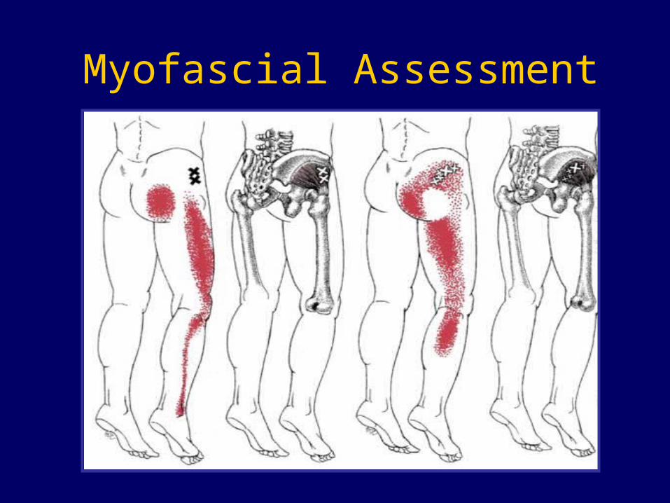
Myofascial Assessment
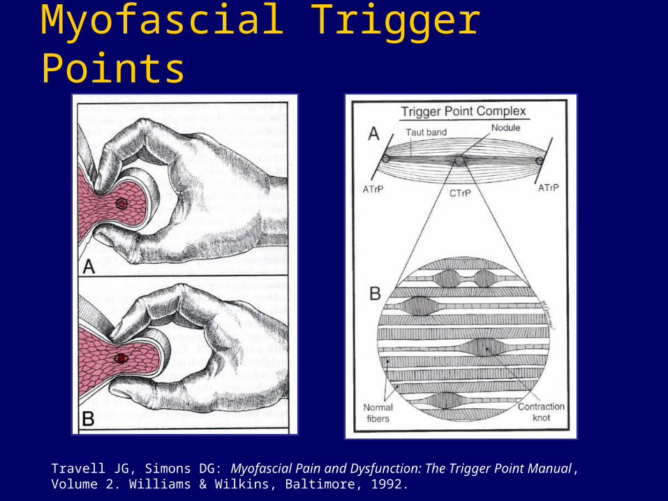
Myofascial Trigger Points
Travell JG, Simons DG: Myofascial Pain and Dysfunction: The Trigger Point Manual, Volume 2. Williams & Wilkins, Baltimore, 1992.
“Muscle pain is not skin pain”
Jay Shah, MD
Myofascial Trigger Points (MTrPs)
Active – cause a clinical pain complaint or other abnormal sensory symptoms
Latent – show all the other characteristics of active MTrPs, except that they’re pain free
Muscle Pain
• Aching and cramping
• Difficult to localize and refers to other deep somatic tissues (fascia, muscle, joints)
• Muscle nociceptive activity is processed differently in the CNS
• Inhibited more strongly by descending pain-modulating pathways than cutaneous pain
Symptoms• Local & referred pain• Pain with iso
contraction• Stiffness, limited
ROM• Muscle weakness• Paresthesia &
numbness• Propriocpetive
disturbance• Autonomic
dysfunction
Physical Findings• Local Tenderness• Single or multiple
muscles• Palpable nodules• Firm or Taut Bands• “twitch response”
(LTR)• Jump sign• Muscle shortening• Limited joint motion• Muscle Weakness

Motor Strength Testing
• 5 = Normal, full ROM vs. gravity, max resistance
• 4 = Good, full ROM vs. gravity, moderate resistance
• 3 = Fair, full ROM vs. gravity,
no resistance• 2 = Poor, full ROM,
gravity eliminated• 1 = Trace• 0 = No activity
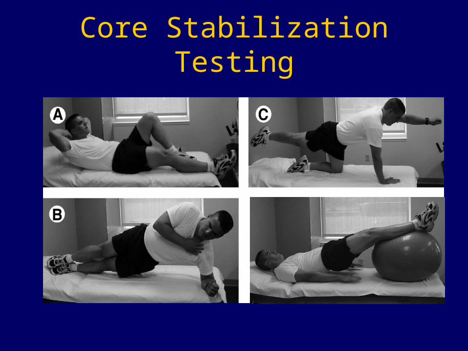
Core Stabilization Testing
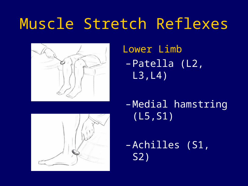
Muscle Stretch Reflexes
Lower Limb
– Patella (L2, L3,L4)
– Medial hamstring (L5,S1)
– Achilles (S1, S2)
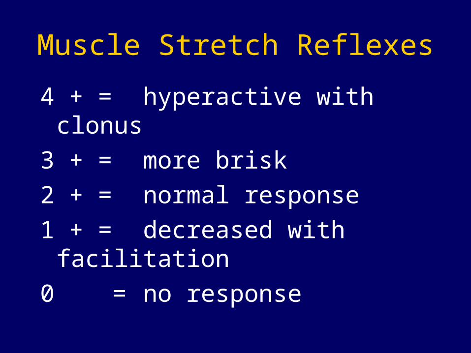
Muscle Stretch Reflexes
4 + = hyperactive with clonus
3 + = more brisk
2 + = normal response
1 + = decreased with facilitation
0 = no response
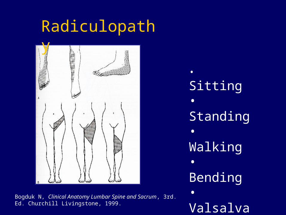
Radiculopathy
Bogduk N, Clinical Anatomy Lumbar Spine and Sacrum, 3rd. Ed. Churchill Livingstone, 1999.
• Sitting• Standing• Walking• Bending• Valsalva or cough
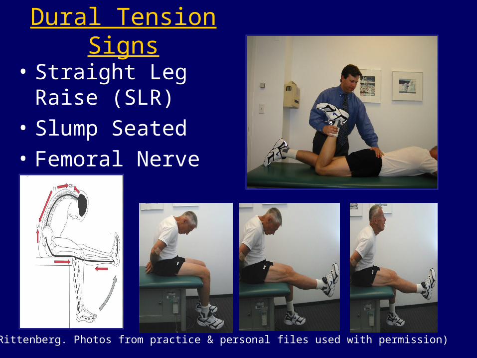
Dural Tension Signs
• Straight Leg Raise (SLR)
• Slump Seated
• Femoral Nerve Stretch
(J. Rittenberg. Photos from practice & personal files used with permission)
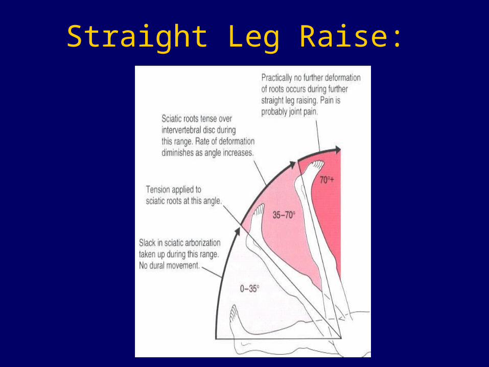
Straight Leg Raise:
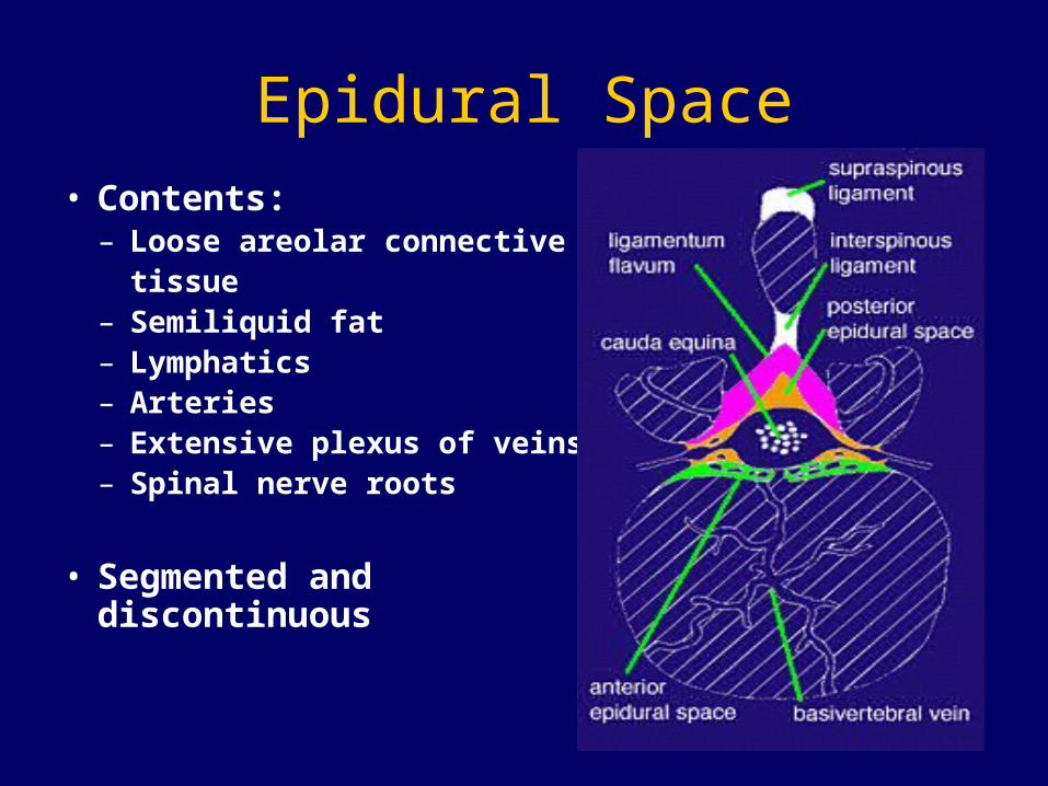
Epidural Space• Contents:
– Loose areolar connective tissue
– Semiliquid fat– Lymphatics– Arteries– Extensive plexus of veins– Spinal nerve roots
• Segmented and discontinuous


Transforaminal Approach
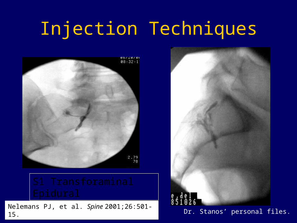
Injection Techniques
S1 Transforaminal Epidural
Dr. Stanos’ personal files.Nelemans PJ, et al. Spine 2001;26:501-15.
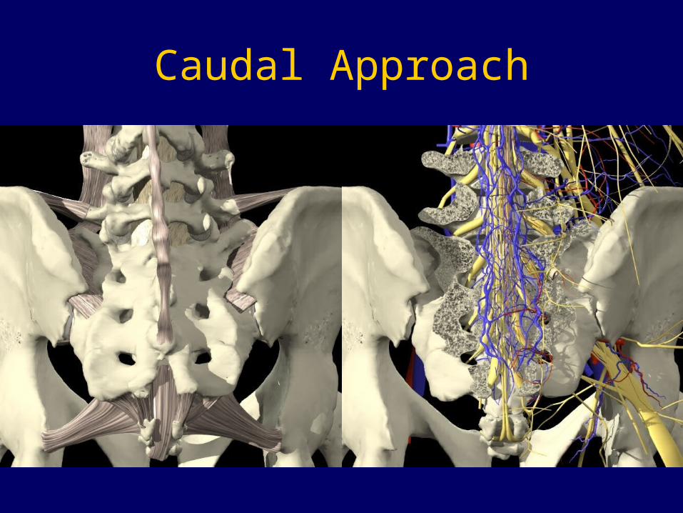
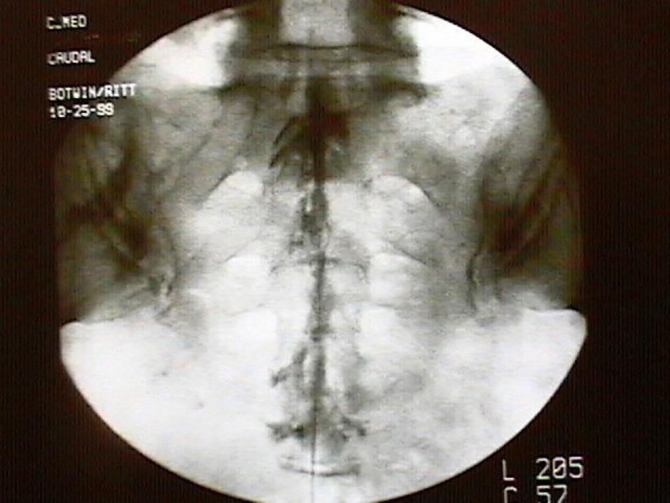
Caudal Approach
Axial Low Back Pain
• Degenerative disc disease (DDD)
• Internal disc derangement (IDD)
• Facet dysfunction
• Myofascial dysfunction
© 2005 Rehabilitatio Institute of Chicago
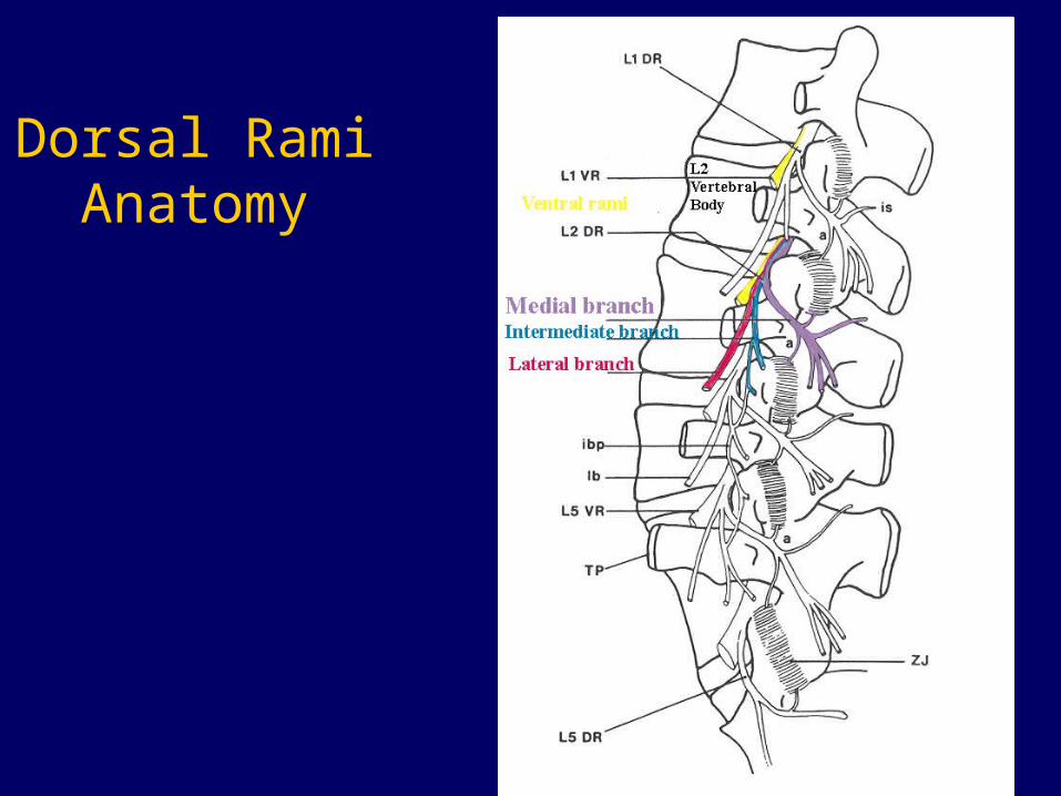
Dorsal Rami Anatomy
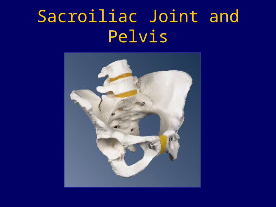
Sacroiliac Joint and Pelvis
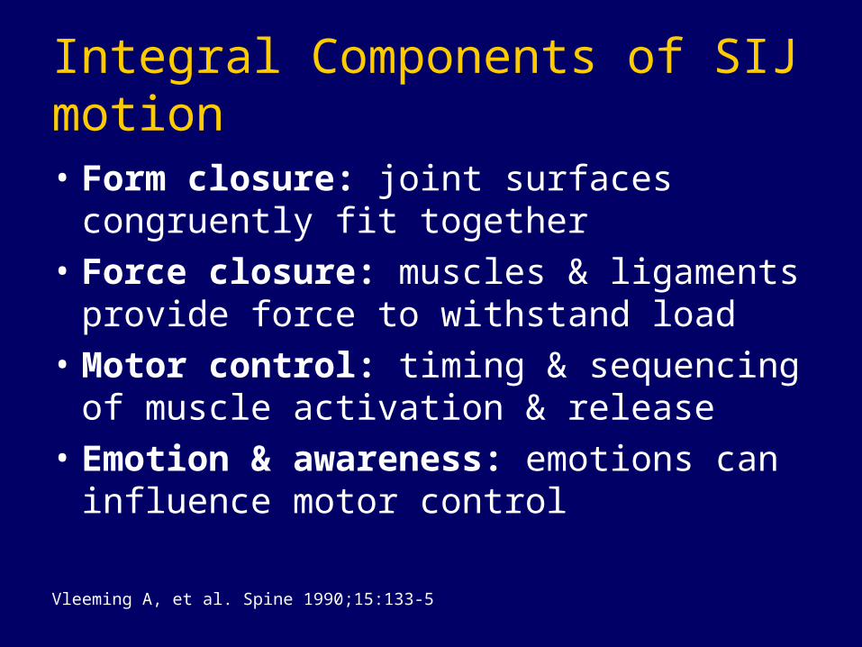
Integral Components of SIJ motion
• Form closure: joint surfaces congruently fit together
• Force closure: muscles & ligaments provide force to withstand load
• Motor control: timing & sequencing of muscle activation & release
• Emotion & awareness: emotions can influence motor control
Vleeming A, et al. Spine 1990;15:133-5
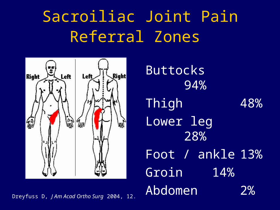
Sacroiliac Joint Pain Referral Zones
Buttocks 94%
Thigh 48%
Lower leg 28%
Foot / ankle13%
Groin 14%
Abdomen 2%
Dreyfuss D, J Am Acad Ortho Surg 2004, 12.
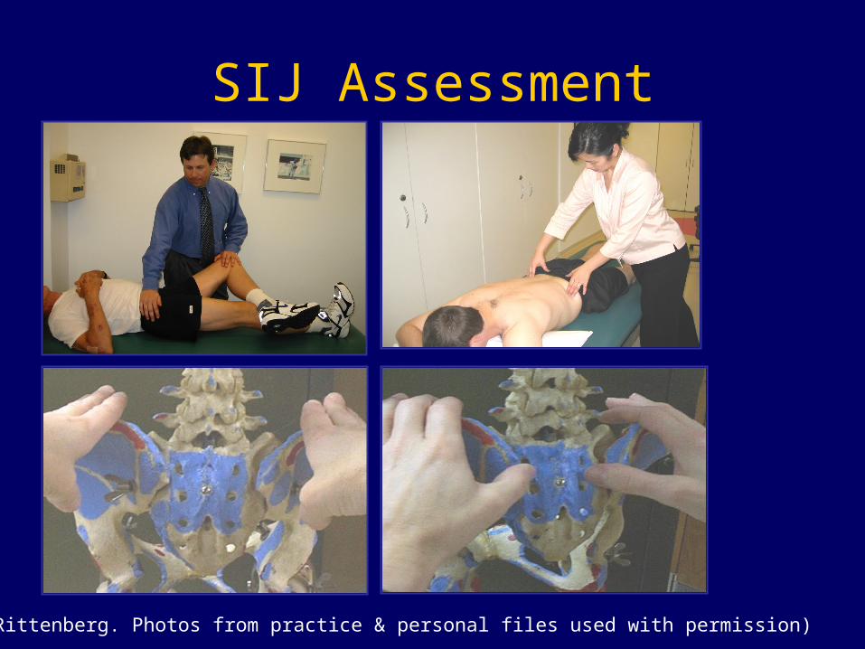
SIJ Assessment
(J.Rittenberg. Photos from practice & personal files used with permission)
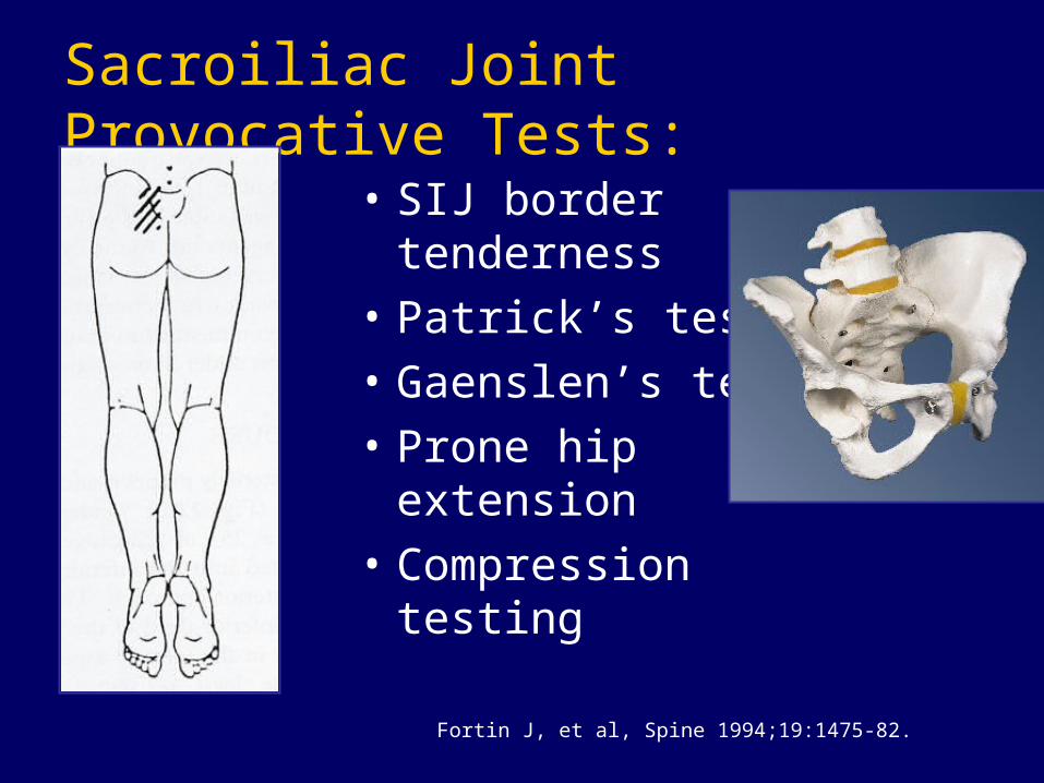
Sacroiliac Joint Provocative Tests:
• SIJ border tenderness
• Patrick’s test
• Gaenslen’s test
• Prone hip extension
• Compression testing
Fortin J, et al, Spine 1994;19:1475-82.
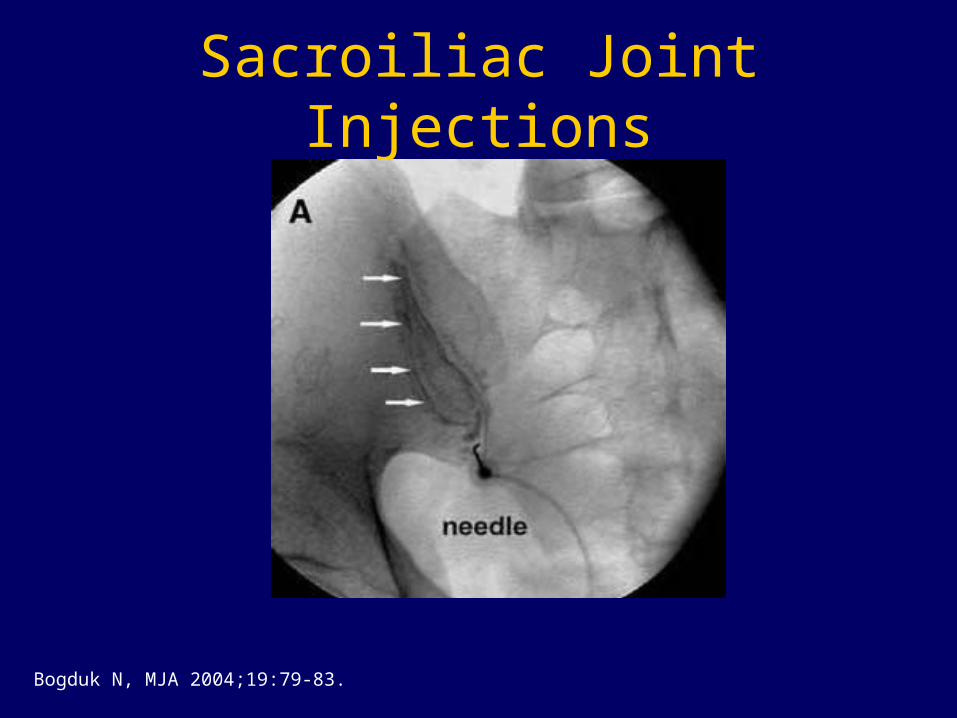
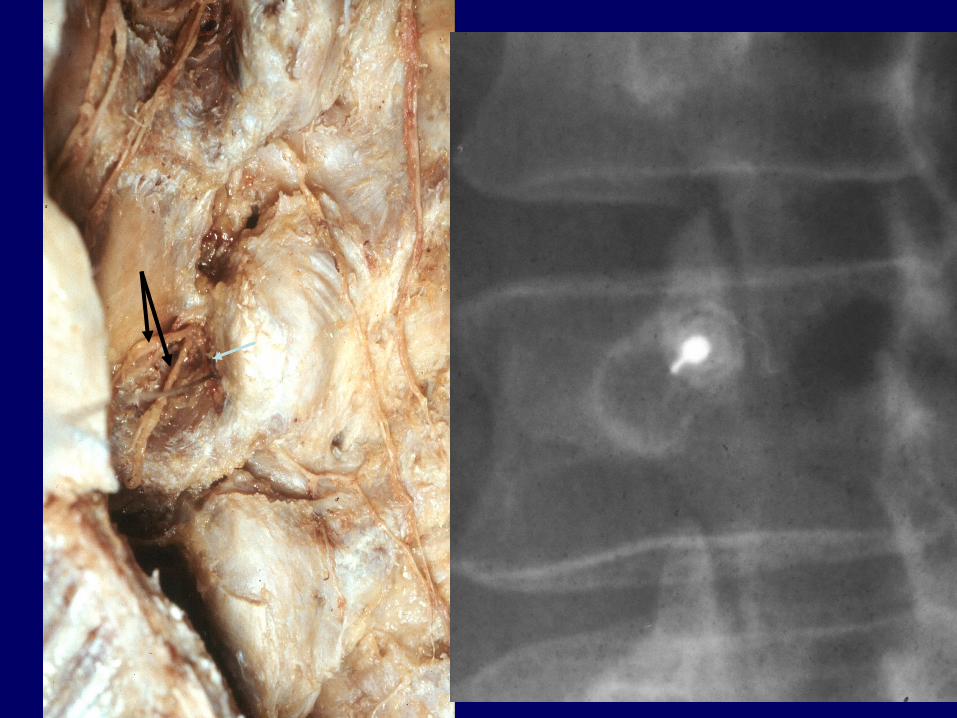
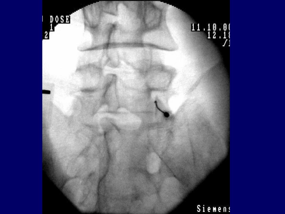
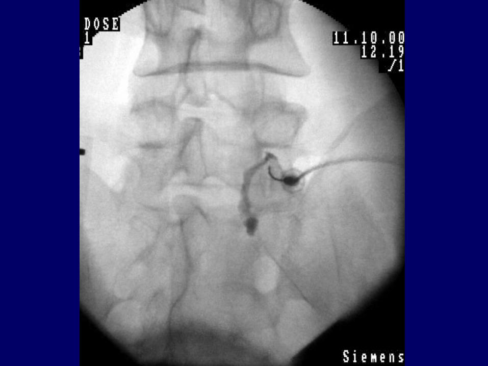
Sacroiliac Joint Injections
Bogduk N, MJA 2004;19:79-83.
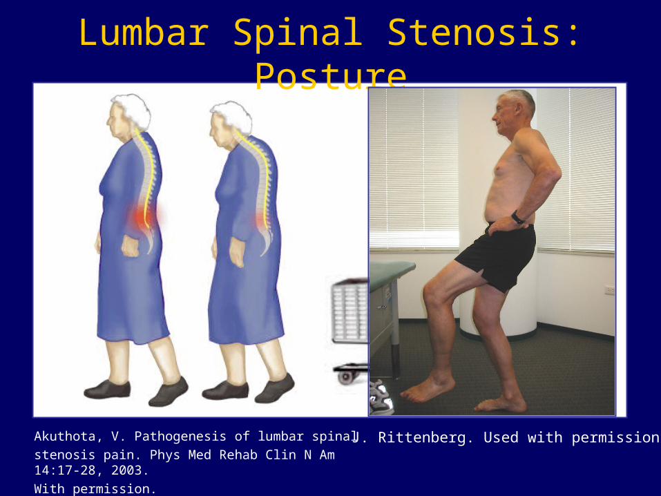
Lumbar Spinal Stenosis: Posture
Akuthota, V. Pathogenesis of lumbar spinal
stenosis pain. Phys Med Rehab Clin N Am 14:17-28, 2003.
With permission.
J. Rittenberg. Used with permission.

BI-Level Central
Neurovascular Claudication
Porter RW. Spine 1996;21:2046-52.
• Onset with walking
• “Heavy” sensation
• Variability
• Attempt to increase flexion
• Stooped posture
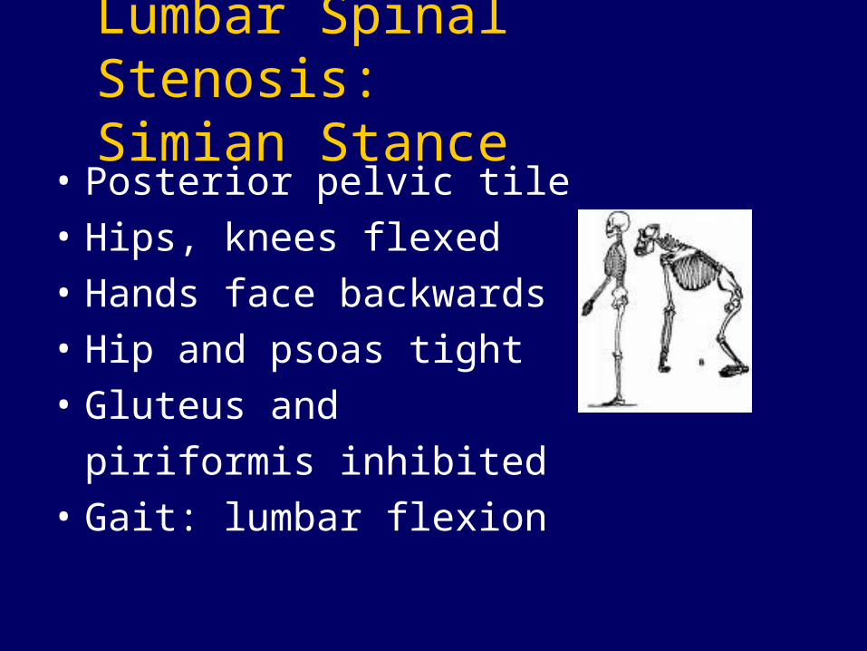
Lumbar Spinal Stenosis: Simian Stance
• Posterior pelvic tile
• Hips, knees flexed
• Hands face backwards
• Hip and psoas tight
• Gluteus and
piriformis inhibited
• Gait: lumbar flexion
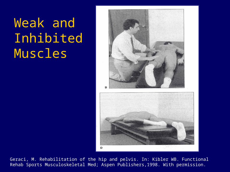
Geraci, M. Rehabilitation of the hip and pelvis. In: Kibler WB. Functional Rehab Sports Musculoskeletal Med; Aspen Publishers,1998. With permission.
Weak and Inhibited Muscles
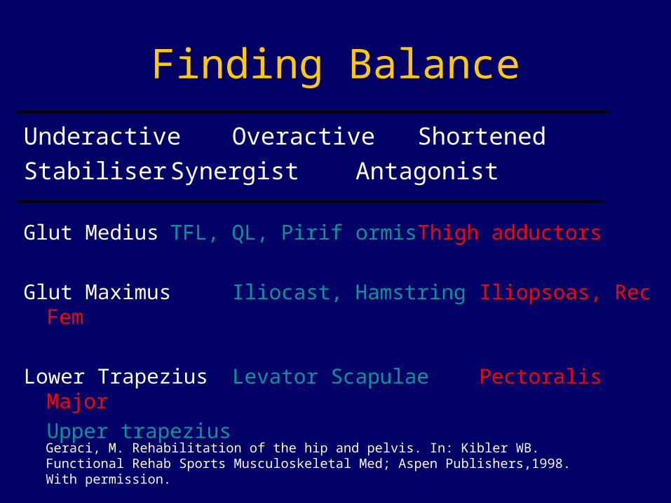
Finding Balance
Underactive Overactive Shortened
Stabiliser Synergist Antagonist
Glut Medius TFL, QL, Piriformis Thigh adductors
Glut Maximus Iliocast, Hamstring Iliopsoas, Rec Fem
Lower Trapezius Levator Scapulae Pectoralis Major
Upper trapezius
Geraci, M. Rehabilitation of the hip and pelvis. In: Kibler WB. Functional Rehab Sports Musculoskeletal Med; Aspen Publishers,1998. With permission.
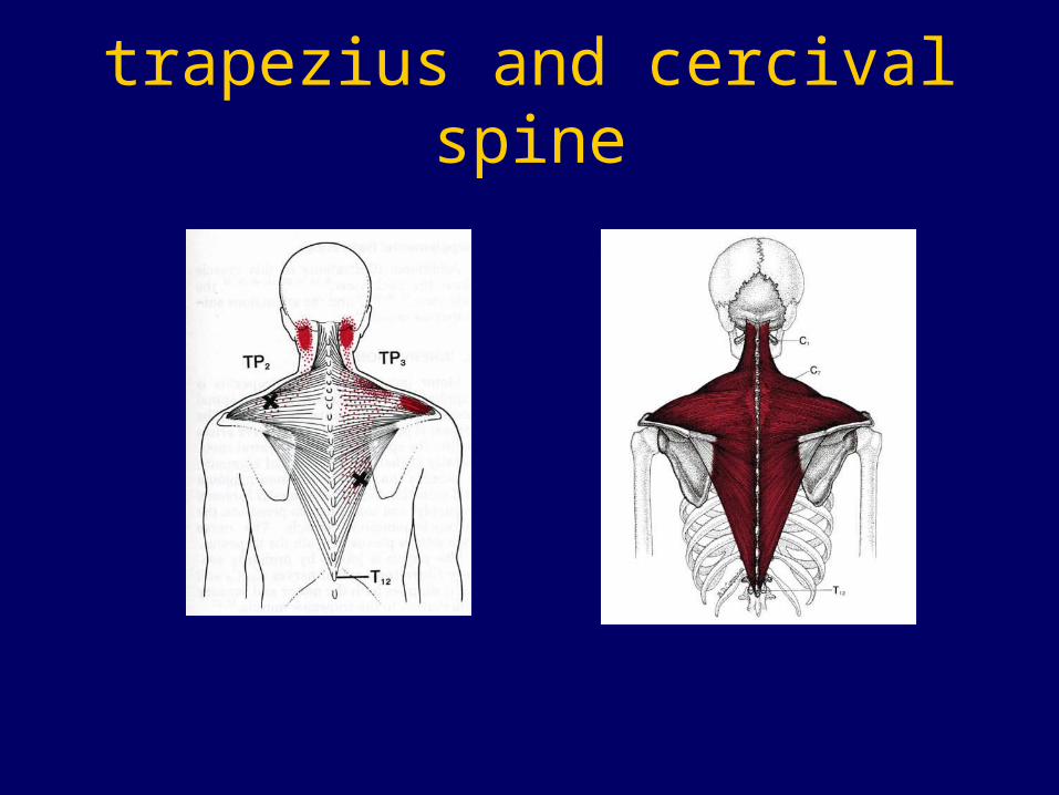
trapezius and cercival spine
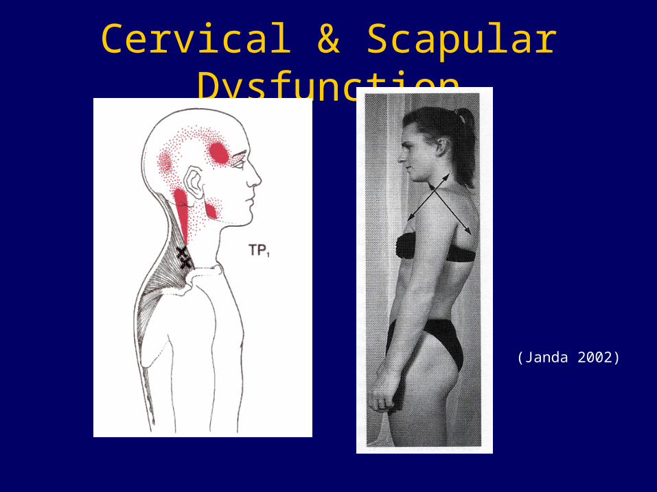
Cervical & Scapular Dysfunction
(Janda 2002)
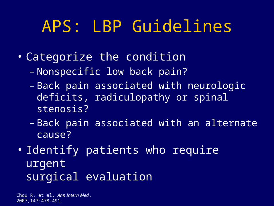
APS: LBP Guidelines
• Categorize the condition– Nonspecific low back pain?– Back pain associated with neurologic deficits,
radiculopathy or spinal stenosis?– Back pain associated with an alternate
cause?
• Identify patients who require urgent surgical evaluation
Chou R, et al. Ann Intern Med. 2007;147:478-491.
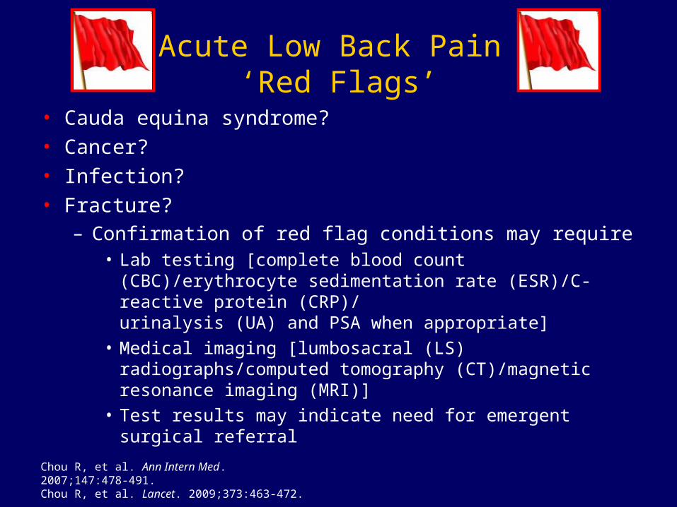
Acute Low Back Pain ‘Red Flags’
• Cauda equina syndrome?
• Cancer?
• Infection?
• Fracture?– Confirmation of red flag conditions may require
• Lab testing [complete blood count (CBC)/erythrocyte sedimentation rate (ESR)/C-reactive protein (CRP)/urinalysis (UA) and PSA when appropriate]
• Medical imaging [lumbosacral (LS) radiographs/computed tomography (CT)/magnetic resonance imaging (MRI)]
• Test results may indicate need for emergent surgical referral
Chou R, et al. Ann Intern Med. 2007;147:478-491.Chou R, et al. Lancet. 2009;373:463-472.
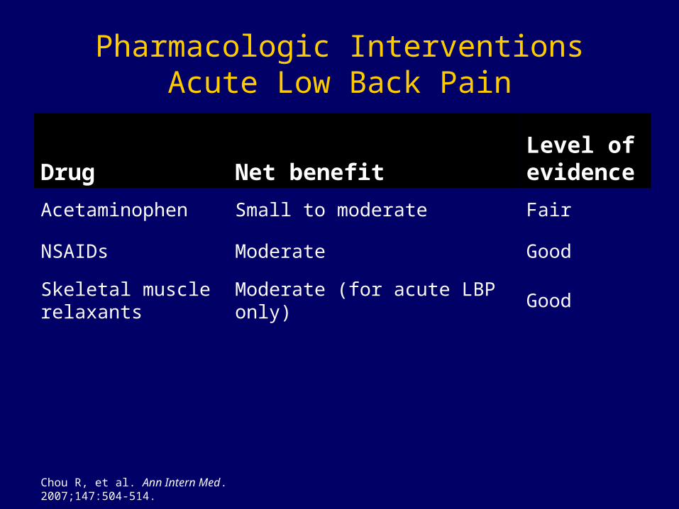
Pharmacologic InterventionsAcute Low Back Pain
Drug Net benefitLevel of evidence
Acetaminophen Small to moderate Fair
NSAIDs Moderate Good
Skeletal muscle relaxants
Moderate (for acute LBP only) Good
Chou R, et al. Ann Intern Med. 2007;147:504-514.

Guideline Highlights
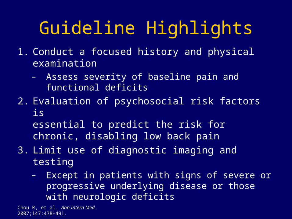
Chou R, et al. Ann Intern Med. 2007;147:478-491.
Guideline Highlights1. Conduct a focused history and physical
examination– Assess severity of baseline pain and
functional deficits
2. Evaluation of psychosocial risk factors is essential to predict the risk for chronic, disabling low back pain
3. Limit use of diagnostic imaging and testing– Except in patients with signs of severe or
progressive underlying disease or those with neurologic deficits

Recommendation 6ACP/APS Guidelines 2007
• Clinicians should consider the use of medications with proven benefits in conjunction with back care information and self-care. Clinicians should assess the severity of baseline pain and functional deficits, potential benefits, risks, and relative lack of long-term efficacy and safety data before initiating therapy. For most patients, first-line medication options are acetaminophen or NSAIDs.(Strong recommendation, moderate-quality evidence)
Chou R, et al. Ann Intern Med. 2007;147:504-514.
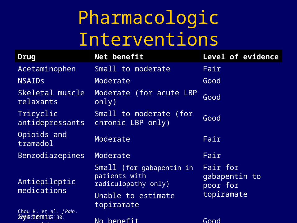
Pharmacologic InterventionsDrug Net benefit Level of evidence
Acetaminophen Small to moderate Fair
NSAIDs Moderate Good
Skeletal muscle relaxants
Moderate (for acute LBP only) Good
Tricyclic antidepressants
Small to moderate (for chronic LBP only)
Good
Opioids and tramadol Moderate Fair
Benzodiazepines Moderate Fair
Antiepileptic medications
Small (for gabapentin in patients with radiculopathy only)
Unable to estimate topiramate
Fair for gabapentin to poor for topiramate
Systemic steroids No benefit Good
Chou R, et al. J Pain. 2009;10:113-130.

Summary
• Comprehensive, but focused• Efficient• Exam should be easy on you and the
patient• Great opportunity to initiate a therapeutic
relationship and dialogue• Use a “good” exam to improve outcomes
and identify deficits or impairments
Thanks