Embed Size (px)
Citation preview

Aspects on bleeding and transfusion in elective orthopaedic surgery
Clinical and experimental studies
Malin S Carling
Institute of Clinical Sciences Department of Orthopaedics
Sahlgrenska Academy at University of Gothenburg

Gothenburg 2015
Cover illustration: Anton Carling (4 years) and Oliver Carling (7 years). Children’s view of the circulatory system and blood vessels.
Aspects on bleeding and transfusion in elective orthopaedic surgery © Malin S Carling 2015 [email protected], [email protected] ISBN 978-91-628-9571-6 (Print) ISBN 978-91-628-9572-3 (PDF) http://hdl.handle.net/2077/39556 Printed in Gothenburg, Sweden 2015 Printed by Ineko AB Illustrations by Pontus Andersson, Pontus Art Production

“The only weapon with which the unconscious patient can immediately retaliate upon the incompetent surgeon is haemorrhage”
William Halsted
(Bulletin of the John Hopkins Hospital 1912; 23: 191)

ABSTRACT
Background: Perioperative bleeding complications are associated with increased morbidity and mortality. One way to minimize perioperative bleeding and transfusion is to identify patients at risk of bleeding. Preoperative fibrinogen plasma concentration and factor XIII (FXIII) activity may be indicators of perioperative bleeding and transfusion. The aim of the thesis was to investigate 1) whether there is a correlation between the levels of preoperative coagulation factors and perioperative bleeding and transfusion in elective orthopaedic surgery patients, 2) transfusion and blood loss in arthroplasty surgery and possible risk factors, and 3) the ability of FXIII to improve clot formation in blood samples from cardiac and scoliosis surgery patients.
Patients and methods: The study described in Paper I, involved 82 patients undergoing idiopathic scoliosis surgery. Preoperative fibrinogen plasma concentration was correlated with perioperative bleeding and red blood cell (RBC) transfusion requirements. The studies described in Papers II and III involved 245 patients undergoing either degenerative spine fusion surgery (52), elective total unilateral primary hip arthroplasty (THA) (114), or knee arthroplasty (TKA) (79). In Paper II perioperative bleeding and transfusion requirement in THA and TKA patients were investigated. In Paper III preoperative fibrinogen plasma concentration and FXIII activity were correlated with perioperative bleeding and RBC transfusion in the three surgery groups. In Paper IV, blood samples from patients undergoing cardiac surgery (9) and scoliosis surgery (10) were supplemented with three increasing doses of FXIII concentrate, alone or in combination with a fixed dose of either fibrinogen concentrate or fresh apheresis platelets. Clot formation was assessed with modified rotational thromboelastometry (ROTEM®).
Results: An association was found between low fibrinogen concentration and large perioperative bleeding and RBC transfusion for scoliosis surgery patients (Paper I). An association was found between low fibrinogen and large perioperative bleeding in spine surgery, but not in THA or TKA patients (Paper III). No association was observed between fibrinogen and RBC transfusion or between FXIII activity and perioperative bleeding and/or RBC transfusion in any of the surgery groups. A lower prevalence of red blood cell transfusion in THA and TKA than previously reported was found (Paper II). Low preoperative hemoglobin levels, low body mass index and long operation time increased the risk for RBC transfusion. In Paper IV EXTEM clotting time was shortened and FIBTEM maximum clot firmness
was increased compared to baseline in both surgery groups when FXIII was added. The effect was more pronounced when fibrinogen concentrate or platelets were added.
Conclusions: Preoperative measurement of fibrinogen plasma concentration, but not preoperative FXIII activity, may be useful to identify patients at risk of perioperative bleeding and transfusion in certain types of elective orthopaedic surgery. In THA and TKA patients, RBC transfusions are relatively rarely given today. Risk factors for large bleeding and red blood cell transfusion in unselected elective THA or TKA patients are low preoperative hemoglobin levels, low body mass index and long operation time. Ex vivo supplementation with clinically relevant doses of FXIII dose-dependently improve clot formation in blood samples from cardiac surgery and idiopathic scoliosis surgery patients, both alone and when given in combination with fibrinogen or platelets.
Keywords: orthopaedic surgery, fibrinogen, factor XIII, surgical bleeding, transfusion
ISBN: 978-91-628-9571-6 (Print)

SAMMANFATTNING PÅ SVENSKA
Bakgrund: Blödningskomplikationer i samband med kirurgi är förenade med ökad sjuklighet och dödlighet. Ett sätt att minimera blödning och blodtransfusioner är att identifiera patienter med risk för blödning under och efter operation. Mätning av koagulationsfaktorerna fibrinogen och faktor XIII (FXIII) innan operation har föreslagits kunna ge information om vilka patienter som har ökad risk för större blödning under kirurgi och om det finns risk för behov av blodtransfusion. Studierna i denna avhandling undersöker om det finns ett samband mellan fibrinogen och FXIII, och blödning och blodtransfusion vid planerad ortopedisk kirurgi. Vidare undersöks blödning och transfusionsbehov, samt riskfaktorer för dessa, vid planerad höft- och knäproteskirurgi. I en experimentell studie studeras effekten av FXIII på koagulationsförmågan i blodprover från patienter som genomgått hjärt- och skolioskirurgi.
Patienter och metoder: I artikel I redovisas en studie som inkluderade 82 unga patienter som opererades på grund av skolios. Patienternas fibrinogennivåer före operationen jämfördes med blödning under och efter kirurgin, samt med behovet av blodtransfusion under vårdtiden. I artikel II och III, studerades 245 patienter som genomgick antingen steloperation i ryggen (n=52), ensidig höftproteskirurgi (n=114), eller knäproteskirurgi (n=79). I artikel II undersöktes blödning under kirurgi och behovet av blodtransfusion under vårdtiden samt riskfaktorer för dessa. I artikel III jämfördes patienternas nivåer av fibrinogen och FXIII före operationen med blödning under och efter kirurgi, samt med behovet av blodtransfusion under vårdtiden, hos de tre patientgrupperna. I artikel IV togs blodprover från nio patienter som genomgick hjärtkirurgi och tio patienter som genomgick skolioskirurgi. I blodproverna tillsattes tre olika doser av FXIII-koncentrat, ensamt eller i kombination med en fast dos av antingen fibrinogen-koncentrat eller färska blodplättar (trombocyter). Blodets koagulationsförmåga bedömdes med metoden modifierad rotationstromboelastometri (ROTEM®).
Resultat: Hos unga skoliospatienter (artikel I) sågs ett samband mellan låga nivåer av fibrinogen före operation och större blödning under och efter kirurgi, samt med större behov av blodtransfusion under vårdtiden. I artikel III sågs ett samband mellan låga fibrinogennivåer före operation och större blödning hos ryggkirurgiska patienter som genomgick steloperation. Detta samband sågs ej vid höft- eller knäproteskirurgi. Inget samband mellan fibrinogennivåer och behovet av blodtransfusion, eller mellan FXIII-aktivitet och blödning eller behov av blodtransfusion, kunde påvisas i någon av

patientgrupperna. I artikel II sågs en lägre förekomst av blodtransfusion vid höft- och knäproteskirurgi än vad som tidigare rapporterats. Lågt blodvärde före operationen, lågt body mass index (BMI) och lång operationstid ökade risken för blodtransfusion. I artikel IV förbättrades blodets koagulationsförmåga när FXIII tillsattes, ensamt eller i kombination med fibrinogen-koncentrat eller blodplättar, mätt med ROTEM®, både i blod från hjärt- och från skoliospatienter.
Slutsatser: Fibrinogen-koncentration uppmätt före operation kan identifiera patienter med ökad risk för blödning och behov av blodtransfusioner under och efter operationen vid vissa typer av planerad ortopedisk kirurgi. Däremot är mätning av FXIII-aktivitet före planerad ortopedisk kirurgi inte till hjälp för att identifiera riskpatienter avseende blödning och/eller blodtransfusion.
Antalet patienter som erhåller blodtransfusion vid total höft- och knäledsproteskirurgi är idag lägre än vad som tidigare rapporterats. Riskfaktorer för stor blödning och blodtransfusion är lågt blodvärde före operationen, lågt BMI och lång operationstid.
Vid tillsatts av rimliga behandlingsdoser av FXIII-koncentrat till blodprover från patienter som genomgår hjärtkirurgi eller skolioskirurgi, kan blodets koagulationsförmåga förbättras. Detta gäller både när FXIII-koncentrat ges ensamt och när det ges i kombination med fibrinogen-koncentrat eller blodplättar.


i
LIST OF PAPERS
This thesis is based on the following studies, referred to in the text by their Roman numerals.
I. Carling MS, Jeppsson A, Wessberg P, Baghaei F, Brisby H. Preoperative fibrinogen plasma concentration is associated with perioperative bleeding and transfusion requirements in scoliosis surgery. Spine (Phila Pa 1976). 2011 Apr 1;36(7):549-55.
II. Carling MS, Jeppsson A, Eriksson BI, Brisby H. Transfusions and blood loss in total hip and knee arthroplasty: a prospective observational study. J Orthop Surg Res. 2015 Mar 28;10(1):48.
III. Carling MS, Zarhoud J, Jeppsson A, Eriksson BI, Brisby H. Preoperative fibrinogen plasma concentration and FXIII activity and perioperative bleeding and transfusions in elective orthopedic surgery: A prospective observational study. Submitted.
IV. Shams Hakimi C, Carling MS, Brisby H, Jeppsson A. Ex vivo effects of factor XIII supplementation on clot formation in blood samples from cardiac and scoliosis surgery patients. In manuscript.

ii
CONTENT
ABBREVIATIONS ............................................................................................. IV 1 INTRODUCTION ........................................................................................... 1
1.1 Bleeding and transfusion in surgery ...................................................... 3 1.1.1 Surgical factors .............................................................................. 3 1.1.2 Patient related factors .................................................................... 4
1.2 Coagulation ........................................................................................... 5 1.2.1 The coagulation cascade ................................................................ 5 1.2.2 What affects coagulation? ............................................................. 8 1.2.3 Fibrinogen .................................................................................... 13 1.2.4 Factor XIII ................................................................................... 14 1.2.5 Coagulation assays ...................................................................... 17
1.3 Patient blood management .................................................................. 24 1.3.1 Transfusion and fluid substitution ............................................... 25 1.3.2 Hemostatic drugs ......................................................................... 26 1.3.3 Indications for transfusion ........................................................... 29 1.3.4 Risks associated with transfusions .............................................. 32 1.3.5 Measures to prevent transfusion .................................................. 33
1.4 Study objectives .................................................................................. 35 2 AIM ........................................................................................................... 37 3 PATIENTS AND METHODS ......................................................................... 39
3.1 Ethics ................................................................................................... 39 3.2 Patients ................................................................................................ 39 3.3 Methods ............................................................................................... 40
3.3.1 Clinical management ................................................................... 40 3.3.2 Blood sampling and analysis ....................................................... 42 3.3.3 Definitions and calculation of bleeding ....................................... 43 3.3.4 Preparation of samples ................................................................ 44 3.3.5 Statistical analysis ....................................................................... 44

iii
4 RESULTS .................................................................................................... 47 4.1 Paper I .................................................................................................. 47 4.2 Paper II ................................................................................................ 51 4.3 Paper III ............................................................................................... 55 4.4 Paper IV ............................................................................................... 58
5 DISCUSSION .............................................................................................. 63 5.1 Bleeding and transfusion in orthopaedic surgery ................................ 63 5.2 Fibrinogen; bleeding and transfusion .................................................. 67 5.3 Factor XIII; bleeding, transfusion and coagulation capacity ............... 69 5.4 Limitations ........................................................................................... 70
6 CONCLUSIONS ........................................................................................... 73 7 FUTURE PERSPECTIVES ............................................................................. 75 ACKNOWLEDGEMENT .................................................................................... 77 REFERENCES .................................................................................................. 79

iv

ABBREVIATIONS
ADP Adenosin diphosphate ALI Acute lung injury aPTT Activated partial tromboplastin time ASA Acetyl salicylic acid BMI Body mass index cFXIII Cellular factor XIII CPB Cardiopulmonary bypass CT Clotting time EBL Estimate blood loss EBV Estimated blood volume ESA European Society of Anaesthesiology FFP Fresh frozen plasma Hb Hemoglobin INR International normalized ratio ISTH International Society on Thrombosis and Haemostasis LMWH Low molecular weight heparin MCF Maximum clot firmness NOAC New oral anticoagulant NSAID Non-steroidal anti-inflammatory drugs OR Odds ratio PCC Prothrombin complex concentrate PCI Percutaneous coronary intervention pFXIII Plasma factor XIII POC Point-of-care PT Prothrombin time RBC Red blood cells SSRI Selective seretonin reuptake inhibitors TF Tissue factor THA Total hip arthroplasty TIC Trauma-induced coagulopathy TKA Total knee arthroplasty TRALI Transfusion-related acute lung injury TXA Tranexamic acid vWF von Willebrand factor WBCT Whole blood clotting time
v

Introduction

Malin S Carling
1
1 INTRODUCTION
Major perioperative bleeding, defined as bleeding during and in relation to the surgical procedure, increases morbidity and mortality. In order to ensure safe and efficient surgical procedures it is important to minimize bleeding and the subsequent need for transfusions but also to optimize the patient’s coagulation competence. [204]
Some types of open orthopaedic surgery are associated with large perioperative blood loss and, as a consequence, frequent use of allogeneic transfusion of blood products. Various measures can be taken, pre-, intra, and postoperatively, to reduce bleeding and the need for transfusions; treatment of preoperative anemia, autologous blood transfusion programs, cell saving techniques, hemostatic drugs, transfusion guidelines and protocols. [22, 204] All these measures are based on knowledge on what increases the risk of large perioperative bleeding and for transfusion, Figure 1.
This thesis was initiated based on findings in other surgical patient groups. Preoperative measurements of coagulation factors, such as fibrinogen and coagulation factor XIII, have been found to be of importance to minimize perioperative bleeding and transfusion requirements. [50, 116] To investigate this in an orthopaedic surgery setting, studies in patient groups historically known to require transfusions, such as arthroplasty patients and patients undergoing spine surgery procedures, were designed and performed. Due to the many changes in the routines of today a separate study of bleeding and transfusion rates in arthroplasty patients was additionally performed.

Aspects on bleeding and transfusion in elective orthopaedic surgery
2
Figure 1. Factors influencing bleeding and transfusion requirements in the surgical setting.
Injury
Procedure
TemperatureCoagulation
disordersPreoperative
drugs
BMI HbHemostatic
drugs
Anaesthesia
Transfusion
Acidosis
Fluids

Malin S Carling
3
1.1 Bleeding and transfusion in surgery Bleeding in surgery is inevitable. In order to take the right measures to minimize bleeding it is important to know what factors influence perioperative bleeding.
The International Society on Thrombosis and Haemostasis (ISTH, www.isth.org) has defined major bleeding in surgical patients. [198] Simplified, this definition states that a major bleeding can be a fatal bleeding, bleeding that is symptomatic and occurs in a critical area or organ or surgical site bleeding that requires a secondary intervention. It can also be a bleeding that causes a drop in hemoglobin (Hb) level by 20 g/L or where the patient receives at least two units of red blood cell (RBC) transfusion.
Another definition by the Swedish Society on Thrombosis and Haemostasis (SSTH, www.ssth.se) for massive bleeding is a bleeding requiring transfusion of >10 units of RBC in the previous 24 hours. [23]
Ginzburg and Dujardin studied orthopaedic surgeons’ definition of major bleeding in 2011. [75] The study showed that surgeons were more concerned about bleeding that caused re-operation than bleeding that caused a drop in Hb level. Transfusion of >2 units of RBC was considered a relevant threshold for major bleeding.
1.1.1 Surgical factors Operation time has been shown to affect perioperative bleeding in orthopaedic and cardiac surgery. [144, 169] However, the cause and effect is difficult to establish. During major orthopaedic surgical procedures there is a continuous bleeding from exposed bone marrow and cancellous bone surfaces. Operation time can be increased because of bleeding that obscures the surgery field.
The type of anesthesia affects perioperative bleeding and transfusion in total hip arthroplasty (THA) and total knee arthroplasty (TKA) surgery. [102, 120, 154, 189] Regional anesthesia, most commonly spinal anesthesia, lowers the mean arterial blood pressure and affects the venous return. [161] This gives a lower perfusion pressure in the surgical field with subsequent lower blood loss.

Aspects on bleeding and transfusion in elective orthopaedic surgery
4
In surgery of the limbs it is easy to minimize intraoperative blood loss by using a tourniquet. Studies on TKA surgery have shown differing results on perioperative bleeding; some studies show lower perioperative bleeding in patients operated in a bloodless field while other studies show no difference. [5, 12, 109, 137] One study by Tetro and Rudan in 2001 even showed a higher perioperative blood loss in the tourniquet group. [228] The same study showed no difference in transfusion requirements between the groups.
The experience of the surgeon might also influence the perioperative bleeding. [251] This relationship is also difficult to establish because of confounding factors.
1.1.2 Patient related factors Anemia is one of the major patient-related risk factors for transfusion during surgery. [9, 18, 30, 82, 92, 144, 157] A low Hb level affects blood coagulation, described in Section 1.2.2. During surgery the patients with an initial low Hb levels reaches critically low Hb faster because of the bleeding and hemodilution during surgery.
The main function of RBC is to deliver oxygen to the tissues. The body has several mechanisms to optimize this delivery, including respiratory and cardiac adaption as well as increased tissue oxygen extraction in anemia. [205] An Hb concentration of 130 g/L in men, and 120 g/L in women is defined as clinical anemia according to the WHO. [2] Anemia can have a number of causes, such as lack of production of RBCs, destruction of RBCs, or acute or chronic bleeding. [79] Women, on average, have a lower Hb concentration and a smaller blood volume.
Studies show that obese patients bleed more during elective orthopaedic surgery. [32, 88, 110] At the same time surgery in obese patients takes longer, and a long operation time has been shown to increase bleeding. To complicate the matter further, a study in cardiac surgery patients shows an increased risk for reexploration for bleeding in patients with low body mass index (BMI). [118]
Other suggested patient-related risk factors for perioperative bleeding and transfusion are high age and gender. [18, 118, 174, 182] Studies differ, however, and the causality between gender and bleeding is not clear.
Malin S Carling
5
1.2 Coagulation The coagulation system is complex and allows for controlled coagulation at the site of injury without the clot spreading throughout the vascular tree. Blood coagulation interacts with many other bodily systems such as the inflammatory system and angiogenesis, as well as medications, infectious agents and foreign materials. The following section provides an overview of blood coagulation and clinically important factors affecting blood coagulation.
1.2.1 The coagulation cascade Knowledge about the ability of the blood to form clots has been known since ancient times. In the 19th century, scientists began to learn more about the different factors that lay behind the thrombus formation. [195, 237]
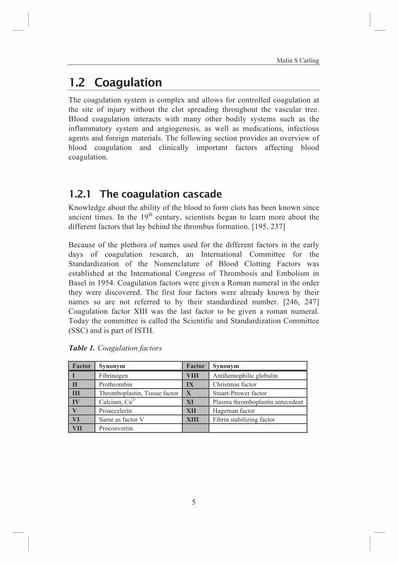
Because of the plethora of names used for the different factors in the early days of coagulation research, an International Committee for the Standardization of the Nomenclature of Blood Clotting Factors was established at the International Congress of Thrombosis and Embolism in Basel in 1954. Coagulation factors were given a Roman numeral in the order they were discovered. The first four factors were already known by their names so are not referred to by their standardized number. [246, 247] Coagulation factor XIII was the last factor to be given a roman numeral. Today the committee is called the Scientific and Standardization Committee (SSC) and is part of ISTH.
Table 1. Coagulation factors
Factor Synonym Factor Synonym I Fibrinogen VIII Antihemophilic globulin II Prothrombin IX Christmas factor III Thromboplastin, Tissue factor X Stuart-Prower factor IV Calcium, Ca2+ XI Plasma thromboplastin antecedent V Proaccelerin XII Hageman factor VI Same as factor V XIII Fibrin stabilizing factor VII Proconvertin

Aspects on bleeding and transfusion in elective orthopaedic surgery
6
Figure 2. Schematic overview of the coagulation cascade. ADP = Adenosine diphosphate, TF = Tissue factor, vWF = von Willebrand factor, APC = Apoprotein C, PS = Protein S, TM = Thrombomodulin, AT = Antithrombin, FVa = activated Factor V, FVIIa = activated Factor VII, FVIIIa = activated Factor VIII, FIXa = activated Factor IX, FXa = activated Factor X, FXIa = activated Factor XI

Malin S Carling
7
Primary hemostasis The coagulation cascade was previously divided into the intrinsic, platelet derived pathway, and the extrinsic, plasma coagulation factor-driven pathway. Even if this categorization is still in use, for instance when defining coagulation assays, a more explanatory way of describing the coagulation cascade is by dividing it into primary and secondary hemostasis. [15]
The endothelial cells in the blood vessel wall are designed so that they do not spontaneously initiate the coagulation cascade. The negatively charged platelets are repelled by an equally negatively charged glycocalyx on the endothelial cells. The endothelial cells produce a small amount of endogenous heparin that together with circulating antithrombin inactivates coagulation factors.
When a blood vessel injury occurs, the vessel constricts as a response to the injury. The antithrombotic glycocalyx is removed at the injury site and the platelets are not repelled but, instead, are pushed towards the vessel wall by fluid dynamics. Subendothelial collagen is exposed and the platelets are activated. The platelets are also activated by circulating thrombin and adenosine diphosphate (ADP). Von Willebrand factor (vWF) secures the platelets to the vessel wall. The platelets then change form and become irregular and form pseudopods, thereby increasing their surface area, on which platelet procoagulant receptors are exposed. The activated platelets form a monolayer over the injury and the coagulation cascade proliferates on the surface.
The activated platelet releases ADP, Ca2+, serotonin, vWF, factor V, factor XIII and fibrinogen, all substances that help the continuing thrombus formation.
Secondary hemostasis Injury to the endothelial cells exposes thromboplastin, also called tissue factor (TF). Tissue factor activates factor VII and, together, they form a complex that in turns activates factor V and factor X, which also form a complex at the site of injury. This FVa/FXa complex is called prothrombinase and it activates prothrombin into thrombin.
Thrombin is a strong procoagulant factor and its main role is to activate fibrinogen into fibrin, which forms the fibrin clot. It also activates platelets and other coagulation factors. Only a small amount of thrombin is needed to

Aspects on bleeding and transfusion in elective orthopaedic surgery
8
initiate coagulation on the surface of the vessel wall. In the next step, the propagation phase, hemostasis takes place on the activated platelets covering the injury.
The thrombin burst, where a large amount of thrombin is activated, and the amplified activation of coagulation factors, is due to enlarged surface of the activated platelets and the large amount of procoagulants expressed on that surface. The coagulation factors link with the surface through Ca2+ bonds. The large amount of thrombin can, in turn, convert large amounts of fibrinogen into fibrin, which forms the fibrin clot. Thrombin also activates FXIII, which stabilizes the clot by forming crosslinks with fibrin.
The surplus of activated factors is released to plasma and is inactivated by antithrombin. The anticoagulant properties of the uninjured vessel walls limit coagulation to the site of injury. For instance, circulating thrombin attaches to thrombomodulin on the vessel wall. This complex activates protein C and protein S which inactivates FV and FVIII and stops their activation of thrombin.
1.2.2 What affects coagulation? Many of the coagulation factors have catalytic functions in the coagulation cascade, and 30% of normal plasma concentration of these factors is assumed to be sufficient to maintain coagulation. Others, like fibrinogen, FXIII and platelets are consumed during coagulation and sufficient levels are critical for clotting. [15]
Patient related factors The most obvious patient-related factors are different types of bleeding disorders. The most common bleeding disorders are hemophilia A and B and von Willebrand disease, all of which are congenital. In 2012, the prevalence of hemophilia A was 7.1 per 100,000 inhabitants, for hemophilia B 1.7 per 100,000 inhabitants, and for von Willebrand disease one in 100 inhabitants. [16, 221]
Platelet dysfunction and thrombocytopenia also affect coagulation. Congenital platelet dysfunctions are rare and diverse. [113] Examples are Bernard-Soulier syndrome, which affects platelet adhesion to the vessel walls, and Glanzmann thrombasteni, which affects platelet binding with

Malin S Carling
9
fibrinogen. Various conditions that affect bone marrow generation and increase consumption of platelets, such as myelodysplastic syndromes, large alcohol consumption or drug-induced thrombocytopenia, cause thrombocytopenia.
In the blood vessel, platelets migrate towards the vessel wall because of hemodynamic and rheological factors. Hemoglobin levels influence this migration, and anemic patients may not have enough red blood cells to push the platelets to the vessel wall, thereby lowering the actual platelet count at the site of injury. [4] The hemoglobin concentration at which coagulation is affected is unclear. [131]
Blood coagulation is also temperature dependent. Even small changes in core temperature (<1°C) increase perioperative blood loss and increases transfusion rates. [187] However, studies are not conclusive and body temperature should be seen as just one of many ways to optimize the patient’s coagulation system. [52, 131]
In vitro studies suggest that coagulation is pH dependent as well. The TF-factor VII-complex has diminished activity in an acidotic environment. [156] The combination of low pH and low body temperature is even more detrimental to coagulation. Patients with large bleeding due to trauma are often cold because of the circumstances during and after trauma, and this is often combined with acidosis. In these cases it is important to reverse the low body temperature and acidosis to optimize coagulation before necessary surgery is carried out. [99]
Calcium is an important part of the coagulation cascade. A plasma concentration of 0.9 mmol/L appears to be required to ensure coagulation. [54] In trauma patients it has been shown that hypocalcemia on admission to hospital was associated with increased mortality and a larger risk of massive transfusion. Hypocalcemia may also be the result of massive transfusion since citrate, used in fresh frozen plasma (FFP) and platelet transfusion as an anticoagulant, binds Ca2+. [216]
Diet and other lifestyle factors may also have an impact on hemostasis. [138, 180] This has been widely studied in terms of relation to cardiovascular disease, but not much is known about how this affects bleeding and transfusion in a surgical setting. Fish oil, as a dietary supplementation to prevent atherosclerotic disease, has been studied, but no effect on bleeding or transfusion rates has been demonstrated. [239]

Aspects on bleeding and transfusion in elective orthopaedic surgery
10
High alcohol consumption can cause thrombocytopenia and liver cirrhosis. Thrombocytopenia in persons with high alcohol consumption both reduces the number of platelets and affects platelet aggregation. [94] Many pro- and anticoagulant factors are produced in the liver, so liver cirrhosis, not only on the basis of alcohol, can cause both bleeding and thrombotic disorders. Even if these patients have a high prothrombin time (PT) they should not be given procoagulants such as prothrombin complex concentrates (PCC) and, in conditions with increased risk of thrombotic events, such as surgery or immobilization, thrombosis prophylaxis should always be considered. [131, 143]
Iatrogenic factors Different drugs affect the coagulation system. Some are supposed to down-regulate the coagulation cascade by inhibiting coagulation factors or the platelets, while others have this as a side effect. This can cause difficulties if a patient on these medications requires acute or planned surgery or if the patient is subjected to major trauma.
The discovery of heparin in 1916 and the development of low molecular weight heparin (LMWH) were milestones in medicine. Heparin allows physicians to treat hypercoagulant and thrombotic diseases and it is also used to prevent thromboembolism in, for example, patients undergoing surgery and in atrial fibrillation.
Because LMWH are administered as subcutaneous injections, warfarin is preferred for long-term treatment. The disadvantages of warfarin are that it must be monitored regularly and that the drug interacts with other medications as well as different types of food. New oral anticoagulants (NOAC) have been developed that allow safe anticoagulation without monitoring. [199] Because NOACs have not been in use for long; the first NOAC still in use, dabigatran, was released in Sweden in 2008. Consequently, studies are needed of NOACs in combination with surgery, especially emergency surgery. [58, 64]
Platelet inhibitors are mainly used in treating patients with cardio- or cerebrovascular disease. The modern-day inhibitors are very potent, leading to larger perioperative bleeding compared to patients without platelet inhibition. [196] Because of the risk of stent thrombosis in patients having undergone percutaneous coronary intervention (PCI) with stent, discontinuation of antiplatelet therapy must be discussed with a cardiologist.

Malin S Carling
11
[37] Acetylsalicylic acid (ASA) is also potent, irreversibly inhibiting the platelets, and there is an effect on perioperative bleeding. [55] Whether this effect is clinically relevant is still debated.
Non-steroidal anti-inflammatory drugs (NSAID) are common over-the-counter drugs to treat conditions such as headaches, fever and pain. Like acetylsalicylic acid they inhibit platelet aggregation and hence reduce blood clotting. Depending on the specific pharmacodynamics properties of the NSAID, their influence on coagulation differs. New NSAID that selectively inhibits cyclo-oxygenase-2 have no impact on platelet aggregation. [158, 191]
Selective serotonin reuptake inhibitors (SSRI) increase the risk of bleeding and transfusion in surgery. [17, 164, 201] Activated platelets expel serotonin into plasma and it is used to help platelet aggregation. Platelets have a similar serotonin transporter to that present on presynaptic neuronal endings, so it is likely that the platelet transporter is inhibited by SSRI in a similar way as in the central nervous system. [150]
In patients experiencing blood loss, the first measure to take is to replace blood with intravenous fluids, colloids or crystalloids. This, together with consumption of coagulation factors caused by bleeding, may result in dilutional coagulopathy. Artificial colloids are known to affect blood clotting more than crystalloids and the reason might be inhibition of fibrin formation since the addition of fibrinogen reverses the effect of colloids on clot formation. [60, 63, 130, 160]

Aspects on bleeding and transfusion in elective orthopaedic surgery
12
Table 2. Antithrombotic drugs and their indications, a selection. [3]
Name Function Indication Heparin Indirect inactivation of
thrombin and FXa Heparin lock in indwelling venous catheters, anticoagulant during extracorporeal circulation
Low Molecular Weight Heparin
Inhibition of activated factor X
Venous thromboembolism, thrombosis prophylaxis
Warfarin Inhibition of vitamin K-dependent coagulation factors II, VII, IX, X
Venous thromboembolism, thrombosis prophylaxis, anticoagulation in patients with valve replacement or atrial fibrillation
Dabigatran Thrombin inhibitor Venous thromboembolism, thrombosis prophylaxis, anticoagulation in patients with atrial fibrillation
Rivaroxaban Inhibition of activated factor X
Venous thromboembolism, thrombosis prophylaxis, anticoagulation in patients with atrial fibrillation, prophylaxis after acute coronary syndrome
Apixaban Inhibition of activated factor X
Venous thromboembolism, thrombosis prophylaxis, anticoagulation in patients with valve replacement or atrial fibrillation
Acetylsalicylic acid
Inhibition of platelet aggregation through cyclo-oxygenase
Acute myocardial infarction, prophylaxis against cardio-vascular complication after acute coronary syndrome, prophylaxis against cerebrovascular disease
Dipyridamol Inhibition of platelet aggregation
Secondary prevention of cerebrovascular disease
Clopidogrel Inhibition of platelet aggregation through inhibition of the P2Y12 receptor
Prevention of atherothrombotic events in patients with myocardial infarction or acute coronary syndrome, treated with or without PCI, anticoagulation in patients with atrial fibrillation
Prasugrel Inhibition of platelet aggregation through inhibition of the ADP receptor
Prevention of atherothrombotic events in patients who have had PCI with stent
Ticagrelor Inhibition of platelet aggregation through inhibition of the ADP receptor
Prevention of atherothrombotic events in patients with myocardial infarction or acute coronary syndrome, treated with or without PCI or coronary bypass surgery

Malin S Carling
13
1.2.3 Fibrinogen The main function of fibrinogen in the coagulation cascade is as the precursor to fibrin, which in turn forms the fibrin clot.
Figure 3. Fibrinogen hexamer. With permission from the Protein Data Bank in Europe. [83]
Fibrinogen is composed of three homologous polypeptide chains – A!, B" and # – forming two identical sets joined together to make a dimer structure. [163] Fibrinogen is primarily expressed and formed in liver cells and it is secreted in its hexamer form. [190] Cleavage of the A!-chain by thrombin initiates the formation of the fibrin clot at the site of bleeding. [162] The structure of the fibrin clot formed is affected by the amount of thrombin available – low levels of thrombin create a tighter fibrin clot. [26] The fibrinogen or fibrin #-chain contains the site for FXIII cross-linking. [209]
The normal fibrinogen plasma concentration is 2.0-4.5 g/L and levels >1.0 g/L are considered enough to maintain hemostasis. [218] In the bleeding patient, recent guidelines suggest that fibrinogen levels should be kept above 1.5-2.0 g/L. [131]
Congenital afibrinogenemia is a rare disease (prevalence 1 in 1,000,000) that usually manifests with umbilical cord bleeding in the neonatal period. [53] In contrast to hemophilia patients, bleeding in different joints is not common in patients with afibrinogenemia. Instead, bleeding occurs in the skin, genitourinary and gastrointestinal tract, and intracranial hemorrhage is a major cause of death in these patients. [178] Fibrinogen is vital for pregnancy and first trimester abortion is common in women with afibrinogenemia. [53]
Patients with hypofibrinogenemia or dysfibrinogenemia are usually asymptomatic. [149, 152] Dysfibrinogenemia patients may experience both bleeding and thromboembolic complications. [152] Defective thrombin

Aspects on bleeding and transfusion in elective orthopaedic surgery
14
binding, leading to increased levels of thrombin, and an abnormal fibrin clot resistant to fibrinolysis are the probable mechanisms behind this paradoxical clinical presentation. [53]
In the bleeding patient, fibrinogen is consumed and it is the first coagulation factor to reach critically low levels in massive blood loss. [98] Compared to some of the other coagulation factors, fibrinogen is used up once it has been transformed into fibrin and incorporated in the fibrin clot. Sufficient plasma concentration is therefore necessary to maintain hemostasis in the bleeding patient. [131, 216]
Fibrinogen is an acute phase reactant and fibrinogen plasma concentration increases for instance due to trauma, surgery, inflammation or infection. [173, 188, 207, 227] In a study comparing postoperative fibrinogen plasma concentration between patients who did versus did not receive intraoperative fibrinogen supplementation, there was no statistical difference between the two groups. [213]
Fibrinogen derived from human plasma is commercially available with the indication congenital hypo- or afibrinogenemia. Today, fibrinogen concentrate is mainly used in patients with acquired hypofibrinogenemia because of massive blood loss and consumption due to trauma or surgery as described above, and its use is increasing. [212] The development of recombinant fibrinogen is desired and is advancing. Using a human cell line transfected with optimized cDNA encoding the fibrinogen Aα, Bβ and γ chains, fibrinogen can be produced in a controlled setting. [186] In an ex vivo setting, recombinant fibrinogen has been shown to have similar pro-coagulant characteristics as plasma-derived fibrinogen on blood samples from cardiac surgery patients. However, further studies are needed before recombinant fibrinogen can be used in clinical practice.
1.2.4 Factor XIII Coagulation factor XIII is a plasma pro-transglutaminase with multiple functions not only in coagulation but also in a wide range of physiological and pathological processes. Factor XIII exists in an extracellular form, circulating in plasma, and an intracellular form. The main function of FXIII is to cross-link with fibrin to stabilize the fibrin clot and protect it from fibrinolysis. [209]

Malin S Carling
15
Figure 4. FXIII tetramer. With permission from the Protein Data Bank in Europe. [83]
Plasma FXIII (pFXIII) consists of two potentially active, catalytic A subunits (FXIII-A) and two inhibitory B subunits (FXIII-B), together forming a tetramer FXII-A2B2. Factor XIII-A is a transglutaminase consisting of 732 amino acids and is mainly synthesized by cells of bone marrow origin. However, when bone marrow function is impaired cells of unknown origin take over the production of FXIII-A. Factor XIII-B is a glycoprotein consisting of 641 amino acids and is synthesized in the liver. [248] The two subunits form their tetrameric complex in the circulating blood. In plasma, FXIII-A circulates only in its fully complexed form, while approximately 50% of FXIII-B circulates in free, non-complexed form. [252] Factor XIII is effectively activated in plasma only on the surface of newly formed fibrin. [206]
Cellular FXIII (cFXIII) is a dimer consisting of only FXIII-A, FXIII-A2; it can be found mainly in platelets and monocytes/macrophages but also in chondrocytes, osteoblasts and osteocytes. [6, 171] As opposed to pFXIII, cFXIII is activated by an increase in cellular Ca2+, a non-proteolytic mechanism. [168]
Congenital FXIII deficiency is a rare bleeding disorder (estimated prevalence 1:1,000,000 to1:5,000,000). [72] Mutations occur in the FXIII-A genome, more than 100 are identified so far, causing either absence of FXIII or
Aspects on bleeding and transfusion in elective orthopaedic surgery
16
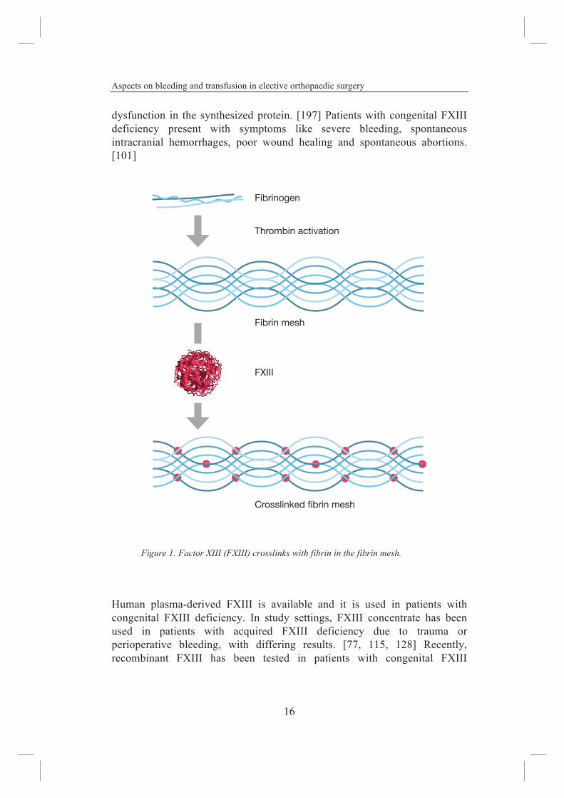
dysfunction in the synthesized protein. [197] Patients with congenital FXIII deficiency present with symptoms like severe bleeding, spontaneous intracranial hemorrhages, poor wound healing and spontaneous abortions. [101]
Figure 1. Factor XIII (FXIII) crosslinks with fibrin in the fibrin mesh.
Human plasma-derived FXIII is available and it is used in patients with congenital FXIII deficiency. In study settings, FXIII concentrate has been used in patients with acquired FXIII deficiency due to trauma or perioperative bleeding, with differing results. [77, 115, 128] Recently, recombinant FXIII has been tested in patients with congenital FXIII
Fibrinogen
Thrombin activation
Fibrin mesh
Crosslinked fibrin mesh
FXIII

Malin S Carling
17
deficiency, with promising results. [106, 123] The recombinant FXIII is manufactured in yeast cells and contains no mammalian or human products, thus minimizing the risk of transferring infectious, or other diseases. [146]
1.2.5 Coagulation assays Various laboratory assays are used to investigate a patient’s coagulation status. The most common are PT and activated partial thromboplastin time (aPTT). These two tests are often routinely taken to assess preoperative coagulation status in patients scheduled for elective surgery. [126, 244] Other ways of measuring a patient’s coagulation capacity are by measuring individual coagulation factors known to influence perioperative hemostasis, such as fibrinogen, and also by using point-of-care (POC) analyses.
Blood coagulation is a dynamic process and conditions can change quickly. The coagulation assays mentioned above are time consuming and are passé by the time the results arrive in the emergency or operating room. Both PT and aPTT only measure part of the coagulation cascade and the measurement ends at clot formation. Point-of-care analyses, such as viscoelastic tests and platelet aggregation assays, can be used to obtain faster information about a patient’s coagulation status, and also provide information on clot quality and fibrinolysis. At the same time, POC analyses require knowledge and experience in how to interpret the results. [69, 245]
Prothrombin time Prothrombin time measures the extrinsic pathway. The assay is affected by the levels of the vitamin K-dependent coagulation factors II (prothrombin), V, VII and X, as well as very low levels of fibrinogen. [124] The test was first described in 1935 by Quick, therefore initially called Quick’s time, but the name was later changed to PT. [184, 185] Different reagents are used when measuring PT, giving different results especially in patients on oral anticoagulation treatment. Prothrombin time is therefore standardized as an International Normalized Ratio (INR). In Scandinavia, laboratories use the method developed by Owren, which only measures levels of factors II, VII and X. [124] Test results for the two tests in the normal range are similar. [100]
A patient with a PT of <1.5 is considered to have a low risk of bleeding during and after surgery. [229] A recent study suggests that the best cut-off point for avoiding major bleeding during surgery is 1.1, which is notably lower than previous recommendations. [223]
Aspects on bleeding and transfusion in elective orthopaedic surgery
18
Activated partial thromboplastin time Activated partial thromboplastin time is a test that roughly studies the intrinsic pathway of the coagulation cascade. In 1913, a test designed to confirm the diagnosis of severe hemophilia, called whole blood clotting time (WBCT) was developed. [139] The test was further refined in steps, until it finally became the test used today. [135, 183] Unlike WBCT, aPTT uses platelet-free plasma from blood-samples taken in citrated tubes. Samples are recalcified, and partial thromboplastin is added. The, often automated, photo-optical apparatus then measures the time from when the test is initiated until a clot is formed. In order to speed up the time before the clot forms, an activator is added, hence the name activated PTT. Activated partial thromboplastin time is measured in seconds (s) and the reference interval for adults is 30-42 s.
Functional deficits in factors VIII, IX, XI, XII, prekallikrein and high molecular weight kininogen, as well as severe deficits in factors V, X, II and fibrinogen can affect aPTT measurements. [124]
In the latest guidelines, neither PT nor aPTT are recommended for routine preoperative testing of a patient’s coagulation status. [131]
Fibrinogen Fibrinogen plasma concentration can be measured in different ways. The most common is the method described by Clauss in 1957. [49] This method actually measures the clotting time in diluted plasma and compares that time with reference plasma with known fibrinogen concentration. The test sample is saturated with thrombin so that the amount of thrombin in the original sample does not influence the clotting time. The method of Clauss is considered the most frequently used assay.
The PT-derived fibrinogen assay measures the patient’s PT in platelet-poor plasma and compares it to the PT in plasma dilutions with known fibrinogen concentration.
To measure protein concentration, various immunological assays, like ELISA or the immune-nephelometric method, may be used. In congenital dysfibrinogenemia there is usually a discrepancy between fibrinogen activity and antigen level. The fibrin clot may also be extracted, cleaned, dried and

Malin S Carling
19
then weighed. This is a time consuming test not often used in the clinical setting.
The different assays are not comparable in terms of measured fibrinogen levels. [148] Fibrinogen assays are also influenced by pharmaceuticals, hemodilution and by the level of fibrinogen in the blood sample. [62, 142, 148]
In the acute situation, fibrinogen levels may be estimated using viscoelastic methods such as thromboelastography or thromboelastometry. There is a correlation between fibrinogen plasma concentration, as measured by Clauss, and thromboelastometry/thromboelastography results, but the viscoelastic results are not yet fully standardized. [159, 214, 231]
Factor XIII The main reasons for analyzing FXIII are to measure FXIII activity in patients with suspected FXIII deficiency and to monitor substitution therapy in patients with congenital FXIII deficiency. [119] There are many different assays, both quantitative and qualitative and, to diagnose a deficiency the assays should be used in combination in order to reach the correct diagnosis and classification. [127]
Quantitative assays activate FXIII with thrombin and Ca2+. In order to eliminate interfering fibrin formation, various measures such as high plasma dilution and fibrin polymerization inhibitory peptide, are used. Amine incorporation assays measure the binding of amine to FXIIIa. This method is highly sensitive and can detect a FXIII activity as low as 1% but the method is time-consuming and, because of the many steps involved the method cannot be automated and standardized. [119]
The recommended screening test for FXIII activity is a quantitative assay based on ammonia release from FXIIIa. It was first described in 1969. [243] Modern assays measure a substrate protein that captures the released ammonia, and the consumption of the protein can be measured with photometric methods. [67, 117] The method is quick, automated and reproducible. Its drawbacks are the low sensitivity, and that it needs a parallel blank sample that can correct for the ammonia produced in the plasma from sources other than FXIIIa. [10]

Aspects on bleeding and transfusion in elective orthopaedic surgery
20
The original test for the clot stabilizing protein was to create a fibrin clot and then to try to dissolve it in a urea or acid solution. [132] Clot solubility tests are still in use today, but because the test is not standardized – different clot activators, clot formation time, solvent, etc. are used – the test is not recommended for testing for FXIII deficiency testing. [127]
In patients with a suspected congenital or acquired FXIII deficiency a low FXIII activity on a quantitative assay requires further investigation into the type of FXIII deficiency present. [127]
In the bleeding patient, FXIII deficiency can be assumed to result from consumption and a fast evaluation of FXIII may be critical. Unlike measurement of fibrinogen, where there is a correlation between regular fibrinogen plasma concentration assays and viscoelastic tests, no such correlation is seen between viscoelastic tests and regular measurement of FXIII activity. [81]
Viscoelastic tests Thromboelastography was designed by H Hartet and described in an article in 1948. [89] The idea is that a blood sample is placed in a cup into which a pin is inserted. The cup oscillates and as the blood coagulates the pin starts to move along with the cup, figure 6. This movement increases as the viscosity of the blood sample increases, and this is recorded and analyzed. (Figure 7) Today, two different devices using the same principle of blood viscosity as a means to measure coagulation are in use – the Thromboelastograph® (TEG®) which is based on Hartet’s original design, and Thromboelastometry® (ROTEM®), which is a further development using the same principle as thromboelastography. [147]
Sonoclot®, which measures the changes in mechanical impedance using ultrasound, is another bedside device first described by von Kaulla et al. in 1975. [238] The set-up is similar to thromboelastography and thromboelastometry, with the difference that the measuring pin moves up and down in the blood sample.
This section will focus on thromboelastometry, since it is the device used in Paper IV. However, thromboelastometry, thromboelastography and Sonoclot® produce largely comparable results.
Malin S Carling
21
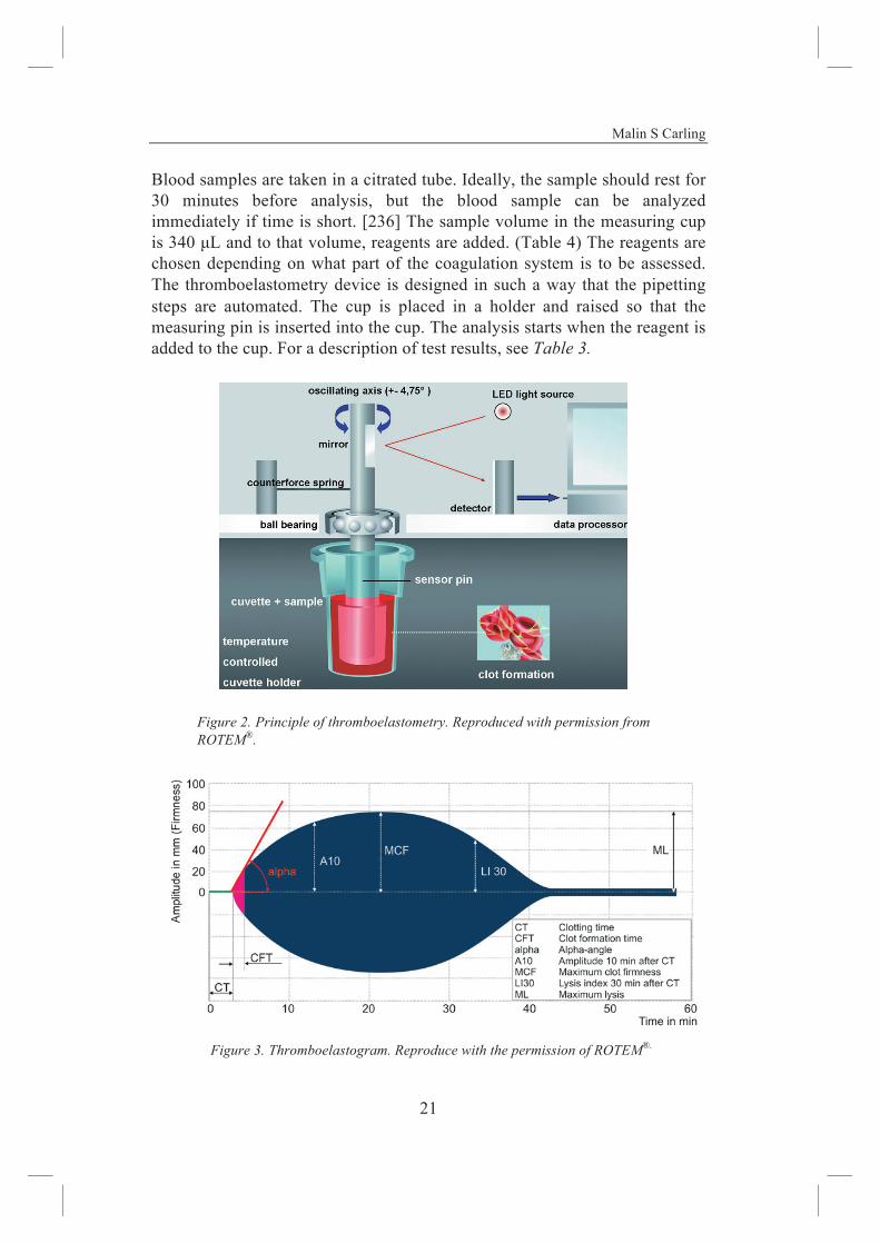
Blood samples are taken in a citrated tube. Ideally, the sample should rest for 30 minutes before analysis, but the blood sample can be analyzed immediately if time is short. [236] The sample volume in the measuring cup is 340 µL and to that volume, reagents are added. (Table 4) The reagents are chosen depending on what part of the coagulation system is to be assessed. The thromboelastometry device is designed in such a way that the pipetting steps are automated. The cup is placed in a holder and raised so that the measuring pin is inserted into the cup. The analysis starts when the reagent is added to the cup. For a description of test results, see Table 3.
Figure 2. Principle of thromboelastometry. Reproduced with permission from ROTEM®.
Figure 3. Thromboelastogram. Reproduce with the permission of ROTEM®.

Aspects on bleeding and transfusion in elective orthopaedic surgery
22
Because of the short time from blood sample to test result, thromboelastometry has become a widely used tool to monitor coagulation in the bleeding patient. In the latest guidelines from the European Society of Anaesthesiology (ESA) on management of severe perioperative bleeding the POC coagulation monitoring with viscoelastic tests is recommended. [131]
Thromboelastometry and thromboelastography are known to be user dependent, results often vary, and test accuracy has not been widely studied. [47, 104] Reference ranges for the different assays have been developed, but results should be analyzed and acted upon based on the combined results from the assays. [134]
Table 3. Test results
Result Description Reference value Clotting time – CT Time from addition of start
reagent until clotting begins INTEM: 137-246 s EXTEM: 42-74 s FIBTEM: 43-69 s
Clot formation time – CFT Time from CT to when a clot is formed
INTEM: 40-100 s EXTEM: 46-148 s
Amplitude 10 minutes after CT – A10
INTEM: 44-68 s EXTEM: 43-65 s FIBTEM: 9-24 s
Maximum clot firmness – MCF
Maximum vertical amplitude of the graph
INTEM: 52-72 mm EXTEM: 49-71 mm FIBTEM: 9-25 s
Maximum lysis – ML Amount of lysis INTEM: 0-12 % EXTEM: 0-18 %
s = seconds, mm = millimeter

Malin S Carling
23
Table 4. List of assays used in thromboelastometry analysis, its reagents and the interpretation of test results.
Test Reagent Interpretation INTEM Ca2+, phospholipids, ellagic
acid Coagulation through the intrinsic pathway
EXTEM Tissue factor Coagulation through the extrinsic pathway HEPTEM Ellagic acid, heparinase The effect of heparin in heparinazed patients;
compare HEPTEM with INTEM FIBTEM Cytochalasin D, tissue factor Coagulation without platelet activation APTEM Aprotinin, tissue factor Faster detection of fibrinolysis
Platelet aggregation Platelet function is essential for blood coagulation but is difficult to evaluate. The standard platelet count only tells us the amount of circulating platelets but does not give any information on the quality and function of the platelets. As mentioned above, many different factors affect platelet function and, at least in patients prescribed platelet inhibitors, it is important to assess thrombocyte function before surgery.
Various POC devices are available to assess platelet drug inhibition. Impedance aggregometry and light transmission aggregometry are two of the methods to measure platelet function in the clinical setting. Whole blood is used in order to avoid the potential activation of the platelets that may occur when the sample is centrifuged to form platelet-rich plasma. In impedance aggregometry a small electric current is transmitted through the blood sample between two platinum electrodes and, as the platelets aggregate on the electrodes, the electric resistance increases. Cardinal and Flower first described this method in 1979. [41] Light transmission aggregometry measures changes in light as platelets aggregate. Today, there are several POC devices available that are similar in design and test results, such as Multiplate® (Roche Diagnostics, Basel, Switzerland), VerifyNow® (Accumetrics, San Diego, CA, USA) or Chrono-Log® (Chrono-log, Havertown, PA, USA).

Aspects on bleeding and transfusion in elective orthopaedic surgery
24
1.3 Patient blood management In 1628, the British physician William Harvey published his book on blood circulation. He described how blood was pumped through the body by the heart and flowed through the vessels. This knowledge formed the foundation for further research on the circulatory system, bleeding and transfusion. There is a debate on who performed the first human-to-human blood transfusion. [136]
Doctor Richard Lower, UK, experimented on dog-to-dog transfusion in 1665, as described in several recordings in the Journal Book of the Royal Society. In 1667 he made a successful dog-to-man blood transfusion. Lamb blood was also used for transfusion but, because of several complications with this practice, the Pope issued a ban on the procedure in 1679. [136]
The first documented human-to-human blood transfusion was performed on 22 December 1818, and was published in 1819 by a British physician and obstetrician named James Blundell. As an obstetrician he saw the consequences of post-partum hemorrhage and saw blood transfusion as a way to treat this condition. [28] From that point on the practice of blood transfusion, although rare at first, became in use.
Two discoveries made blood transfusion feasible and integrated in everyday clinical practice. The first was the discovery of the AB0 blood groups in 1901 by Landsteiner. [133] The second was the introduction of sodium citrate to prevent the blood from clotting thereby enabling storage and blood-banking. [141] Two World Wars led to further development in transfusion practices and how to organize blood-banking and donors so as to ensure a constant supply of blood products. [31]
At the start of transfusion history, whole blood was given. Today fresh whole blood transfusion has seen a revival in war medicine. The harsh conditions in medical facilities close to the battlefield do not provide the infrastructure needed for the modern blood-bank. At the same time, massive hemorrhage is one of the leading causes of death in warfare, and prompt transfusion can be life-saving. Fresh whole blood transfusion has therefor been in practice in several war situations with good results. [20, 217]
Today, blood transfusions are a part of the clinical setting. In Sweden, approximately 100,000 people receive blood transfusion every year and, in 2014, 462,629 blood units were donated. [1] However, the use of blood transfusions is declining, both because of increasing knowledge about

Malin S Carling
25
potential risks associated with transfusion but also because of better understanding about when and how to transfuse.
1.3.1 Transfusion and fluid substitution In modern transfusion medicine, whole blood is divided into three components; erythrocytes, plasma and platelets, and transfusions are based on what the patient needs. Donated whole blood is centrifuged and the blood components are layered and thereafter stored into different storage bags.
Red blood cells Red blood cells (RBC) is the most common blood component to be transfused. After separation from the other blood components, RBC are stored in an additive solution, in +2° - +6° C for up to 42 days. [1] Over time, the quality of the RBC decreases and changes in RBC takes place. This phenomenon is referred to as storage lesions and there are discussions on how they affect patients. [14, 56]
Plasma Plasma transfusion has many potential advantages in the bleeding patient. It is a source of coagulation factors, a volume expander and a means to reverse the anticoagulant effect of drugs. Plasma is obtained either from whole blood donors, through separation, or from apheresis with plasma extracted from one donor. [21] Most of the plasma is frozen to -18°C within 6 to 8 hours after donation and is labeled fresh frozen plasma (FFP). When needed, FFP is thawed. Plasma can also be stored as liquid, non-frozen plasma in +2° - +6°C for up to 14 days. [170] However, studies on FFP show that it has little effect on bleeding and transfusion in patients undergoing surgery. [129]
Platelets Platelets are responsible for the primary hemostasis and are first to arrive at the site of injury to the vessel wall. They are easily activated however and donated platelets, platelet concentrate, are stored at room temperature and in constant agitation. [167] Because of the risk of bacterial growth platelets cannot be stored for more than 5 to 7 days. [234] There are two methods to

Aspects on bleeding and transfusion in elective orthopaedic surgery
26
prepare platelet concentrate; either pooled buffy coat platelets extracted from 4 to 6 whole blood donors or apheresis platelets extracted from one donor. The advantage with apheresis platelets is that the recipients are exposed to less antigen stimulation. [234]
Fluids The body is adaptable and can function with a certain amount of anemia as long as there is a sufficient blood pressure to ensure tissue perfusion. Consequently, the recommendation is to start replacing blood loss with crystalloid or colloid fluids. [131, 216] It is important to be aware that colloids can affect blood coagulation. [63]
1.3.2 Hemostatic drugs There are many different drugs that can be given before, during and after surgery to boost blood coagulation. Depending on the type of surgery and the patient’s coagulation status the drugs can reduce perioperative bleeding and transfusion requirements.
Table 5. List of hemostatic agents, information derived from www.fass.se [3]
Hemostatic drug Sales name Content Indication Prothrombin complex concentrate (PCC)
Ocplex® , Confidex®
Human FII, FIX, FVII, FX, Protein C, Protein S
Reversal of anticoagulation with vitamin K inhibitors
Fibrinogen Riastap® Human fibrinogen Congenital fibrinogen dysfunction or hypofibrinogenemia
FXIII Cluvot® Human FXIII Congenital FXIII deficiency
Recombinant FVIIa
Novoseven® Eptacog alfa Congenital or acquired hemophilia
Desmopressin Octostim® Desmopressin Congenital or acquired platelet dysfunction
Tranexamic acid Pilexam® , Cyklokapron®
Tranexamic acid Prevention and treatment of hyperfibrinolysis
Local hemostatics For instance Tachosil®, Tiseel®, Evisel®
Fibrinogen, thrombin, aprotinin, FXIII
Local hemostasis

Malin S Carling
27
Coagulation factors Fresh frozen plasma contains a variety of coagulation factors, but their concentration in plasma might not be sufficient to restore coagulation. Concentrated, specific coagulation factors are therefore available. Many of them are primarily produced to treat patients with coagulation disorders, but they are also used to treat acquired coagulation disorders/dysfunctions.
Prothrombin complex concentrate (PCC) contains vitamin K-dependent coagulation factors and is indicated for patients on oral anticoagulation with vitamin K inhibitors. [177] When reversing the anticoagulant effect in trauma or surgery patients, PCC should be administered in combination with vitamin K. The effect of PCC lasts for 6 to 8 hours and by then the reversal with vitamin K should have taken place.
In patients with traumatic or surgical bleeding, the most critical coagulation factor is fibrinogen. Several studies show that fibrinogen substitution in patients with major bleeding and signs of fibrinogen deficit reduces bleeding and transfusion requirements. [61, 232, 249]
Factor XIII is suggested to reduce bleeding and improve coagulation in surgery patients, even though results vary. [85, 115, 128, 203] In vitro studies show that FXIII increases clot firmness, but these results have not been confirmed in a clinical setting. [230, 231]
Recombinant factor VIIa (Novoseven®) has been shown to reduce bleeding and transfusion requirements, but not mortality, in trauma patients. [93] In cardiac surgery patients, recombinant FVIIa reduces bleeding but increases the risk of thrombotic events. [74] Results in studies vary however, and current guidelines do not suggest routine use of recombinant factor VIIa other than as a last resort when other treatments have failed to control the bleeding. [131, 216]
Desmopressin is a synthetic hormone that increases the release of von Willebrand factor on endothelial cells, thereby increasing platelet activation and aggregation. The intended use for desmopressin is in patients with mild hemophilia type A and von Willebrand disease. Studies show a small effect on transfusion requirements in surgery patients treated with desmopressin. [51]

Aspects on bleeding and transfusion in elective orthopaedic surgery
28
Tranexamic acid Dr Blundell, who established blood transfusions, was an obstetrician trying to save women from post-partum hemorrhage. [28] Tranexamic acid (TXA) was also developed by obstetricians, but they were looking for a treatment for women with heavy menstrual bleeding. In the 1960s two research groups, in Japan and Sweden, simultaneously found the antifibrinolytic properties of trans-4-aminomethyl-cyclohexanecarboxylic acid, in short tranexamic acid. [155, 175] Another early use was to reduce bleeding during surgery in patients with bleeding disorders, but soon more and more applications for TXA were uncovered. [225] Today, TXA is used in different surgical settings such as gynecology and obstetrics, orthopaedics and cardiac surgery. It is also used as a local hemostatic in oral extraction and in nose bleeds.
Figure 4. Working mechanism of tranexamic acid. T-PA=tissue plasmin activator.

Malin S Carling
29
Tranexamic acid binds to, and blocks, the plasminogen lysine binding site. Plasminogen is the precursor to plasmin, which facilitates the decomposition of the fibrin clot by fibrinolysis. When TXA hinders plasminogen from binding to fibrin at the lysine binding site, plasminogen cannot be activated to plasmin and fibrinolysis is slowed down. [225]
Both intravenous and topical TXA are known to reduce perioperative bleeding and the number of transfusions in many different surgical settings where major blood loss is expected. [38, 73, 122, 225] Tranexamic acid is also a cheap drug, and by reducing the number of transfusions, the use of TXA in surgery is highly cost-effective. [111] The use of TXA has also been evaluated in trauma patients in a large randomized, placebo-controlled study, CRASH-2, including 20,211 patients. The results showed that mortality independent of cause and mortality from hemorrhage was significantly lower in patients receiving TXA compared to placebo. [176]
Questions have been raised on the safety of TXA with regards to thromboembolic events. The CRASH-2 study showed no difference in death from vascular occlusion. [176] A meta-analysis from 2012 on knee arthroplasty patients showed no increase in thromboembolic disease in patients receiving TXA. [250] In patients with ongoing thromboembolic disease and risk factors for thromboembolic events TXA should be given with caution and more studies are required in these patient groups. [131]
Local hemostatic agents There are different types of agents for obtaining a local hemostatic effect. Some create a larger surface area on which coagulation can occur, while some also contain coagulation factors. [222] The role of local hemostatic agents in orthopaedic surgery is not established and studies differ as to the effect on bleeding and transfusion requirements. [8, 25, 42, 140]
1.3.3 Indications for transfusion As stated above, RBCs transport oxygen to the tissues and assist somewhat in blood coagulation. Transfusion should therefore be given when the patients lacks sufficient oxygen transportation and is affected by anemia. Transfusions should also be considered as one of many measures to ensure sufficient oxygen transportation.

Aspects on bleeding and transfusion in elective orthopaedic surgery
30
Not many years ago, RBC transfusions were seen as a way to replenish the patient and also to speed up recovery. Transfusions were given liberally, and apart from rare immunological reactions, transfusions were considered safe and without side effects. In the 1980s, this perception changed with the appearance of HIV and several transfusion-related transmissions of the virus. Even though screening tests for HIV had been developed, the debate about the safety and necessity for transfusion had begun. [204]
It is important to differentiate between the bleeding patient and the non-bleeding patient. In a bleeding patient, indications for transfusion are much more liberal, and in massive-bleeding patients, other methods than Hb levels are used to determine how the patient should be transfused. [131]
The bleeding patient – intraoperative During surgery, bleeding is ongoing and the blood that is lost contains not only oxygen transporters but also important coagulation factors. The surgical trauma creates a consumption of coagulation factors and triggers fibrinolysis. [179]
Under normal circumstances there is an overcapacity in oxygen transport, which means that the patient can handle a certain amount of bleeding as long as blood is substituted with fluids to maintain sufficient tissue perfusion. [23, 131] Both colloids and crystalloids can be used, but it is important to consider the anticoagulant effect in synthetic colloids.
With continuing bleeding, the blood loss reaches critical levels and transfusions are required to maintain tissue oxygenation. In a bleeding patient recommendations are to maintain the following parameters: [23, 131, 216]
• Hb level > 70-90 g/L • Platelet count >100 x 109 • Fibrinogen >2.0 g/L • Normothermia • Normal pH ~7.2 • Calcium concentration, Ca 2+ > 1 mmol/L
The rule of thumb is to give the patient what is lost through bleeding, which is a mixture of blood products that equals whole blood. In massive bleeding, transfusion packs are used with a predefined ratio of blood products, for instance 4 units of RBC, 4 units of FFP and one unit of platelets, or one unit

Malin S Carling
31
each of RBC, FFP and platelets. [131, 216] The optimal relationship between the different blood products is not fully established and more studies are needed.
In a patient with an ongoing large bleeding, fibrinogen is consumed and reaches critical levels. Early supplementation with fibrinogen concentrate is therefore needed, so to avoid compromising formation of the fibrin clot. The recommendation is to give 2-4 g of fibrinogen concentrate to a patient that requires large transfusion. [131, 216] Often fibrinogen is part of a transfusion pack.
It is not only critical to transfuse the right blood products; timing is also important. In order to maximize the effect of a transfusion pack, in combination with hemostatic drugs such as fibrinogen, patients need to be given the transfusions all together and as fast as the patient can tolerate. Red blood cells are needed for oxygen transport but also for helping platelets to migrate towards the vessel wall. Fibrinogen concentrate needs the coagulation factors in FFP to become activated to fibrin. Platelets are required to initiate coagulation and as a scaffold for propagation of clot formation. [23]
Trauma induced coagulopathy (TIC), pathophysiology and treatment have attracted increasing interest. [13] In trauma patients, the nature of the injury mechanism affects coagulation. Body temperature is reduced. Acidosis occurs due to tissue damage and compromised tissue perfusion. Fracture and large wounds creates major activation of the coagulation cascade with consumption and a subsequent reduction in coagulation factors. Massive bleeding also reduces coagulation factors and compromise tissue oxygenation through hypotension and anemia. [40]
Non-bleeding patients – postoperative A patient in a postoperative setting has a different need for coagulation management than in the intraoperative setting. The first large-scale attempt to establish transfusion triggers in critical care was the Transfusion Requirement in Critical Care trial, published in 1999. [96] In the study, 838 patients were either assigned to a restrictive transfusion strategy or to a liberal transfusion strategy. The restrictive transfusion group had an Hb threshold for transfusion of 70 g/L, while the liberal transfusion group had a threshold of 100 g/L. The study showed no difference in mortality and morbidity between

Aspects on bleeding and transfusion in elective orthopaedic surgery
32
these groups and the number of RBC units transfused were 54% lower in the restrictive transfusion group.
This breakthrough study began a debate on when to transfuse. Several randomized studies have been carried out, reaching similar conclusion. A study on hip fracture patients saw no difference in death or ability to walk across a room 60 days after surgery between a liberal or restrictive transfusion protocol. [45] Similarly, a post-hoc analysis on elective hip or knee arthroplasty patients saw no difference in morbidity and mortality as well as Quality of Life and fatigue scores depending on transfusion regiment. [211] A Cochrane review from 2012 suggests that in non-bleeding patients without acute coronary heart disease, Hb levels of 70-80 g/L do not require RBC transfusion. [44] However, these suggestions were recently challenged by a randomized study in cardiac surgery patients where a more liberal transfusion regimen, Hb <90 g/L compared to Hb <75 g/L, was associated with lower mortality. [165]
1.3.4 Risks associated with transfusions Transfusion of blood products may be lifesaving but may also be associated with increased morbidity and mortality in patients undergoing surgery. [39, 70] It is however, difficult to distinguish between the risk of the underlying cause, for example excessive bleeding, and the risk of the blood transfusion per se. Known risks associated with transfusion are transmission of infectious microorganisms, immunological reactions and transfusion-related acute lung injury (TRALI). Today, measures are taken to minimize these hazards but patients still experience adverse effects from transfusions.
The transmission of infections through transfusions has been known since the 1940s and many measures, including screening and control of blood donors, have been taken to minimize these risks. [166] In modern transfusion practice, these measures have reduced the risk to extremely low levels.
As part of the procedures relating to transfusions, immunological testing of the recipient and the blood given is performed. Since the discovery of the AB0 blood groups, several other blood groups, like Rhesus antigen, have been revealed. In an acute situation, where there is no time for sufficient blood group testing, group 0 RBC and AB plasma should be used. [179]
Transfusion-related acute lung injury is nowadays considered a major cause of adverse events following transfusion. The first cases were described in
Malin S Carling
33
1983, and the connection with HLA antibodies was described, but it was not until 2003 and 2004 that the condition was defined. [151, 181] If acute lung injury (ALI) occurs within six hours of a plasma-containing blood component transfusion in a patient without previous ALI, it is considered to be TRALI. The definition for ALI is new onset hypoxemia (blood oxygen saturation <90% on room air), bilateral pulmonary infiltrates on chest x-rays, and without volume overload.
TRALI is mostly associated with transfusion of FFP or pooled platelets, especially if the blood donor is female. [151]
Studies indicate an increased risk of postoperative infection in patients who have received perioperative transfusion. [224] Other studies indicate an increased risk of recurrence of cancer in patients with perioperative transfusion at the first cancer surgery. The rationale behind these findings could be a transfusion-related immune modulation, but this theory is debated. [46, 235]
1.3.5 Measures to prevent transfusion
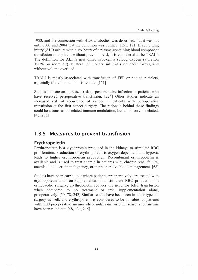
Erythropoietin Erythropoietin is a glycoprotein produced in the kidneys to stimulate RBC proliferation. Production of erythropoietin is oxygen-dependent and hypoxia leads to higher erythropoietin production. Recombinant erythropoietin is available and is used to treat anemia in patients with chronic renal failure, anemia due to certain malignancy, or in preoperative blood management. [68]
Studies have been carried out where patients, preoperatively, are treated with erythropoietin and iron supplementation to stimulate RBC production. In orthopaedic surgery, erythropoietin reduces the need for RBC transfusion when compared to no treatment or iron supplementation alone, preoperatively. [59, 78, 242] Similar results have been seen in other types of surgery as well, and erythropoietin is considered to be of value for patients with mild preoperative anemia where nutritional or other reasons for anemia have been ruled out. [48, 131, 215]

Aspects on bleeding and transfusion in elective orthopaedic surgery
34
Autologous blood transfusion Worries about the spread of hepatitis in the 60s and 70s saw an increase in autologous blood transfusion, i.e. the practice of a patient giving blood prior to elective surgery that can be used, if needed, during surgery. This practice was again increased when HIV was discovered. [256]
Today, autologous transfusions should be considered as one of many ways to reduce allogeneic RBC transfusion. Many studies show that the indiscriminate use of preoperative autologous donation may not be cost-effective and that many blood units are wasted. [24, 29, 92] Even though the number of allogeneic transfusions is decreasing, the overall number of transfusions is increasing, probably due to the risk of anemia after donation. [97, 121] However, for a selected group of patients with a high risk of transfusion, autologous transfusion is an option. [153] The recommendation is that these patients should be treated with erythropoietin after blood donation to avoid preoperative anemia. Studies show that the use of autologous transfusion is decreasing. [253, 254] In 2002 only 0.13% of all transfusions in Sweden were autologous. [76]
Cell salvage Autotransfusion of shed blood during surgery was first described in 1860 during an amputation. [35] Since then, the technique has been refined and is in clinical use in many settings, both intraoperatively and postoperatively. Several studies and meta-analysis have shown that the cell salvage technique reduces the use of allogeneic RBC transfusion. [33, 43, 103] There is a risk, however, that the salvaged RBC hemolyzes and that the suction blood impairs hemostasis. [84, 220] It is also considered that cell salvage should not be used in surgery for malignancy or infection, since there might be a risk of spreading malignant cells or infectious materials to the circulating blood. [57] These considerations are now under scrutiny and, at least for gastrointestinal surgery, one study has showed that intraoperative cell salvage is a safe practice. [34]

Malin S Carling
35
1.4 Study objectives Blood coagulation during surgery has not been widely studied in the orthopaedic surgery setting. This is in spite of the fact that orthopaedic surgery is one of the surgical fields with the highest transfusion rates. As stated above transfusion of blood products is not without complications and there are other measures can be taken to prevent transfusion. Both blood coagulation status as well as other patient- or procedure-related factors could influence perioperative bleeding and transfusion requirements.
In Paper I, the goal was to identify preoperative indicators that could predict perioperative bleeding and transfusion requirements in a healthy group of patients undergoing a standardized orthopaedic surgical procedure, scoliosis surgery, known to be associated with relatively large bleeding volumes. Studies in other surgical fields, such as cardiac surgery, had shown that low fibrinogen was a risk factor for large bleeding and transfusion. Similar studies had not been carried out on orthopaedic surgery patients.
Transfusion requirements vary, both between studies but also between different orthopaedic surgical procedures. The trend over recent years has been to minimize surgical trauma by, for example, using smaller incisions and using drainage less frequently. Paper II was initiated to study the perioperative bleeding and transfusion requirements, and the associated risk factors, in the everyday surgical setting in total hip and knee arthroplasty patients.
The role of FXIII activity on perioperative bleeding and transfusion in surgery has not been established in orthopaedic surgery. In Paper III the objective was to investigate whether preoperative fibrinogen plasma concentration and FXIII activity could be useful tests for identifying patients at risk of perioperative bleeding, and transfusion requirements, in different groups of orthopaedic surgery patients.
The effect of FXIII supplementation on clot formation has been studied but not in blood samples from patients undergoing surgery and not in clinically relevant doses. Paper IV was therefore designed to investigate whether ex vivo FXIII concentrate supplementation can improve clot formation and whether the combination with fibrinogen concentrate or platelet supplementation alters the effect of the added FXIII concentrate.

Aim

Malin S Carling
37
2 AIM
The overall aim of the thesis was to investigate whether there is a relationship between coagulation factors and perioperative bleeding and transfusion in elective orthopaedic surgery patients.
The specific aims were:
• To study the relationship between preoperative fibrinogen plasma concentration and perioperative bleeding and transfusion requirements in idiopathic scoliosis surgery patients, degenerative spine surgery patients and arthroplasty patients. (Papers I and III)
• To describe perioperative bleeding and transfusion in hip and knee arthroplasty patients. (Paper II)
• To identify risk factors for excessive bleeding and transfusion in hip and knee arthroplasty patients. (Paper II)
• To study the relationship between fibrinogen and coagulation factor XIII activity and perioperative bleeding and transfusion requirements in hip and knee arthroplasty and degenerative spine fusion surgery patients. (Paper III)
• To study the ex vivo effect of FXIII supplementation, alone or in combination with fibrinogen or platelets, on clot formation in blood samples from cardiac surgery and idiopathic scoliosis surgery patients. (Paper IV)

Patients and
methods

Malin S Carling
39
3 PATIENTS AND METHODS
3.1 Ethics All studies were approved by the Research Ethics Committee at the University of Gothenburg.
3.2 Patients
Paper I Eighty-two otherwise healthy patients with adolescent idiopathic scoliosis, undergoing posterior scoliosis surgery were included in the study. Patients were recruited and underwent surgery at Sahlgrenska University Hospital.
Paper II and III The studies involved 245 patients undergoing either spine fusion surgery involving two or more spinal segments (n=52), elective total unilateral primary hip arthroplasty (THA) (n=114), or knee arthroplasty (TKA) (n=79). In Paper II only the hip and knee arthroplasty patients were investigated. Patients were recruited from a university hospital (Sahlgrenska University Hospital, Gothenburg, Sweden) and a municipal hospital (Kungälv Hospital, Kungälv, Sweden) between October 2009 and January 2011. Hip and knee patients underwent at both hospital, and spine patients solely at the university hospital.
The exclusion criteria were known liver disease and/or coagulation disorder. Patients on acetylsalicylic acid (ASA) or non-steroidal anti-inflammatory drugs (NSAID) were urged to discontinue these medications at least three days before surgery, according to the local guidelines. Patients on warfarin treatment were bridged with low molecular weight heparin (LMWH).

Aspects on bleeding and transfusion in elective orthopaedic surgery
40
Paper IV Nine patients undergoing cardiac surgery with cardiopulmonary bypass (CPB) and ten patients undergoing scoliosis surgery for adolescent idiopathic scoliosis were included in the study. Patients were recruited and underwent surgery at Sahlgrenska University Hospital. For cardiac surgery patients, clopidogrel or ticagrelor was discontinued at least five days before surgery. None of the scoliosis surgery patients were on any medication influencing coagulation or platelet function.
Table 6. Patient characteristics
Paper I Paper II and III Paper IV Type of surgery
Scoliosis surgery n = 82
THA n = 114
TKA n = 79
Spine surgery n = 52
Cardiac surgery n = 9
Scoliosis surgery n = 10
Gender (Female)
70 (85%) 52 (46%) 47 (59%) 27 (52%) 0 8 (80%)
Age (years)
15 ±3 66 ±12 69 ±10 62 ±12 67 ±2 16 ±2
BMI (kg/m2)
20.8 ±4.4 27.2 ±4.1 30.0 ±4.4 26.8 ±4.4 27.5 ±0.9 18.7 ±1.1
BMI = body mass index, THA = total hip arthroplasty surgery, TKA = Total knee arthroplasty surgery
3.3 Methods
3.3.1 Clinical management
Type of surgery In Papers I and IV, all scoliosis surgery patients underwent posterior instrumented fusion. In Papers II and III, in the THA group, 55 patients (56%) received cemented prostheses, 25 (25%) received uncemented prostheses, 9 (9%) underwent a hybrid procedure (with an uncemented acetabular component and a cemented femoral component), and 10 patients (10%) underwent a reverse hybrid procedure (with a cemented acetabular component and an uncemented femoral component). All the patients in the TKA group received cemented knee prostheses, and all operations were carried out in bloodless field. The spine patients all underwent an instrumented lumbar or thoracolumbar fusion, and the number of segments fused ranged from 2 to 15, median 4.
Malin S Carling
41
In Paper IV, the cardiac surgery patients underwent coronary artery bypass surgery.
Anesthesia In Papers I and IV, scoliosis surgery patients underwent surgery under total intravenous anesthesia. In Papers II and III, spinal anesthesia was used for 160 of the patients (95 THA and 65 TKA), and general anesthesia was used for the remaining patients, including all the spine patients.
In Paper IV, all cardiac surgery patients were operated with CPB. Before cannulation, heparin (Lövens, Ballerup, Denmark), 300 IU/kg, was given and supplemented as required to maintain an activated clotting time of more than 480 seconds. CPB was performed with a hollow-fiber membrane oxygenator. The operations were performed with standard nonpulsatile CPB technique in normothermia and hemodilution (hematocrit 20-30%). A standard flow of 2.4 L × min × m-2 was used. Cardioprotection was achieved with cold blood cardioplegia. No topical cooling was used. Weaning off CPB was performed at a temperature of at least 36°C. Protamine (1 mg protamine per 100 units of heparin) was given to reverse the effect of heparin.
Tranexamic acid In Paper I, all patients received 1 g of tranexamic acid (TXA) intravenously at the initiation of surgery. In Papers II and III, all THA and TKA patients received TXA perioperatively according to local routine. Seventeen (33%) of spine patients received TXA either before surgery or when bleeding, according to the individual surgeons preference. In Paper IV, all cardiac surgery patients received 2 g of TXA before initiation of surgery and after wound closure. In scoliosis surgery five patients received TXA at the initiation of surgery.
Thrombosis prophylaxis In Paper I, patients received thrombosis prophylaxis with either LMWH or colloid infusion. Fifty-six patients received LMWH for seven days or until mobilized, with the first dose the day before surgery. The remaining 28 patients received Macrodex® 500 mL on the day of surgery and the following five days.

Aspects on bleeding and transfusion in elective orthopaedic surgery
42
In Papers II and III, the new oral anticoagulant (NOAC) dabigatran was used as thrombosis prophylaxis in 104 THA patients and 75 TKA patients while the rest of the patients including all spine patients received LMWH. The first dose of dabigatran was administered in the evening after surgery while the first dose of LMWH was given the evening before surgery.
In Paper IV, all scoliosis surgery patients received LMWH with the first dose administered the evening before surgery. Cardiac surgery patients received LMWH, the first dose administered the morning after surgery.
Transfusion In Paper I, red blood cell (RBC) transfusion was given when the hemoglobin (Hb) level decreased to <80 g/L. In Papers II and III, no specific transfusion protocol or transfusion triggers were used. In Paper I a cell saver was used for all patients and intraoperative autotransfusion of processed RBC was performed if the cell saver volume exceeded 400 mL.
3.3.2 Blood sampling and analysis
Sampling In Paper I, blood samples were collected within 24 hours before surgery for analysis of Hb, platelet count, prothrombin time (PT), activated partial thromboplastin time (aPTT) and fibrinogen plasma concentration.
In Papers II and III blood samples were taken from a peripheral vein within 24 hours before surgery. Samples for PT, aPTT, fibrinogen and FXIII were collected in citrated tubes (0.129 M à 2.7 mL). The samples for fibrinogen and FXIII were centrifuged and the plasma was stored in -80°C for further analysis. Samples for Hb analysis and platelet count were collected in EDTA tubes (1.8 g/L).
In Paper IV blood samples were collected from an arterial line except in two scoliosis surgery patients where blood was collected from a peripheral venous catheter dedicated for blood sampling. Preoperative blood samples for both cardiac and scoliosis surgery patients were collected after the induction of anesthesia but before surgery started. In cardiac surgery patients, postoperative blood samples were collected when the patient was off CPB and after heparin had been neutralized with protamine. In scoliosis surgery

Malin S Carling
43
patients, blood samples were collected immediately on completion of surgery. Samples for ROTEM®, fibrinogen and FXIII analysis were collected in citrated tubes (0.129 M à 2.7 mL). The samples for fibrinogen and FXIII analysis were centrifuged and the plasma was stored in -80°C for further analysis. Samples for Hb analysis and platelet count were collected in EDTA tubes (1.8 g/L).
Analysis In Papers I-IV Hb, platelet count, PT and aPTT were analyzed using clinical standard methods.
In Papers I, III and IV fibrinogen plasma concentration was measured by the modified method by Clauss (STA®-R; Diagnostica Stago, Asnieres, France), with a reference value of 2.0-4.5 g/L. [49] In Papers III and IV the FXIII activity was measured using a photometric method with a reference value of 0.7-1.4 kIU/L or 70-140% (Berichrom FXIII/Dade Behring, Marburg, Germany). [67]
In Paper IV clot formation was analyzed with thromboelastometry (ROTEM®, Pentapharm GmbH, Munich, Germany).
3.3.3 Definitions and calculation of bleeding In Papers I, III and IV, perioperative bleeding in scoliosis and spine surgery patients and cardiac surgery patients was defined as intraoperative bleeding calculated from wound suction plus the estimated amount of blood in the cloths used, and drainage volumes in combination with postoperative bleeding which were drainage volumes during the first 24 h.
In Papers II and III, THA and TKA patients did not receive any postoperative drains and, for the TKA patients, bloodless field was also used, which meant that total bleeding could not be measured as described above. In these patients, perioperative bleeding was estimated instead based on the drop in Hb between the preoperative measurements and the measurements 24–48 h postoperatively, according to a formula developed by Brecher. [36] The formula used was as follows:

Aspects on bleeding and transfusion in elective orthopaedic surgery
44
Estimated blood loss (EBL) = ((‘estimated blood volume (EBV)’ × ‘hematocrit preoperatively’ - EBV × ‘hematocrit postoperatively’) + (‘RBC transfusion’ × 200 + ‘intraoperative cell salvage’ × 0.55)) / 0.35.
For EBV, the formula is as follows: EBV = (0.0235 × (height in cm)0.42246 × (weight in kg)0.51456) × k, where k = 2.430 for women and 2.530 for men (12).
In Paper I, extensive bleeding was defined as bleeding in the upper quartile (>1920 mL), and extensive transfusion was defined as >2 units of RBC. Patients with a bleeding of ≤1920 mL were defined as normal bleeders.
In Paper II patients in the upper 75th percentile of EBL/kg in the THA and TKA group respectively, were defined as excessive bleeders while all other patients were considered to be non-excessive bleeders.
In Paper III large bleeding was defined as >2,000 mL and large transfusion was defined as RBC >2 units and/or transfusion of ≥500 mL cell saver-processed blood. [19, 200]
3.3.4 Preparation of samples In Paper IV, preoperative clot formation was analyzed in whole blood without additives. Postoperatively, ten different samples were prepared for each study subject; one baseline and three with increasing doses of FXIII concentrate (Fibrogammin®, CSL Behring, Marburg, Germany) (+20, +40 and +60%) alone or in combination with a fixed dose of fibrinogen concentrate (+1.0 g/L) (Riastap®, CSL Behring) or freshly made apheresis platelets (+92 × 109 × L-1) from the institutional blood bank. Phosphate-buffered saline (PBS), 140 mM NaCl, 10 mM Na3PO4, pH 7.4) was used in different volumes to keep the same hemodilution in the samples. All samples for clot formation analysis had a total volume of 987 µL thereof 760 µL citrated whole blood.
3.3.5 Statistical analysis In Papers I-IV, statistical significance was defined as a p-value of <0.05. Data are presented as mean and standard deviation, median and range, or numbers and percentage where appropriate. Continuous variables were compared with Student t-test or Mann-Whitney U-test, depending on

Malin S Carling
45
distribution, and categorical variables were compared with Chi-square test. In the statistical analyses PT was considered a continuous variable.
In Paper I, correlation between laboratory variables and bleeding were calculated with Pearson test.
In Paper II, statistical models for identification of predictors of blood loss and transfusion were made with logistic regression analysis. For multiple regression analysis, stepwise logistic regression analysis was used.
In Paper III, baseline and perioperative variables in the three surgery groups were compared using an ANOVA or Kruskal-Wallis test, depending on distribution. Changes within a group were compared with paired t-test or Wilcoxon rank-sum test, as appropriate.
In Paper IV, factor analysis was used to identify the difference in ROTEM® results between the different blood samples prepared. Calculations were done on logarithmic data in order to produce a normal distribution. Factor XIII dose was used as a fixed factor while patient and treatment, FXIII alone or in combination with fibrinogen or platelets was used as random factors.
For statistical calculations, SPSS software version 18.0 was used for Paper I and version 20.0 for Papers II-IV. (IBM Corp., Armonk, NY, USA)

Results

Malin S Carling
47
4 RESULTS
4.1 Paper I Patient characteristics, hemostatic variables and bleeding and transfusion data are presented in Table 7. Patients defined as bleeders (perioperative bleeding >1920 mL) had a longer operation time, were more likely to be on colloid prophylaxis and had a lower preoperative fibrinogen concentration compared to normal bleeders (perioperative bleeding ≤1920 mL), Table 8. Patients needing extensive transfusion, defined as >2 units of RBC transfusion, had a higher total bleeding volume, a lower preoperative fibrinogen concentration, were more likely to be on colloid prophylaxis, were more likely female, and had a lower body mass index (BMI).
The positive and negative predicting values for extensive total bleeding for a preoperative fibrinogen concentration cut-off level of 2.8 g/L, were 47% and 94%, respectively. The corresponding values for extensive transfusion were 30% and 88%, respectively.
Total bleeding volume correlated significantly with preoperative fibrinogen concentration (r= -0.31, p=0.005) but not with platelet count, activated partial thromboplastin time (aPTT) nor prothrombin time (PT) (p=0.61, 0.46 and 0.57, respectively), Figure 9.

Aspects on bleeding and transfusion in elective orthopaedic surgery
48
Table 7. Patient hemostatic variables, and bleeding and transfusion data for patients in Papers I-III.
Paper I Paper II and III Type of surgery Scoliosis
surgery n = 82
THA n = 114
TKA n = 79
Spine surgery n = 52
Hb (g/L)
137 ±12 136 ±12.7 133 ±12.3 124 ±14.0
Platelet count (109/L)
308 ±70 283 ±64.7 280 ±72.0 263 ±70.2
PT (INR)
1.1 ±0.1 1.0 ±0.1 1.0 ±0.1 1.1 ±0.093
aPTT (s)
35 ±3 34 ±3.8 33 ±3.3 36 ±4.7
Fibrinogen (g/L)
3.0 ±0.7 3.3 ±0.67 3.4 ±0.58 3.0 ±0.78
Factor XIII (kIU/L)
- 1.17 ±0.24 1.25 ±0.24 1.06 ±0.23
Operation time (min)
194 ±50 125 ±28.0 113 ±28.2 247 ±77.5
Total bleeding (mL)
1552 ±1019 984 (0-4,751)
758 (0-1,927)
1,715 (400-7,420)
Perioperative RBC transfusion
30 (37%) 21 (18%) 9 (11%) 35 (67%)
Extensive transfusion (> 2 RBC Units)
13 (16%) 2 (2%) 1 (1%) 18 (35%)
Large transfusion (> 2 RBC Units or > 500 ml cell salvage)
- 4 (3%) 1 (1%) 25 (48%)
THA = total hip arthroplasty surgery, TKA = total knee arthroplasty surgery, Hb = hemoglobin level, PT = prothrombin time, aPTT = activated partial thromboplastin time, RBC transfusion = red blood cell transfusion

Malin S Carling
49
Table 8. Pre- and perioperative variables in bleeders (>1920 mL) vs. normal bleeders (≤1920 mL) and patients with extensive transfusion (>2 units of RBC) vs. patients with no or limited transfusion.
Bleeders n = 20
Normal bleeders n = 62
Extensive RBC transfusion
n = 13
No or limited RBC transfusion
n= 69 Age (years)
15 ±3 15 ±3 15 ±2 16 ±3
Female (n)
15 (75%) 55 (89%) 12 (92%)*** 58 (84%)***
BMI (kg/m2)
20 ±3 21 ±5 18 ±1* 21 ±5*
Operation time (s)
221 ±56** 156 ±45** 205 ±54 192 ±49
Colloid prophylaxis
13 (65%)** 15 (24%)** 9 (69%)** 19 (28%)**
Fibrinogen (g/L)
2.6 ±0.6*** 3.1 ±0.6* 2.5 ±0.5** 3.1 ±0.6**
PT (INR)
1.1 ±0.1 1.1 ±0.1 1.1 ±0.1 1.1 ±0.1
aPTT (s)
36 ±3 36 ±3 36 ±4 36 ±3
Platelet count (109/L)
301 ±63 310 ±71 285 ±62 312 ±70
Hb (g/L)
138 ±9 136 ±13 136 ±8 137 ±12
RBC transfusion
3.0 ±2.4*** 0.4 ±0.9*** - -
Total bleeding (ml)
- - 2964 ±1382** 1286 ±669**
* p < 0.05, **p < 0.01, ***p < 0.001 vs. excessive bleeding or extensive transfusion. BMI = body mass index, Hb = hemoglobin level, PT = prothrombin time, aPTT = activated partial thromboplastin time, RBC transfusion = red blood cell transfusion

Aspects on bleeding and transfusion in elective orthopaedic surgery
50
Figure 5. Correlation between total bleeding volume and preoperative fibrinogen plasma concentration (A), prothrombin time (B), activated thromboplastin time (C), and platelet count (D). The equation for the logarithmic fit line in (A) is y = 3319 - 3807*log10(x).

Malin S Carling
51
4.2 Paper II Patient characteristics are presented in Table 7, page 48. All patients survived the perioperative period and there were no major complications except in one total hip arthroplasty (THA) patient who was re-operated because of inappropriate stem placement, and one total knee arthroplasty (TKA) patient who was re-operated because of early, deep infection.
Observed median intraoperative bleeding for THA patients was 450 mL (150-3,000 mL) and for TKA patients 0 mL (0-600 mL). Median estimated blood loss (EBL) was 984 and 758 mL for THA and TKA patients, respectively. There was a significant difference between observed perioperative bleeding and EBL for both THA and TKA patients, Figure 10.
Figure 6. Observed and estimated median perioperative bleeding in patients undergoing total hip arthroplasty (THA) or total knee arthroplasty (TKA).
Univariate regression analysis results are presented in Table 9 and Table 10 for THA and TKA patients, respectively.
For THA patients a multiple regression analysis, female gender (odds ratio (OR) 3.91, 95% CI 1.41-10.88 per kg, p=0.009), low BMI (OR 0.82, 95% CI 0.71-0.94 per unit, p=0.005), and long operation time (OR 1.03, 95% CI 1.01-1.05 per min, p=0.004) increased the risk for excessive bleeding.
0
200
400
600
800
1000
1200
THA TKA
mL Observed
Estimated
p<0.001
p<0.001

Aspects on bleeding and transfusion in elective orthopaedic surgery
52
Preoperative Hb was the only factor significantly predictive of allogeneic RBC transfusion (OR 0.87, 95% CI 0.82-0.93 per g/L, p<0.001).
For TKA patients a multiple regression analysis, low BMI (OR 1.1, 95% CI 0.64-0.93 per unit, p=0.006) and high preoperative Hb (OR 1.11, 95% CI 1.04-1.17 per g/L, p=0.001) increased the risk of excessive bleeding. In multiple regression analysis, low BMI (OR 0.71 95% CI 0.51-0.98 per unit, p=0.034), low preoperative Hb (OR 0.71 95% CI 0.51-0.98 per g/L, p=0.015), and long operation time (OR 1.04 95% CI 1.00-1.07 per minute, p=0.036), increased the risk for RBC transfusion.

Malin S Carling
53
Table 9. Univariate risk factor analysis in THA patients for excessive bleeding and transfusion
Excessive bleeding (EBL/kg >17.1 ml/kg)
Allogeneic RBC transfusion
OR 95% CI P value OR 95% CI P value
Age (years)
1.00 0.99 - 1.07 0.11 1.05 1.01 - 1.10 0.022
Sex(F)
3.39 1.37 - 8.38 0.008 7.04 2.19 - 22.63 0.001
BMI (kg/m2)
0.83 0.73 - 0.95 0.006 0.90 0.79 - 1.03 0.13
Weight (kg)
0.92 0.89 - 0.96 <0.001 0.94 0.91 - 0.98 0.003
NSAID (No)
1.42 0.51 - 3.93 0.50 0.66 0.24 - 1.83 0.42
ASA (No)
1.17 0.35 - 3.89 0.80 0.75 0.22 - 2.57 0.65
SSRI (No)
0.45 0.12 - 1.73 0.24 0.29 0.08 - 1.15 0.079
Hemoglobin (g/L)
0.97 0.94 - 1.01 0.12 0.87 0.82 - 0.93 <0.001
Platelet count (109/L)
1.01 1.00 - 1.01 0.17 1.00 1.00 - 1.01 0.52
PT (INR)
0.76 0.001 - 70.9 0.90 0.03 0.00 - 7.71 0.21
aPTT (s)
1.01 0.90 - 1.13 0.87 1.05 0.93 - 1.17 0.45
Operation time (minutes)
1.02 1.00 - 1.04 0.017 1.02 1.00 - 1.03 0.065
Anesthesia (Spinal)
0.89 0.29 - 2.75 0.85 0.82 0.24-2.77 0.75
EBL/kg
n.a. 1.17 1.09 - 1.26 <0.001
aPTT = activated partial thromboplastin time, BMI = body mass index, EBL = estimated blood loss, LMWH = low molecular weight heparin, NSAID = non-steroidal anti-inflammatory drugs, PT = prothrombin time, RBC transfusion = red blood cell transfusion, SSRI = selective serotonin receptor inhibitor

Aspects on bleeding and transfusion in elective orthopaedic surgery
54
Table 10. Univariate risk factor analysis in TKA patients for excessive bleeding and transfusion
Excessive bleeding (EBL/kg >12.7 ml/kg)
Allogeneic RBC transfusion
OR 95% CI p-value OR 95% CI p-value
Age (years)
1.02 0.97 - 1.08 0.45 0.99 0.92 - 1.06 0.79
Sex (F)
0.69 0.24 - 1.95 0.49 0.29 0.068 - 1.28 0.10
BMI (kg/m2)
0.81 0.69 - 0.95 0.010 0.78 0.62 - 0.98 0.034
Weight (kg)
0.96 0.92 - 1.0 0.046 0.97 0.92 - 1.02 0.26
NSAID (No)
0.94 0.26 - 3.34 0.92 0.88 0.16 - 4.68 0.88
ASA (No)
1.14 0.32 - 4.0 0.84 2.57 0.3 - 22.02 0.39
SSRI (No)
1.12 0.21 - 5.93 0.89 1.03 0.11 - 9.37 0.98
Hemoglobin (g/l)
1.09 1.03 - 1.16 0.001 0.91 0.84 - 0.97 0.007
Platelet count (109/L)
1.0 0.99 - 1.01 0.56 1.001 0.99 - 1.01 0.88
PT (INR)
0.001 0.00 - 1.03 0.051 0.036 0 - 220.4 0.46
aPTT (s)
0.91 0.78 - 1.07 0.26 1.34 1.03 - 1.74 0.028
Operation time (minutes)
1.00 0.98 - 1.02 0.87 1.03 1.00 - 1.06 0.034
Anesthesia (Spinal)
2.12 0.43 - 10.48 0.35 0.72 0.13 - 3.92 0.71
EBL/kg
n.a. 1.020 0.88 – 1.18 0.795
aPTT = activated partial thromboplastin time, BMI = body mass index, EBL = estimated blood loss, LMWH = low molecular weight heparin, NSAID = non-steroidal anti-inflammatory drugs, PT = prothrombin time, RBC transfusion = red blood cell transfusion, SSRI = selective serotonin receptor inhibitor

Malin S Carling
55
4.3 Paper III The three groups of patients, total hip arthroplasty (THA), total knee arthroplasty (TKA) and spine fusion surgery patients, differed in several baseline variables and coagulation parameters; age, BMI, Hb, PT, aPTT, fibrinogen and FXIII. Based on these findings the three surgical groups were analyzed separately. Preoperative fibrinogen plasma concentration and FXIII activity was higher in women than in men (3.4 ±0.7 vs. 3.1 ±0.7, p=0.001 and 1.24 ±0.23 vs. 1.10 ±0.24 kIU/L, p<0.001, respectively). One THA, one TKA and two spine patients were reoperated, none because of bleeding complications.
Information on perioperative bleeding and transfusion is presented in Table 7, page 48. Ten (9%) of THA patients, no TKA patients and 33 (64%) spine patients received intraoperative cell saver autotransfusion. No THA or TKA patients and 49 (94%) had postoperative drainage. There was a significant difference in bleeding volumes between the three study groups, p<0.001. In spine patients, there was no direct correlation between preoperative fibrinogen concentration and perioperative bleeding (r=-0.15, p=0.29) but a fibrinogen concentration ≤2.5 g/L was associated with a significantly larger perioperative bleeding (2,430 (400-6,560) mL vs. 1,390 (400-7,240) mL, Table 11. Accordingly, the proportion of spine surgery patients with a bleeding volume of >2,000 mL was significantly higher in patients with low fibrinogen concentration (61% vs. 32%, p=0.046). No such correlation could be seen for THA or TKA surgery patients.
Spine patients with fibrinogen ≤2.5 g/L were younger and had a lower BMI and preoperative FXIII activity while PT and aPTT were marginally longer. Number of segments fused and operation time did not differ significantly, Table 13. In a univariate analysis comparing patients with or without large bleeding, the only preoperative variables significantly associated with large bleeding were fibrinogen concentration >2.5 g/L (41% vs. 20%, p=0.046), and number of segments fused (5.5 (2-13) vs. 3 (2-15), p<0.001).
There was no correlation between preoperative fibrinogen plasma concentration and transfusion requirements for any of the surgery groups.
Low preoperative FXIII activity (<1.0 kIU/L) tended to correlate with large bleeding in spine surgery patients (p=0.055). There was no correlation between large bleeding and FXIII activity in THA and TKA patients. There was no correlation between FXIII activity and transfusion requirements in any of the surgery groups, Table 12.

Aspects on bleeding and transfusion in elective orthopaedic surgery
56
Table 11. Bleeding and transfusions in THA, TKA and spine surgery patients with preoperative fibrinogen concentration ≤2.5 or >2.5 g/L. Median and range or numbers (%).
THA TKA Spine surgery Fibrinogen (g/L)
≤2.5 n = 14
>2.5 n = 100
≤2.5 n = 4
>2.5 n = 75
≤2.5 n = 18
>2.5 n = 34
Bleeding (mL)
1,036 (91-2,393)
972 (0-4,571)
922 (620-1,313)
758 (0-1,927)
2,430 (400-6,560)
1,390 (400-7,420)
p = 0.56 p = 0.61 p = 0.029 Large bleeding (n)
6 (6%) 2 (14%) 0 0 11 (61%) 11 (32%)
p = 0.26 p = 0.046
Large transfusion (n)
0 4 (28%) 0 1 (1%) 11 (61%) 14 (41%)
p = 0.45 p = 0.82 p = 0.17
Large bleeding = >2,000 mL, large transfusion = RBC transfusion >2 units and/or transfusion of ≥500 mL cell saver-processed blood
Table 12. Bleeding and transfusion in THA, TKA and spine surgery patients with FXIII concentration ≤1.0 or >1.0 kIE/L. Median and range or numbers (%).
THA TKA Spine surgery FXIII (kIU/L)
≤1.0 n = 28
>1.0 n = 86
≤1.0 n = 12
>1.0 n = 67
≤1.0 n = 21
>1.0 n = 31
Bleeding (mL)
1,011 (34-3,191)
964 (0-4,571)
626 (368-1,393)
782 (0-1,927)
2,040 (400-7,420)
1,380 (400-4,100)
p = 0.88 p = 0.11 p = 0.055 Large bleeding (n)
3 (11%) 5 (6%) 0 0 11 (52%) 11 (35%)
p = 0.40 n.a. p = 0.26
Large transfusion (n)
2 (11%) 2 (2%) 0 1 (1 %) 10 (48%) 15 (48%)
p = 0.23 p = 0.67 p = 0.96
Large bleeding = >2,000 mL, large transfusion = RBC transfusion >2 units and/or transfusion of ≥500 mL cell saver-processed blood

Malin S Carling
57
Table 13. Pre- and intraoperative variables in spine surgery patients with fibrinogen concentration ≤2.5g/L or >2.5g/L. Mean and standard deviation, median and range or number (%).
Fibrinogen ≤2.5g/L (n=18)
Fibrinogen >2.5g/L (n=34)
p-value
Female gender (n)
6 (33%) 13 (38%) 0.73
Age (years)
56 SD 12 65 SD 10 0.004
BMI (kg/m2)
24.7 SD 3.9 27.9 SD 4.3 0.011
Hb (g/L)
127 SD 11 122 SD 15 0.81
Platelet count (109/L)
250 SD 64 270 SD 73 0.33
PT (INR)
1.05 SD 0.09 1.12 SD 0.09 0.004
aPTT (s)
38 SD 5.9 35 SD 3.7 0.028
Factor XIII (kIU/L)
0.91 SD 0.19 1.13 SD 0.21 <0.001
Tranexamic acid (n)
8 (44%) 9 (26%) 0.19
Number of segments (n)
5 (2-13) 4 (3-15) 0.60
Operation time (min)
254 (124-516) 217 (146-362) 0.20
Proportion of patients with drain (n)
17(94%) 32 (94%) 0.96
BMI = body mass index, Hb = hemoglobin level, PT = prothrombin time, aPTT = activated partial thromboplastin time

Aspects on bleeding and transfusion in elective orthopaedic surgery
58
4.4 Paper IV Patient characteristics are presented in Table 7, page 48. There was a significant difference in age, gender and BMI (p<0.001) for cardiac and scoliosis surgery patients. Perioperative blood analysis is presented in Table 14.
In both cardiac and scoliosis surgery patients, supplementation with increasing doses of FXIII shortened clotting time (CT) and increased maximum clot firmness (MCF). Supplementation with FXIII in combination with a fixed dose of fibrinogen or platelets shortened CT and increased MCF more compared with only FXIII, Figure 11 and Figure 12.
In a factorial analysis the dose-dependent effect of increasing doses of FXIII on CT and MCF was unaffected by the supplementation of fibrinogen or platelets in both cardiac and scoliosis surgery patients. In cardiac surgery patients there was a significant decrease in CT between the lowest and the highest dose of FXIII (-4.2%, 95% CI -0.4% - -8.1%, p=0.030) and a significant increase in MCF between the lowest and the highest dose of FXIII (+7.5%, 95% CI 2.2% - 12.5%, p=0.007). In scoliosis surgery patients there was a significant increase in MCF between the lowest and the highest dose of FXIII (+9.3%, 95% CI 4.2% - 14.1%, p<0.001). There was no significant decrease in CT between the lowest and the highest dose of FXIII (-1.5%, 95% CI -6.6% - 3.4%, p=0.55).

Malin S Carling
59
Table 14. Perioperative blood analysis
Cardiac surgery n=9
Scoliosis surgery n=10
p-value between groups
Hemoglobin (g/L) Preoperative Postoperative
128 ± 3.3 110 ± 4.3***
118 ± 2.8 107 ± 3.0**
0.032 0.64
Platelet count (×109/L) Preoperative Postoperative
215 ± 18 145 ± 9.7**
256 ± 18 236 ± 14
0.13 <0.001
Fibrinogen (g/L) Preoperative Postoperative
3.1 ± 0.16 2.7 ± 0.20*
2.4 ± 0.15 2.0 ± 0.14**
0.004 0.011
Factor XIII (%) Preoperative Postoperative
90 ± 5 78 ± 5*
76 ± 4 64 ± 4*
0.035 0.036
EXTEM-CT (s) Preoperative Postoperative#
45 (42-68) 77 (60-101)**
55 (41-67) 69 (48-89)**
0.079 0.18
FIBTEM-MCF (mm) Preoperative Postoperative#
17 (14-26) 12 (8-18)**
12 (10-19) 7 (6-14)**
0.004 0.020
EXTEM-CT = EXTEM clotting time; FIBTEM-MCF = FIBTEM maximum clot firmness *=p<0.05, **=p<0.01, ***p<0.001, # = After dilution to maintain the same hemodilution in all samples.

Aspects on bleeding and transfusion in elective orthopaedic surgery
60
Figure 7. Cardiac surgery patients. Absolute changes in EXTEM-clotting time (Panel A) and FIBTEM-maximum clot firmness (Panel B) after addition of increasing doses of Factor XIII (+20, +40 and +60%, respectively) alone or in combination with a fixed dose of fibrinogen or platelets.. * = p<0.05,**=p<0.01 in comparison to baseline.

Malin S Carling
61
Figure 8. Scoliosis surgery patients. Absolute changes in EXTEM-clotting time (Panel A) and FIBTEM-maximum clot firmness (Panel B) after addition of increasing doses of Factor XIII (+20, +40 and +60%, respectively) alone or in combination with a fixed dose of fibrinogen or platelets. See Methods for details. * = p<0.05,**=p<0.01 in comparison to baseline.

Discussion

Malin S Carling
63
5 DISCUSSION
In modern orthopaedic surgery, many surgical procedures are carried out with minimal invasive methods, thereby minimizing bleeding and subsequently transfusion requirements. There are however, surgical procedures that require large exposures, like spine surgery, hip and knee arthroplasty surgery or trauma surgery. A large number of different factors can influence bleeding volumes and transfusion requirement and in order to minimize perioperative bleeding and transfusion requirements it is always important to make a plan to optimize the patients coagulation status and take appropriate measures.
5.1 Bleeding and transfusion in orthopaedic surgery
In Papers I and III perioperative blood loss was similar when comparing idiopathic scoliosis surgery, mean 1,552 mL ±1019 mL, and degenerative surgery patients, median 1,715 mL (range 400-7,420 mL). These numbers are similar with findings in other studies on bleeding in spine surgery. [241, 255] There was a large variation in bleeding in Paper I despite the similarity in surgical procedure, indicating that both patient-related and procedure-related factors influence perioperative blood loss. It is important to preoperatively identify the patients and procedures at risk of large bleeding in order minimize bleeding.
In Paper II, total hip arthroplasty (THA) patients had a median visual perioperative bleeding of 450 mL (150-3,000 mL) and in total knee arthroplasty (TKA) patients 0 mL (0-600 mL). However, the calculated perioperative bleeding was larger and there was a significant difference between the two types of bleeding measurements. This can easily be understood in TKA patients since the surgery was done in bloodless field. However, a substantial hidden blood loss also occurred in THA patients and the reason could be that no patients in the present study received drainage postoperatively. The extent of occult bleeding or hidden blood loss in THA and TKA surgery has previously been described and should be taken into consideration in perioperative blood management. [145, 202]
A study by Smorgick et al. shows hidden blood loss also occurs in spine surgery. [210] Paper III showed a good correlation and no significant difference between calculated blood loss and observed bleeding for spine surgery patients. A majority of the patients had postoperative drainage and

Aspects on bleeding and transfusion in elective orthopaedic surgery
64
this regimen made it easier to assess the observed bleeding. At the same time drainage could increase perioperative bleeding by removing tissue pressure and allow for bleeding to continue.
Previous studies show a wide range of transfusion requirements in arthroplasty patients, between 21-70%, with the majority of studies reporting numbers in the middle range. [39, 108, 193, 219] The main finding in Paper II was that the rate of transfusion was lower than previously reported in unselected patients undergoing THA or TKA surgery. In the study, 18% of THA and 11% of TKA patients received perioperative red blood cell (RBC) transfusion. These transfusion rates are similar to studies where a transfusion protocol was used. [30, 114, 153, 192, 211] Some studies that use preoperative autologous blood donation programs still describe allogeneic transfusion rates of 9.6% and 13% in arthroplasty patients. [30, 153] Transfusion requirements have been decreasing the last decades and this seems to be a global trend.
The reasons behind the low transfusion rate in the present study can only be speculative, since indications for the transfusions given have not been studied. One reason could be that the patients had a normal preoperative hemoglobin (Hb) level which is known to decrease the risk of transfusion. Mean preoperative Hb levels were 136 ±12.7 g/L in THA patients and 133 ±12.3 g/L in TKA patients. Another reason could be the use of tranexamic acid (TXA), which is known to lower transfusion rates in orthopaedic surgery.
In Papers I and III, spine surgery patients had a high transfusion rate, 37% in idiopathic scoliosis surgery patients and 67% in degenerative spine surgery patients. Similar transfusion requirements, 18-60%, can be seen in other studies on spine surgery patients. [33, 255] In the present studies, the higher transfusion rate in spine surgery, compared to THA and TKA surgery, is reflected in a lower perioperative bleeding. At the same time, TXA was not used in all spine surgery patients in Paper III compared to the patients in Paper I and THA and TKA surgery patients, which could also explain the high transfusion rate in these patients.
There can be many reasons why there is a large difference in transfusion practice in general. One simple reason could be that some attending physicians are not aware of the risks of transfusion, thinking more about the risk of a postoperative low Hb level. Better knowledge about the disadvantages of transfusion – spread of infectious disease, transfusion related acute lung injury (TRALI) and immunosuppression – might reduce

Malin S Carling
65
transfusion rates. There are studies suggesting changes in attitudes towards lower transfusion thresholds. [95, 172] More studies need to be made on how both hospital staff, but also patients, perceive transfusion of blood products.
Fast-track surgery with perioperative protocols has reduced the length of hospital stay in arthroplasty surgery. In order to get the patient home quicker it might be tempting to treat general discomfort, dizziness and tiredness after surgery with a RBC transfusion. Studies on fast-track THA and TKA surgery indicate that RBC transfusion in fact increases length of stay. [105, 107] Since RBC transfusion is strongly correlated with pre- and postoperative anemia, which also is associated with prolonged hospital stay, it is however difficult to determine cause and effect. [205]
A transfusion protocol should include predefined Hb levels for transfusion as well as pre- and intraoperative measures to minimize bleeding and transfusion. Another important measure is to inform all the staff involved in patient care about the transfusion guidelines and about why measures are taken to minimize transfusions. [226] This training needs to be continuous. [233]
In Paper II, the low transfusion rates were not accompanied with any high complication rates when compared to the literature. Since the large study on transfusion requirements in critical care in 1999, several other studies in the surgical setting have been made to identify transfusion triggers for different patients. [96] In 2011 a randomized, prospective study on patients with a history of or risk factors for cardiovascular disease, undergoing hip fracture surgery a transfusion trigger of Hb level <80 g/L or <100 g/L had no impact on death or ability to walk independently 60 days after surgery. [45] Another retrospective study on patients who underwent major surgical procedures showed no increased risk of complications in patients with a restrictive transfusion trigger (Hb level <70 g/L) compared to a liberal transfusion trigger. [125] However, there is presently an ongoing discussion in this field. [66]
Using a low Hb level as a transfusion trigger in all patients is not an end in itself. In a recent study examining frail elderly patients undergoing hip fracture surgery there was an increased 90-day mortality for nursing home residents in the restrictive transfusion group compared to the liberal transfusion group. [80] The Hb levels used in this study were higher than in the studies previously mentioned; <97 g/L in the restrictive group and <113 g/L in the liberal group. In order to maintain a high acceptance for the

Aspects on bleeding and transfusion in elective orthopaedic surgery
66
decided transfusion guidelines, a constant evaluation of the transfusion thresholds and their consequences in clinical practice is needed. [233]
In patients who cannot receive RBC transfusions, for religious or medical reasons, blood management protocols enable safe and successful arthroplasty or spine surgery without the need for transfusions. [90, 91, 112] With the known risks following transfusion, a suggestion worth to consider ought to be that all patients should be treated as if they refused transfusion and transfusion should only be used when all other measures – preoperative, intraoperative and postoperative – fail to sustain sufficient oxygen delivery.
The idiopathic scoliosis surgery patients studied in Paper I showed a correlation between perioperative bleeding and operation time and also with type of thrombosis prophylaxis; longer operation time gave a larger bleeding and patients with colloid prophylaxis bleed more. These risk factors for bleeding have been previously presented. [130, 255]
The causality between bleeding and operation time is not clear. Operation time may increase because the patient is bleeding which can make it difficult to proceed with the surgery. On the other hand, a more complex operation takes longer time and can also be expected to cause more bleeding. Of course the operation time should not be prolonged more than is necessary for a safe procedure to be carried out. Preoperative planning, for instance the approach, the reduction method, the choice of implant, may reduce operation time and this could thereby, indirectly, reduce the bleeding.
Artificial colloid infusions are known to impair hemostasis and this anticoagulant effect can be used as thrombosis prophylaxis. [63] Because of the increased bleeding risk, also described in Paper I, colloids should not routinely be used as thrombosis prophylaxis, especially since safer alternatives are now available. [71] Colloids also need to be distributed intravenously, which may extend the length of hospital stay.
Paper I did not show a correlation between preoperative Hb levels and extensive transfusion. This correlation has been shown in previous studies in different surgical settings. [9, 18, 30, 82, 92, 144, 157] The reason could be that the patients had a sufficient preoperative Hb level, 137 ±12 g/L. Since there was no correlation with bleeding, another reason could be that the patients with low Hb bleed less and therefore did not acquire a low enough Hb to be subject for transfusion. On the other hand, in Paper II, low preoperative Hb levels were a risk factor for transfusion in both THA and TKA patients. The difference in results could be explained by a difference in

Malin S Carling
67
patient management and the lack of a standardized transfusion protocol. There are many risk factors for perioperative bleeding and transfusion requirements. In order to minimize bleeding and transfusion, each patient should be addressed according to their individual needs.
Low body mass index (BMI) was associated with RBC transfusion in both idiopathic scoliosis surgery patients as well as in THA and TKA surgery patients in Papers I and II. The association between bleeding and transfusion, and BMI and obesity differs in studies. [32, 88, 110, 118] A patient with a low BMI has a smaller blood volume and is therefore more vulnerable to bleeding. Blood loss in a person with a higher BMI and a larger blood volume may not require transfusion but in a smaller person the same numeral blood loss might be enough to lower the Hb level below the transfusion threshold. On the other hand, surgery in obese patients could be a challenge, with the need for larger incisions with larger wound surfaces increasing the bleeding. Operation time is also known to be longer in obese patients compared to normal-weight patients undergoing similar surgical procedures. [88, 110] These factors combined could lead to an increased risk of large perioperative bleeding and thereby increased risk for transfusion in obese patients whereas factors unrelated to bleeding per se can increase the risk for transfusion in patients with low BMI as described above.
It is well known that TXA reduces perioperative blood loss and RBC transfusion. [38, 73, 122, 225] However, TXA is not always a routine in major elective orthopaedic surgery, although there are convincing data demonstrating that a correct regimen of TXA does not lead to an increased risk of thromboembolic events. [38, 250]
5.2 Fibrinogen; bleeding and transfusion Many studies have shown the correlation between preoperative fibrinogen concentration and perioperative bleeding in other surgical settings such as cardiac surgery, neurosurgery and in liver transplant surgery. [7, 11, 116, 240] Papers I and III show a similar association in spine surgery patients. No such association could be seen in THA and TKA patients. The reason could be that THA and TKA patients did not have a sufficiently large perioperative bleeding for fibrinogen to reach critically low levels. The median perioperative bleeding for THA and TKA patients was 984 mL and

Aspects on bleeding and transfusion in elective orthopaedic surgery
68
758 mL respectively, compared to 1,715 mL in spine surgery patients. Mean perioperative bleeding in scoliosis surgery patients was 1,552 mL.
As with fibrinogen and bleeding, a correlation between preoperative fibrinogen plasma concentration and perioperative transfusion requirements has been demonstrated in different types of surgery, for instance cardiac surgery and liver transplant surgery. [50, 116] These are surgical procedures considered to be coupled with large transfusion requirements. In both Papers I and III there was a considerable amount of perioperative transfusions, 37% in scoliosis surgery patients and 67% in degenerative spine surgery patients. Nevertheless, a correlation was only found in idiopathic scoliosis surgery patients. This difference in results could be caused by the fact that fibrinogen concentration is only one of many factors that influence transfusion requirements in surgery.
Another reason for the diverging results could be the lack of a standardized transfusion protocol during the study period. In Paper I, RBC transfusion was prescribed when the Hb level was <80 g/L. In Paper III, RBC transfusion was prescribed at the discretion of the attending physician. The recommendation today is to use transfusion protocols as a tool in prescribing transfusions. [86, 131]
Even though the results in Papers I and III are not conclusive, they indicate that preoperative fibrinogen concentration should be one factor to consider in the care of these patients. In patients scheduled for surgery with a high risk of large bleeding, preoperative fibrinogen concentration gives more information on the patient’s coagulation status than prothrombin time (PT) and activated partial thromboplastin time (aPTT) does. [131] Adding fibrinogen concentration to the routine blood analysis in these patient groups could identify patients with low fibrinogen concentration and in the event of large intraoperative bleeding, supplementation with fibrinogen concentrate could be initiated.
If fibrinogen concentration is not measured preoperatively, it is still important to bear in mind that fibrinogen is always consumed in the bleeding patient. In a situation with large intraoperative blood loss, the surgeon could be the first to observe if the patient’s coagulation is impaired and should notify the anesthesia staff so they can take measures. During a surgical procedure with large bleeding, the use of viscoelastic methods to evaluate the coagulation system is a fast tool for guidance on what type of blood products or hemostatic drugs should be given to the patient in that specific situation.

Malin S Carling
69
5.3 Factor XIII; bleeding, transfusion and coagulation capacity
In Paper III no correlation could be seen between bleeding and preoperative FXIII activity for any of the patient groups. Studies on the effect of FXIII activity on perioperative bleeding have previously demonstrated diverse results. [7, 27, 203, 227] For instance, Blome et al. showed no correlation between preoperative FXIII activity and postoperative drain volume in cardiac surgery patients. [27] In contrast, Ternström et al. showed a correlation between both pre- and postoperative FXIII activity and perioperative blood loss in cardiac surgery patients. [227] These results in combination with the present study indicate a need for more research to be done on FXIII and perioperative bleeding and blood loss.
It could be that FXIII activity would be more valuable as a postoperative marker to give an impression of how much FXIII is available to stabilize postoperative clot formation, as is indicated in the study by Ternström et al. [227] Factor XIII also influences wound healing and, if FXIII has been consumed during surgery and has reached critically low levels, supplementation might be important to improve wound healing.
In Paper IV the main finding was that FXIII supplementation independently decreased clotting time (CT) and increased maximum clot firmness (MCF) in both cardiac and idiopathic scoliosis surgery patients in an ex vivo setting. The dose-dependent effect of FXIII on MCF was independent of the addition of fibrinogen or platelets.
The study in Paper IV was made with clinically relevant doses of FXIII, fibrinogen and platelets, all of which may be used in the clinical setting to treat acquired coagulation disorders. [65, 131] Previous studies on ex vivo FXIII supplementation have used supraphysiological doses of FXIII concentrate and blood samples from intensive care patients or diluted samples from healthy volunteers. [87, 194, 208, 230]
In Paper IV none of the cardiac surgery patients and only one of the scoliosis surgery patients had a postoperative FXIII activity below 0.50 kIU/L. Despite almost normal FXIII activity, supplementation with FXIII alone decreased CT and increased MCF. The effect was however limited. Compared to baseline CT decreased approximately 10% and MCF increased about 20% for

Aspects on bleeding and transfusion in elective orthopaedic surgery
70
both surgery groups. The effect of fibrinogen or platelet supplementation on CT and MCF were markedly higher than the effect of FXIII alone. This finding is in line with current guidelines suggesting that FXIII supplementation should only be considered in patients with adequate fibrinogen levels or when fibrinogen supplementation is not adequate to restore coagulation. [65, 131] It is fibrinogen that is the critical coagulation factor in bleeding patients, and supplementation should focus on restoring fibrinogen concentration.
In the only large scale, randomized, clinical trial with FXIII supplementation in cardiac surgery patients no effect was seen on bleeding and transfusion with recombinant FXIII. [115] When combining the results from Papers III and IV and also previous ex vivo studies on the effect of fibrinogen on CT and MCF, the effect of FXIII concentrations seems to be low compared with the effect of fibrinogen. [60] Consequently, the indication to use measurement of FXIII in clinical practice in patients undergoing orthopaedic surgery does not seem to be relevant in patients without coagulation disorders.
5.4 Limitations The strength of Papers I-III is also a major limitation; the studies are made on consecutive patients in an everyday clinical setting without selection bias. In Paper III, there is a large variation in the types of spine surgery performed. In Papers II and III, the studied patients are restricted to hip and knee arthroplasty but a number of different surgeons performed the surgeries and different types of implants were used in the hip arthroplasty patients.
Another limitation is that the definitions for large bleeding are based on the upper quartile of perioperative bleeding in Paper I and II. This makes comparison with other studies, using other definitions, difficult.
Because of few studies on preoperative fibrinogen and FXIII in correlation with perioperative bleeding and transfusion requirements, and also the large variation in bleeding and transfusion requirements in the literature, power calculations have been difficult to make. The study population in Paper III might be too small to detect a correlation and also makes subgroup analysis difficult.
In Papers I, II and III, a limitation is that even though different risk factors for large perioperative bleeding and transfusion are presented, the causality is

Malin S Carling
71
not proven. The results may be caused by other factors that covary with, for instance, fibrinogen.
Paper IV examined clot formation in an ex vivo setting, and it is not clear if the results could be related to the clinical setting. The ex vivo model does not include the influence of the vascular wall and the endothelium and blood flow on clot formation.

Conclusions
Malin S Carling
73
6 CONCLUSIONS
• There was a correlation between preoperative fibrinogen plasma concentration and perioperative bleeding and red blood cell transfusion requirements in idiopathic scoliosis surgery patients. (Paper I)
• Red blood cell transfusions in unselected elective total hip and knee arthroplasty patients were found to be lower than previously reported. (Paper II)
• Risk factors for excessive bleeding and red blood cell transfusion in arthroplasty patients were low preoperative hemoglobin levels, low body mass index and long operation time. (Paper II)
• There was an association between preoperative fibrinogen plasma concentration and perioperative bleeding but not with red blood cell transfusion in degenerative spine fusion surgery. No such correlation could be seen in hip or knee arthroplasty surgery patients. (Paper III)
• There was no correlation between preoperative FXIII activity and perioperative bleeding or red blood cell transfusion in spine surgery, nor in hip or knee arthroplasty surgery. (Paper III)
• Ex vivo supplementation with clinically relevant doses of FXIII dose-dependently improved clot formation in blood samples from cardiac surgery and idiopathic scoliosis surgery patients, both alone, and when given in combination with fibrinogen or platelets. (Paper IV)

Future
perspectives

Malin S Carling
75
7 FUTURE PERSPECTIVES
Large perioperative bleeding, and subsequent transfusion of blood products, is now known to be a risk factor in surgery. Measures to minimize bleeding needs to be studied further, both from an efficacy and a safety perspective. We need to find out more about which patients are at risk of large bleeding during and after surgery, and focus our effort on these patients. Since the perioperative care of the patient involves many different categories of staff and also different medical specialties, communication about and consensus on the patient’s treatment needs to be established and reviewed.
The role of fibrinogen, appropriate preoperative plasma concentration for safe surgery as well as indications for perioperative supplementation, is not fully established in orthopaedic surgery. Studies with greater patient numbers could give more information on fibrinogen and its role in perioperative coagulation management. It is also of interest to study fibrinogen in the orthopaedic trauma setting such as for hip fracture patients, who are often frail and need thorough perioperative care to survive both the surgical procedure and the postoperative rehabilitation.
Since this is the first study on the effect of preoperative FXIII activity on bleeding and transfusion requirements in orthopaedic surgery more studies ought to be performed, preferably on larger study populations. Another orthopaedic aspect of FXIII is its role in bone and cartilage formation. Intracellular FXIII is present in chondrocytes, osteoblasts and osteocytes and FXIII is also involved in wound healing. Could some form of FXIII supplementation, systemically or locally, improve bone healing and cartilage regeneration?
Bearing in mind the risks of transfusion, it is important to know when this, sometimes lifesaving, treatment should be given and which patients benefit the most. More and larger studies need to be carried out. Larger studies are also required to establish the relationship between transfusion and postoperative infection, an issue that is very important in, particularly, arthroplasty surgery.

Acknowledgement

77
ACKNOWLEDGEMENT
Tack till Avdelningen för Ortopedi, Institutionen för kliniska vetenskaper, Sahlgrenska akademin vid Göteborgs universitet, för att jag fått möjligheten att bedriva forskarstudier.
Helena Brisby, handledare. Det finns inte ord stora nog att beskriva det stöd du har varit för mig under hela min forskarutbildning.
Bengt I Eriksson, handledare. Du har alltid varit där när jag behövt dig. Tack för din hjälp och för ditt genuina intresse.
Anders Jeppsson, inofficiell handledare. Tack för all hjälp med studier och skrivande, och tack för att du tar hand om Helena.
Lotta Falkendal, Anita Klasson and Anita Henriksson. Studiesköterskor. Tack för att ni samlat, centrifugerat, fryst, inkluderat, fixat. Jag skulle inte ha klarat detta utan er.
Forskningsgruppen på toraxkirurgen, Sahlgrenska. Tack för att ni tagit hand om mig även om jag “bara” är en ortoped.
Linda Johansson, administratör. Tack för att du håller reda på alla papper, regler, personer. Hur skulle man överleva som doktorand utan dig?
Caroline Shams Hakimi, medförfattare och “lab-partner”. Du lärde mig pipettera och att lura ROTEM® när den krånglade. Lycka till med din avhandling!
Josef Zarhoud, medförfattare och “prova-på-forskning”-AT. Tack för all hjälp med att få ordning på datafilerna och jag hoppas att jag inte skrämt dig från att börja forska själv.
Vladimir Radulovic, medförfattare och min personliga “koagulations-expert”. Tack för allt du har lärt mig om koagulation.
Emma Svensson, Fariba Baghei, Per Wessberg, Anita Henriksson, medförfattare. Tack för bra samarbete under artikelskrivandet.
Jòn Karlsson, professor. Tack för god hjälp, både med forskning och klinik.

Aspects on bleeding and transfusion in elective orthopaedic surgery
78
Magnus Karlsson, verksamhetschef, ortopedi SU. Tack för att kliniken har gett mig möjligheten att fullfölja mina forskarstudier.
Pontus Andersson, illustratör. Tack för alla fina bilder som gjort min avhandling lite mer begriplig.
Lesli Walke, språkgranskning. Tack för att du gjort min engelska begriplig.
Alla mina kollegor på ortopeden, SU. Tack för allt stöd och alla glada tillrop under min doktorandtid.
Andy, Harald, Jonna, Maria, Peter, STUP-gruppen. Det är en ynnest att ha er som vänner.
Anna Rubenson. Hur skulle det gå om du inte fixade schemat?
Emma-Johanna Samuelsson. Tack för att du får mig att inse vad som är viktigt i livet.
Annette Källsbo, När det är som jobbigast finns du alltid där för terapidrinkar och samtal om livets väsentligheter.
Anita Älgemon & Bo Andersson, mamma och pappa. Tack för att ni alltid har trott på mig.
Karin Carling. Min allra bästaste syster! Nu är jag också doktor.
David, Oliver, Anton. Mina pojkar! Mitt liv!
“Can’t you tell I got news for you
Sun is shining and so are you!” Axwell ∧ Ingrosso

79
REFERENCES
1. Geblod.nu. [cited 2015 September 15th]; Available from: http://www.geblod.nu.
2. Haemoglobin concentrations for the diagnosis of anaemia and assessment of severity. Vitamin and Mineral Nutrition Information System., 2011.
3. FASS. 2015 [cited 2015 October 8]; Available from: http://www.fass.se.
4. Aarts, P.A.M.M., Van den Broek, S.A.T., Prins, G.W., Kuiken, G.D.C., Sixma, J.J., and Heethaar, R.M., Blood platelets are concentrated near the wall and red blood cells, in the center in flowing blood. Arteriosclerosis, 1988. 8(6): p. 819-824.
5. Abdel-Salam, A. and Eyres, K.S., Effects of tourniquet during total knee arthroplasty. A prospective randomised study. Journal of Bone and Joint Surgery - Series B, 1995. 77(2): p. 250-253.
6. Adany, R., Kiss, A., and Muszbek, L., Factor XIII: A marker of mono- and megakaryocytopoiesis. Br. J. Haematol., 1987. 67(2): p. 167-172.
7. Adelmann, D., Klaus, D.A., Illievich, U.M., Krenn, C.G., Krall, C., Kozek-Langenecker, S., and Schaden, E., Fibrinogen but not factor XIII deficiency is associated with bleeding after craniotomy. Br. J. Anaesth., 2014. 113(4): p. 628-633.
8. Aguilera, X., Martinez-Zapata, M.J., Bosch, A., Urrútia, G., González, J.C., Jordan, M., . . . Fernández, J.A., Efficacy and safety of fibrin glue and tranexamic acid to prevent postoperative blood loss in total knee arthroplasty: a randomized controlled clinical trial. The Journal of bone and joint surgery. American volume, 2013. 95(22): p. 2001-2007.
9. Ahmed, I., Chan, J.K.K., Jenkins, P., Brenkel, I., and Walmsley, P., Estimating the transfusion risk following total knee arthroplasty. Orthopedics, 2012. 35(10): p. e1465-e1471.
10. Ajzner, É. and Muszbek, L., Prophylactic and perioperative replacement therapy for acquired factor XIII deficiency: A rebuttal. J. Thromb. Haemost., 2004. 2(11): p. 2075-2077.
11. Akamatsu, N., Sugawara, Y., Nakazawa, A., Nishioka, Y., Kaneko, J., Aoki, T., . . . Kokudo, N., Hemostatic status in liver transplantation: Association between preoperative procoagulants/anticoagulants and postoperative hemorrhaging/thrombosis. Liver Transpl., 2015. 21(2): p. 258-265.
12. Alcelik, I., Pollock, R.D., Sukeik, M., Bettany-Saltikov, J., Armstrong, P.M., and Fismer, P., A Comparison of Outcomes With and Without a Tourniquet in Total Knee Arthroplasty. A Systematic Review and Meta-analysis of Randomized Controlled Trials. J. Arthroplasty, 2012. 27(3): p. 331-340.

Aspects on bleeding and transfusion in elective orthopaedic surgery
80
13. Allen, S.R. and Kashuk, J.L., Unanswered questions in the use of blood component therapy in trauma. Scand. J. Trauma Resusc. Emerg. Med., 2011. 19.
14. Almizraq, R., Tchir, J.D.R., Holovati, J.L., and Acker, J.P., Storage of red blood cells affects membrane composition, microvesiculation, and in vitro quality. Transfusion (Paris), 2013. 53(10): p. 2258-2267.
15. Antovic, J.P. and Blombäck, M., Essential Guide to Blood Coagulation. Essential Guide to Blood Coagulation. 2010.
16. Armstrong, E., Astermark, J., Baghaei, F., Berntorp, E., Brodin, E., Clausen, N., . . . Önundarson, P., Nordic Hemophilia Guidelines. 2015.
17. Auerbach, A.D., Vittinghoff, E., Maselli, J., Pekow, P.S., Young, J.Q., and Lindenauer, P.K., Perioperative use of selective serotonin reuptake inhibitors and risks for adverse outcomes of surgery. JAMA Internal Medicine, 2013. 173(12): p. 1075-1081.
18. Barr, P.J., Donnelly, M., Cardwell, C., Alam, S.S., Morris, K., Parker, M., and Bailie, K.E.M., Drivers of Transfusion Decision Making and Quality of the Evidence in Orthopedic Surgery: A Systematic Review of the Literature. Transfus. Med. Rev., 2011. 25(4): p. 304-316.e6.
19. Barr, P.J., Donnelly, M., Morris, K., Parker, M., Cardwell, C., and Bailie, K.E.M., The epidemiology of red cell transfusion. Vox Sang., 2010. 99(3): p. 239-250.
20. Beckett, A., Callum, J., Da Luz, L.T., Schmid, J., Funk, C., Glassberg, E., and Tien, H., Fresh whole blood transfusion capability for special operations forces. Can. J. Surg., 2015. 58(3): p. S153-S156.
21. Benjamin, R.J. and McLaughlin, L.S., Plasma components: Properties, differences, and uses. Transfusion (Paris), 2012. 52(SUPPL. 1): p. 9S-19S.
22. Bergqvist, D., Björck, M., Holst, J., Nyman, R., Skiöldebrand, C., and Takolander, R., Svårkontrollerad blödning vid kirurgi- praktiska åtgärder. Lakartidningen, 2007. 104(6): p. 407-411.
23. Berntorp, E., Albert, J., Astermark, J., Axelsson, C.G., Baghaei, F., Bruzelius, M., . . . Östlund, A., Hemostas vid allvarlig blödning. 2014.
24. Bierbaum, B.E., Callaghan, J.J., Galante, J.O., Rubash, H.E., Tooms, R.E., and Welch, R.B., An analysis of blood management in patients having a total hip or knee arthroplasty. J. Bone Joint Surg., 1999. 81(1): p. 2-10.
25. Björses, K. and Holst, J., Topical haemostatics in renal trauma--an evaluation of four different substances in an experimental setting. The Journal of trauma, 2009. 66(3): p. 602-611.
26. Blombäck, B., Carlsson, K., Fatah, K., Hessel, B., and Procyk, R., Fibrin in human plasma: Gel architectures governed by rate and nature of fibrinogen activation. Thromb. Res., 1994. 75(5): p. 521-538.

81
27. Blome, M., Isgro, F., Kiessling, A.H., Skuras, J., Haubelt, H., Hellstern, P., and Saggau, W., Relationship between factor XIII activity, fibrinogen, haemostasis screening tests and postoperative bleeding in cardiopulmonary bypass surgery. Thromb. Haemost., 2005. 93(6): p. 1101-7.
28. Blundell, J., Some account of a case of obstinate vomiting, in which an attempt was made to prolong life by the injection of blood into the veins. Med. Chir. Trans., 1819. 10: p. 296-311.
29. Bong, M.R., Patel, V., Chang, E., Issack, P.S., Hebert, R., and Di Cesare, P.E., Risks associated with blood transfusion after total knee arthroplasty. J. Arthroplasty, 2004. 19(3): p. 281-287.
30. Borghi, B. and Casati, A., Incidence and risk factors for allogenic blood transfusion during major joint replacement using an integrated autotransfusion regimen. Eur. J. Anaesthesiol., 2000. 17(7): p. 411-417.
31. Boulton, F., Blood transfusion and the World Wars. Med. Confl. Surviv., 2015. 31(1): p. 57-68.
32. Bowditch, M.G. and Villar, R.N., Do obese patients bleed more? A prospective study of blood loss at total hip replacement. Ann. R. Coll. Surg. Engl., 1999. 81(3): p. 198-200.
33. Bowen, R.E., Gardner, S., Scaduto, A.A., Eagan, M., and Beckstead, J., Efficacy of intraoperative cell salvage systems in pediatric idiopathic scoliosis patients undergoing posterior spinal fusion with segmental spinal instrumentation. Spine (Phila Pa 1976), 2010. 35(2): p. 246-251.
34. Bower, M.R., Ellis, S.F., Scoggins, C.R., McMasters, K.M., and Martin, R.C.G., Phase II comparison study of intraoperative autotransfusion for major oncologic procedures. Ann. Surg. Oncol., 2011. 18(1): p. 166-173.
35. Brainard, D., Amputation of the thigh for disease of the knee joint. Chicago Medical Journal, 1860. 18: p. 116-117.
36. Brecher, M.E., Monk, T., and Goodnough, L.T., A standardized method for calculating blood loss. Transfusion (Paris), 1997. 37(10): p. 1070-4.
37. Brilakis, E.S., Patel, V.G., and Banerjee, S., Medical management after coronary stent implantation: A review. JAMA - Journal of the American Medical Association, 2013. 310(2): p. 189-198.
38. Brown, J.R., Birkmeyer, N.J.O., and O'Connor, G.T., Meta-analysis comparing the effectiveness and adverse outcomes of antifibrinolytic agents in cardiac surgery. Circulation, 2007. 115(22): p. 2801-2813.
39. Browne, J.A., Adib, F., Brown, T.E., and Novicoff, W.M., Transfusion rates are increasing following total hip arthroplasty: Risk factors and outcomes. J. Arthroplasty, 2013. 28(8 SUPPL): p. 34-37.

Aspects on bleeding and transfusion in elective orthopaedic surgery
82
40. Cardenas, J.C., Wade, C.E., and Holcomb, J.B., Mechanisms of trauma-induced coagulopathy. Curr. Opin. Hematol., 2014. 21(5): p. 404-409.
41. Cardinal, D.C. and Flower, R.J., The 'electronic platelet aggregometer'. Br. J. Pharmacol., 1979. 66(1).
42. Carless, P.A., Henry, D.A., and Anthony, D.M., Fibrin sealant use for minimising peri-operative allogeneic blood transfusion. Cochrane database of systematic reviews (Online), 2003(2).
43. Carless, P.A., Henry, D.A., Moxey, A.J., O'Connell, D., Brown, T., and Fergusson, D.A., Cell salvage for minimising perioperative allogeneic blood transfusion. Cochrane database of systematic reviews (Online), 2010. 3.
44. Carson, J.L., Carless, P.A., and Hebert, P.C., Transfusion thresholds and other strategies for guiding allogeneic red blood cell transfusion. Cochrane database of systematic reviews (Online), 2012. 4.
45. Carson, J.L., Terrin, M.L., Noveck, H., Sanders, D.W., Chaitman, B.R., Rhoads, G.G., . . . Magaziner, J., Liberal or restrictive transfusion in high-risk patients after hip surgery. N. Engl. J. Med., 2011. 365(26): p. 2453-2462.
46. Cata, J.P., Wang, H., Gottumukkala, V., Reuben, J., and Sessler, D.I., Inflammatory response, immunosuppression, and cancer recurrence after perioperative blood transfusions. Br. J. Anaesth., 2013. 110(5): p. 690-701.
47. Chitlur, M., Sorensen, B., Rivard, G.E., Young, G., Ingerslev, J., Othman, M., . . . Lusher, J., Standardization of thromboelastography: A report from the TEG-ROTEM working group. Haemophilia, 2011. 17(3): p. 532-537.
48. Christodoulakis, M. and Tsiftsis, D.D., Preoperative epoetin alfa in colorectal surgery: A randomized, controlled study. Ann. Surg. Oncol., 2005. 12(9): p. 718-725.
49. Clauss, A., Rapid physiological coagulation method in determination of fibrinogen. Acta Haematol., 1957. 17(4): p. 237-246.
50. Costa, M., Dalmau, A., Sabate, A., Koo, M., Aparicio, I., and Contreras, L., Low plasma fibrinogen levels and blood product transfusion in liver transplantation. Minerva Anestesiol., 2014. 80(5): p. 568-573.
51. Crescenzi, G., Landoni, G., Biondi-Zoccai, G., Pappalardo, F., Nuzzi, M., Bignami, E., . . . Zangrillo, A., Desmopressin reduces transfusion needs after surgery: A meta-analysis of randomized clinical trials. Anesthesiology, 2008. 109(6): p. 1063-1076.
52. Dan, M., Martos, S.M., Beller, E., Jones, P., Randle, R., and Liu, D., Blood loss in primary total knee arthroplasty-body temperature is not a significant risk factor-a prospective, consecutive, observational cohort study. J. Orthop. Surg. Res., 2015. 10(1).
53. De Moerloose, P. and Neerman-Arbez, M., Treatment of congenital fibrinogen disorders. Expert Opin. Biol. Ther., 2008. 8(7): p. 979-992.

83
54. De Robertis, E., Kozek-Langenecker, S.A., Tufano, R., Romano, G.M., Piazza, O., and Zito Marinosci, G., Coagulopathy induced by acidosis, hypothermia and hypocalcaemia in severe bleeding. Minerva Anestesiol., 2015. 81(1): p. 65-75.
55. Devereaux, P.J., Mrkobrada, M., Sessler, D.I., Leslie, K., Alonso-Coello, P., Kurz, A., . . . Yusuf, S., Aspirin in patients undergoing noncardiac surgery. N. Engl. J. Med., 2014. 370(16): p. 1494-1503.
56. Edgren, G., Kamper-Jørgensen, M., Eloranta, S., Rostgaard, K., Custer, B., Ullum, H., . . . Nyrén, A.O., Duration of red blood cell storage and survival of transfused patients. Transfusion (Paris), 2010. 50(6): p. 1185-1195.
57. Ereth, M.H., Oliver Jr, W.C., and Santrach, P.J., Perioperative interventions to decrease transfusion of allogeneic blood products. Mayo Clin. Proc., 1994. 69(6): p. 575-586.
58. Faraoni, D., Levy, J.H., Albaladejo, P., Samama, C.M., Jebara, S., Steib, A., . . . Roullet, S., Updates in the perioperative and emergency management of non-vitamin K antagonist oral anticoagulants. Critical Care, 2015. 19(1).
59. Feagan, B.G., Wong, C.J., Kirkley, A., Johnston, D.W.C., Smith, F.C., Whitsitt, P., . . . Lau, C.Y., Erythropoietin with iron supplementation to prevent allogeneic blood transfusion in total hip joint arthroplasty. A randomized, controlled trial. Ann. Intern. Med., 2000. 133(11): p. 845-854.
60. Fenger-Eriksen, C., Anker-Møller, E., Heslop, J., Ingerslev, J., and Sørensen, B., Thrombelastographic whole blood clot formation after ex vivo addition of plasma substitutes: Improvements of the induced coagulopathy with fibrinogen concentrate. Br. J. Anaesth., 2005. 94(3): p. 324-329.
61. Fenger-Eriksen, C., Lindberg-Larsen, M., Christensen, A.Q., Ingerslev, J., and Sorensen, B., Fibrinogen concentrate substitution therapy in patients with massive haemorrhage and low plasma fibrinogen concentrations. Br. J. Anaesth., 2008. 101(6): p. 769-773.
62. Fenger-Eriksen, C., Moore, G.W., Rangarajan, S., Ingerslev, J., and S√∏rensen, B., Fibrinogen estimates are influenced by methods of measurement and hemodilution with colloid plasma expanders. Transfusion (Paris), 2010. 50(12): p. 2571-2576.
63. Fenger-Eriksen, C., Tønnesen, E., Ingerslev, J., and Sørensen, B., Mechanisms of hydroxyethyl starch-induced dilutional coagulopathy. J. Thromb. Haemost., 2009. 7(7): p. 1099-1105.
64. Ferrandis, R., Castillo, J., de Andrés, J., Gomar, C., Gómez-Luque, A., Hidalgo, F., . . . Torres, L.M., The perioperative management of new direct oral anticoagulants: A question without answers. Thromb. Haemost., 2013. 110(3): p. 515-522.
65. Ferraris, V.A., Brown, J.R., Despotis, G.J., Hammon, J.W., Reece, T.B., Saha, S.P., . . . Shann, K.G., 2011 update to the society of thoracic surgeons and the society
Aspects on bleeding and transfusion in elective orthopaedic surgery
84
of cardiovascular anesthesiologists blood conservation clinical practice guidelines. Ann. Thorac. Surg., 2011. 91(3): p. 944-982.
66. Ferraris, V.A. and Martin, J.P., Is limiting transfusion safe? The devil is in the details. J. Thorac. Cardiovasc. Surg., 2015.
67. Fickenscher, K., Aab, A., and Stuber, W., A photometric assay for blood coagulation factor XIII. Thromb. Haemost., 1991. 65(5): p. 535-540.
68. Fisher, J.W., Erythropoietin: Physiology and pharmacology update. Exp. Biol. Med., 2003. 228(1): p. 1-14.
69. Fries, D. and Streif, W., Point-of-Care Testing in Critically Ill Patients. Semin. Thromb. Hemost., 2014. 41(1): p. 75-83.
70. Ganz, M.L., Wu, N., Rawn, J., Pashos, C.L., and Strandberg-Larsen, M., Clinical and economic outcomes associated with blood transfusions among elderly Americans following coronary artery bypass graft surgery requiring cardiopulmonary bypass. Blood Transfusion, 2014. 12(SUPPL.1): p. s90-s99.
71. Geerts, W.H., Bergqvist, D., Pineo, G.F., Heit, J.A., Samama, C.M., Lassen, M.R., and Colwell, C.W., Prevention of venous thromboembolism: American College of Chest Physicians evidence-based clinical practice guidelines (8th edition). Chest, 2008. 133(6 SUPPL. 6): p. 381S-453S.
72. Gerlach, R., Raabe, A., Zimmermann, M., Siegemund, A., and Seifert, V., Factor XIII deficiency and postoperative hemorrhage after neurosurgical procedures. Surg. Neurol., 2000. 54(3): p. 260-266.
73. Gilbody, J., Dhotar, H.S., Perruccio, A.V., and Davey, J.R., Topical Tranexamic Acid Reduces Transfusion Rates in Total Hip and Knee Arthroplasty. J. Arthroplasty, 2013.
74. Gill, R., Herbertson, M., Vuylsteke, A., Olsen, P.S., Von Heymann, C., Mythen, M., . . . Schmidt, T.A., Safety and efficacy of recombinant activated factor VII: A randomized placebo-controlled trial in the setting of bleeding after cardiac surgery. Circulation, 2009. 120(1): p. 21-27.
75. Ginzburg, E. and Dujardin, F., Physicians' perceptions of the definition of major bleeding in major orthopedic surgery: Results of an international survey. J. Thromb. Thrombolysis, 2011. 31(2): p. 188-195.
76. Glenngård, A.H., Persson, U., and Söderman, C., Costs associated with blood transfusions in Sweden - The societal cost of autologous, allogeneic and perioperative RBC transfusion. Transfus. Med., 2005. 15(4): p. 295-306.
77. Godje, O., Gallmeier, U., Schelian, M., Grünewald, M., and Mair, H., Coagulation factor XIII reduces postoperative bleeding after coronary surgery with extracorporeal circulation. Thorac. Cardiovasc. Surg., 2006. 54(1): p. 26-33.
78. Goldberg, M.A., McCutchen, J.W., Jove, M., Di Cesare, P., Friedman, R.J., Poss, R., . . . Young, D., A safety and efficacy comparison study of two dosing regimens of

85
epoetin alfa in patients undergoing major orthopedic surgery. Am. J. Orthop. (Belle Mead NJ), 1996. 25(8): p. 544-552.
79. Greenburg, A.G., Pathophysiology of anemia. Am. J. Med., 1996. 101(2 A): p. 2A7S-2A11S.
80. Gregersen, M., Borris, L.C., and Damsgaard, E.M., Postoperative blood transfusion strategy in frail, anemic elderly patients with hip fracture. Acta Orthop., 2015. 86(3): p. 363-372.
81. Grossmann, E., Akyol, D., Eder, L., Hofmann, B., Haneya, A., Graf, B.M., . . . Raspé, C., Thromboelastometric detection of clotting factor XIII deficiency in cardiac surgery patients. Transfus. Med., 2013. 23(6): p. 407-415.
82. Guerin, S., Collins, C., Kapoor, H., McClean, I., and Collins, D., Blood transfusion requirement prediction in patients undergoing primary total hip and knee arthroplasty. Transfus. Med., 2007. 17(1): p. 37-43.
83. Gutmanas, A., Alhroub, Y., Battle, G.M., Berrisford, J.M., Bochet, E., Conroy, M.J., . . . Kleywegt, G.J., PDBe: Protein Data Bank in Europe. Nucleic Acids Res., 2014. 42(Database issue): p. D285-91.
84. Gäbel, J., Hakimi, C.S., Westerberg, M., Radulovic, V., and Jeppsson, A., Retransfusion of cardiotomy suction blood impairs haemostasis: Ex vivo and in vivo studies. Scand. Cardiovasc. J., 2013. 47(6): p. 368-376.
85. Gödje, O., Haushofer, M., Lamm, P., and Reichart, B., The effect of Factor XIII on bleeding in coronary surgery. Thorac. Cardiovasc. Surg., 1998. 46(5): p. 263-267.
86. Görlinger, K., Dirkmann, D., and Hanke, A.A., Potential value of transfusion protocols in cardiac surgery. Curr. Opin. Anaesthesiol., 2013. 26(2): p. 230-243.
87. Haas, T., Fries, D., Velik-Salchner, C., Reif, C., Klingler, A., and Innerhofer, P., The in vitro effects of fibrinogen concentrate, factor XIII and fresh frozen plasma on impaired clot formation after 60% dilution. Anesth. Analg., 2008. 106(5): p. 1360-1365.
88. Hardesty, C.K., Poe-Kochert, C., Son-Hing, J.P., and Thompson, G.H., Obesity negatively affects spinal surgery in idiopathic scoliosis spine. Clin. Orthop. Relat. Res., 2013. 471(4): p. 1230-1235.
89. Hartert, H., Blutgerinnungsstudien mit der Thrombelastographie, einem neuen Untersuchungsverfahren. Klin. Wochenschr., 1948. 26(37-38): p. 577-583.
90. Harwin, S.F., Issa, K., Naziri, Q., Johnson, A.J., and Mont, M.A., Results of Primary Total Knee Arthroplasty in Jehovah's Witness Patients. J. Arthroplasty, 2013. 28(1): p. 49-55.
91. Harwin, S.F., Pivec, R., Naziri, Q., Issa, K., and Mont, M.A., Is total hip arthroplasty a successful and safe procedure in Jehovah's Witnesses? Mean five-year results. Hip Int., 2014. 24(1): p. 69-76.

Aspects on bleeding and transfusion in elective orthopaedic surgery
86
92. Hatzidakis, A.M., Mendlick, R.M., McKillip, T., Reddy, R.L., and Garvin, K.L., Preoperative autologous donation for total joint arthroplasty: An analysis of risk factors for allogenic transfusion. Journal of Bone and Joint Surgery - Series A, 2000. 82(1): p. 89-100.
93. Hauser, C.J., Boffard, K., Dutton, R., Bernard, G.R., Croce, M.A., Holcomb, J.B., . . . Group, C.S., Results of the CONTROL trial: efficacy and safety of recombinant activated Factor VII in the management of refractory traumatic hemorrhage. The Journal of trauma, 2010. 69(3): p. 489-500.
94. Haut, M.J. and Cowan, D.H., The effect of ethanol on hemostatic properties of human blood platelets. The American Journal of Medicine, 1974. 56(1): p. 22-33.
95. Hébert, P.C., Fergusson, D.A., Stather, D., McIntyre, L., Martin, C., Doucette, S., . . . Graham, I.D., Revisiting transfusion practices in critically ill patients. Crit. Care Med., 2005. 33(1): p. 7-12.
96. Hébert, P.C., Wells, G., Blajchman, M.A., Marshall, J., Martin, C., Pagliarello, G., . . . Yetisir, E., A multicenter, randomized, controlled clinical trial of transfusion requirements in critical care. N. Engl. J. Med., 1999. 340(6): p. 409-417.
97. Henry, D.A., Carless, P.A., Moxey, A.J., O'Connell, D., Forgie, M.A., Wells, P.S., and Fergusson, D., Pre-operative autologous donation for minimising perioperative allogeneic blood transfusion. Cochrane database of systematic reviews (Online), 2002(2).
98. Hiippala, S.T., Myllyla, G.J., and Vahtera, E.M., Hemostatic factors and replacement of major blood loss with plasma-poor red cell concentrates. Anesth. Analg., 1995. 81(2): p. 360-365.
99. Holcomb, J.B., Jenkins, D., Rhee, P., Johannigman, J., Mahoney, P., Mehta, S., . . . Hess, J.R., Damage control resuscitation: Directly addressing the early coagulopathy of trauma. Journal of Trauma - Injury, Infection and Critical Care, 2007. 62(2): p. 307-310.
100. Horsti, J., Agreement of Owren and quick prothrombin times: Effects of citrate and calcium concentrations and international sensitivity index correction. Clin. Chem., 2001. 47(5): p. 940-944.
101. Hsieh, L. and Nugent, D., Factor XIII deficiency. Haemophilia, 2008. 14(6): p. 1190-1200.
102. Hu, S., Zhang, Z.Y., Hua, Y.Q., Li, J., and Cai, Z.D., A comparison of regional and general anaesthesia for total replacement of the hip or knee: A meta-analysis. Journal of Bone and Joint Surgery - Series B, 2009. 91(7): p. 935-942.
103. Huët, C., Salmi, R., Fergusson, D., Koopman-Van Gemert, A.W.M.M., Rubens, F., and Laupacis, A., A meta-analysis of the effectiveness of cell salvage to minimize perioperative allogeneic blood transfusion in cardiac and orthopedic surgery. Anesth. Analg., 1999. 89(4): p. 861-869.

87
104. Hunt, H., Stanworth, S., Curry, N., Woolley, T., Cooper, C., Ukoumunne, O., . . . Hyde, C., Thromboelastography (TEG) and rotational thromboelastometry (ROTEM) for trauma induced coagulopathy in adult trauma patients with bleeding. The Cochrane database of systematic reviews, 2015. 2: p. Cd010438.
105. Husted, H., Holm, G., and Jacobsen, S., Predictors of length of stay and patient satisfaction after hip and knee replacement surgery: Fast-track experience in 712 patients. Acta Orthop., 2008. 79(2): p. 168-173.
106. Inbal, A., Oldenburg, J., Carcao, M., Rosholm, A., Tehranchi, R., and Nugent, D., Recombinant factor XIII: A safe and novel treatment for congenital factor XIII deficiency. Blood, 2012. 119(22): p. 5111-5117.
107. Jans, O., Jorgensen, C., Kehlet, H., and Johansson, P.I., Role of preoperative anemia for risk of transfusion and postoperative morbidity in fast-track hip and knee arthroplasty. Transfusion (Paris), 2013.
108. Jans, O., Kehlet, H., Hussain, Z., and Johansson, P.I., Transfusion practice in hip arthroplasty - a nationwide study. Vox Sang., 2011. 100(4): p. 374-380.
109. Jarolem, K.K., Scott, D.F., Jaffe, W.K., Stein, K.S., Jaffe, F.F., and Atik, T., A comparison of blood loss and transfusion requirements in total knee arthroplasty with and without arterial tourniquet. Am. J. Orthop., 1995. 24(12): p. 906-909.
110. Jiang, J., Teng, Y., Fan, Z., Khan, S., and Xia, Y., Does obesity affect the surgical outcome and complication rates of spinal surgery? A meta-analysis. Clin. Orthop. Relat. Res., 2014. 472(3): p. 968-975.
111. Johansson, T., Pettersson, L.G., and Lisander, B., Tranexamic acid in total hip arthroplasty saves blood and money: A randomized, double-blind study in 100 patients. Acta Orthop., 2005. 76(3): p. 314-319.
112. Joseph, S.A., Berekashvili, K., Mariller, M.M., Rivlin, M., Sharma, K., Casden, A., . . . Neuwirth, M., Blood conservation techniques in spinal deformity surgery: A retrospective review of patients refusing blood transfusion. Spine (Phila Pa 1976), 2008. 33(21): p. 2310-2315.
113. Jurk, K. and Kehrel, B.E., Inherited and acquired disorders of platelet function. Transfusion Medicine and Hemotherapy, 2007. 34(1): p. 6-19.
114. Karam, J.A., Bloomfield, M.R., DiIorio, T.M., Irizarry, A.M., and Sharkey, P.F., Evaluation of the Efficacy and Safety of Tranexamic Acid for Reducing Blood Loss in Bilateral Total Knee Arthroplasty. J. Arthroplasty, 2013.
115. Karkouti, K., Von Heymann, C., Jespersen, C.M., Korte, W., Levy, J.H., Ranucci, M., . . . Song, H.K., Efficacy and safety of recombinant factor XIII on reducing blood transfusions in cardiac surgery: A randomized, placebo-controlled, multicenter clinical trial. J. Thorac. Cardiovasc. Surg., 2013. 146(4): p. 927-939.
116. Karlsson, M., Ternström, L., Hyllner, M., Baghaei, F., Nilsson, S., and Jeppsson, A., Plasma fibrinogen level, bleeding, and transfusion after on-pump

Aspects on bleeding and transfusion in elective orthopaedic surgery
88
coronary artery bypass grafting surgery: A prospective observational study. Transfusion (Paris), 2008. 48(10): p. 2152-2158.
117. Karpati, L., Penke, B., Katona, E., Balogh, I., Vamosi, G., and Muszbek, L., A modified, optimized kinetic photometric assay for the determination of blood coagulation factor XIII activity in plasma. Clin. Chem., 2000. 46(12): p. 1946-1955.
118. Karthik, S., Grayson, A.D., McCarron, E.E., Pullan, D.M., and Desmond, M.J., Reexploration for bleeding after coronary artery bypass surgery: Risk factors, outcomes, and the effect of time delay. Ann. Thorac. Surg., 2004. 78(2): p. 527-534.
119. Katona, E., Pénzes, K., Molnár, É., and Muszbek, L., Measurement of factor XIII activity in plasma. Clin. Chem. Lab. Med., 2012. 50(7): p. 1191-1202.
120. Keith, I., Anaesthesia and blood loss in total hip replacement. Anaesthesia, 1977. 32(5): p. 444-450.
121. Kennedy, C., Leonard, M., Devitt, A., Girardi, F.P., and Cammisa, F.P., Efficacy of preoperative autologous blood donation for elective posterior lumbar spinal surgery. Spine (Phila Pa 1976), 2011. 36(26): p. E1736-E1743.
122. Ker, K., Edwards, P., Perel, P., Shakur, H., and Roberts, I., Effect of tranexamic acid on surgical bleeding: Systematic review and cumulative meta-analysis. BMJ (Online), 2012. 344(7858).
123. Kerlin, B., Brand, B., Inbal, A., Halimeh, S., Nugent, D., Lundblad, M., and Tehranchi, R., Pharmacokinetics of recombinant factor XIII at steady state in patients with congenital factor XIII A-subunit deficiency. J. Thromb. Haemost., 2014. 12(12): p. 2038-2043.
124. Khan, F., Michael Snyder, L., and Liberto Pechet, M.D., The laboratory of coagulation: A review of present laboratory techniques. J. Thromb. Thrombolysis, 1998. 5(1): p. 83-88.
125. Kim, Y., Spolverato, G., Lucas, D.J., Ejaz, A., Xu, L., Wagner, D., . . . Pawlik, T.M., Red Cell Transfusion Triggers and Postoperative Outcomes After Major Surgery. J. Gastrointest. Surg., 2015.
126. Kitchens, C.S., To bleed or not to bleed? Is that the question for the PTT? J. Thromb. Haemost., 2005. 3(12): p. 2607-2611.
127. Kohler, H.P., Ichinose, A., Seitz, R., Ariens, R.A.S., and Muszbek, L., Diagnosis and classification of factor XIII deficiencies. J. Thromb. Haemost., 2011. 9(7): p. 1404-1406.
128. Korte, W.C., Szadkowski, C., Gahler, A., Gabi, K., Kownacki, E., Eder, M., . . . Schnider, T., Factor XIII substitution in surgical cancer patients at high risk for intraoperative bleeding. Anesthesiology, 2009. 110(2): p. 239-45.
129. Kozek-Langenecker, S., Sorensen, B., Hess, J.R., and Spahn, D.R., Clinical effectiveness of fresh frozen plasma compared with fibrinogen concentrate: a systematic review. Crit. Care, 2011. 15(5): p. R239.

89
130. Kozek-Langenecker, S.A., Influence of fluid therapy on the haemostatic system of intensive care patients. Best Practice and Research: Clinical Anaesthesiology, 2009. 23(2): p. 225-236.
131. Kozek-Langenecker, S.A., Afshari, A., Albaladejo, P., Santullano, C.A.A., De Robertis, E., Filipescu, D.C., . . . Wyffels, P., Management of severe perioperative bleeding: Guidelines from the European Society of Anaesthesiology. Eur. J. Anaesthesiol., 2013. 30(6): p. 270-382.
132. Laki, K. and Lóránd, L., On the solubility of fibrin clots. Science, 1948. 108(2802): p. 280.
133. Landsteiner, K., Ueber Agglutinationserscheinungen normalen mensclinchen Blutes. Wiener Klinische Wochenschrift., 1901. 14: p. 1132-1134.
134. Lang, T., Bauters, A., Braun, S.L., Pötzsch, B., Von Pape, K.W., Kolde, H.J., and Lakner, M., Multi-centre investigation on reference ranges for ROTEM thromboelastometry. Blood Coagul. Fibrinolysis, 2005. 16(4): p. 301-310.
135. Langdell, R.D., Wagner, R.H., and Brinkhous, K.M., Effect of antihemophilic factor on one-stage clotting tests. A presumptive test for hemophilia and a simple one-stage antihemophilic factor assay procedure. The Journal of Laboratory and Clinical Medicine, 1953. 41(4): p. 637-647.
136. Learoyd, P., The history of blood transfusion prior to the 20th century - Part 1. Transfus. Med., 2012. 22(5): p. 308-314.
137. Ledin, H., Aspenberg, P., and Good, L., Tourniquet use in total knee replacement does not improve fixation, but appears to reduce final range of motion. Acta Orthop., 2012. 83(5): p. 499-503.
138. Lee, K.W. and Lip, G.Y.H., Effects of Lifestyle on Hemostasis, Fibrinolysis, and Platelet Reactivity: A Systematic Review. Arch. Intern. Med., 2003. 163(19): p. 2368-2392.
139. Lee, R.I. and White, P.D., A clinical study of the coagulation time of blood. Am. J. Med. Sci., 1913. 145: p. 495-503.
140. Levy, O., Martinowitz, U., Oran, A., Tauber, C., and Horoszowski, H., The use of fibrin tissue adhesive to reduce blood loss and the need for blood transfusion after total knee arthroplasty. A prospective, randomized, multicenter study. Journal of Bone and Joint Surgery - Series A, 1999. 81(11): p. 1580-1588.
141. Lewishon, R., Blood transfusion by the citrate method. Surgical, Gynecology and Obstetrics, 1915. 21: p. 37-47.
142. Lindahl, T.L., Baghaei, F., Blixter, I.F., Gustafsson, K.M., Stigendal, L., Sten-Linder, M., . . . Hillarp, A., Effects of the oral, direct thrombin inhibitor dabigatran on five common coagulation assays. Thromb. Haemost., 2011. 105(2): p. 371-8.

Aspects on bleeding and transfusion in elective orthopaedic surgery
90
143. Lisman, T., Caldwell, S.H., Burroughs, A.K., Northup, P.G., Senzolo, M., Stravitz, R.T., . . . Porte, R.J., Hemostasis and thrombosis in patients with liver disease: The ups and downs. J. Hepatol., 2010. 53(2): p. 362-371.
144. Liu, B., Belboul, A., Larsson, S., and Roberts, D., Factors influencing haemostasis and blood transfusion in cardiac surgery. Perfusion, 1996. 11(2): p. 131-143.
145. Liu, X., Zhang, X., Chen, Y., Wang, Q., Jiang, Y., and Zeng, B., Hidden Blood Loss After Total Hip Arthroplasty. J. Arthroplasty, 2011. 26(7): p. 1100-1105.e1.
146. Lovejoy, A.E., Reynolds, T.C., Visich, J.E., Butine, M.D., Young, G., Belvedere, M.A., . . . Nugent, D.J., Safety and pharmacokinetics of recombinant factor XIII-A2 administration in patients with congenital factor XIII deficiency. Blood, 2006. 108(1): p. 57-61.
147. Luddington, R.J., Thrombelastography/thromboelastometry. Clin. Lab. Haematol., 2005. 27(2): p. 81-90.
148. Mackie, I.J., Lawrie, A.S., Kitchen, S., Gaffney, P.J., Howarth, D., Lowe, G.D.O., . . . Rumley, A., A performance evaluation of commercial fibrinogen reference preparations and assays for clauss and PT-derived fibrinogen. Thromb. Haemost., 2002. 87(6): p. 997-1005.
149. Maghzal, G.J., Brennan, S.O., Homer, V.M., and George, P.M., The molecular mechanisms of congenital hypofibrinogenaemia. Cell. Mol. Life Sci., 2004. 61(12): p. 1427-1438.
150. Mago, R., Mahajan, R., and Thase, M.E., Medically serious adverse effects of newer antidepressants. Current Psychiatry Reports, 2008. 10(3): p. 249-257.
151. Makar, R.S., Powers, A., and Stowell, C.P., Reducing Transfusion-Related Acute Lung Injury Risk: Evidence for and Approaches to Transfusion-Related Acute Lung Injury Mitigation. Transfus. Med. Rev., 2012. 26(4): p. 305-320.
152. Martinez, J., Congenital dysfibrinogenemia. Curr. Opin. Hematol., 1997. 4(5): p. 357-365.
153. Martinez, V., Monsaingeon-Lion, A., Cherif, K., Judet, T., Chauvin, M., and Fletcher, D., Transfusion strategy for primary knee and hip arthroplasty: impact of an algorithm to lower transfusion rates and hospital costs. Br. J. Anaesth., 2007. 99(6): p. 794-800.
154. McKenzie, P.J. and Loach, A.B., Local anaesthesia for orthopaedic surgery. Br. J. Anaesth., 1986. 58(7): p. 779-789.
155. Melander, B., Gliniecki, G., Granstrand, B., and Hanshoff, G., Biochemistry and toxicology of amikapron; the antifibrinolytically active isomer of AMCHA. (A comparative study with epsilon-aminocaproic acid). Acta Pharmacol. Toxicol. (Copenh.), 1965. 22(4): p. 340-352.

91
156. Meng, Z.H., Wolberg, A.S., Monroe Iii, D.M., and Hoffman, M., The Effect of Temperature and pH on the Activity of Factor VIIa: Implications for the Efficacy of High-Dose Factor VIIa in Hypothermic and Acidotic Patients. Journal of Trauma - Injury, Infection and Critical Care, 2003. 55(5): p. 886-891.
157. Mesa-Ramos, F., Mesa-Ramos, M., Maquieira-Canosa, C., and Carpintero, P., Predictors for blood transfusion following total knee arthroplasty: A prospective randomised study. Acta Orthop. Belg., 2008. 74(1): p. 83-89.
158. Meunier, A., Lisander, B., and Good, L., Effects of celecoxib on blood loss, pain, and recovery of function after total knee replacement: A randomized placebo-controlled trial. Acta Orthop., 2007. 78(5): p. 661-667.
159. Meyer, M.A.S., Ostrowski, S.R., Sørensen, A.M., Meyer, A.S.P., Holcomb, J.B., Wade, C.E., . . . Stensballe, J., Fibrinogen in trauma, an evaluation of thrombelastography and rotational thromboelastometry fibrinogen assays. J. Surg. Res., 2015. 194(2): p. 581-590.
160. Mittermayr, M., Streif, W., Haas, T., Fries, D., Velik-Salchner, C., Klingler, A., . . . Innerhofer, P., Hemostatic Changes After Crystalloid or Colloid Fluid Administration During Major Orthopedic Surgery: The Role of Fibrinogen Administration. Anesth. Analg., 2007. 105(4): p. 905-917.
161. Modig, J., Malmberg, P., and Karlstrom, G., Effect of epidural versus general anaesthesia on calf blood flow. Acta Anaesthesiol. Scand., 1980. 24(4): p. 305-309.
162. Mosesson, M.W., Fibrinogen and fibrin structure and functions. J. Thromb. Haemost., 2005. 3(8): p. 1894-1904.
163. Mosesson, M.W., Siebenlist, K.R., and Meh, D.A., The structure and biological features of fibrinogen and fibrin, in Ann. N. Y. Acad. Sci. 2001. p. 11-30.
164. Movig, K.L.L., Janssen, M.W.H.E., De Waal Malefijt, J., Kabel, P.J., Eeufkens, H.G.M., and Egberts, A.C.G., Relationship of Serotonergic Antidepressants and Need for Blood Transfusion in Orthopedic Surgical Patients. Arch. Intern. Med., 2003. 163(19): p. 2354-2358.
165. Murphy, G.J., Pike, K., Rogers, C.A., Wordsworth, S., Stokes, E.A., Angelini, G.D., and Reeves, B.C., Liberal or restrictive transfusion after cardiac surgery. N. Engl. J. Med., 2015. 372(11): p. 997-1008.
166. Murphy, H.M., The transmission of infectious hepatitis by blood transfusion. Gastroenterology, 1945. 5: p. 449-56.
167. Murphy, S., Sayar, S.N., and Gardner, F.H., Storage of platelet concentrates at 22 degrees C. Blood, 1970. 35(4): p. 549-557.
168. Muszbek, L., Haramura, G., and Polgar, J., Transformation of cellular factor XIII into an active zymogen transglutaminase in thrombin-stimulated platelets. Thromb. Haemost., 1995. 73(4): p. 702-705.

Aspects on bleeding and transfusion in elective orthopaedic surgery
92
169. Najafi, A., Moharari, R.S., Orandi, A.A., Etezadi, F., Sanatkar, M., Khajavi, M.R., . . . Orandi, A.H., Prophylactic administration of fibrinogen concentrate in perioperative period of total hip arthroplasty: A randomized clinical trial study. Acta Med. Iran., 2014. 52(11): p. 804-810.
170. Norda, R. and Tynell, E., Use of plasma: Clinical indications and types of plasma components in Sweden. Transfus. Clin. Biol., 2007. 14(6): p. 560-563.
171. Nurminskaya, M.V., Recheis, B., Nimpf, J., Magee, C., and Linsenmayer, T.F., Transglutaminase factor XIIIA in the cartilage of developing avian long bones. Dev. Dyn., 2002. 223(1): p. 24-32.
172. Nuttall, G.A., Stehling, L.C., Beighley, C.M., and Faust, R.J., Current Transfusion Practices of Members of the American Society of Anesthesiologists: A Survey. Anesthesiology, 2003. 99(6): p. 1433-1443.
173. Oberweis, B.S., Cuff, G., Rosenberg, A., Pardo, L., Nardi, M.A., Guo, Y., . . . Berger, J.S., Platelet aggregation and coagulation factors in orthopedic surgery. J. Thromb. Thrombolysis, 2014.
174. Oberweis, B.S., Nukala, S., Rosenberg, A., Guo, Y., Stuchin, S., Radford, M.J., and Berger, J.S., Thrombotic and bleeding complications after orthopedic surgery. Am. Heart J., 2013. 165(3): p. 427-433.e1.
175. Okamoto, S., Sato, S., Takada, Y., and Okamoto, U., AN ACTIVE STEREO-ISOMER (TRANS-FORM) OF AMCHA AND ITS ANTIFIBRINOLYTIC (ANTIPLASMINIC) ACTION IN VITRO AND IN VIVO. The Keio journal of medicine, 1964. 13: p. 177-185.
176. Olldashi, F., Kerçi, M., Zhurda, T., Ruçi, K., Banushi, A., Traverso, M.S., . . . Chengo, C., Effects of tranexamic acid on death, vascular occlusive events, and blood transfusion in trauma patients with significant haemorrhage (CRASH-2): A randomised, placebo-controlled trial. The Lancet, 2010. 376(9734): p. 23-32.
177. Pabinger, I., Brenner, B., Kalina, U., Knaub, S., Nagy, A., Ostermann, H., . . . Tiede, A., Prothrombin complex concentrate (Beriplex® P/N) for emergency anticoagulation reversal: A prospective multinational clinical trial. J. Thromb. Haemost., 2008. 6(4): p. 622-631.
178. Peyvandi, F., Kaufman, R.J., Seligsohn, U., Salomon, O., Boltoon-Maggs, P.H.B., Spreafico, M., . . . Mannucci, P.M., Rare bleeding disorders. Haemophilia, 2006. 12(SUPPL. 3): p. 137-142.
179. Pham, H.P. and Shaz, B.H., Update on massive transfusion. Br. J. Anaesth., 2013. 111(SUPPL.1): p. i71-i82.
180. Pieters, M. and de Maat, M.P.M., Diet and haemostasis - A comprehensive overview. Blood Rev., 2015. 29(4): p. 231-241.

93
181. Popovsky, M.A., Abel, M.D., and Moore, S.B., Transfusion-related acute lung injury associated with passive transfer of antileukocyte antibodies. Am. Rev. Respir. Dis., 1983. 128(1): p. 185-189.
182. Prasad, N., Padmanabhan, V., and Mullaji, A., Blood loss in total knee arthroplasty: An analysis of risk factors. Int. Orthop., 2007. 31(1): p. 39-44.
183. Proctor, R.R. and Rapaport, S.I., The partial thromboplastin time with kaolin. A simple screening test for first stage plasma clotting factor deficiencies. Am. J. Clin. Pathol., 1961. 36: p. 212-219.
184. Quick, A.J., The development and use of the prothrombin tests. Circulation, 1959. 19(1): p. 92-96.
185. Quick, A.J., Stanley-Brown, M., and Bancroft, F.W., Am. J. Med. Sci., 1935. 190: p. 501-511.
186. Radulovic, V., Baghaei, F., Blixter, I.F., Samuelsson, S., and Jeppsson, A., Comparable effect of recombinant and plasma-derived human fibrinogen concentrate on ex vivo clot formation after cardiac surgery. J. Thromb. Haemost., 2012. 10(8): p. 1696-1698.
187. Rajagopalan, S., Mascha, E., Na, J., and Sessler, D.I., The effects of mild perioperative hypothermia on blood loss and transfusion requirement. Anesthesiology, 2008. 108(1): p. 71-77.
188. Rangarajan, K., Subramanian, A., Gandhi, J.S., Saraf, N., Sharma, V., and Farooque, K., Coagulation studies in patients with orthopedic trauma. Journal of Emergencies, Trauma and Shock, 2010. 3(1): p. 4-8.
189. Rashiq, S. and Finegan, B.A., The effect of spinal anesthesia on blood transfusion rate in total joint arthroplasty. Can. J. Surg., 2006. 49(6): p. 391-396.
190. Redman, C.M. and Xia, H., Fibrinogen biosynthesis: Assembly, intracellular degradation, and association with lipid synthesis and secretion, in Ann. N. Y. Acad. Sci. 2001. p. 480-495.
191. Riendeau, D., Percival, M.D., Brideau, C., Charleson, S., Dubé, D., Ethier, D., . . . Chan, C.C., Etoricoxib (MK-0663): Preclinical profile and comparison with other agents that selectively inhibit cyclooxygenase-2. J. Pharmacol. Exp. Ther., 2001. 296(2): p. 558-566.
192. Robinson, P.M., Obi, N., Harison, T., and Jeffery, J., Changing transfusion practice in total hip arthroplasty: Observational study of the reduction of blood use over 6 years. Orthopedics, 2012. 35(11): p. e1586-e1591.
193. Rosencher, N., Kerkkamp, H.E., Macheras, G., Munuera, L.M., Menichella, G., Barton, D.M., . . . Abraham, I.L., Orthopedic Surgery Transfusion Hemoglobin European Overview (OSTHEO) study: blood management in elective knee and hip arthroplasty in Europe. Transfusion (Paris), 2003. 43(4): p. 459-69.

Aspects on bleeding and transfusion in elective orthopaedic surgery
94
194. Schlimp, C.J., Cadamuro, J., Solomon, C., Redl, H., and Schöchl, H., The effect of fibrinogen concentrate and factor XIII on thromboelastometry in 33% diluted blood with albumin, gelatine, hydroxyethyl starch or saline in vitro. Blood Transfusion, 2013. 11(4): p. 510-517.
195. Schmidt, A., Neue Untersuchungen über die Faserstoffgerinnung. Pflüger, Archiv für die Gesammte Physiologie des Menschen und der Thiere, 1872. 6(1): p. 413-538.
196. Schotola, H., Bräuer, A., Meyer, K., Hinz, J., Schöndube, F.A., Bauer, M., . . . Popov, A.F., Perioperative outcomes of cardiac surgery patients with ongoing ticagrelor therapy: Boon and bane of a new drug. Eur. J. Cardiothorac. Surg., 2014. 46(2): p. 198-205.
197. Schroeder, V. and Kohler, H.P., New developments in the area of factor XIII. J. Thromb. Haemost., 2013. 11(2): p. 234-244.
198. Schulman, S., Anger, S.U., Bergqvist, D., Eriksson, B., Lassen, M.R., and Fisher, W., Definition of major bleeding in clinical investigations of antihemostatic medicinal products in surgical patients. J. Thromb. Haemost., 2010. 8(1): p. 202-204.
199. Schulman, S. and Crowther, M.A., How I treat with anticoagulants in 2012: New and old anticoagulants, and when and how to switch. Blood, 2012. 119(13): p. 3016-3023.
200. Schulman, S. and Kearon, C., Definition of major bleeding in clinical investigations of antihemostatic medicinal products in non-surgical patients. J. Thromb. Haemost., 2005. 3(4): p. 692-694.
201. Schutte, H.J., Jansen, S., Schafroth, M.U., Goslings, J.C., Van Der Velde, N., and De Rooij, S.E.J.A., SSRIs increase risk of blood transfusion in patients admitted for hip surgery. PLoS One, 2014. 9(5).
202. Sehat, K.R., Evans, R.L., and Newman, J.H., Hidden blood loss following hip and knee arthroplasty: Correct management of blood loss should take hidden loss into account. Journal of Bone and Joint Surgery - Series B, 2004. 86(4): p. 561-565.
203. Shainoff, J.R., Estafanous, F.G., Yared, J.P., DiBello, P.M., Kottke-Marchant, K., and Loop, F.D., Low factor XIIIA levels are associated with increased blood loss after coronary artery bypass grafting. J. Thorac. Cardiovasc. Surg., 1994. 108(3): p. 437-445.
204. Shander, A., Hofmann, A., Isbister, J., and Van Aken, H., Patient blood management-The new frontier. Best Practice and Research: Clinical Anaesthesiology, 2013. 27(1): p. 5-10.
205. Shander, A., Javidroozi, M., Ozawa, S., and Hare, G.M.T., What is really dangerous: Anaemia or transfusion? Br. J. Anaesth., 2011. 107(SUPPL. 1): p. i41-i59.

95
206. Shemirani, A.H., Haramura, G., Bagoly, Z., and Muszbek, L., The combined effect of fibrin formation and factor XIII A subunit Val34Leu polymorphism on the activation of factor XIII in whole plasma. Biochimica et Biophysica Acta - Proteins and Proteomics, 2006. 1764(8): p. 1420-1423.
207. Shen, J., Ran, Z.H., Zhang, Y., Cai, Q., Yin, H.M., Zhou, X.T., and Xiao, S.D., Biomarkers of altered coagulation and fibrinolysis as measures of disease activity in active inflammatory bowel disease: A gender-stratified, cohort analysis. Thromb. Res., 2009. 123(4): p. 604-611.
208. Shenkman, B., Livnat, T., Lubetsky, A., Tamarin, I., Budnik, I., Einav, Y., and Martinowitz, U., The in-vitro effect of fibrinogen, factor XIII and thrombin-activatable fibrinolysis inhibitor on clot formation and susceptibility to tissue plasminogen activator-induced fibrinolysis in hemodilution model. Blood Coagul. Fibrinolysis, 2012. 23(5): p. 370-378.
209. Siebenlist, K.R., Meh, D.A., and Mosesson, M.W., Protransglutaminase (factor XIII) mediated crosslinking of fibrinogen and fibrin. Thromb. Haemost., 2001. 86(5): p. 1221-1228.
210. Smorgick, Y., Baker, K.C., Bachison, C.C., Herkowitz, H.N., Montgomery, D.M., and Fischgrund, J.S., Hidden blood loss during posterior spine fusion surgery. Spine Journal, 2013. 13(8): p. 877-881.
211. So-Osman, C., Nelissen, R., Brand, R., Faber, F., Slaa, R.T., Stiggelbout, A., and Brand, A., The impact of a restrictive transfusion trigger on post-operative complication rate and well-being following elective orthopaedic surgery: A post-hoc analysis of a randomised study. Blood Transfusion, 2013. 11(2): p. 289-295.
212. Solomon, C., Gröner, A., Ye, J., and Pendrak, I., Safety of fibrinogen concentrate: Analysis of more than 27 years of pharmacovigilance data. Thromb. Haemost., 2015. 113(4): p. 759-771.
213. Solomon, C., Hagl, C., and Rahe-Meyer, N., Time course of haemostatic effects of fibrinogen concentrate administration in aortic surgery. Br. J. Anaesth., 2013. 110(6): p. 947-956.
214. Song, J.G., Jeong, S.M., Jun, I.G., Lee, H.M., and Hwang, G.S., Five-minute parameter of thromboelastometry is sufficient to detect thrombocytopenia and hypofibrinogenaemia in patients undergoing liver transplantation. Br. J. Anaesth., 2014. 112(2): p. 290-297.
215. Sowade, O., Warnke, H., Scigalla, P., Sowade, B., Franke, W., Messinger, D., and Gross, J., Avoidance of allogeneic blood transfusions by treatment with epoetin beta (recombinant human erythropoietin) in patients undergoing open-heart surgery. Blood, 1997. 89(2): p. 411-418.
216. Spahn, D.R., Bouillon, B., Cerny, V., Coats, T.J., Duranteau, J., Fernández-Mondéjar, E., . . . Rossaint, R., Management of bleeding and coagulopathy following major trauma: An updated European guideline. Critical Care, 2013. 17(2).

Aspects on bleeding and transfusion in elective orthopaedic surgery
96
217. Spinella, P.C., Warm fresh whole blood transfusion for severe hemorrhage: U.S. military and potential civilian applications. Crit. Care Med., 2008. 36(7 Suppl): p. S340-345.
218. Stainsby, D., MacLennan, S., Thomas, D., Isaac, J., and Hamilton, P.J., Guidelines on the management of massive blood loss. Br. J. Haematol., 2006. 135(5): p. 634-641.
219. Stanworth, S.J., Cockburn, H.A.C., Boralessa, H., and Contreras, M., Which groups of patients are transfused? A study of red cell usage in London and southeast England. Vox Sang., 2002. 83(4): p. 352-357.
220. Sullivan, I.J. and Faulds, J.N., Assessment of intra-operative cell salvage haemolysis in the obstetric and orthopaedic clinical setting, in comparison with allogeneic blood. Transfus. Med., 2014.
221. Sveinsdottir, S.V., Hillarp, A., Astermark, J., Svensson, P.J., and Berntorp, E., Von willebrands sjukdom - från biokemi till klinisk praxis. Lakartidningen, 2010. 107(34): p. 10.
222. Szpalski, M., Gunzburg, R., and Sztern, B., An overview of blood-sparing techniques used in spine surgery during the perioperative period. Eur. Spine J., 2004. 13(SUPPL. 1): p. S18-S27.
223. Tamim, H., Habbal, M., Saliba, A., Musallam, K., Al-Taki, M., Hoballah, J., . . . Taher, A., Preoperative INR and postoperative major bleeding and mortality: A retrospective cohort study. J. Thromb. Thrombolysis, 2015.
224. Teng, Z., Zhu, Y., Liu, Y., Wei, G., Wang, S., Du, S., and Zhang, X., Restrictive blood transfusion strategies and associated infection in orthopedic patients: A meta-analysis of 8 randomized controlled trials. Sci. Rep., 2015. 5.
225. Tengborn, L., Blombäck, M., and Berntorp, E., Tranexamic acid - An old drug still going strong and making a revival. Thromb. Res., 2015. 135(2): p. 231-242.
226. Ternström, L., Hyllner, M., Backlund, E., Schersten, H., and Jeppsson, A., A structured blood conservation programme reduces transfusions and costs in cardiac surgery. Interact. Cardiovasc. Thorac. Surg., 2014. 19(5): p. 788-794.
227. Ternström, L., Radulovic, V., Karlsson, M., Baghaei, F., Hyllner, M., Bylock, A., . . . Jeppsson, A., Plasma activity of individual coagulation factors, hemodilution and blood loss after cardiac surgery: A prospective observational study. Thromb. Res., 2010. 126(2): p. e128-e133.
228. Tetro, A.M. and Rudan, J.F., The effects of a pneumatic tourniquet on blood loss in total knee arthroplasty. Can. J. Surg., 2001. 44(1): p. 33-38.
229. Thachil, J., Gatt, A., and Martlew, V., Management of surgical patients receiving anticoagulation and antiplatelet agents. Br. J. Surg., 2008. 95(12): p. 1437-1448.

97
230. Theusinger, O.M., Baulig, W., Asmis, L.M., Seifert, B., and Spahn, D.R., In vitro factor XIII supplementation increases clot firmness in rotation thromboelastometry (ROTEM®). Thromb. Haemost., 2010. 104(2): p. 385-391.
231. Theusinger, O.M., Schröder, C.M., Eismon, J., Emmert, M.Y., Seifert, B., Spahn, D.R., and Baulig, W., The influence of laboratory coagulation tests and clotting factor levels on rotation thromboelastometry (ROTEM®) during major surgery with hemorrhage. Anesth. Analg., 2013. 117(2): p. 314-321.
232. Thorarinsdottir, H.R., Sigurbjornsson, F.T., Hreinsson, K., Onundarson, P.T., Gudbjartsson, T., and Sigurdsson, G.H., Effects of fibrinogen concentrate administration during severe hemorrhage. Acta Anaesthesiol. Scand., 2010. 54(9): p. 1077-1082.
233. Tinmouth, A.T., Hare, G.M.T., and Mazer, C.D., The “sticky” business of “adherence” to transfusion guidelines. Intensive Care Med., 2010. 36(7): p. 1107-1109.
234. Tynngård, N., Preparation, storage and quality control of platelet concentrates. Transfus. Apher. Sci., 2009. 41(2): p. 97-104.
235. Uccella, S., Ghezzi, F., Cromi, A., Bogani, G., Formenti, G., Donadello, N., . . . Bolis, P., Perioperative allogenic blood transfusions and the risk of endometrial cancer recurrence. Arch. Gynecol. Obstet., 2013. 287(5): p. 1009-1016.
236. Vig, S., Chitolie, A., Bevan, D.H., Halliday, A., and Dormandy, J., Thromboelastography: A reliable test? Blood Coagul. Fibrinolysis, 2001. 12(7): p. 555-561.
237. Virchow, R., Zur pathologischen Physiologie des Bluts. Archiv für Pathologische Anatomie und Physiologie und für Klinische Medicin, 1847. 1(3): p. 547-563.
238. Von Kaulla, K.N., Ostendorf, P., and Von Kaulla, E., The impedance machine: a new bedside coagulation recording device. J. Med., 1975. 6(1): p. 73-87.
239. Wachira, J.K., Larson, M.K., and Harris, W.S., N-3 Fatty acids affect haemostasis but do not increase the risk of bleeding: Clinical observations and mechanistic insights. Br. J. Nutr., 2014. 111(9): p. 1652-1662.
240. Waldén, K., Jeppsson, A., Nasic, S., Backlund, E., and Karlsson, M., Low preoperative fibrinogen plasma concentration is associated with excessive bleeding after cardiac operations. Ann. Thorac. Surg., 2014. 97(4): p. 1199-1206.
241. Wass, C.T., Long, Timothy R., Faust, Ronald J., Yaszemski, Michael J., and Joyner, Michael J., Changes in red blood cell transfusion practice during the past two decades: a retrospective analysis, with the Mayo database, of adult patients undergoing major spine surgery. Transfusion (Paris), 2007. 47(6): p. 1022-1027.
242. Weber, E.W.G., Slappendel, R., Hémon, Y., Mähler, S., Dalén, T., Rouwet, E., . . . Van Der Ark, P., Effects of epoetin alfa on blood transfusions and postoperative

Aspects on bleeding and transfusion in elective orthopaedic surgery
98
recovery in orthopaedic surgery: The European Epoetin Alfa Surgery Trial (EEST). Eur. J. Anaesthesiol., 2005. 22(4): p. 249-257.
243. Wenzel, E. and Christ, G., The formation of ammonia in citrated plasma following recalcification. Thromb. Diath. Haemorrh., 1969. 22(3): p. 575-576.
244. White, M.J., Hazard, S.W., III, Frank, S.M., Boyd, J.S., Wick, E.C., Ness, P.M., and Tobian, A.A.R., The evolution of perioperative transfusion testing and blood ordering. Anesth. Analg., 2015. 120(6): p. 1196-1203.
245. Whiting, P., Al, M., Westwood, M., Ramos, I.C., Ryder, S., Armstrong, N., . . . Kleijnen, J., Viscoelastic point-of-care testing to assist with the diagnosis, management and monitoring of haemostasis: A systematic review and cost-effectiveness analysis. Health Technol. Assess., 2015. 19(58): p. 1-228.
246. Wright, I.S., Nomenclature of blood clotting factors; acceptance by the International Committee on nomenclature of four factors, their characteristics and international number. Can. Med. Assoc. J., 1958. 80(8): p. 659-661.
247. Wright, I.S., The nomenclature of blood clotting factors. Can. Med. Assoc. J., 1962. 86: p. 373-374.
248. Wölpl, A., Lattke, H., Board, P.G., Arnold, R., Schmeiser, T., Kubanek, B., . . . Goldmann, S.F., Coagulation factor XIII A and B subunits in bone marrow and liver transplantation. Transplantation, 1987. 43(1): p. 151-153.
249. Yamamoto, K., Usui, A., and Takamatsu, J., Fibrinogen concentrate administration attributes to significant reductions of blood loss and transfusion requirements in thoracic aneurysm repair. J. Cardiothorac. Surg., 2014. 9(1).
250. Yang, Z.G., Chen, W.P., and Wu, L.D., Effectiveness and safety of tranexamic acid in reducing blood loss in total knee arthroplasty: A meta-analysis. Journal of Bone and Joint Surgery - Series A, 2012. 94(13): p. 1153-1159.
251. Yasunaga, H., Tsuchiya, K., Matsuyama, Y., and Ohe, K., High-volume surgeons in regard to reductions in operating time, blood loss, and postoperative complications for total hip arthroplasty. J. Orthop. Sci., 2009. 14(1): p. 3-9.
252. Yorifuji, H., Anderson, K., Lynch, G.W., Van de Water, L., and McDonagh, J., B protein of factor XIII: Differentiation between free B and complexed B. Blood, 1988. 72(5): p. 1645-1650.
253. Yoshihara, H. and Yoneoka, D., National Trends in the Utilization of Blood Transfusions in Total Hip and Knee Arthroplasty. J. Arthroplasty, 2014. 29(10): p. 1932-1937.
254. Yoshihara, H. and Yoneoka, D., Trends in the utilization of blood transfusions in spinal fusion in the United States from 2000 to 2009. Spine (Phila Pa 1976), 2014. 39(4): p. 297-303.
255. Zheng, F., Cammisa, F.P., Jr., Sandhu, H.S., Girardi, F.P., and Khan, S.N., Factors predicting hospital stay, operative time, blood loss, and transfusion in

99
patients undergoing revision posterior lumbar spine decompression, fusion, and segmental instrumentation. Spine (Phila Pa 1976), 2002. 27(8): p. 818-24.
256. Zuck, T.F. and Carey, P.M., Autologous transfusion practice. Controversies about current fashions and real needs (I). Vox Sang., 1990. 58(3): p. 234-242.

Aspects on bleeding and transfusion in elective orthopaedic surgery
100