Embed Size (px)
Citation preview

ACTAUNIVERSITATIS
UPSALIENSISUPPSALA
2015
Digital Comprehensive Summaries of Uppsala Dissertationsfrom the Faculty of Medicine 1084
Aspects of Bacterial Resistance to Silver
SUSANNE SÜTTERLIN
ISSN 1651-6206ISBN 978-91-554-9205-2urn:nbn:se:uu:diva-247472

Dissertation presented at Uppsala University to be publicly examined in Hörsal, Departmentof clinical microbiology, Dag Hammarskjölds väg 17, Uppsala, Friday, 8 May 2015 at13:00 for the degree of Doctor of Philosophy (Faculty of Medicine). The examination willbe conducted in Swedish. Faculty examiner: Professor Roland Möllby (Institutionen formikrobiologi, tumör- och cellbiologi, Karolinska institutet).
AbstractSütterlin, S. 2015. Aspects of Bacterial Resistance to Silver. Digital ComprehensiveSummaries of Uppsala Dissertations from the Faculty of Medicine 1084. 64 pp. Uppsala: ActaUniversitatis Upsaliensis. ISBN 978-91-554-9205-2.
Bacterial resistance to antibiotics has increased rapidly within recent years, and it has becomea serious threat to public health. Infections caused by multi-drug resistant bacteria entail highermorbidity, mortality, and a burden to health care systems. The use of biocides, including silvercompounds, may affect the resistance to both biocides and antibiotics and, thereby, can be adriving factor in this development.
The aim of the following thesis was to investigate the frequency of silver resistance andthe effects of silver exposure on bacterial populations being of clinical significance and fromgeographically different parts of the world. Furthermore, it explored the genetic background ofsilver resistance, and if silver could select directly or indirectly for antibiotic resistance.
By a range of methods, from culture in broth to whole genome sequencing, bacterialpopulations from humans, birds and from the environment were characterized.
The studies showed that sil genes, encoding silver resistance, occurred at a high frequency.Sil genes were found in 48 % of Enterobacter spp., in 41 % of Klebsiella spp. and in 21 %of all human Escherichia coli isolates with production of certain types of extended-spectrumbeta-lactamases (CTX-M-14 and CTX-M-15). In contrast, silver resistance was not found inbird isolates or in bacterial species, such as Pseudomonas aeruginosa and Legionella spp., withwet environments as their natural habitat. One silver-resistant Enterobacter cloacae strain wasisolated from a chronic leg ulcer after only three weeks of treatment with silver-based dressings.The in-vivo effects of these dressings were limited, and they failed to eradicate both Gram-positive and Gram-negative bacteria. The activity of silver nitrate in vitro was bacteriostaticon Gram-positive species such as S. aureus and bactericidal on Gram-negative species. InEnterobacteriaceae, sil genes were associated with silver resistance phenotypes in all but onecase. Using whole genome sequencing, single nucleotide polymorphisms in the silS gene werediscovered after silver exposure in isolates with expressed silver resistance. This resistancecould co-select for resistance to beta-lactams, co-trimoxazole and gentamicin.
The findings of this thesis indicate that silver exposure may cause phenotypic silverresistance, and it may reduce the susceptibility to mainly beta-lactams and select for bacteriawith resistance to clinically important antibiotics.
Keywords: Antimicrobial resistance, Silver resistance
Susanne Sütterlin, Department of Medical Sciences, Clinical Microbiology and InfectiousMedicine, Akademiska sjukhuset, Uppsala University, SE-75185 Uppsala, Sweden.
© Susanne Sütterlin 2015
ISSN 1651-6206ISBN 978-91-554-9205-2urn:nbn:se:uu:diva-247472 (http://urn.kb.se/resolve?urn=urn:nbn:se:uu:diva-247472)

To my family


List of Papers
This thesis is based on the following papers which are referred to in the text by their Roman numerals.
I Sütterlin, S., Tano E., Bergsten, A., Tallberg, A.-B., Melhus, Å. (2012) Effects of silver-based wound dressings on the bacterial flora in chronic leg ulcers and its susceptibility in vitro to silver. Acta Dermato-Venerologica, 92: 34–39.
II Sütterlin, S., Edquist, P., Sandegren, L., Adler, M., Tängdén, T., Drobni, M., Olsen, B., Melhus, Å. (2014) Silver resistance genes are overrepresented among Escherichia coli isolates with CTX-M pro-duction. Applied and Environmental Microbiology, 80 (22): 6863–69.
III Sütterlin, S. and Yin, H., Zhang, X.-J., Li, L.-H., Sun, L.-W., Melhus, Å. (2015) High carriage rate of CTX-M-producing Escherichia coli in Chinese preschool children. Submitted manuscript.
IV Sütterlin, S., Dahlö, M., Tellgren-Roth, C., Melhus, Å. (2015) High frequency of silver resistance in invasive isolates belonging to genera Klebsiella and Enterobacter. Manuscript.
Reprints were made with the permission by the respective publishers.


Contents
Preface .......................................................................................................... 11
Introduction .................................................................................................. 13 Bacteria of interest and the infections they can cause ............................. 13
The Enterobacteriaceae family ........................................................... 13 Primary wound pathogens ................................................................... 15 Environmental bacteria ....................................................................... 15
Antimicrobial resistance .......................................................................... 16 Antimicrobial resistance due to global response systems ................... 16 Antimicrobial resistance due to genetic exchange .............................. 17 Circulation of resistant bacterial clones .............................................. 18 Lack of One Health perspective on antibiotic resistance .................... 19 The success story of CTX-M-producing Enterobacteriaceae ............ 21
Silver ........................................................................................................ 22 Role of silver in human medicine ....................................................... 22 Antibacterial activity of silver ............................................................ 23 Bacterial resistance to silver ............................................................... 23 Risks associated with the use of silver ................................................ 25
Co-selection of antibiotic and heavy metal resistance ............................. 25 Cross-resistance .................................................................................. 25 Co-resistance ....................................................................................... 26 Co-regulation ...................................................................................... 27
Aims of this Doctoral Thesis ........................................................................ 28
Materials and Methods ................................................................................. 29 Bacteria .................................................................................................... 29 Silver resistance ....................................................................................... 31
Susceptibility testing to silver nitrate .................................................. 31 Exposure of bacteria to silver in vitro ................................................. 31 Growth curves ..................................................................................... 31 Detection of genes in the sil operon .................................................... 32 Sanger sequencing of the silS gene ..................................................... 32 Next generation sequencing ................................................................ 32
Antibiotic resistance ................................................................................ 32 Antibiotic susceptibility testing .......................................................... 32

Amplification and characterisation of resistance genes ...................... 33 Outer membrane protein profiles ........................................................ 33
Epidemiological typing ............................................................................ 33 PCR-based fingerprinting ................................................................... 33 MLST .................................................................................................. 33 PCR-detection of the O25b-ST131 clone ........................................... 34 Clonality with BURST ........................................................................ 34
Statistical analyses ................................................................................... 34
Results .......................................................................................................... 35 Antibacterial activity of silver ................................................................. 35
Antibacterial activity of silver in vivo (I) ............................................ 35 Silver nitrate MICs and MBCs ........................................................... 36
Frequency of silver resistance ................................................................. 37 Frequency of phenotypical resistance to silver nitrate ........................ 37 Genetic resistance to silver ................................................................. 37
The sil operon and phenotypic silver resistance ...................................... 40 Sil genes and phenotypic resistance to silver nitrate ........................... 40 Fitness of silver-resistant strains ......................................................... 40 SNPs in the silS gene .......................................................................... 41
Co-selection of antibiotics and silver ...................................................... 42 Co-selection in isolates without in-vitro exposure to silver ................ 42 Co-selection in isolates with in-vitro silver resistance ....................... 42 Effect on outer membrane proteins ..................................................... 43 Association of sil genes and CTX–M production in E. coli ............... 43 Association of sil genes and antibiotics .............................................. 44 Association of sil genes and merA genes ........................................... 44
Discussion .................................................................................................... 45 Antibacterial effects of silver .................................................................. 45 Distribution of silver resistance ............................................................... 46 Genetic background to silver resistance .................................................. 48 Co-selection of antibiotic and silver resistance ....................................... 48
Conclusions .................................................................................................. 50
Sammanfattning på svenska ......................................................................... 51
Acknowledgements ...................................................................................... 53
References .................................................................................................... 55
Abbreviations
ESBL Extended spectrum beta-lactamase MIC Minimal inhibition concentration MBC Minimal bactericidal concentration cfu Colony forming unit SNP Single nucleotide polymorphism AP-PCR Arbitrarily-primed polymerase chain reaction MLST Multi locus sequence typing ST Sequence type TEM Temoniera beta-lactamase SHV Sulfhydryl variable beta-lactamase CTX-M Cefotaximase-Munich beta-lactamase BURST Based upon related sequence type Omp Outer membrane protein MRSA Methicillin-resistant Staphylococcus aureus


11
Preface
Among the most commonly prescribed drugs, antibiotics are undoubtedly the number one. We use them for a wide range of bacterial infections, from quite simple forms with high spontaneous recovery rates to life-threatening conditions with no chance of survival without antibiotic treatment. Antibiot-ics are a prerequisite to more advanced medicine, including transplantations and oncologic treatments. Their use, overuse and misuse have, however, brought us rapidly closer to the post-antibiotic era, with antibiotic resistance having become one of the greatest challenges in modern medicine.
Ever since their introduction, the incidence of bacterial resistance to anti-biotics has continuously increased. The result of this evolution is a higher frequency of treatment failure, prolonged hospitalisation periods and higher morbidity and mortality rates. The decreased effectiveness of pre-operative antibiotic prophylaxis and an increased need for combination antimicrobial chemotherapy in advanced intensive care have also caused a cost explosion for health care systems.
Even though we have not yet reached the post-antibiotic era, we are just getting its first foretaste. About 25,000 patients die every year in Europe as a result of infections caused by resistant bacteria, and both national and inter-national surveillance programs are facing an exponential growth in the inci-dence of multi-drug resistance in most human bacterial pathogens.
As a consequence of the development described above, local, national and global initiatives have been taken to combat antibiotic resistance. Two main targets have been identified: Lowering the consumption of antibiotics and reduction of the dissemination of resistant bacteria by providing better infection control. There is broad approach to this with a One Health perspec-tive, with humans, animals and the environment thereby being considered in the same way. An example of how this can work is that both human and veterinary care personnel should be encouraged to use antibiotics under stricter and more rational aspects, and that access to antibiotics by non-health care professionals must be restricted.
In Europe, the Scandinavian countries play a pioneering and leading role. In Sweden, intervention studies have shown that it is possible to drastically reduce antibiotic usage. However, despite a decline (or at least no further increase) in the number of antibiotic prescriptions, what is disappointing, is that the rate of bacterial resistance in clinical isolates is not on the wane.

12
Furthermore, it has been extremely difficult to stop the spread of multi-drug resistant bacteria in the community. It is, therefore, possible that some fac-tors other than the selective pressure from just antibiotics may contribute to the current situation.
A report published by the European Commission in 2009 identifies bio-cides as a potential risk factor for the development and spread of antibiotic resistance. Biocides are chemical substances with antimicrobial activity sim-ilar to antibiotics, and they are widely used in health care as disinfectants and preservatives. Although they might have the same effect as antibiotics, they are under less control, and, as the European Commission stated, there is a significant lack of knowledge in this field.
A popular biocide in the pre-antibiotic era was silver. With the introduc-tion of antibiotics, silver was more or less forgotten with one exception: many Swedish citizens had their eyes treated with silver nitrate shortly after birth to prevent the dreaded gonococcal conjunctivitis, even in the 1960s.
Along with a growing number of antibiotic treatment failures, silver has experienced some revival. Silver is often presented as an alternative or a complement to antibiotics, and it has been suggested that resistance to silver does not occur. However, history tells us a different story, and the question is rather not if a bacterium will develop resistance to an antimicrobial sub-stance but when it will do so. Starting to use silver in clothes, shoes, tooth-brushes, pacifiers, etc. to combat bacteria without careful thought, might even aggravate a serious problem that already exists. The above considera-tions form the basis for the following thesis.

13
Introduction
Bacteria are considered to be the oldest form of life on earth, and they ap-peared more than 3.5 billion years ago. Already at the time of their appear-ance, the environment was extremely hostile. For their survival, they had to evolve certain strategies to manage the toxic natural elements and chemical compounds they found in their vicinity. In addition, it was vital to find ap-propriate ecological niches to avoid desiccation, high osmotic pressure, radi-ation, and extreme pH changes. Although microscopic, bacteria harbour a machinery that is impressively versatile in its simplicity. Meanwhile, they have adapted to most global conditions and can live in areas where only a very small number of other organisms could survive.
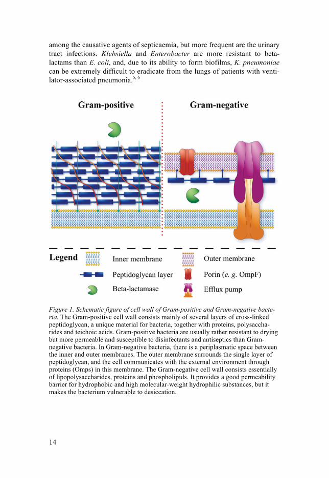
Bacteria of interest and the infections they can cause The Enterobacteriaceae family Several genera can be found in the Enterobacteriaceae family. These Gram-negative facultative anaerobic rods are widely distributed in soil, water, plants and intestines of animals and humans. For structure of the Gram-negative cell wall, see Figure 1.
The type species is Escherichia coli. It is the predominant facultative spe-cies in the bowel of humans, and it is present in the gut microbiota of nearly all vertebrates.1, 2 If it is found in a water supply system, it indicates a con-tinuing faecal contamination. There are several recognised categories of diarrheagenic E. coli: enterohemorrhagic (EHEC), enterotoxigenic (ETEC), enteropathogenic (EPEC), enteroinvasive (EIEC), and enteroaggregative E. coli (EAEC). To identify these categories PCR-methods are usually used. Apart from diarrhoea, E. coli is a leading cause of urinary tract infections, septicaemia, and neonatal meningitis.3 E. coli represents a great reservoir for antibiotic resistance, and the emergence of ESBL-producing strains have increased the fatality rates.4
Other important members of the family are the genera Klebsiella and En-terobacter. Klebsiella and Enterobacter can be found as commensals in hu-mans but have their natural inhabits in soils and on plants. Both genera can also cause infectious diseases, often in nosocomial contexts. Klebsiel-la pneumoniae and Enterobacter cloacae rank between fifth and ninth
14
among the causative agents of septicaemia, but more frequent are the urinary tract infections. Klebsiella and Enterobacter are more resistant to beta-lactams than E. coli, and, due to its ability to form biofilms, K. pneumoniae can be extremely difficult to eradicate from the lungs of patients with venti-lator-associated pneumonia.5, 6
Figure 1. Schematic figure of cell wall of Gram-positive and Gram-negative bacte-ria. The Gram-positive cell wall consists mainly of several layers of cross-linked peptidoglycan, a unique material for bacteria, together with proteins, polysaccha-rides and teichoic acids. Gram-positive bacteria are usually rather resistant to drying but more permeable and susceptible to disinfectants and antiseptics than Gram-negative bacteria. In Gram-negative bacteria, there is a periplasmatic space between the inner and outer membranes. The outer membrane surrounds the single layer of peptidoglycan, and the cell communicates with the external environment through proteins (Omps) in this membrane. The Gram-negative cell wall consists essentially of lipopolysaccharides, proteins and phospholipids. It provides a good permeability barrier for hydrophobic and high molecular-weight hydrophilic substances, but it makes the bacterium vulnerable to desiccation.

15
Primary wound pathogens Pyogenic streptococci and Staphylococcus aureus are Gram positive, facul-tative anaerobic bacteria with complex nutritional requirements. Together, they cause the majority of the skin and soft tissue infections in humans.
Streptococcus pyogenes, or the group A streptococcus, is the most viru-lent of the pyogenic streptococci. It is an exclusive human pathogen, and the mucosa of the upper respiratory tract and non-intact skin are preferred sites for colonisation and ports for entry. S. pyogenes is equipped with a large number of virulence factors, and can cause a broad spectrum of infections, including impetigo, acute otitis media, tonsillitis, erysipelas, lymphangitis, necrotizing fasciitis, toxic shock syndrome, and septicemia. It can hide intra-cellularly and form biofilm. It is still susceptible to penicillin, after more than 70 years of exposure.6
The main habitat of S. aureus is the skin of primates. The relationship with the host is usually benign, but when the epithelial barrier is destroyed and/or medical devices are implanted severe infections can be the result. S. aureus is the leading agent of post-operative infections and infections associated with foreign bodies. It is also an important cause of acute endo-carditis, joint and bone infections, and community-acquired septicemia. De-pending on the type of exotoxins it expresses, it can cause food poisoning, necrotizing pneumonia, toxic shock syndrome, and scalded skin syndrome.6, 7 Its production of biofilms is a therapeutic problem, and in late years dissem-ination of MRSA in the community has become more frequent. Without full susceptibility to methicillin or similar drugs, the outcome is less certain.8
Environmental bacteria Pseudomonas aeruginosa is Gram-negative rod with a strictly aerobic res-piratory metabolism. It is a soil organism that can utilise a wide range of nutrients. Since its requirements are simple, it can grow in almost any moist environment, including sink drains, liquid soaps, eye-drops, humidifiers, and antiseptic solutions. The bacterium does not usually colonise healthy hu-mans, but can cause severe pneumonia in patients with mechanical ventila-tion, neutropenia or cystic fibrosis. It is one of the leading causes of burn wound infections, and it is frequently found in chronic ulcers. It is the most studied of all biofilm formers.9 P. aeruginosa is naturally resistant to several antibiotics and can rapidly develop resistance during antibiotic therapy. It is thereby difficult to treat.6
The Legionellaceae consist of the single genus Legionella. They are all strict aerobic and nutritionally fastidious Gram-negative rods. They are nor-mally found in aqueous environments and form biofilms. Legionella pneu-mophila is the clinically most important species. It is the leading cause of Legionnaires’ disease, a form of pneumonia. The infection can be mild to

16
life-threatening. In its natural environment, L. pneumophila is a facultative intracellular parasite of free-living amoebae. When infecting humans, it at-tacks primarily alveolar macrophages, which have many features in common with amoebae. When living intracellular, the bacterium is protected from biocides.10
Antimicrobial resistance The underlying mechanisms for the development and spread of antibiotic resistance are complex.11, 12 Most resistance mechanisms are pre-existent. In order to become clinically significant they need to become incorporated into a pathogen. This can be achieved by means of genetic exchange, by transla-tion of pre-existing genes that may be activated by selection or induction, or when mutational events extend the substrate range of resistance-mediating enzymes.12
Antimicrobial resistance due to global response systems Bacterial cells have wide spectrum of mechanisms to use when reacting acutely to sudden environmental changes. The resistance mechanisms at play on the cellular level are, however, divided into only four groups: 1) reduced permeability of the cell wall (porin loss, active efflux), 2) modifica-tion of the antimicrobial substance (beta-lactamases), 3) modification of the target protein (PBP-changes), and 4) altered metabolic route. For silver, the first two mechanisms, a loss of porins or an activation of efflux pumps, are of most interest.
Porins, or outer membrane proteins (Omps), are channels in the bacterial cell membrane of Gram-negatives that allow substances needed for the bac-terial cell metabolism to penetrate into the cell (Figure 1). Some antimicro-bial substances enter the cell through the same porins, and a transcriptional down-regulation of these Omps reduces the intracellular concentration of antimicrobial agents and thus, results in decreased susceptibility.13
Efflux pumps are transport proteins that often use active transport to clear the cell from antibiotics or other harmful substances (Figure 1). Multidrug efflux pumps have been described, e.g. like AcrB in E. coli, that rather un-specifically clears a wide range of substances.14
Sometimes bacteria combine these two resistance mechanisms. An exam-ple of this is the multiple antibiotic resistance (Mar) phenotype. It is charac-terised by decreased susceptibility to multiple antibiotics, caused by a com-bination of porin losses and increased efflux that is activated by the mar operon.15 The Mar phenotype is not only induced by antibiotics,16 it is also

17
induced by drugs like diazepam,17 illustrating the complexity and cross-reactivity of bacterial response systems.
Other bacterial species, like P. aeruginosa, use the production of biofilms as a defence strategy. Several mechanisms contribute to a reduced suscepti-bility to antibiotics among biofilm producers: Firstly, a biofilm acts as a physical barrier which impedes the permeability of antibiotics into the cell.18 Secondly, the majority of bacterial cells within the biofilm are in stationary growth phase. Antibiotics like the beta-lactams, that require a high bacterial division rate for their activity, become inefficient.18, 19 Furthermore, biofilm facilitates horizontal gene transfer between bacteria,20 and it increases the mutation frequency.19
Antimicrobial resistance due to genetic exchange The bacterial genome is characterised by a remarkable plasticity that is caused by horizontal gene transfer, genome rearrangements and the activity of mobile DNA elements. A common differentiation is made between the ability of mobilising of genetic elements within a cell and between two dif-ferent cells. Horizontal gene transfer means the transfer of genetic elements between bacteria by cell-to-cell contact through conjugation and transduc-tion, or without cell-to-cell contact through transformation or phages.
Pre-existing antibiotic resistance genes may become mobilised from the chromosomes of bacterial species with limited clinical significance and get introduced into important human pathogens. For instance, two plasmid-mediated AmpC beta-lactamases have been mobilised from Aeromonas spp. and Citrobacter freundii and are now frequently isolated from clinical E. coli isolates.21 The complexity of exchange of genetic material between different species is illustrated in Figure 2.
In order to accomplish horizontal gene transfer, genetic elements have to be mobilised. Most mobile elements, like transposons, integrons or genomic islands, can be integrated into other genetic elements, but not all are mobile by themselves. For instance, despite the fact that integrons can integrate themselves by an integrase, their mobilisation is achieved indirectly, often as parts of integron cassettes that are incorporated into transposons.22, 23 Trans-posons are able to move vertically, between the chromosome and extra-chromosomal DNA, i.e. plasmids. Horizontal transfer of transposons is ac-complished by conjugative plasmids or phages.24

18
Figure 2. Illustration of intraspecies genetic exchange (adapted from Tenover et al.12).
While the vast majority of integrons are embedded in chromosomes, class 1 integrons are the most wide-spread variant in clinical isolates. It has been postulated that clinical class 1 integrons may have evolved from envi-ronmental class 1 integrons from Betaproteobacteria species.22 Gene cas-settes from clinical isolates frequently contain genes conferring resistance to quaternary ammonium compounds (qacEΔ), sulphonamide (sul1), trime-thoprim (dhfr) and streptomycin (aad).25 The mobilisation of class 1 in-tegrons in Gram-negative bacteria is usually associated with transposons of Tn21 and Tn402 types.22, 26
Circulation of resistant bacterial clones Bacterial clones are bacteria that share identical genetic and phenotypical properties resulting from a common origin.27, 28 In a global perspective, a tool for the determination of clonality of an isolate is multilocus sequence typing (MLST). This technique uses genetic sequence variations, based on usually seven representative housekeeping genes.29 Clonal relationships between sequence types are often determined using the BURST (based upon related sequence types) minimal spanning tree algorithm.30, 31 There is at least one MLST-scheme for most human pathogens.
For E. coli, there are two highly virulent sequence types, ST131 and ST405. Other sequence types, like ST10, ST69 and ST23, have been associ-ated with acquired resistance.28 The most well-known K. pneumoniae clones are ST14 and ST15. They are both part of the largest eBURST group.28 Among P. aeruginosa, the virulent clones ST235, ST111 and ST175 are globally spread and represent the majority of the multi-drug resistant P. aeruginosa strains worldwide.32 However, as the MLST categorisation of

19
bacteria is based on seven housekeeping genes, little is known about the properties that distinguish the above mentioned successful clones from non-related sequence types.
Figure 3. Illustration of the complexity of horizontal and vertical gene transfer be-tween the two species E. coli and K. pneumoniae. By incorporating genes into the chromosome, antimicrobial resistance can become a permanent part of a successful clone. Furthermore, vertical gene transfer contribute to preserve the reservoir of antimicrobial resistance genes.
The above globally successful clones are likely to act as a reservoir and host for mobile genetic elements and thereby contribute to the dissemination of multiresistance28 (Figure 3).
Lack of One Health perspective on antibiotic resistance Several studies have attempted to reduce the rate of antibiotic resistance by restricting the prescriptions of antibiotics for humans. Unfortunately, these studies have not been successful; the resistance to specific antibiotics have remained the same, and not even a decrease of the overall resistance to anti-biotics has been shown.33, 34 Possible underlying mechanisms are the pres-ence of gene cassettes coding for multi-drug resistance and the persistence of resistance genes once they have been acquired.35
Antibiotic resistance in human pathogens may be stabilised by a continu-ous low level of antibiotic exposure and give the pathogens the possibility to adapt to their hosts.36 Under continuous selective antimicrobial pressure,
20
adaptive mutations allow the bacteria to regain their original fitness while maintaining their antibiotic resistance.12, 36
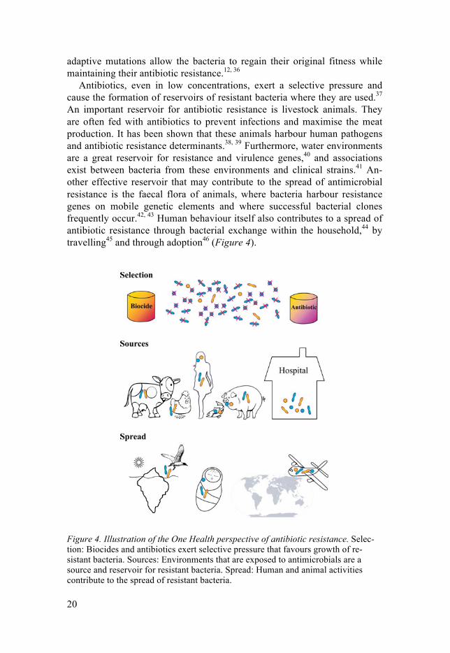
Antibiotics, even in low concentrations, exert a selective pressure and cause the formation of reservoirs of resistant bacteria where they are used.37 An important reservoir for antibiotic resistance is livestock animals. They are often fed with antibiotics to prevent infections and maximise the meat production. It has been shown that these animals harbour human pathogens and antibiotic resistance determinants.38, 39 Furthermore, water environments are a great reservoir for resistance and virulence genes,40 and associations exist between bacteria from these environments and clinical strains.41 An-other effective reservoir that may contribute to the spread of antimicrobial resistance is the faecal flora of animals, where bacteria harbour resistance genes on mobile genetic elements and where successful bacterial clones frequently occur.42, 43 Human behaviour itself also contributes to a spread of antibiotic resistance through bacterial exchange within the household,44 by travelling45 and through adoption46 (Figure 4).
Figure 4. Illustration of the One Health perspective of antibiotic resistance. Selec-tion: Biocides and antibiotics exert selective pressure that favours growth of re-sistant bacteria. Sources: Environments that are exposed to antimicrobials are a source and reservoir for resistant bacteria. Spread: Human and animal activities contribute to the spread of resistant bacteria.

21
The success story of CTX-M-producing Enterobacteriaceae In Gram-negative bacteria, beta-lactamases with extended spectrum have emerged as a significant public health problem.47 In the 1990s, the SHV and TEM were the predominant ESBL-types in a global perspective, but today, CTX-M enzymes stand for the majority of the ESBL-production.47, 48
The putative progenitors of the CTX-M family, are chromosomally en-coded cefotaximases of Kluyvera spp. (blaklu/CTX-M). Insertion sequences are frequently found upstream of the chromosomal cefotaximase, and they can be mobilised under stress conditions.49 The evolution of this mobilisation process occurred independently in different geographical regions and has resulted in phylogenetically diverse CTX-M clusters.50 The most dissemi-nated CTX-types are currently CTX-M-14 and CTX-M-15, both frequently present in humans, animals and the environment in both densely populated but also in remote areas.51
Maintenance and dissemination of CTX-M genotypes occurs to a signifi-cant extent by plasmids of incompatibility group FII.52 IncFII plasmids are well-adapted to members of the Enterobacteriaceae family,53 and persis-tence and spread of resistance determinants like CTX-M-15 are facilitated after their incorporation into these plasmids. This might also explain why CTX-M enzymes are predominantly found in Enterobacteriaceae but not to the same extent in P. aeruginosa.
Figure 5. Hierarchy of genetic structures participating in gene transfer, mainte-nance and expression of resistance genes. The column on the right gives typical examples.

22
Several clones, especially of E. coli or K. pneumoniae, frequently express CTX-M enzymes. E. coli clone ST131 often produces CTX-M-1554 and frequently carries IncFII plasmids. Other virulent clones that frequently ex-press CTX-M are ST405 and ST69.28 K. pneumoniae clone ST11, which is often isolated in Asia, harbours CTX-M-14 or CTX-M-15.51 Despite the above statements, there is no strict link between CTX-M enzymes and cer-tain clones. It is rather the local conditions that allow CTX-M-producing bacteria to emerge.4, 28
In the context of CTX-M producing species, the concept of 'genetic capi-talism' is frequently mentioned. It describes the observation that several clones, once they had acquired a resistance mechanism, were more prone to accumulate additional resistance and had a greater likelihood to become multi-drug resistant.51
Silver Role of silver in human medicine The growing and serious threat of antibiotic resistance in modern medicine has renewed the interest in silver compounds. The antibacterial properties of silver nitrate have been known since at least the Middle Ages. Historically, silver nitrate has a long tradition in the treatment of chronic ulcers and other types of wounds. The hard form of silver nitrate was known as lapis infer-nalis or 'lunar caustic', referring to the pain associated with silver treatment and the use of silver as a metaphor for the moon.55
Figure 6. Examples of frequently used silver-based dressings.
23
Beside silver nitrate, silver has been used in other combinations like sil-ver sulphadiazine, a substance frequently used in treatment of burns. Recent silver-products contain silver in the form of nanoparticles.56, 57 The ancient tradition of silver treatment of wounds is thereby continued, and a great va-riety of silver-based dressings are available on the market.
Silver can now be found in a wide variety of medical devices, e. g. central venous catheters, endotracheal tubes and urinary catheters, to prevent noso-comial infections.56 Furthermore, silver is widely used in consumer products in order to prevent unwanted microbial growth, although neither data on antimicrobial efficacy nor sufficient risk assessments are available.56, 57
Antibacterial activity of silver The mode of action of silver is not known in detail. There are, however, indications that silver is bound to different cell wall structures58-60 as well as DNA molecules and damage these.58, 61 The cell membrane has recently been pointed out as one of the more important targets.62, 63
Bacterial resistance to silver Since the reintroduction of silver products as treatment alternatives for burn wounds, there has been an increasing number of reports on bacterial re-sistance to silver.64-66 Silver-resistant bacteria have mainly been isolated from patients in burn care centres,59, 67-69 but some have also been isolated from the environment.70
There are indications that several bacterial species can accumulate the metal,71, 72 thereby removing it from solutions. Electron microscopy studies have shown, that silver accumulates on cell surfaces.72
Efflux has been suggested as an important mechanism of bacterial re-sistance in Gram-negative species.73, 74 In 1999, Gupta et al. described the sil operon, the genetic and molecular basis of silver resistance found in a Sal-monella typhimurium isolate.75 The operon codes for the silver binding pro-teins SilE and presumably SilF, two efflux pumps SilCBA and SilP, and the regulator proteins SilSR (Figure 7). The sil operon was found on plasmids that also harboured antibiotic resistance genes.76, 77
Data on the frequency of sil genes in different bacterial species are lim-ited. Although silver resistance has been found in a variety of species, pres-ence of the sil operon has mainly been reported in E. cloacae isolates: sil genes were found in 103 out of 164 clinical E. cloacae isolates from a Ger-man hospital.78 Another study reported sil carriage in six out of ten wound isolates.79
In E. coli, the sil operon and the chromosomal copper/silver efflux system cus both contribute to silver resistance and also interact with each other.80

Fi
gure
7. T
he si
l ope
ron
and
its p
ropo
sed
tran
scri
ptio
nal p
rodu
cts.
Top:
pro
pose
d fu
nctio
n of
gen
es fr
om th
e si
l ope
ron
(ada
pted
from
Gup
ta
et a
l.75,
Ran
dall
et a
l.80).
Bot
tom
: the
sil o
pero
n of
pla
smid
pU
UH
239.
2. O
M –
out
er m
embr
ane,
IM –
inne
r mem
bran
e.

25
Apart from active efflux of silver, silver-resistant E. coli isolates are fre-quently porin-deficient.73, 80
In contrast to Gram-negative bacteria, little is known about silver re-sistance of Gram-positive bacteria. Genes from the sil operon have been described in three MRSA isolates, but none of them were phenotypically resistant.81 Not even after in-vitro attempts did resistance to silver nitrate develop.62
Risks associated with the use of silver The use of silver in products has almost exploded since the turn of the centu-ry. Now, words of warning have appeared from researchers that emphasise the risks of silver usage. Concern is raised regarding the toxicity of silver for humans, animals and the environment. Also potential links to antibiotic re-sistance are mentioned.
Frequent intake of silver leads to deposition of silver in tissues, particu-larly the skin and those rich in fat. In the former case, the combination of silver depositions and sunlight can cause argyria or argyrosis. Silver nano-particles are known to convey cytotoxicity to a number of cells, including fibroblasts, hepatocytes, osteoblasts or bone-marrow cells.57, 82, 83 In analogy with humans, silver exerts toxic effects on animals and the environment, and the exposure is mainly due to emissions from the industry.84
Constant exposure of bacteria to low concentrations of silver may not on-ly cause silver resistance, but may also result in co-resistance to antibiotics.64, 85
Co-selection of antibiotic and heavy metal resistance The mechanisms responsible for co-selection of antibiotic and metal re-sistance can be classified as follows:
Cross-resistance Cross-resistance occurs when resistance to different compounds is mediated by the same structure, but only one of these compounds activates the mecha-nism. Classical examples are the multidrug efflux pumps that are found in many members of the Enterobacteriaceae or in P. aeruginosa. For instance, the resistance-nodulation-division (RND) efflux pumps play an important role in innate resistance14 but also, when overexpressed, for the multi-drug resistance phenotype of clinical isolates.86, 87 In E. coli, the AcrB efflux pump is a multidrug efflux pump with penicillins, fluoroquinolones, chlo-ramphenicol, detergents and cationic dyes as substrates.14 Furthermore,

26
E. coli expresses the copper(I)/silver(I) resistance efflux transporter CusCF-BA that is the only known heavy metal specific RND transporter.88 In con-trast to AcrB, CusCFBA is far more specific: in addition to cop-per(I)/silver(I), cross-resistance was only found for the drugs dinitrobenzene, dinitrophenol and ethionamide.89
Another mechanism conferring cross-resistance is deficiency of outer membrane channels. Common ways to alter permeability are alteration of porin size and loss of porin proteins. In P. aeruginosa, resistance to car-bapenems is mostly mediated by a combination of porin loss and active ef-flux.90 Furthermore, deficiency of porins has been described as a cause of resistance to different beta-lactams in Gram-negative bacteria.91-93 In an in-vitro study, silver-resistant E. coli isolates were porin-deficient and thus, resistant to cephalosporins.73
Co-resistance The possibility of co-resistance is present when genes coding for resistances are located together on (mobile) genetic elements like plasmids, transposons and integrons. Due to this association, co-selection of resistance determi-nants might occur.
One of the best-documented systems for co-resistance is, maybe, that of mercury and antibiotics. The mer operon, determining mercury resistance, has been found on plasmids and in chromosomes, frequently in the context of Tn21 and Tn21-like transposons.94 These transposons harbour mer genes and genes conferring resistance to spectinomycin-streptomycin aadA.95 A primate study showed that mercury from dental amalgam fillings caused a selection for plasmids carrying mercury and antibiotic resistance genes.96 Thus, it has been postulated that mercury may be a driving force for the se-lection of antibiotic resistance genes.97, 98
Another example, that is very illustrative of the necessity of a One Health perspective where antimicrobial resistance is concerned, is the co-resistance of copper and zinc. Copper, zinc and antibiotics are all used as growth pro-motors or disinfectants in commercial swine herds. There are several studies which suggest that there are links between these determinants. The usage of zinc in a pig nursery was associated with the selection of MRSA isolates.99 In another study, copper as food supplement contributed to the selection of resistance to antibiotics.100
Although the silver resistance determinant sil has been described in mo-bile genetic elements like IncH plasmids76 and can be found in plasmids from strains involved in hospital outbreaks75, 77, 78 and in a mobile island from copper-resistant E. coli strains,101 data on co-resistance between silver and antibiotics is lacking.

27
Co-regulation Transcriptional and translational regulation systems can be activated in re-sponse to bacterial stress.15, 86, 102, 103
In P. aeruginosa the CzcRS system is a transcriptional regulator involved in the regulation of quorum sensing, the resistance to the metals zinc, cad-mium and cobalt, and it also mediates antibiotic resistance.104-106 Although the multiple antibiotic resistance regulator MarR of E. coli is regulated by copper,107 cross-resistance of copper and antibiotics has not been document-ed so far.
28
Aims of this Doctoral Thesis
With the major problem of multiresistant bacteria and the increasing use of silver in health care and consumer products as a background, the overall intention of this thesis was to fill in knowledge gaps concerning the occur-rence, the mechanisms and the possible collateral damage of silver re-sistance. In order to achieve this aim, we sought • to investigate the antimicrobial effects of silver in vivo and in vitro on
bacteria with Gram-positive or Gram-negative cell walls and different environmental niches.
• to investigate the distribution of genetic and phenotypic silver resistance in isolates from infected patients, human and avian carriers and from the environment.
• to investigate the genetic background to phenotypic silver resistance.
• to investigate if there are links between resistance to antibiotics and resistance to silver through co-selection.

29
Materials and Methods
Bacteria The bacteria referred to in this thesis were clinical isolates from patients at Uppsala University Hospital and Changchun Children’s Hospital. There was also a collection of isolates from wild birds. The main focus was put on hu-man pathogens belonging to the Enterobacteriaceae family, i.e. E. coli, En-terobacter spp. and Klebsiella spp., with P. aeruginosa and Gram-positive bacteria like S. aureus or beta-hemolytic streptococci also having been in-cluded. An overview of the strain collection is given in Table 1.
Study I Wound samples were collected from 14 patients with chronic leg ulcers. All patients had undergone wound treatment at Uppsala University Hospital from November 2006 to September 2007. The patients were categorized into two groups: Group 1 was treated with silver dressings for a period of 3–5 weeks, and Group 2 received treatment with silver dressings for at least 2 months. In addition, 14 Enterobacteriaceae and P. aeruginosa strains with different antibiotic resistance profiles, including multi-resistance, were cho-sen to evaluate their ability to develop resistance to silver.
Study II The bacterial collection of this study consisted of human (n = 105) and avian (n = 111) E. coli isolates from faecal samples. The human as well as the avian study populations were composed of national (Swedish) and interna-tional isolates and included both producers and non-producers of ESBL.
Study III Faecal samples were collected during a two-week period at Changchun Children’s Hospital, China, in 2009 from forty children aged 0–3 years and admitted to a neonatology or a gastroenterology ward.
Study IV The presence of silver resistance and genes encoding silver resistance was investigated in a total of 752 blood isolates collected at Uppsala University Hospital during the years 1990–2010. The species distribution was as fol-lows: E. coli (n = 223), Enterobacter spp. (n = 165), Klebsiella spp.
30
(n = 208) and P. aeruginosa (n = 156). Furthermore, 87 Legionella isolates, mainly derived from the hospital water pipeline system, were included. Bac-teria were identified to the species level with standard laboratory procedures, and, when needed, by VITEK 2 (Biomerieux, USA) or MALDI-TOF (Bruker Daltonics, Germany). All isolates were stored at -70 °C.
Table 1. Overview over the study strains.
Strains Properties Study
S. aureus (n = 14) Clinical isolates derived from chronic leg ulcers. I
E. coli (n = 4) E. cloacae (n = 5) K. pneumonia (n = 2) P. aeruginosa (n = 3)
Randomly chosen isolates with different antibiotic pro-files used for further in-vitro investigation of silver expo-sure.
I
E. coli (n = 216) Faecal E. coli isolates from the following defined popu-lations: Human source: • Patients with diarrhoea, non-ESBL-producing iso-
lates (n = 52). • ESBL-producing E. coli, Uppsala University Hospi-
tal screening routines (n = 34).108 • ESBL-producing E. coli, ESBL-screening of
healthy travellers outside Scandinavia (n = 19).45 Avian source: • Herring gulls, Commander Islands, Bering Strait,
Russia (non-ESBL-producing E. coli n = 25, ESBL-producing E. coli n = 1).43
• Yellow-legged gulls, Southern France (non-ESBL producing E. coli (n = 25), ESBL-producing E. coli (n = 16).109
• Mainly mallards, Uppsala (non-ESBL-producing E. coli n = 17)
• Black headed gulls, Kalmar (non-ESBL-producing E. coli n = 25, ESBL-producing E. coli n = 2).110
II
E. coli (n = 27) Faecal screening isolates from children admitted to Changchun Children’s Hospital, China, ESBL-producing E. coli.
III
E. coli (n = 223) K. pneumonia (n = 129) K. oxytoca (n = 79) E. cloacae (n = 131) E. aerogenes (n = 32) E. agglomerans (n = 2) P. aeruginosa (n = 156)
Blood-stream isolates collected at Uppsala University Hospital during the years 1990–2010.
IV
Legionella spp. (n = 87)
Isolates from the water supply system of Uppsala Uni-versity Hospital.
IV

31
Silver resistance Susceptibility testing to silver nitrate MIC and MBC of silver nitrate was carried out according to the guidelines of the Swedish Reference Group for Antibiotics. Bacteria were suspended in IsoSensitest broth (Oxoid Ltd., UK) containing silver nitrate at concentra-tions ranging from 4–512 mg/L with a final bacterial concentration of 105 cfu/mL. After 18–20 h of incubation, MIC was defined as the lowest concentration yielding no visible growth. A silver nitrate MIC of > 512 mg/L, classified the bacterium as silver-resistant. The lowest concen-tration of silver nitrate killing 99.9 % of a bacterial inoculum was termed the MBC.
Exposure of bacteria to silver in vitro To induce silver-resistance, a stepwise selection procedure following MIC testing was performed. Ten µL of the bacterial suspension was inoculated into a series of tubes, each containing 1 mL of IsoSensitest broth supple-mented with increasing concentrations of silver nitrate (4–512 mg/L). The tubes were incubated at 37 °C overnight, and from the tube with the highest silver nitrate concentration and still visible growth, the new inoculum was taken. The experiment was repeated until a MIC of silver nitrate > 512 mg/L was reached, or after 10 passages had been performed. If a strain developed resistance to silver at least five sub-cultivations on blood or CLED agar were performed. After each passage, ≥ 5 cfu were tested if they still grew in Iso-Sensitest broth containing silver nitrate at a concentration of 512 mg/L.
Growth curves Growth curves were obtained using a BioscreenC reader (Labsystems, Fin-land). The bacteria were grown in IsoSensitest broth with or without silver nitrate (128 mg/L). The bacterial inocula (250 µL of each strain at a concen-tration of 5 x 10³ cfu/mL) were suspended in a honeycomb plate and imme-diately placed in the BioscreenC at 37 °C for 24 h. The optical density was determined every 10 min at 600 nm, after shaking the plate for 10 s at max-imum amplitude. Each growth curve represented the mean of two independ-ent experiments in triplicate.

32
Detection of genes in the sil operon DNA was prepared by boiling bacteria in PCR-water for at least 10 min, and amplification was carried out in a GeneAmp PCR system 9700 cycler (PE Applied Biosystems, USA) using Taqman Mastermix (Qiagen, Germa-ny). Gene specific primers for sil genes and appropriate annealing tempera-tures were used as previously described.78, 111 PCR-products were separated by gel electrophoresis and analysed visually.
Sanger sequencing of the silS gene Sequencing of the silS amplicons was performed on an ABI 3730 XL Auto-mated Sequencer (Applied Biosystems, USA). SNP calling was carried out with novoSNP.112 As reference, the silS gene from pUUH239.2 (NC_016966) was used.
Next generation sequencing DNA was prepared using QIAquick PCR Purification Kit (Qiagen, Germa-ny). The DNA was thereafter sequenced in an IonTorrentTM with a read length of 400 bp, according to the manufacturer’s instructions (LifeTechnol-ogies, USA). The reads were assembled into a draft genome using the As-semblerSPAdes plugin in TorrentSuite 4.2 with LifeTechnologies’ recom-mended settings. Databases were created for each of the assembled ge-nomes.
Antibiotic resistance Antibiotic susceptibility testing Susceptibility testing was performed according to the recommendations of the Swedish Reference Group for Antibiotics or the European Committee on Antimicrobial Susceptibility Testing. Isolates were tested by disc diffusion, and, when indicated, by MIC-determination using Etest (AB Biodisk, Swe-den).
Isolates with reduced susceptibility to cefpodoxime, ceftazidime and/or cefotaxime were tested for ESBL-production by a modified double disc dif-fussion synergy test.113 The plates were incubated for 16–24 h at 35 °C in room atmosphere.

33
Amplification and characterisation of resistance genes The DNA was prepared and amplified as described above for the sil genes. Investigated resistance genes were merA (mercury resistance),114 blaCTX-M, blaTEM and blaSHV (beta-lactamases),108 and qnr115 and aac(6)-lb116 (plasmid-mediated quinolone resistance). Genes encoding beta-lactamases were fur-ther typed by sequencing using an ABI 3130 instrument (Applied Biosys-tems, USA). The sequences obtained were compared with published se-quences, employing the NCBI Basic Local Alignment Search Tool (BLAST).108, 117
Outer membrane protein profiles Outer membrane proteins were extracted from late logarithmic phase cul-tures at 37 °C in Mueller-Hinton broth. The bacterial cells were washed, lysed with lysozyme, and, after adding RNase and DNase, disrupted by five freeze-thaw cycles. Membrane pellets were received after ultracentrifuga-tion, treated with N-lauroylsarcosine and resuspended in Laemmli sample buffer. The proteins were stained with bromophenol blue and subjected to polyacrylamide gel electrophoresis.
Epidemiological typing PCR-based fingerprinting Fingerprints of bacterial isolates were produced using AP-PCR. The primers used in the studies were ERIC-1R,118 ERIC-2,119 A70-9, 208 and 272.120 Amplified products were analysed with gel electrophoresis and interpreted visually. Two isolates with identical band patterns were considered to be the same strain.
MLST MLST was performed for E. coli, K. pneumoniae and E. cloacae isolates using established protocols.121-123 For E. coli, the seven housekeeping genes adk, fumC, gyrB, icd, mdh, purA, and recA were amplified and sequenced. The analysis on the latter two species was carried out in silico using whole genome sequence data. Chromatograms were edited with the se-qtrace software,124 and ST analysis was performed using the MLST web-sites for E. coli (http://mlst.warwick.ac.uk/mlst/dbs/Ecoli), K. pneumonia (http://bigsdb.web.pasteur.fr/klebsiella/klebsiella.html) and for E. cloacae (www.pubmlst.org).

34
PCR-detection of the O25b-ST131 clone For detection of the E. coli O25B-ST131 clone, an allele-specific PCR am-plifying the pabB gene was used. The PCR was carried out as described by Clermont et al.125 with slight modifications.
Clonality with BURST Genetic relationship was determined using the BURST (based upon related sequence type) algorithm as implemented in eBURST (version 3; http://eburst.mlst.net)30 and in the goeBURST software.31 The stringent group definition of clonal complexes (CCs) was used, i. e. only sequence types that shared identical allels at ≥ 6 of 7 loci were grouped. Population snapshots were based on group definitions with 0/7 identical alleles in eBURST, displaying related and unrelated sequence types.
Statistical analyses
Where appropriate, differences in the distributions were analysed with Fisher’s exact test. A difference was considered statistically significant for p ≤ 0.05.
35
Results
Antibacterial activity of silver Antibacterial activity of silver in vivo (I) Wound treatment with silver dressings was not able to eradicate the primary wound pathogens S. aureus, beta-hemolytic streptococci and P. aeruginosa (I). In 9 out of 14 ulcers, S. aureus continued to grow after at least three weeks of treatment. Likewise, cultures remained positive for P. aeruginosa (3/14) and beta-hemolytic streptococci (3/14) after treatment.
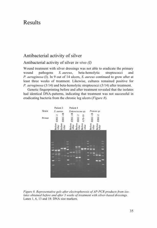
Genetic fingerprinting before and after treatment revealed that the isolates had identical DNA-patterns, indicating that treatment was not successful in eradicating bacteria from the chronic leg ulcers (Figure 8).
Figure 8. Representative gels after electrophoresis of AP-PCR products from iso-lates obtained before and after 3 weeks of treatment with silver-based dressings. Lanes 1, 6, 13 and 18: DNA size markers.

36
Silver nitrate MICs and MBCs The reference strain E. coli ATCC 25922 yielded a MIC of 16 mg/L ± one dilution step at all times. Independent of cell wall structure, MIC-values for silver nitrate ranged from 8–32 mg/L for the majority of the tested strains (n = 464), indicating that this is the range for the wild type. For the MIC distribution, see (Figure 9).
Figure 9. Distribution of silvernitrate MICs among the Enterobacteriaceae family in studies I, II and IV. Grey – sil-positive strains, white – sil-negative strains.
To determine the MIC was difficult for Enterobacter spp. They had a tendency to randomly jump over certain concentrations of silver nitrate. A representative example of this phenomenon is shown in (Figure 10). When cells from the tubes containing " 64 mg/L silver nitrate were used for deter-mining the MIC, they always grew in silver nitrate concentrations of > 512 mg/L.
Figure 10. Result of the silver nitrate MIC determination for E. cloacae strain B8275034. The phenomenon of jumping concentrations in the dilution series can be observed in tube 5 (32 mg/L) and tubes 9 (512 mg/L). The concentrations of silver nitrate is 2–512 mg/L in increasing order from left to right.

37
The MBC determination showed that silver nitrate had a bactericidal ef-fect on Gram-negative bacteria. The MBC-values for these bacteria were in the same range as the MIC-values (16–32 mg/L). In contrast, silver nitrate exhibited only a bacteriostatic effect on Gram-positive bacteria. All tested Gram-positive isolates had MBCs of ≥ 512 mg/L.
Frequency of silver resistance Frequency of phenotypical resistance to silver nitrate Phenotypical resistance to silver nitrate predominated in E. cloacae (n = 16) but was also found in E. aerogenes (n = 2), K. pneumonia (n = 2) and K. oxytoca (n = 2) (I, IV).
During the treatment of a chronic ulcer with a dressing containing silver (Aquacel Ag®), an E. cloacae isolate (SM0700965 II) resistant to silver ni-trate was found (≥ 512 mg/L). Before treatment, no E. cloacae was isolated, and when this isolate was detected the wound was treated with silver-based dressings over a period of three weeks (I).
MIC-testing for silver nitrate on an extensive strain collection of Entero-bacteriacae (n = 443), revealed elevated MIC-values (≥ 64 mg/L) to silver nitrate in E. cloacae (15/99, 15 %), E. aerogenes (2/29, 7 %), K. pneumoniae (2/95, 2 %) and K. oxytoca (2/59, 3 %) (IV). None of the tested E. coli isolates expressed resistance to silver nitrate without in-vitro exposure to the substance (I, II, IV).
Genetic resistance to silver Frequency of sil genes in isolates from infected patients and carriers Genes of the sil operon were only found in species belonging to the Entero-bacteriaceae family. No sil genes were detected in P. aeruginosa, Legionel-la spp., Enterococcus spp., beta-haemolysing streptococci or S. aureus.
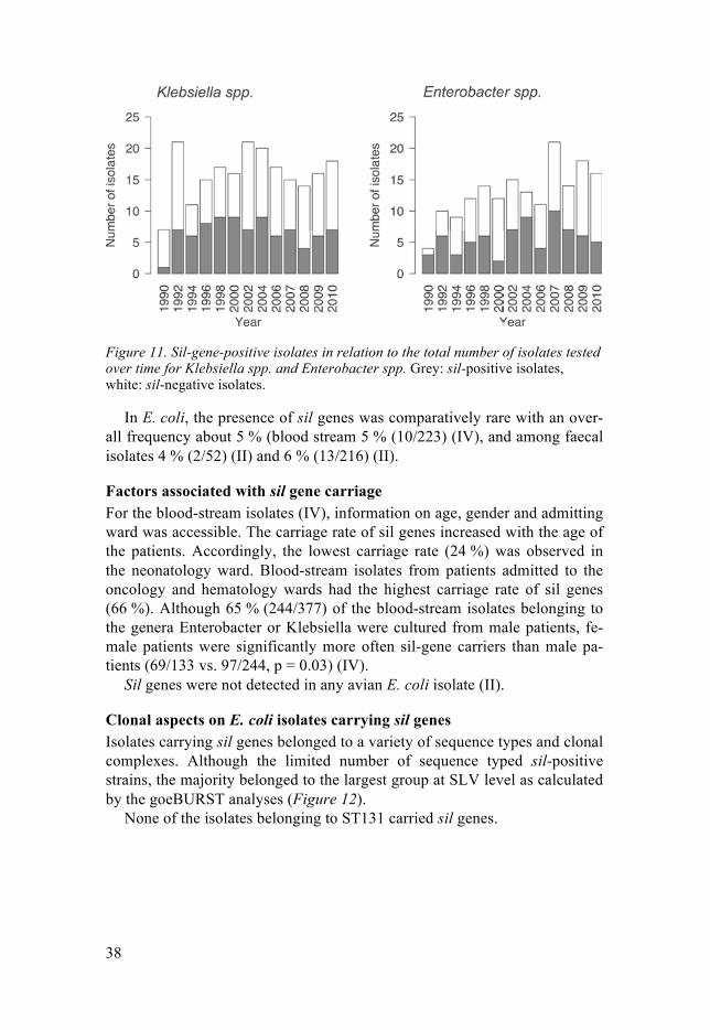
The silver-resistant wound isolate SM0700695 II carried sil genes (I). Out of 839 blood-stream isolates, 176 (21 %) harboured sil genes. These genes were most frequent in Enterobacter spp. (80/165; 48 %) and Klebsiella spp. (86/208; 41 %). Of the investigated species, the highest frequency was found in E. cloacae (76/131; 58 %) and K. oxytoca (39/79; 49 %) (IV). No differ-ence in frequency of sil genes in blood-stream isolates was noted during a time period of 20 years (Figure 11).
38
Figure 11. Sil-gene-positive isolates in relation to the total number of isolates tested over time for Klebsiella spp. and Enterobacter spp. Grey: sil-positive isolates, white: sil-negative isolates.
In E. coli, the presence of sil genes was comparatively rare with an over-all frequency about 5 % (blood stream 5 % (10/223) (IV), and among faecal isolates 4 % (2/52) (II) and 6 % (13/216) (II).
Factors associated with sil gene carriage For the blood-stream isolates (IV), information on age, gender and admitting ward was accessible. The carriage rate of sil genes increased with the age of the patients. Accordingly, the lowest carriage rate (24 %) was observed in the neonatology ward. Blood-stream isolates from patients admitted to the oncology and hematology wards had the highest carriage rate of sil genes (66 %). Although 65 % (244/377) of the blood-stream isolates belonging to the genera Enterobacter or Klebsiella were cultured from male patients, fe-male patients were significantly more often sil-gene carriers than male pa-tients (69/133 vs. 97/244, p = 0.03) (IV).
Sil genes were not detected in any avian E. coli isolate (II).
Clonal aspects on E. coli isolates carrying sil genes Isolates carrying sil genes belonged to a variety of sequence types and clonal complexes. Although the limited number of sequence typed sil-positive strains, the majority belonged to the largest group at SLV level as calculated by the goeBURST analyses (Figure 12).
None of the isolates belonging to ST131 carried sil genes.

39
Table 2. Summary of E. coli types carrying sil genes according to MLST.
Strain Sequence type Specimen ESBL production, CTX-M type if known
Study
S8 58 Faeces CTX-M-14 II S10 940 Faeces CTX-M-14 II S11 10 Faeces CTX-M-14 II P9 388 Faeces CTX-M-15 II P14 205 Faeces CTX-M-15 II P18 127 Faeces CTX-M-15 II P21 1312 Faeces CTX-M-15 II R7 424 Faeces CTX-M-15 II R9 940 Faeces CTX-M-15 II R20 155 Faeces CTX-M-15 II F32 10 Faeces No ESBL production II F32 409 Faeces No ESBL production II B0909531 540 Blood No ESBL production IV B1011268 410 Blood ESBL production IV B0804035 1011 Blood No ESBL production IV B0607370 23 Blood No ESBL production IV P12-1 Not typeable (clonal com-
lex 10) Faeces CTX-M-14 III
Figure 12. Minimal spanning tree illustration of sequence types of sil-positive E. coli isolates from the studies (calculated by goeBURST algoritm). Light red dots: sil-positive isolates.

40
The sil operon and phenotypic silver resistance Sil genes and phenotypic resistance to silver nitrate In all strains that showed phenotypic resistance to silver nitrate without a previous silver exposure, sil genes were detected (I, IV). Furthermore, dur-ing in-vitro exposure, all sil positive strains developed resistance to silver nitrate (IV). Carriage of sil genes was, however, not a prerequisite to silver resistance, since there was one strain lacking sil genes that developed re-sistance (I).
In-vitro resistance was unstable after at least five subcultivations for one isolate without sil genes (I), none out of 13 sil-positive isolates (II) and 4/17 sil-positive isolates (IV).
In whole genome-sequenced isolates, the sil operon was complete and all genes were in the same order compared with reference from plasmid pUUH239.2.
Fitness of silver-resistant strains The development of silver resistance in vivo and in vitro had a fitness cost. The cost differed between the strains (Figure 13).
Figure 13. Growth curves of silver-resistant isolates after in-vivo selection. (A) E. cloacae B8275034 and (B) K. pneumoniae B0910808. Dashed line: after selec-tion, solid line: during selective pressure of silver nitrate (128 mg/L).

41
SNPs in the silS gene To explore the genetic events taking place in the sil operon in the two strains E. cloacae B09014770 and K. pneumoniae B1018747 during the exposure to silver, the whole genome sequencing was carried out before (AgS) and after (AgR) the exposure. Upon comparison of the genomes (AgS-AgR), SNPs in the silS gene were observed. In E. cloacae strain B09014770, there was a SNP at T965A, whereas K. pneumoniae strain B1018747 had one at G629A.
To examine how common these SNPs were, 17 additional, silS genes were sequenced before and after the emergence of silver resistance. SNPs were found in 12 out of 17 isolates. They were distributed over the whole length of the gene, but a certain accumulation of mutations was noted in two segments, 629–725 bp and 919–1054 bp (Figure 14).
Figure 14. Localisation of SNPs in the silS gene after in-vivo and in-vitro selection. Sequence of silS from pUUH239.2 was used as reference.
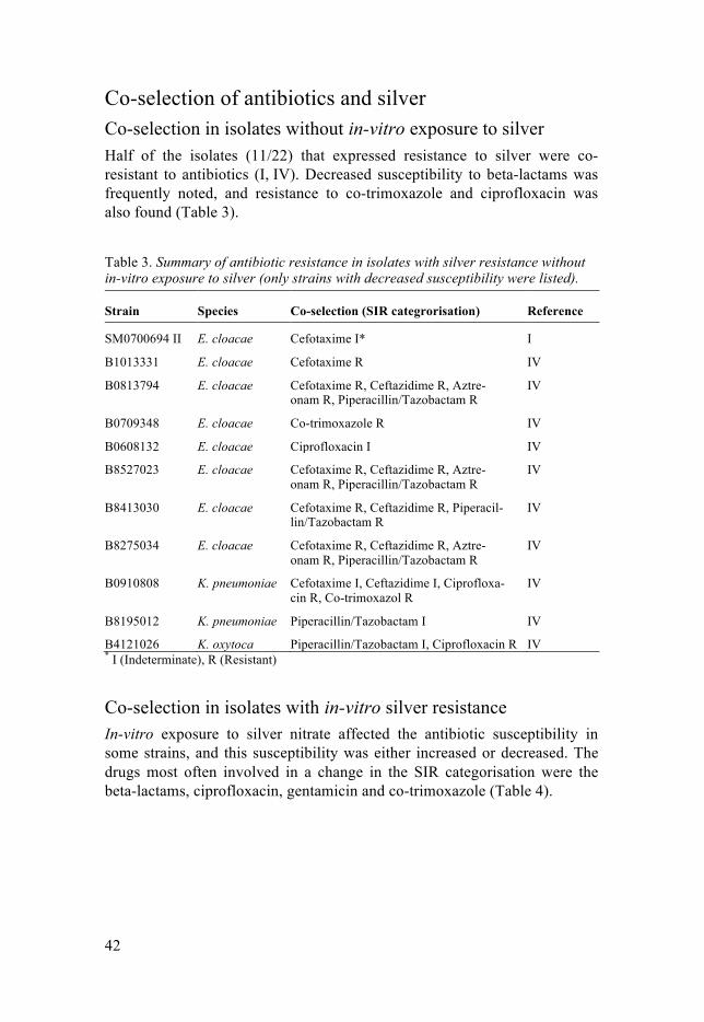
42
Co-selection of antibiotics and silver Co-selection in isolates without in-vitro exposure to silver Half of the isolates (11/22) that expressed resistance to silver were co-resistant to antibiotics (I, IV). Decreased susceptibility to beta-lactams was frequently noted, and resistance to co-trimoxazole and ciprofloxacin was also found (Table 3).
Table 3. Summary of antibiotic resistance in isolates with silver resistance without in-vitro exposure to silver (only strains with decreased susceptibility were listed).
Strain Species Co-selection (SIR categrorisation) Reference
SM0700694 II E. cloacae Cefotaxime I* I
B1013331 E. cloacae Cefotaxime R IV
B0813794 E. cloacae Cefotaxime R, Ceftazidime R, Aztre-onam R, Piperacillin/Tazobactam R
IV
B0709348 E. cloacae Co-trimoxazole R IV
B0608132 E. cloacae Ciprofloxacin I IV
B8527023 E. cloacae Cefotaxime R, Ceftazidime R, Aztre-onam R, Piperacillin/Tazobactam R
IV
B8413030 E. cloacae Cefotaxime R, Ceftazidime R, Piperacil-lin/Tazobactam R
IV
B8275034 E. cloacae Cefotaxime R, Ceftazidime R, Aztre-onam R, Piperacillin/Tazobactam R
IV
B0910808 K. pneumoniae Cefotaxime I, Ceftazidime I, Ciprofloxa-cin R, Co-trimoxazol R
IV
B8195012 K. pneumoniae Piperacillin/Tazobactam I IV
B4121026 K. oxytoca Piperacillin/Tazobactam I, Ciprofloxacin R IV * I (Indeterminate), R (Resistant)
Co-selection in isolates with in-vitro silver resistance In-vitro exposure to silver nitrate affected the antibiotic susceptibility in some strains, and this susceptibility was either increased or decreased. The drugs most often involved in a change in the SIR categorisation were the beta-lactams, ciprofloxacin, gentamicin and co-trimoxazole (Table 4).

43
Table 4. Summary of developed antibiotic resistance after exposure to silver nitrate in vitro.
Strain Species Changes in susceptibility Reference
S4279/06 E. cloacae
Imipenem S to R* I
R07** E. coli Piperacillin/Tazobactam S to I II
R09** E. coli Ceftibuten I to R II
R20** E. coli Ceftibuten I to R II
S11** E. coli Ciprofloxacin R to S II
P03** E. coli Ceftibuten S to R, Ciprofloxacin R to S, Piperacillin/Tazobactam S to I
II
P21** E. coli Ceftibuten I to R II
P14** E. coli Piperacillin/Tazobactam S to R, Co-trimoxazol R to I
II
F23 E. coli Piperacillin/Tazobactam S to R, Gentami-cin S to R, Co-trimoxazol S to R
II
*S (Susceptible), I (Indeterminate), R (Resistant) **ESBL-producer
Effect on outer membrane proteins Outer membrane protein profiles were analysed on E. coli isolates before and after silver-exposure experiments. After silver exposure, two out of six isolates lost OmpC expression, whereas one isolate lost OmpF expression. Loss of OmpC porin was most common (II).
There was no obvious pattern matching the antibiotic susceptibility changes.
Association of sil genes and CTX–M production in E. coli Sil genes were more often found in ESBL-producing E. coli than in non-ESBL-producing E. coli (II). The frequency of sil genes in CTX-M-producing E. coli varied depending on the source. While sil genes were rela-tively common in ESBL-producing E. coli from stool samples collected in Sweden (21 %, 11/53) (II), the frequency of sil genes in ESBL-producing E. coli from children admitted to Changchun Children ́s Hospital (4 %, 1/27) (III) or birds (0/9) (II) was very low or zero (Table 5).

44
Table 5. Overview over strain collections with CTX-M producing E. coli.
Source of strain* Host Frequency of sil genes Reference
Uppsala University Hospital screening routines (n = 34)
Mainly adults 24 % (n = 8) II
ESBL screening of healthy travel-lers outside Scandinavia (n = 19)
Mainly adults 16 % (n = 3) II
Yellow legged gulls, Southern France (n = 9)
Birds 0 II
Screening isolates from children admitted to Changchun Children’s Hospital, China (n = 27)
Children 4 % (n = 1) III
* All isolates originated from stool samples. Out of all investigated CTX-M types, sil genes were only present in CTX-
M-15 (31 %, 8/26) and CTX-M-14 (17 %, 4/23). None of the other strains with CTX-M types carried sil genes (55 (n = 17), 9 (n = 9), 27 (n = 2), 64 (n = 1), 101 (n = 1)) (II, III).
Association of sil genes and antibiotics The association of sil genes and antibiotics was investigated in human feacal E. coli isolates with and without ESBL production. Isolates with silE gene were more likely to be resistant to co-trimoxazole (92 % vs. 40 %, P = 0.0005) and gentamicin (46 % vs. 17 %, p = 0.022) than silE-negative isolates (II).
Association of sil genes and merA genes In fecal E. coli, merA gene was present in 22 out of 216 isolates (10 %), more merA genes were detected in human E. coli (16 %, 17/105) compared with avian E. coli (5 %, 5/111) (p = 0.004) (II). In feacal samples from chil-dren admitted to Changchun Children’s Hospital, merA gene was found in two samples (III). Only one isolate had sil and merA genes simultaneously, the isolate was derived from a patient in a surveillance-programme at Uppsa-la University Hospital (II).
In blood-stream isolates, merA was most common in Klebsiella spp. (34/208 isolates; 16 %), followed by E. coli (26/223 isolates; 12 %), P. aeruginosa (17/156 isolates; 11 %), and Enterobacter spp. (8/165 iso-lates; 5 %).
45
Discussion
Antibacterial effects of silver The first products that appeared on the market and used silver as a biocide were silver-based dressings for wound treatment. Therefore, we investigated the antibacterial effects of topical silver treatment on the bacterial wound flora. We found that treatment of 14 chronic leg ulcers with silver-based dressings for at least three weeks did not eradicate primary wound pathogens or prevent wound colonisation with secondary wound pathogens. Further-more, after only three weeks of topical treatment, we isolated a silver-resistant E. cloacae strain from the wound of one patient.
These findings are worrying since the use of silver-based dressings on chronic leg ulcers has increased62 to such an extent as to become very costly despite lacking clinical evidence of their efficacy.126, 127 We noted that S. aureus and pyogenic streptococci had high silver nitrate MBCs. This sug-gests that silver exerts only a bacteriostatic effect on the primary wound pathogens. Similar findings have been described by Feng et al.58 and Randall et al.62 when they investigated the effects of silver on S. aureus.
To determine silver nitrate MICs or MBCs is not always easy, and some research groups, therefore, seem to have chosen some alternatives to stand-ard laboratory procedures used for antibiotics. The preferred route is to use more unconventional definitions of bactericidal activity and to develop new tests that are optimised in one way or another to show bactericidal effects of silver in vitro.60 The value of these tests in clinical settings can, however, be questioned. In contrast to the results of custom-made tests, we did not find a bactericidal effect of silver on S. aureus or pyogenic streptococci, and that was independent of the conditions (in vitro or in vivo). Therefore, we believe that it is crucial to use established laboratory procedures and modify them as little as possible. This way, other groups can repeat the experiments, and results can be compared.
In contrast to the findings in Gram-positive bacteria, the silver nitrate MBCs were almost identical to the MICs in Gram-negative bacteria, i. e. Gram-negative bacteria are definitely killed by silver. This finding is inter-esting since it is usually the other way around for commonly used disinfect-ants and antiseptics. However, despite our in-vitro findings, members of the Enterobacteriaceae family and P. aeruginosa were not eradicated by topical silver treatment in vivo. Several reasons for this have been suggested, includ-

46
ing insufficient release concentrations of silver from topical dressings64 and a high binding rate of silver ions to halides and proteins that may further reduce the fraction of free (active) silver ions in a complex wound environ-ment.56 This may contribute to the failure to eradicate wound pathogens and colonisers in the majority of the ulcers investigated.
The results of in-vitro killing kinetics for the different types of silver-based dressings available suggest that the bactericidal effect of silver on Gram-negative bacteria is achieved within 4 h.128, 129 These findings indicate that there is only limited benefit of prolonged treatment, and, moreover, long-term treatment might increase the risk to select for resistant Gram-negative species. Thus, the necessity of long-term treatment with silver-based dressings is questionable.
Distribution of silver resistance In the present thesis, we used a standard laboratory method to investigate the silver nitrate MIC distribution in a strain collection consisting of members of the Enterobacteriaceae family, P. aeruginosa and Legionella spp. Notewor-thy was that phenotypical resistance, in terms of increased MICs without prior exposure to silver in vitro, was only found in bacteria belonging to the two genera Enterobacter and Klebsiella.
The species with the highest frequency of silver resistance was E. cloacae, a finding which is in accordance with other studies.59, 67, 79 As many as 15 % of the invasive E. cloacae isolates were phenotypically silver-resistant.
In congruence with the phenotypical findings, genetic determinants of silver resistance, i. e. sil genes, were exclusively found in members of the Enterobacteriaceae family. Even in the genetic context, sil genes were most frequent in Enterobacter spp. (48 %) and Klebsiella spp. (41 %). For E. cloacae, we found a sil gene carriage rate of 58 %, a figure which is com-parable with the 63 % found in a recent study of clinical isolates at a German hospital.78
Compared with Enterobacter and Klebsiella, few human E. coli isolates carried sil genes. However, in Swedish human E. coli isolates with produc-tion of CTX-M-14 and -15, we found an elevated frequency of sil genes (up to 21 %). If sil-positive E. coli isolates were few in Swedes, they were close to non-existent in other populations. Sil genes were not found in any avian E. coli isolate, although the isolates were collected from diverse geograph-ical regions, and the birds lived in some cases in areas with a high human activity.109, 110 Almost as surprising was, that despite a very high isolation frequency of CTX-M-producing E. coli, only a single Chinese child carried a strain with sil genes. It is possible that one of the most important sources of

47
CTX-M genes in China, i.e. chicken,130 lacks exposure to silver, as wild birds seem to do.
Interestingly, sil genes were not detected in invasive P. aeruginosa iso-lates or in the Legionella isolates that were included in the study. These bac-teria prefer wet environments and are both excellent biofilm formers. Le-gionella spp. can, in addition, hide inside eukaryotic cells. In their natural habitat they ought to come in contact with silver, albeit at low concentra-tions. Its toxic effects can probably be avoided by the strategies mentioned above. Carriage of sil genes is, therefore, not necessary.
Accumulation of resistance genes has been described as an adaptive mechanism to environmental requirements that is driven by selective pres-sure.42 Silver is a rare but naturally occurring metal that can be detected at low concentrations in rivers and lakes, even in pristine unpolluted areas.84 A recent investigation of Gullberg et al.36 showed that very low concentrations of heavy metals were able to exert selective pressure. Nevertheless, silver ions are very reactive, and they bind rapidly to proteins and other molecules in the surrounding. The antibacterial effect in natural habitats might, there-fore, be limited or short-lived.
Kremer et al. suggested another possibility; that silver resistance could be a potential fitness factor contributing to the successful establishment of an outbreak strain in the hospital environment.78 We found sil genes more often in isolates from the hematology ward, a ward with intensive use of chemo-therapeutics, disinfectants and antibiotics, and sil genes may represent a selective advantage in hospital environments.
Remarkably, there was a higher rate of silver resistance in bacteria from females than from males. Silver is used as preservative in cosmetics and as a disinfectant of vegetables and salads. It has also been offered as a food sup-plement.56, 131 Although this is highly speculative, it is possible that the fe-male lifestyle may expose women to silver to a higher extent than men.
The distribution of silver resistance in different bacterial populations and species raises more questions about the source(s) of sil genes and the driving forces involved in the acquisition. If additional bacterial populations are investigated in the future, it would be of value to change the primers used for the sil gene screening. Results of the whole genome sequencing showed that there were mismatches between the primers designed by Percival et al.111 and some strains from our strain collection. With the primers used by Kre-mer et al.,78 the number of isolates positive for different sil genes increased.
48
Genetic background to silver resistance We found silver resistance in Enterobacter and Klebsiella without prior exposure to silver in vitro. All these isolates with phenotypical resistance had sil genes. Inversely, phenotypical resistance to silver nitrate developed easily after in-vitro silver exposure for all isolates that carried sil genes. The selected resistance phenotype was stable for the majority of sil-positive iso-lates. One isolate lacking sil genes developed silver resistance after provoca-tion. This phenotype was instable after subcultivation.
We found that bacterial cells selected by the exposure experiments had mutations in the silS gene. This gene encodes a sensor kinase that activates the response regulator silR.75, 80 Mutations in silS gene occur in silver-exposed strains and result in an activation of the silCFBA efflux pump.80 Randall et al. show that derepression of silCFBA alone was not sufficient to achieve a phenotype with a high level of resistance to silver. In addition to the active clearance of silver from the cytoplasm with the silCFBA efflux pump, cells prevent increasing silver concentrations in the cytoplasm by loss of porins and an up-regulation of the periplasmatic silver-binding protein silE.80
Randall et al. found that both OmpC and OmpF had to be absent in order to achieve a resistance phenotype.80 According to our results, this is not a prerequisite to silver resistance in sil-positive E. coli. The loss of porins has, however, a fitness cost.
Co-selection of antibiotic and silver resistance Sil genes were overrepresented in CTX-M-producing E. coli, indicating a potential co-selecting mechanism. Interestingly, in our material, sil genes were solely found in E. coli that produced CTX-M-15 and CTX-M-14, the most frequent CTX-M types.50, 51 This finding indicates that the sil genes may contribute to the dissemination of these CTX-M types or vice versa.
However, the present material has some limitations. Both CTX-M-15 and -14 were mainly isolated from adults in Sweden, whereas other CTX-M types were, with few exceptions, derived from birds from South France or Chinese children. As mentioned earlier, the rate of CTX-M-producing E. coli in Chinese children was quite high, but the most prevalent CTX-M type (CTX-M-55) lacked sil genes. Noteworthy was that the only Chinese sil-positive isolate was producing CTX-M-15. With the chicken as an important reservoir for CTX-M-producing isolates in China,130 and it is likely that other factors than silver drives the selec-tion of CTX-M-producing isolates in this country.132, 133
Silver resistance was associated with resistance to beta-lactams, irrespec-tive of CXT-M enzymes, co-trimoxazole, gentamicin and quinolones. We found this pattern in all investigated isolates of the Enterobacteriaceae fami-

49
ly with phenotypic resistance, genotypic resistance, and after in-vitro selec-tion of silver resistance. In addition, we could also show that porin losses were linked to silver resistance.73, 80 Even if these losses were not solely responsible for a reduced susceptibility or a resistance to beta-lactams, they may contribute to beta-lactam resistance not caused by CTX-M production.
Although silver resistance was associated with CTX-M-producing iso-lates and resistance to certain clinically important antibiotics, no association was found between silver resistance and mercury resistance. This finding was unexpected, as dental amalgam, an alloy consisting of mercury and sil-ver, is a well-documented source for both metals, is present in many human mouths all over the world, and has been found to select for mercury re-sistance.96, 97, 134 A common selective source for mercury and silver re-sistance seems not that likely.
50
Conclusions
The antibacterial effect in vitro of silver on Gram-positive bacteria was bac-teriostatic, whereas it was bactericidal on Gram-negative bacteria. The in-vivo activity of silver-based wound dressings was limited, as they failed to eradicate both Gram-positive and Gram-negative bacteria. Genetic and phenotypic silver resistance was only found in members of the Enterobacteriaceae family. Most frequent was silver resistance in Entero-bacter spp. and Klebsiella spp. Genetic silver resistance was most frequently found in human isolates. It was not observed in bacteria of avian origin or in species with wet environ-ments as their natural habitat. In Enterobacteriaceae, sil genes were associated with a silver-resistant phe-notype in all cases but one. Selected mutants expressing silver resistance had SNPs in the silS gene, which is part of the regulon of the silver efflux pump silCFBA. Possible links for co-selection were found for beta-lactams, co-trimoxazole, gentamicin and silver. In contrast, no associations between silver and mercu-ry resistance were found.

51
Sammanfattning på svenska
Multiresistenta bakterier är ett allvarligt och snabbt ökande problem i dagens sjukvård, och de är huvudsakligen ett resultat av en mångårig och hög kon-sumtion av antibiotika. Bakterier med antibiotikaresistens kan även selekte-ras av biocider, kemiska substanser med antibakteriell effekt. En biocid som har fått en renässans under senare år är den giftiga tungmetallen silver, som ofta marknadsförs som ett alternativ till antibiotika. Risken finns att en okontrollerad användning av silverprodukter bidrar till spridningen av anti-biotikaresistens. Mekanismer som leder till bakteriell silverresistens är mångfaldiga, och en viktig genetisk markör för silverresistens är de sil-gener som ingår i sil-operonet, ett kluster av gener som kodar för en silverpump och funktionellt relaterade proteiner. Förekomsten av silverresistens hos bakterier och sil-genernas betydelse för denna resistens är sparsamt under-sökta och vilar mest på fallrapporter.
I avhandlingen undersöktes förekomsten av silverresistens i ett antal bak-teriepopulationer från människor, djur och olika miljöer. Vidare undersöktes silvrets roll som potentiell co-selektor för antibiotikaresistens. Speciell vikt lades på sil-operonet och dess roll vid silverresistens. Två av arbetena utför-des i samarbete med personal vid Sårcentrum, Akademiska sjukhuset, och vid Barnsjukhuset i Changchun City, Kina.
Resultaten i avhandlingen togs fram med hjälp av såväl gamla, klassiska som nyare molekylärbiologiska tekniker. Odlingsbaserade metoder användes för artidentifikation, resistensbestämning och beskrivning av egenskaper som fitness hos isolaten. En central metod i avhandlingen var resistensbe-stämning mot silvernitrat med buljonspädning samt ett protokoll för selekt-ion av silverresistens. Molekylärbiologiska metoder omfattade målsekvens-baserad PCR, epidemiologiska typningsmetoder (bl. a. AP-PCR, MLST, integrontypning, CTX-M-typning) samt sekvensering enligt Sanger och helgenomsekvensering med IonTorrent. För att analysera sekvensdata till-lämpades en omfattande arsenal bioinformatiska programvaror.
I en klinisk undersökning av sårfloran hos patienter med kroniska bensår som behandlades med silverförband, observerades en silverresistent Entero-bacter cloacae (MIC-värde > 512 mg/L för silvernitrat) efter enbart tre veckors behandling. Detta blev den första gången en silverresistent bakterie dokumenterades i Norden. I samband med denna studie noterades att silver-nitrat hade huvudsakligen en baktericid effekt på medlemmar i familjen
52
Enterobacteriaceae. Hos stafylokocker och streptokocker var silvrets effekt däremot bakteriostatisk, vilket kan förklara varför viktiga sårpatogener inte eradikerades under den lokala silverbehandlingen. Det noterades även en koppling mellan silverresistens och betalaktamaser av CTX–M-typ, enzymer som bryter ner några av de kliniskt viktigaste grupperna av antibiotika, samt mellan silverresistens och antibiotikakänslighet; vid silvernitratexponering reducerades känsligheten för såväl silver som för karbapenemer och pipera-cillin/tazobaktam.
I en stamkollektion som gick över artgränser, observerades silverresistens enbart hos isolat från människor men inte från fåglar. Dessutom var sil-generna ånyo överrespresenterade i CTX-M-producerande Escherichia coli, den tarmbakterie som orsakar flest urinvägsinfektioner. I och med att dessa multiresistenta bakterier är mycket vanliga i Kina, gjorde vi en studie på Changchuns barnsjukhus. Trots att frekvensen CTX-M-producerare låg runt 70 %, registrerades endast ett enda isolat som sil-positivt. Således betydligt lägre frekvens än i Sverige.
Vi gick därefter vidare och studerade ett flertal gramnegativa arter isole-rade under en 20-års period (1990-2010) från 734 blododlingar. Vi noterade att andelen isolat med genetisk silverresistens var hög hos Klebsiella-arter (41 %) och Enterobacter-arter (48 %), och den fenotypiska silverresistensen var vanligare hos Enterobacter cloacae än resistensen mot kliniskt viktiga antibiotika som aminoglykosider, kinoloner och trimsulfa. Med hjälp av s.k. multilocus sequence typing (MLST) kunde vi påvisa att mer än hälften av isolaten som bar på sil-gener tillhörde högriskkloner av global betydelse. Helgenomsekvensering gav belägg för att hypermutabla regioner i den regu-latoriska genen silS i sil-operonet sannolikt spelar en viktig roll vid utveckl-ingen av resistensfenotypen.
Sammanfattningsvis, avhandlingens resultat tyder på att silver kan bidra direkt och indirekt till antibiotikaresistens.

53
Acknowledgements
I should like to express my sincere gratitude and appreciation to all those many people who have contributed to this thesis by rendering their personal and professional support. My particular thanks go to: Åsa Melhus Åsa, you have been, you are and you will remain an inspiring and challeng-ing tutor. Introducing me into the field of research, on the one hand, and raising irresistible scientific curiosity in me, on the other, is what I cannot thank you enough for. Your wise advice and guidance both at clinics and in research have left their marks on me to know how to solve (microbiological) issues to obtain sustainable results. Also, thank you for giving me the neces-sary skills and knowledge and for encouraging me to forge ahead. Björn Olsen, my co-supervisor, who implanted the idea of globalism in mi-crobiology into me. You belong to those people who have the ability to stimulate others to take the right choice for the future. Eva Tano, together with you, I started my career as a doctoral candidate, and together we are now completing this chapter in my life. You were always ready to share with me your skills and experience of life, no matter whether in terms of viable counts, organising conference trips or cheering me up whenever I was down within the time I spent on this work. Petra Edquist who opened up the door for me into the large and initially foreign world of molecular biology. Hong Yin, Markus Klint, Ylva Molin, Karolina Eriksson-Gonzales, Hilde Riedel, Matti Karvanen, all of you knew how to respond to my methodological con-siderations at any time. Angela Lagerqvist Vidh who unfortunately had to pass away from us much too early. You left me an unbelievably wide repertoire of practical skills and a lot of knowledge on bacteria and on our Department of Clinical Microbi-ology.

54
Gänget på "labbet" who gave me their support and were forbearing when I was saving one or the other gel while some readings were being taken, or when I was collecting some strains for research, when bacterial cultures had to be put in the fridge during the weekend, or when new cultures had to be started, whenever a VITEK needed to be run, or when agar plates had just to be cast until the next day, or, or, or… Anna Schwan, Anna Heydecke, Birgitta Lysty, Eva Hjelm, Kenneth Nilsson and Björn Herrmann, my Department colleagues, who kept my options open and the lab running while I was busy with my research work. Eva Haxton, a pool full of insider tips that never seemed to run dry, and the great editor of the other manuscript. Martin Dahlö and Christian Tellgren-Roth, for their help in bioinformatic analysis. My father-in-law, Wolfgang, for his invaluable help to improve the English and layout of this book. My parents who gave me the idea that is behind all this, and who encouraged me to keep on going to the bitter end. My dear hubby Robert, I am so endlessly grateful for your saintly patience and immense power! Without your incredible efforts to help me to keep on struggling with informatics and with the English language, this book would never have turned out what actually has become of it. Theodor och Mathilda, nu är boken om “Eschan” äntligen klar!
55
References
1 Eckburg PB, Bik EM, Bernstein CN, et al. Diversity of the human intestinal microbial flora. Science 2005; 308: 1635-8
2 Tenaillon O, Skurnik D, Picard B, Denamur E. The population genetics of commensal Escherichia coli. Nature reviews Microbiology 2010; 8: 207-17
3 Kaper JB, Nataro JP, Mobley HL. Pathogenic Escherichia coli. Nature reviews Microbiology 2004; 2: 123-40
4 Woodford N, Ward ME, Kaufmann ME, et al. Community and hospital spread of Escherichia coli producing CTX-M extended-spectrum beta-lactamases in the UK. The Journal of antimicrobial chemotherapy 2004; 54: 735-43
5 Torres A, Carlet J. Ventilator-associated pneumonia. European Task Force on ventilator-associated pneumonia. Eur Respir J 2001; 17: 1034-45
6 Murray PR, Baron EJ, Jorgensen JH, Landry ML, Pfaller MA. Manual of Clinical Microbiology. 9 Edn. Washington, DC, USA: American Society for Microbiology, 2007
7 Otto M. Staphylococcus aureus toxins. Current opinion in microbiology 2014; 17: 32-7
8 Köck R, Becker K, Cookson B, et al. Methicillin-resistant Staphylococcus aureus (MRSA): burden of disease and control challenges in Europe. Euro surveillance 2010; 15: 19688
9 Monds RD, O'Toole GA. The developmental model of microbial biofilms: ten years of a paradigm up for review. Trends in microbiology 2009; 17: 73-87
10 Swanson MS, Hammer BK. Legionella pneumophila pathogesesis: a fateful journey from amoebae to macrophages. Annual review of microbiology 2000; 54: 567-613
11 Cohen J. Confronting the threat of multidrug-resistant Gram-negative bacteria in critically ill patients. The Journal of antimicrobial chemotherapy 2013; 68: 490-1
12 Tenover FC. Development and spread of bacterial resistance to antimicrobial agents: an overview. Clinical infectious diseases : an official publication of the Infectious Diseases Society of America 2001; 33 Suppl 3: S108-15
13 Livermore DM. Interplay of impermeability and chromosomal beta-lactamase activity in imipenem-resistant Pseudomonas aeruginosa. Antimicrobial agents and chemotherapy 1992; 36: 2046-8
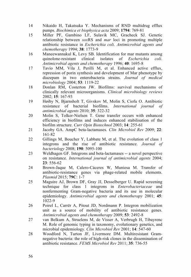
56
14 Nikaido H, Takatsuka Y. Mechanisms of RND multidrug efflux pumps. Biochimica et biophysica acta 2009; 1794: 769-81
15 Miller PF, Gambino LF, Sulavik MC, Gracheck SJ. Genetic relationship between soxRS and mar loci in promoting multiple antibiotic resistance in Escherichia coli. Antimicrobial agents and chemotherapy 1994; 38: 1773-9
16 Maneewannakul K, Levy SB. Identification for mar mutants among quinolone-resistant clinical isolates of Escherichia coli. Antimicrobial agents and chemotherapy 1996; 40: 1695-8
17 Tavío MM, Vila J, Perilli M, et al. Enhanced active efflux, repression of porin synthesis and development of Mar phenotype by diazepam in two enterobacteria strains. Journal of medical microbiology 2004; 53: 1119-22
18 Donlan RM, Costerton JW. Biofilms: survival mechanisms of clinically relevant microorganisms. Clinical microbiology reviews 2002; 15: 167-93
19 Høiby N, Bjarnsholt T, Givskov M, Molin S, Ciofu O. Antibiotic resistance of bacterial biofilms. International journal of antimicrobial agents 2010; 35: 322-32
20 Molin S, Tolker-Nielsen T. Gene transfer occurs with enhanced efficiency in biofilms and induces enhanced stabilisation of the biofilm structure. Curr Opin Biotechnol 2003; 14: 255-61
21 Jacoby GA. AmpC beta-lactamases. Clin Microbiol Rev 2009; 22: 161-82
22 Gillings M, Boucher Y, Labbate M, et al. The evolution of class 1 integrons and the rise of antibiotic resistance. Journal of bacteriology 2008; 190: 5095-100
23 Weldhagen GF. Integrons and beta-lactamases -- a novel perspective on resistance. International journal of antimicrobial agents 2004; 23: 556-62
24 Brown-Jaque M, Calero-Cáceres W, Muniesa M. Transfer of antibiotic-resistance genes via phage-related mobile elements. Plasmid 2015; 79C: 1-7
25 Maguire AJ, Brown DF, Gray JJ, Desselberger U. Rapid screening technique for class 1 integrons in Enterobacteriaceae and nonfermenting Gram-negative bacteria and its use in molecular epidemiology. Antimicrobial agents and chemotherapy 2001; 45: 1022-9
26 Poirel L, Carrër A, Pitout JD, Nordmann P. Integron mobilization unit as a source of mobility of antibiotic resistance genes. Antimicrobial agents and chemotherapy 2009; 53: 2492-8
27 van Belkum A, Struelens M, de Visser A, Verbrugh H, Tibayrenc M. Role of genomic typing in taxonomy, evolutionary genetics, and microbial epidemiology. Clin Microbiol Rev 2001; 14: 547-60
28 Woodford N, Turton JF, Livermore DM. Multiresistant Gram-negative bacteria: the role of high-risk clones in the dissemination of antibiotic resistance. FEMS Microbiol Rev 2011; 35: 736-55
57
29 Urwin R, Maiden MC. Multi-locus sequence typing: a tool for global epidemiology. Trends Microbiol 2003; 11: 479-87
30 Feil EJ, Li BC, Aanensen DM, Hanage WP, Spratt BG. eBURST: Inferring Patterns of Evolutionary Descent among Clusters of Related Bacterial Genotypes from Multilocus Sequence Typing Data. J Bacteriol 2004; 186: 1518-30
31 Francisco AP, Bugalho M, Ramirez M, Carriço JA. Global optimal eBURST analysis of multilocus typing data using a graphic matroid approach. BMC Bioinformatics 2009; 10: 152
32 Mulet X, Cabot G, Ocampo-Sosa AA, et al. Biological markers of Pseudomonas aeruginosa epidemic high-risk clones. Antimicrobial agents and chemotherapy 2013; 57: 5527-35
33 Arason VA, Gunnlaugsson A, Sigurdsson JA, Erlendsdottir H, Gudmundsson S, Kristinsson KG. Clonal spread of resistant pneumococci despite diminished antimicrobial use. Microbial drug resistance 2002; 8: 187-92
34 Sundqvist M, Granholm S, Naseer U, et al. Within-population distribution of trimethoprim resistance in Escherichia coli before and after a community-wide intervention on trimethoprim use. Antimicrobial agents and chemotherapy 2014; 58: 7492-500
35 Salyers AA, Amábile-Cuevas CF. Why are antibiotic resistance genes so resistant to elimination? Antimicrobial agents and chemotherapy 1997; 41: 2321-5
36 Gullberg E, Albrecht LM, Karlsson C, Sandegren L, Andersson DI. Selection of a multidrug resistance plasmid by sublethal levels of antibiotics and heavy metals. MBio 2014; 5: e01918-14
37 Gullberg E, Cao S, Berg OG, et al. Selection of resistant bacteria at very low antibiotic concentrations. PLoS pathogens 2011; 7: e1002158
38 Liebana E, Batchelor M, Hopkins KL, et al. Longitudinal farm study of extended-spectrum beta-lactamase-mediated resistance. Journal of clinical microbiology 2006; 44: 1630-4
39 Wu G, Ehricht R, Mafura M, et al. Escherichia coli isolates from extraintestinal organs of livestock animals harbour diverse virulence genes and belong to multiple genetic lineages. Vet Microbiol 2012; 160: 197-206
40 Ouyang WY, Huang FY, Zhao Y, Li H, Su JQ. Increased levels of antibiotic resistance in urban stream of Jiulongjiang River, China. Applied Microbiology and Biotechnology 2015
41 Di Cesare A, Pasquaroli S, Vignaroli C, et al. The marine environment as a reservoir of enterococci carrying resistance and virulence genes strongly associated with clinical strains. Environ Microbiol Rep 2014; 6: 184-90
42 Österblad M, Norrdahl K, Korpimäki E, Huovinen P. Antibiotic resistance. How wild are wild mammals? Nature 2001; 409: 37-8
43 Hernandez J, Bonnedahl J, Eliasson I, et al. Globally disseminated human pathogenic Escherichia coli of O25b-ST131 clone,

58
harbouring blaCTX-M-15 , found in Glaucous-winged gull at remote Commander Islands, Russia. Environ Microbiol Rep 2010; 2: 329-32
44 Valverde A, Grill F, Coque TM, et al. High rate of intestinal colonization with extended-spectrum-beta-lactamase-producing organisms in household contacts of infected community patients. Journal of clinical microbiology 2008; 46: 2796-9
45 Tängdén T, Cars O, Melhus Å, Löwdin E. Foreign travel is a major risk factor for colonization with Escherichia coli producing CTX-M-type extended-spectrum beta-lactamases: a prospective study with Swedish volunteers. Antimicrob Agents Chemother 2010; 54: 3564-8
46 Tandé D, Boisramé-Gastrin S, Münck MR, et al. Intrafamilial transmission of extended-spectrum-beta-lactamase-producing Escherichia coli and Salmonella enterica Babelsberg among the families of internationally adopted children. J Antimicrob Chemother 2010; 65: 859-65
47 Cantón R, Coque TM. The CTX-M beta-lactamase pandemic. Curr Opin Microbiol 2006; 9: 466-75
48 Livermore DM, Cantón R, Gniadkowski M, et al. CTX-M: changing the face of ESBLs in Europe. The Journal of antimicrobial chemotherapy 2007; 59: 165-74
49 Lartigue MF, Poirel L, Aubert D, Nordmann P. In vitro analysis of ISEcp1B-mediated mobilization of naturally occurring beta-lactamase gene blaCTX-M of Kluyvera ascorbata. Antimicrobial agents and chemotherapy 2006; 50: 1282-6
50 Zhao WH, Hu ZQ. Epidemiology and genetics of CTX-M extended-spectrum beta-lactamases in Gram-negative bacteria. Crit Rev Microbiol 2013; 39: 79-101
51 Cantón R, González-Alba JM, Galán JC. CTX-M Enzymes: Origin and diffusion. Front Microbiol 2012; 3: 110
52 Coque TM, Novais Â, Carattoli A, et al. Dissemination of clonally related Escherichia coli strains expressing extended-spectrum beta-lactamase CTX-M-15. Emerg Infect Dis 2008; 14: 195-200
53 Datta N, Dacey S, Hughes V, et al. Distribution of genes for trimethoprim and gentamicin resistance in bacteria and their plasmids in a general hospital. J Gen Microbiol 1980; 118: 495-508
54 Nicolas-Chanoine MH, Blanco J, Leflon-Guibout V, et al. Intercontinental emergence of Escherichia coli clone O25:H4-ST131 producing CTX-M-15. J Antimicrob Chemother 2008; 61: 273-81
55 Klasen HJ. Historical review of the use of silver in the treatment of burns. I. Early uses. Burns 2000; 26: 117-30
56 Silver S. Bacterial silver resistance: molecular biology and uses and misuses of silver compounds. FEMS Microbiol Rev 2003; 27: 341-53

59
57 Schäfer B, Brocke JV, Epp A, et al. State of the art in human risk assessment of silver compounds in consumer products: a conference report on silver and nanosilver held at the BfR in 2012. Arch Toxicol 2013; 87: 2249-62
58 Feng QL, Wu J, Chen GQ, Cui FZ, Kim TN, Kim JO. A mechanistic study of the antibacterial effect of silver ions on Escherichia coli and Staphylococcus aureus. J Biomed Mater Res 2000; 52: 662-8
59 Rosenkranz HS, Coward JE, Wlodkowski TJ, Carr HS. Properties of silver sulfadiazine-resistant Enterobacter cloacae. Antimicrob Agents Chemother 1974; 5: 199-201
60 Jung WK, Koo HC, Kim KW, Shin S, Kim SH, Park YH. Antibacterial activity and mechanism of action of the silver ion in Staphylococcus aureus and Escherichia coli. Applied and Environmental Microbiology 2008; 74: 2171-8
61 Modak SM, Fox CL. Binding of silver sulfadiazine to the cellular components of Pseudomonas aeruginosa. Biochemical Pharmacology 1973; 22: 2391-404
62 Randall CP, Oyama LB, Bostock JM, Chopra I, O'Neill AJ. The silver cation (Ag+): antistaphylococcal activity, mode of action and resistance studies. J Antimicrob Chemother 2013; 68: 131-8
63 Morones-Ramirez JR, Winkler JA, Spina CS, Collins JJ. Silver enhances antibiotic activity against Gram-negative bacteria. Sci Transl Med 2013; 5: 190ra81
64 Chopra I. The increasing use of silver-based products as antimicrobial agents: a useful development or a cause for concern? J Antimicrob Chemother 2007; 59: 587-90
65 Klasen HJ. A historical review of the use of silver in the treatment of burns. II. Renewed interest for silver. Burns 2000; 26: 131-8
66 Moyer CA, Brentano L, Gravens DL, Margraf HW, Monafo WW, Jr. Treatment of large human burns with 0.5 per cent silver nitrate solution. Arch Surg 1965; 90: 812-67
67 Gayle WE, Mayhall CG, Lamb VA, Apollo E, Haynes BW, Jr. Resistant Enterobacter cloacae in a burn center: the ineffectiveness of silver sulfadiazine. J Trauma 1978; 18: 317-23
68 Hendry AT, Stewart IO. Silver-resistant Enterobacteriaceae from hospital patients. Can J Microbiol 1979; 25: 915-21
69 Kaur P, Vadehra DV. Mechanism of resistance to silver ions in Klebsiella pneumoniae. Antimicrob Agents Chemother 1986; 29: 165-7
70 Haefeli C, Franklin C, Hardy K. Plasmid-determined silver resistance in Pseudomonas stutzeri isolated from a silver mine. Journal of bacteriology 1984; 158: 389-92
71 Shakibaie MR, Kapadnis BP, Dhakephalker P, Chopade BA. Removal of silver from photographic wastewater effluent using Acinetobacter baumannii BL54. Can J Microbiol 1999; 45: 995-1000

60
72 Slawson RM, Trevors JT, Lee H. Silver accumulation and resistance in Pseudomonas stutzeri. Arch Microbiol 1992; 158: 398-404
73 Li XZ, Nikaido H, Williams KE. Silver-resistant mutants of Escherichia coli display active efflux of Ag+ and are deficient in porins. J Bacteriol 1997; 179: 6127-32
74 Starodub ME, Trevors JT. Silver resistance in Escherichia coli R1. J Med Microbiol 1989; 29: 101-10
75 Gupta A, Matsui K, Lo J-F, Silver S. Molecular basis for resistance to silver cations in Salmonella. Nat Med 1999; 5: 183-8
76 Gupta A, Phung LT, Taylor DE, Silver S. Diversity of silver resistance genes in IncH incompatibility group plasmids. Microbiology 2001; 147: 3393-402
77 Sandegren L, Linkevicius M, Lytsy B, Melhus Å, Andersson DI. Transfer of an Escherichia coli ST131 multiresistance cassette has created a Klebsiella pneumoniae-specific plasmid associated with a major nosocomial outbreak. J Antimicrob Chemother 2012; 67: 74-83
78 Kremer AN, Hoffmann H. Subtractive Hybridization Yields a Silver Resistance Determinant Unique to Nosocomial Pathogens in the Enterobacter cloacae Complex. J Clin Microbiol 2012; 50: 3249-57
79 Woods EJ, Cochrane CA, Percival SL. Prevalence of silver resistance genes in bacteria isolated from human and horse wounds. Vet Microbiol 2009; 138: 325-9
80 Randall CP, Gupta A, Jackson N, Busse D, O'Neill AJ. Silver resistance in Gram-negative bacteria: a dissection of endogenous and exogenous mechanisms. J Antimicrob Chemother 2015
81 Loh JV, Percival SL, Woods EJ, Williams NJ, Cochrane CA. Silver resistance in MRSA isolated from wound and nasal sources in humans and animals. Int Wound J 2009; 6: 32-8
82 Mytych J, Wnuk M. Nanoparticle technology as a double-edged sword: Cytotoxic, genotoxic and epigenetic effect on living cells. Journal of Biomaterials and Nanobiotechnology 2013; 4: 53-63
83 Poon VK, Burd A. In vitro cytotoxity of silver: implication for clinical wound care. Burns : journal of the International Society for Burn Injuries 2004; 30: 140-7
84 WHO. Silver and Silver Compounds: Environmental Aspects. Geneva, 2002
85 SCENIHR. Assessment of the Antibiotic Resistance Effects of Biocides. European Commission, Brussels, 2009
86 Hocquet D, Roussel-Delvallez M, Cavallo JD, Plésiat P. MexAB-OprM- and MexXY-overproducing mutants are very prevalent among clinical strains of Pseudomonas aeruginosa with reduced susceptibility to ticarcillin. Antimicrobial agents and chemotherapy 2007; 51: 1582-3
87 Keeney D, Ruzin A, Bradford PA. RamA, a transcriptional regulator, and AcrAB, an RND-type efflux pump, are associated

61
with decreased susceptibility to tigecycline in Enterobacter cloacae. Microbial drug resistance 2007; 13: 1-6
88 Delmar JA, Su CC, Yu EW. Bacterial multidrug efflux transporters. Annu Rev Biophys 2014; 43: 93-117
89 Conroy O, Kim EH, McEvoy MM, Rensing C. Differing ability to transport nonmetal substrates by two RND-type metal exporters. FEMS microbiology letters 2010; 308: 115-22
90 Hancock RE, Brinkman FS. Function of pseudomonas porins in uptake and efflux. Annual review of microbiology 2002; 56: 17-38
91 Charrel RN, Pagès JM, De Micco P, Malléa M. Prevalence of outer membrane porin alteration in beta-lactam-antibiotic-resistant Enterobacter aerogenes. Antimicrob Agents Chemother 1996; 40: 2854-8
92 Hernández-Allés S, Benedí VJ, Martínez-Martínez L, et al. Development of resistance during antimicrobial therapy caused by insertion sequence interruption of porin genes. Antimicrob Agents Chemother 1999; 43: 937-9
93 Martínez-Martínez L, Conejo MC, Pascual A, et al. Activities of imipenem and cephalosporins against clonally related strains of Escherichia coli hyperproducing chromosomal beta-lactamase and showing altered porin profiles. Antimicrob Agents Chemother 2000; 44: 2534-6
94 Osborn AM, Bruce KD, Strike P, Ritchie DA. Distribution, diversity and evolution of the bacterial mercury resistance (mer) operon. FEMS Microbiol Rev 1997; 19: 239-62
95 Liebert CA, Hall RM, Summers AO. Transposon Tn21, flagship of the floating genome. Microbiol Mol Biol Rev 1999; 63: 507-22
96 Summers AO, Wireman J, Vimy MJ, et al. Mercury released from dental "silver" fillings provokes an increase in mercury- and antibiotic-resistant bacteria in oral and intestinal floras of primates. Antimicrob Agents Chemother 1993; 37: 825-34
97 Österblad M, Leistevuo J, Leistevuo T, et al. Antimicrobial and mercury resistance in aerobic Gram-negative bacilli in fecal flora among persons with and without dental amalgam fillings. Antimicrob Agents Chemother 1995; 39: 2499-502
98 Skurnik D, Ruimy R, Ready D, et al. Is exposure to mercury a driving force for the carriage of antibiotic resistance genes? J Med Microbiol 2010; 59: 804-7
99 Slifierz MJ, Friendship RM, Weese JS. Methicillin-resistant Staphylococcus aureus in commercial swine herds is associated with disinfectant and zinc usage. Applied and Environmental Microbiology 2015
100 Hasman H, Aarestrup FM. tcrB, a gene conferring transferable copper resistance in Enterococcus faecium: occurrence, transferability, and linkage to macrolide and glycopeptide resistance. Antimicrobial agents and chemotherapy 2002; 46: 1410-6

62
101 Lüthje FL, Hasman H, Aarestrup FM, Alwathnani HA, Rensing C. Genome Sequences of Two Copper-Resistant Escherichia coli Strains Isolated from Copper-Fed Pigs. Genome Announc 2014; 2
102 Norton MD, Spilkia AJ, Godoy VG. Antibiotic resistance acquired through a DNA damage-inducible response in Acinetobacter baumannii. Journal of bacteriology 2013; 195: 1335-45
103 Painter KL, Strange E, Parkhill J, Bamford KB, Armstrong-James D, Edwards AM. Staphylococcus aureus adapts to oxidative stress by producing H2O2-resistant small colony variants via the SOS response. Infection and immunity 2015
104 Perron K, Caille O, Rossier C, Van Delden C, Dumas JL, Köhler T. CzcR-CzcS, a two-component system involved in heavy metal and carbapenem resistance in Pseudomonas aeruginosa. The Journal of biological chemistry 2004; 279: 8761-8
105 Dieppois G, Ducret V, Caille O, Perron K. The transcriptional regulator CzcR modulates antibiotic resistance and quorum sensing in Pseudomonas aeruginosa. PLoS One 2012; 7: e38148
106 Conejo MC, García I, Martínez-Martínez L, Picabea L, Pascual Á. Zinc eluted from siliconized latex urinary catheters decreases OprD expression, causing carbapenem resistance in Pseudomonas aeruginosa. Antimicrobial agents and chemotherapy 2003; 47: 2313-5
107 Hao Z, Lou H, Zhu R, et al. The multiple antibiotic resistance regulator MarR is a copper sensor in Escherichia coli. Nature chemical biology 2014; 10: 21-8
108 Lytsy B, Sandegren L, Tano E, Torell E, Andersson DI, Melhus Å. The first major extended-spectrum beta-lactamase outbreak in Scandinavia was caused by clonal spread of a multiresistant Klebsiella pneumoniae producing CTX-M-15. APMIS 2008; 116: 302-8
109 Bonnedahl J, Drobni M, Gauthier-Clerc M, et al. Dissemination of Escherichia coli with CTX-M Type ESBL between Humans and Yellow-Legged Gulls in the South of France. PLoS One 2009; 4: e5958
110 Bonnedahl J, Drobni P, Johansson A, et al. Characterization, and comparison, of human clinical and black-headed gull (Larus ridibundus) extended-spectrum beta-lactamase-producing bacterial isolates from Kalmar, on the southeast coast of Sweden. J Antimicrob Chemother 2010; 65: 1939-44
111 Percival SL, Woods E, Nutekpor M, Bowler P, Radford A, Cochrane C. Prevalence of silver resistance in bacteria isolated from diabetic foot ulcers and efficacy of silver-containing wound dressings. Ostomy Wound Manage 2008; 54: 30-40
112 Weckx S, Del-Favero J, Rademakers R, et al. novoSNP, a novel computational tool for sequence variation discovery. Genome Res 2005; 15: 436-42

63
113 Jarlier V, Nicolas MH, Fournier G, Philippon A. Extended broad-spectrum beta-lactamases conferring transferable resistance to newer beta-lactam agents in Enterobacteriaceae: hospital prevalence and susceptibility patterns. Rev Infect Dis 1988; 10: 867-78
114 Novais Â, Cantón R, Valverde A, et al. Dissemination and Persistence of blaCTX-M-9 Are Linked to Class 1 Integrons Containing CR1 Associated with Defective Transposon Derivatives from Tn402 Located in Early Antibiotic Resistance Plasmids of IncHI2, IncP1-α, and IncFI Groups. Antimicrob Agents Chemother 2006; 50: 2741-50
115 Robicsek A, Strahilevitz J, Sahm DF, Jacoby GA, Hooper DC. qnr prevalence in ceftazidime-resistant Enterobacteriaceae isolates from the United States. Antimicrob Agents Chemother 2006; 50: 2872-4
116 Park CH, Robicsek A, Jacoby GA, Sahm D, Hooper DC. Prevalence in the United States of aac(6')-Ib-cr encoding a ciprofloxacin-modifying enzyme. Antimicrob Agents Chemother 2006; 50: 3953-5
117 Altschul SF, Gish W, Miller W, Myers EW, Lipman DJ. Basic local alignment search tool. J Mol Biol 1990; 215: 403-10
118 Struelens MJ, Bax R, Deplano A, Quint WG, Van Belkum A. Concordant clonal delineation of methicillin-resistant Staphylococcus aureus by macrorestriction analysis and polymerase chain reaction genome fingerprinting. Journal of clinical microbiology 1993; 31: 1964-70
119 Grundmann HJ, Towner KJ, Dijkshoorn L, et al. Multicenter study using standardized protocols and reagents for evaluation of reproducibility of PCR-based fingerprinting of Acinetobacter spp. J Clin Microbiol 1997; 35: 3071-7
120 Mahenthiralingam E, Campbell ME, Foster J, Lam JS, Speert DP. Random amplified polymorphic DNA typing of Pseudomonas aeruginosa isolates recovered from patients with cystic fibrosis. J Clin Microbiol 1996; 34: 1129-35
121 Wirth T, Falush D, Lan R, et al. Sex and virulence in Escherichia coli: an evolutionary perspective. Mol Microbiol 2006; 60: 1136-51
122 Miyoshi-Akiyama T, Hayakawa K, Ohmagari N, Shimojima M, Kirikae T. Multilocus sequence typing (MLST) for characterization of Enterobacter cloacae. PLoS One 2013; 8: e66358
123 Diancourt L, Passet V, Verhoef J, Grimont PA, Brisse S. Multilocus sequence typing of Klebsiella pneumoniae nosocomial isolates. J Clin Microbiol 2005; 43: 4178-82
124 Stucky BJ. SeqTrace: a graphical tool for rapidly processing DNA sequencing chromatograms. J Biomol Tech 2012; 23: 90-3
125 Clermont O, Dhanji H, Upton M, et al. Rapid detection of the O25b-ST131 clone of Escherichia coli encompassing the CTX-M-15-producing strains. J Antimicrob Chemother 2009; 64: 274-7
126 Bergin SM, Wraight P. Silver based wound dressings and topical agents for treating diabetic foot ulcers. Cochrane Database Syst Rev 2006: CD005082
64
127 SBU. Silverförband vid behandling av kroniska sår. SBU Alert-rapporter (Statens beredning for medicinsk utvärdering) 2010; 2010-02
128 Fraser JF, Bodman J, Sturgess R, Faoagali J, Kimble RM. An in vitro study of the anti-microbial efficacy of a 1% silver sulphadiazine and 0.2% chlorhexidine digluconate cream, 1% silver sulphadiazine cream and a silver coated dressing. Burns : journal of the International Society for Burn Injuries 2004; 30: 35-41
129 Ip M, Lui SL, Poon VK, Lung I, Burd A. Antimicrobial activities of silver dressings: an in vitro comparison. Journal of medical microbiology 2006; 55: 59-63
130 Rao L, Lv L, Zeng Z, et al. Increasing prevalence of extended-spectrum cephalosporin-resistant Escherichia coli in food animals and the diversity of CTX-M genotypes during 2003-2012. Vet Microbiol 2014; 172: 534-41
131 Scalzo M, Cerretou F, Orlandi C, Simonetti N. Utilization of electrochemical silver ions as preservative agent in cosmetic dispersions. Int J Cosmet Sci 1997; 19: 27-36
132 Denkel LA, Schwab F, Kola A, et al. The mother as most important risk factor for colonization of very low birth weight (VLBW) infants with extended-spectrum beta-lactamase-producing Entero-bacteriaceae (ESBL-E). J Antimicrob Chemother 2014; 69: 2230-7
133 Dubois V, De Barbeyrac B, Rogues AM, et al. CTX-M-producing Escherichia coli in a maternity ward: a likely community importation and evidence of mother-to-neonate transmission. J Antimicrob Chemother 2010; 65: 1368-71
134 Liebert CA, Wireman J, Smith T, Summers AO. The impact of mercury released from dental "silver" fillings on antibiotic resistances in the primate oral and intestinal bacterial flora. Met Ions Biol Syst 1997; 34: 441-60


Acta Universitatis UpsaliensisDigital Comprehensive Summaries of Uppsala Dissertationsfrom the Faculty of Medicine 1084
Editor: The Dean of the Faculty of Medicine
A doctoral dissertation from the Faculty of Medicine, UppsalaUniversity, is usually a summary of a number of papers. A fewcopies of the complete dissertation are kept at major Swedishresearch libraries, while the summary alone is distributedinternationally through the series Digital ComprehensiveSummaries of Uppsala Dissertations from the Faculty ofMedicine. (Prior to January, 2005, the series was publishedunder the title “Comprehensive Summaries of UppsalaDissertations from the Faculty of Medicine”.)
Distribution: publications.uu.seurn:nbn:se:uu:diva-247472
ACTAUNIVERSITATIS
UPSALIENSISUPPSALA
2015