Embed Size (px)
Citation preview

Plant Physiol. (1977) 60, 235-239
Asparaginase and Asparagine Transaminase in Soybean Leavesand Root Nodules1
Received for publication February 8, 1977 and in revised form April 21, 1977
JOHN G. STREETERDepartment ofAgronomy, Ohio Agricultural Research and Development Center, Wooster, Ohio 44691
ABSTRACT
Aspanginase activity (si jumomg protein hr) was detected in ex-tracts of soybean (Glycine max [L.] Men.) leaf blades, but, even afterefforts to optimize extraction and assay of the enzyme, specific activitywas not suffient to metabolize the estimated amount of a netranslocated to leaves. Asparagine trnamin activity with gyoxylateor pyruvate was at least 52 and 62 nmol/mg protein hr, respectively.This estimate of transaminase activity is based on the analysis of thereaction product a-ketosuccinamate. Formation of glycine and laninewas confirmed by amino acid analysis. a-Ketosncdnamate deamidasehad a specific actit of 85 nmol/mg protein * hr in leaf blade extracts.A large amount of se (300-500 nmol/mg protein* hr) was
found in root nodules. The enzyme is stable in 75% ethanol at roomtemperature, has a Km of 5 jaM for rparne, and was six times moreactive (protein basis) in bacteroids than cytosol. The relatively highactivity, stability, and Km of the enzyme complicate efforts to studyasparagie synthesis in the nodule, an organ known to export laqeamounts of this amino acd.
Asparagine was the principal component of stem exudatecollected from field-grown soybean plants (21) and is also themajor component in the bleeding sap of excised soybean rootnodules (27). It is clear that ASN2 plays a central role in nitrogentranslocation and in the nitrogen nutrition of the soybean plant.The extensive studies of Pate and co-workers (see [1] and refer-ences therein) suggest that this generality also applies to otherlegumes.
Evidence for the soybean plant suggests the presence of anactive asparagine-synthesizing system in root nodules. Numer-ous attempts to demonstrate the formation of labeled ASN innodule tissue by feeding a variety of radioactive precursorsunder a wide range of experimental conditions have failed(Streeter, unpublished). These failures are probably related tothe' presence of a very active asparaginase in nodules, as de-scribed in this report.
Since ASN does not accumulate in soybean leaves or stems,except in seedlings (22), shoots apparently possess an activesystem for metabolism of ASN. While asparaginase could bedetected in extracts of soybean leaves, ASN breakdown in someexperiments could not be accounted for by aspartate formed.Pursuit of this problem led to the realization that leaf extractscontained much more ASN transaminase than asparaginase ac-tivity. A preliminary account of this portion of the work wasreported previously (24).
1 Approved for publication as Journal Article 7-77 of the Ohio Agri-cultural Research and Development Center.
2 Abbreviations: ASN: asparagine; AKS: a-ketosuccinamate.
MATERIALS. AND METHODS
Plant Material. Soybean plants (Glycine max [L.] Merr., cv.Beeson), inoculated with a commercial source of Rhizobiumjaponicum, were grown in a greenhouse in silica sand using anitrogen-free nutrient solution. Tissue from 30- to 70-day-oldplants was used in all experiments.
Extraction and Assay of Asparaginase in Leaf Blades. Tooptimize extraction of asparaginase, Na-phosphate (pH 7.3),tris-HCl (pH 8), and Tricine (pH 8) buffers were compared andTricine was found superior. Addition of 1 mm EDTA, 0.5% (v/v) Triton X-100, 1 mg/ml BSA, or insoluble (Polyclar AT,thoroughly washed, approximately 0.5 g dry material/g fresh wt)to 10 mM Tricine buffer (pH 8) did not significantly improve theextraction of asparaginase activity. However, use of Polyclar ATin the extraction buffer reduced the loss of enzyme activity instorage, so the practice was continued.Leaf blade tissue was weighed, chilled, and ground with a mortarand pestle in 50 mm Tricine (pH 8.2); Polyclar was mixed in andthe slurry was allowed to stand 5 to 10 min before filtrationthrough four layers of cheesecloth. Crude extract was centri-fuged at 40,000g for 15 min and 10-ml portions of supernatantwere passed through columns of Sephadex G-25 (23) using 20mM Tricine (pH 8.2). All operations prior to the assay ofenzymes were carried out at 2 C.
Asparaginase activity was determined by measuring the con-version of uniformly labeled [14C]asparagine to [14C]aspartate.[14CJASN was purchased from several sources and all lots con-tained small amounts of contaminants which interfered withassays and which were removed by passage of the materialthrough small columns of Dowex 1 (200-400 mesh) ion ex-change resin in the formate form.
Tris, HEPES, Tricine, and phosphate buffers (0.1 M, pH 8.2)were compared and reaction mixtures containing Tricine re-sulted in twice as much enzyme activity as any other buffer.Addition of 5 ,umol dithiothreitol to the reaction mixture did notincrease enzyme activity. Reaction mixtures contained 0.3 mlgel-filtered enzyme preparation (less than 1 mg protein, in 20mM Tricine, pH 8.2) and about 1 ,uCi [14C]ASN (10-20 nmol).The control consisted of boiled enzyme preparation. Reactionmixtures were incubated for 60 min at 30 C, after which thereaction was stopped by placing tubes in a boiling water bath for10 min.
Initially, the assay of Martin (12) was adapted to determine[14CJaspartate formation. The method involves binding of aspar-tate to DEAE-cellulose paper discs (DE81 discs, Reeve Angel& Co.) which do not bind ASN at neutral pH. The assay isusable but will give slightly different results depending on type ofbuffer, buffer concentration, and pH used in reaction mixtures.Most of the work reported here was done using the assay de-scribed by Prusiner and Milner (15), involving small columns ofion exchange resin. The accuracy of both assays was regularlychecked with two-dimensional TLC or paper chromatography(23).
235
Dow
nloaded from https://academ
ic.oup.com/plphys/article/60/2/235/6075597 by guest on 30 Septem
ber 2021
Plant Physiol. Vol. 60, 1977
Asparagine Transaminase Assay. Keto acids were purchasedfrom Sigma Chemical Co. and were used as the sodium salts. a-Ketosuccinamate (AKS) was synthesized (13, 20). Reactionmixtures contained 0.90 ml gel-filtered enzyme preparation (in50 mm Tricine buffer, pH 8.2), about 0.5 IuCi [14C]ASN (1,umol ASN), and 1 ,umol of keto acid in a total volume of 1 ml.Controls lacking keto acid or protein, or containing boiled ex-tract, were employed. Mixtures were incubated for 1 hr at 30 C.
Attempts were made to analyze radioactive AKS directly byTLC or paper chromatography or by passage of mixturesthrough columns of Dowex 50-H+. Chromatography of reactionmixtures yielded more than one radioactive spot (in addition toASN), presumably due to the formation of AKS dimer (20), andASN was not well separated from AKS in chromatographysystems tried.A satisfactory assay for AKS formation involves the formation
of AKS dinitrophenylhydrazone. After incubation, 20 to 40 ,gof unlabeled AKS and 0.2 ml of a saturated solution of 2,4-dinitrophenylhydrazine in 3 N HCl were added to each tube andmixtures were incubated for another hr at 30 C. Hydrazoneswere purified by extraction with four 1-ml portions of ethylacetate, extraction of combined ethyl acetate fractions with four1-ml portions of 10% (w/v) NaHCO3, acidification of the com-bined bicarbonate fractions with cold concentrated HCl, andextraction with three 1-ml portions of ethyl acetate. Radioactiv-ity in the final ethyl acetate fraction is an accurate representationof radioactivity in AKS as determined by paper chromatographyof dinitrophenylhydrazones (19). Amino acids formed in trans-amination reactions were analyzed qualitatively by descendingpaper chromatography using 1-butanol-acetic acid-water(12:3:5, v/v/v) as a solvent and ninhydrin for location of spots.a-Ketosuccinamate Deamidase Assay. Addition of oxaloace-
tate and NADH to gel-filtered enzyme preparations resulted inrapid decline in A340, indicating the presence of malate dehydro-genase. Endogenous malate dehydrogenase was used to measurethe amount of oxaloacetate formed by AKS deamidase in areaction mixture containing 65,umol of Tricine buffer (pH 8), 5to 10,umol of AKS, 0.5 mol of NADH, and 1.5 ml of enzymepreparation (2-3 mg protein) in a total volume of 3 ml. Controlslacked AKS. AA340 was monitored for 5 to 20 min, during whichAA was linear.
Extraction and Assay of Enzymes in Nodules. Extraction ofnodules was the same as described for leaf blades, except that 10mM Tricine (pH 8.2) was used for gel filtration.
Asparaginase was assayed in a reaction mixture containing 5,umol Tricine buffer (pH 8.2), about 0.5,uCi [14C]ASN (80-100nmol ASN), and enzyme preparation (0.1-0.4 mg protein) in atotal volume of 0.30 ml. [14C]Aspartate formed was determinedusing resin columns (15) or TLC (23).
Asparagine transaminase was assayed as described above.Glutamine synthetase was measured by the y-glutamyltrans-
ferase as"ay described by Kurz et al. (8). The amount of extractused in tiue assay must be carefully adjusted to avoid completeconsumption of ADP; generally 10,ul extract and a 15 minincubation were used.Glutamate synthase assay was similar to that described by
Dunn and Klucas (3). Reaction mixtures contained 200,mol ofHEPES (pH 7.5), 10,mol of a-ketoglutarate, 3,umol ofEDTA, 0.5,umol of NADH, 20,umol of glutamine, and enzymepreparation (0.5-4 mg protein) in a total volume of 2.7 ml.Controls lacked glutamine. £A340 was determined with two ormore protein concentrations.
Glutamate dehydrogenase was assayed in a reaction mixturecontaining 200,umol of HEPES (pH 7.5), 20,umol of a-ketoglutarate 100,umol of NH4Cl, 0.5,umol of NADH andenzyme preparation (0.5-4 mg protein) in a total volume of 2.4ml. Controls lacked NH4CI and A340 was determined at two ormore protein concentrations.
Glutaminase was assayed in a reaction mixture containing 20,umol of HEPES (pH 7.5), about 0.5 MCi (2.6 umol) of uni-formly labeled [14C]glutamine and enzyme preparation (0.1-0.6mg protein) in a total volume of 0.25 ml. Boiled enzyme wasused as a control. After incubation for 1 hr at 30 C, mixtureswere boiled and radioactivity in glutamate was determined usingTLC (23).
Invertase was assayed in reaction mixtures containing 20 ,Mmolof HEPES (pH 7.5), 10 ,umol of sucrose, and enzyme prepara-tion (0.2-1.5 mg protein) in a total volume of 0.40 ml. Boiledenzyme was used in controls. After incubation for 30 min at30 C, mixtures were boiled and portions were analyzed by gaschromatography (25). Since our "pure" sucrose contained tracesof glucose, the quantity of fructose formed was used to estimateenzyme activity.
In all experiments reported, protein concentration of extractswas determined by method of Lowry et al. (11).
RESULTS
Asparagine Metabolism in Leaves. The response of asparagi-nase to pH and substrate concentration was checked in earlyexperiments. The pH response was determined in 50 mm Tricinebuffers having a range of pH values from 7.3 to 9.1. Maximumactivity was observed at pH 8.2 but the peak was not pro-nounced; activity at pH 7.3 and 9.1 was about 75% of theactivity at pH 8.2. Response to substrate (ASN) concentrationwas determined in reaction mixtures having a range of substrateconcentration from 8 to 340 uM. Reciprocal substrate concen-tration (1I[S]) and velocity (1IV) values were calculated. Theregression of 1/V on 1/[S] was calculated and the value of 1/[S]when 1/V = 0 was used to estimate a Km = 16 Mm. Thecorrelation coefficient relating 1I[S] and 1/V values was 0.986.When portions of crude extract (before gel filtration) were
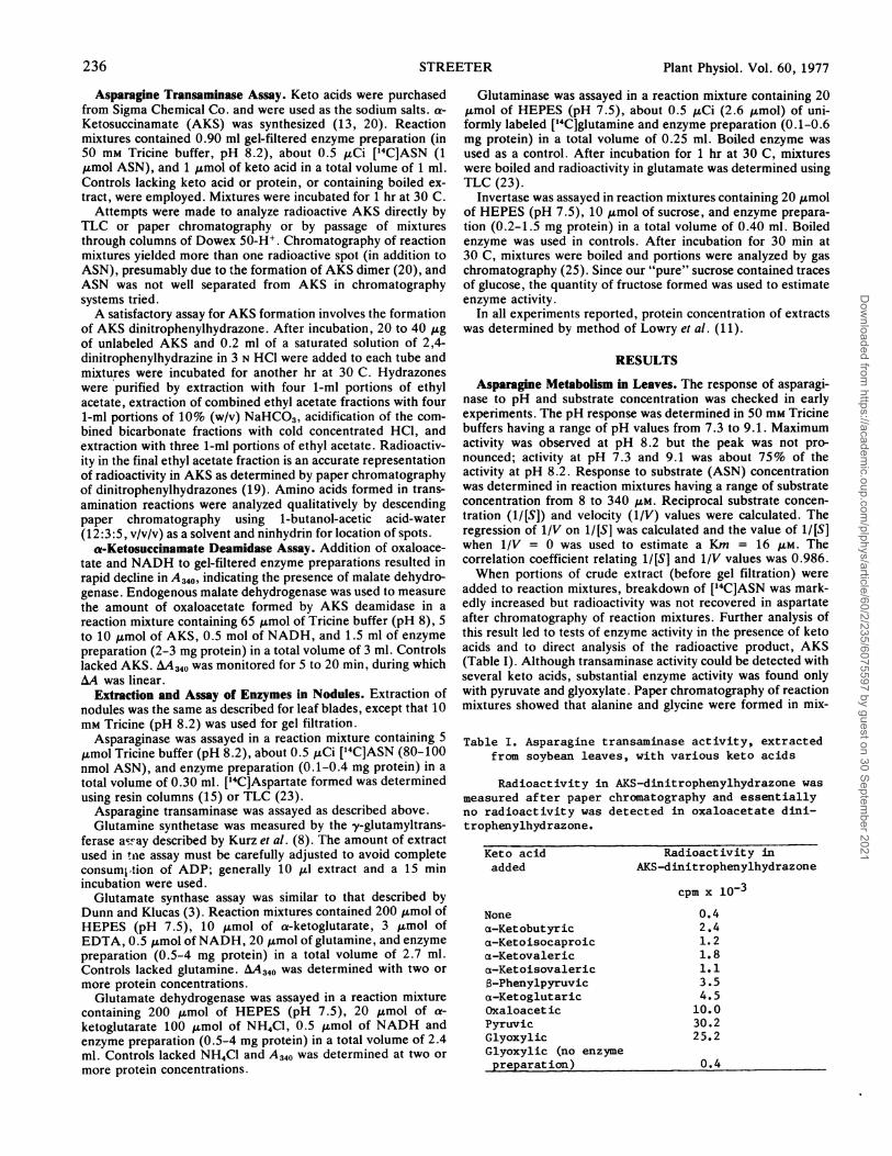
added to reaction mixtures, breakdown of [14C]ASN was mark-edly increased but radioactivity was not recovered in aspartateafter chromatography of reaction mixtures. Further analysis ofthis result led to tests of enzyme activity in the presence of ketoacids and to direct analysis of the radioactive product, AKS(Table I). Although transaminase activity could be detected withseveral keto acids, substantial enzyme activity was found onlywith pyruvate and glyoxylate. Paper chromatography of reactionmixtures showed that alanine and glycine were formed in mix-
Table I. Asparagine transaminase activity, extractedfrom soybean leaves, with various keto acids
Radioactivity in AKS-dinitrophenylhydrazone wasmeasured after paper chromatography and essentiallyno radioactivity was detected in oxaloacetate dini-trophenylhydrazone.
Keto acidadded
Nonea-Ketobutyrica-Ketoisocaproica-Ketovalerica-Ketoisovaleric8-Phenylpyruvica-KetoglutaricOxaloaceticPyruvicGlyoxylicGlyoxylic (no enzymepreparation)
Radioact ivity inAKS-dinitrophenylhydrazone
cpm x 10-30.42.41.21.81.13.54.5
10.030.225.2
0.4
236 STREETER
Dow
nloaded from https://academ
ic.oup.com/plphys/article/60/2/235/6075597 by guest on 30 Septem
ber 2021

ASPARAGINE METABOLISM
tures containing pyruvate and glyoxylate, respectively. Oxaloac-etate spontaneously decarboxylates to pyruvate in solution. Forthis reason, plus the fact that the formation of aspartate was notconfirmed in mixtures containing oxaloacetate, I suspect thatformation of radioactive AKS with oxaloacetate may be due tothe presence of pyruvate.
In spite of efforts to optimize the extraction and assay ofasparaginase, activity was generally about 0.7 nmol/mg pro-tein - hr. In contrast, activity of asparagine transaminase waseasily measured. Since the extraction and assay of the transami-nases may not have been optimum, transaminase activity insoybean leaves would appear to be at least 50 times greater thanthe asparaginase activity (Table II). a-Ketosuccinamate deami-dase (AKS -- oxaloacetate + NH3) was found with a level ofactivity similar to the level of transaminase.An attempt was made to confirm the activity of ASN transam-
inase and AKS deamidase in vivo by feeding radioactive ASN tosoybean leaves. After incubation for 0, 30, 60 or 120 min, leaveswere ground in 75% (v/v) ethanol, and a portion of the extractwas immediately reacted with saturated solution of 2,4-dinitro-phenylhydrazine in 3 N HCI. After purification and chromatog-raphy of hydrazone (19), no accumulation of label was found inAKS or in oxaloacetate. Analysis of amino acids and organicacids (23) revealed slightly more radioactivity in malate thanaspartate but it was not possible to tell which metabolite waslabeled first.
Asparagine Metabolism in Nodules. Radioactive metabolitessuch as pyruvate, succinate, acetate, aspartate, and glyceratehave been supplied to soybean nodules by injection of wholenodules using a ,ul syringe, vacuum infiltration of whole nodules,or incubation of nodule slices on blotter paper saturated with asolution of radioactive metabolite. Tissue was routinely ex-tracted with 75% (v/v) ethanol in these experiments. Results(unpublished) of these studies indicated rapid synthesis of or-ganic acids and amino acids but essentially no synthesis of la-
Table II. Activity of four enzymes extracted from soybean leaves
Activity of each enzyme is the highest observed in severaldifferent experiments; i.e. all four enzymes were not assayed inthe same extract.
Enzyme Enzyme activitynmol mg protein-l hr1
Asparaginase 1.0Asparagine-pyruvate transaminase 52.0Asparagine-glyoxylate transaminase 62.0a-Ketosuccinamate deamidase 85.0
beled asparagine. The importance of asparaginase in soybeannodules became apparent when [14C]ASN was supplied to nod-ule tissue and the result was a rapid hydrolysis of ASN toaspartate. Nearly quantitative conversion of ASN to aspartateoccurred even when nodule tissue was ground in 75% ethanolprior to mixing the extract with ['4C]ASN. After trying severalextraction media, it was found that ethanol containing 10% (v/v)acetic acid or formic acid or 15 A.mol HCl/ml will rapidly destroyasparaginase activity. There was no significant acid-catalyzedhydrolysis in 75% ethanol containing as much as 65 ,umol HCl/ml.Experiments with [14C]ASN indicated the presence in soybean
nodules of asparaginase which retains activity in 75% (v/v)ethanol at room temperature. This finding led to extraction andassay of the enzyme as described under "Materials and Meth-ods." The stability of asparaginase in ethanol was confirmed byadding increasing amounts of absolute ethanol to gel-filtered,buffer extracts of nodules. Assay of the enzyme after exposure toethanol concentrations as high as 50% (v/v) at room tempera-ture for 30 min resulted in only small decreases in specificactivity. Heating a preparation containing 67% ethanol (v/v) to40 C for a few min resulted in complete loss of activity. Thus,extraction of nodules with warm ethanol in in vivo studies ofasparagine synthesis may inactivate asparaginase as efficiently asdilute HCI.The amount of asparaginase activity in nodules can be judged
by comparing it to the activity of other enzymes commonlyassayed in nodule extracts (Table III). There was essentially noasparagine transaminase activity in nodules. Nodule extractscontained more asparaginase than glutamate synthase or gluta-minase and there was several hundred times as much asparagi-nase in nodules as in soybean leaves (Table II). The concentra-tion of asparaginase in bacteroids was approximately six times asgreat as the concentration in cytosol (Table III). Recovery ofbacteroids by the methods used is variable and incomplete, but itis possible to estimate that total asparaginase activity in cytosolwas about double the activity in bacteroids.Response of nodule asparaginase to substrate concentration
was determined in reaction mixtures having a range of substrateconcentrations from 1.4 to 20 ,UM. It was necessary to use a verylow protein concentration (40 ,g protein/assay) and short incu-bations (20 min) in order to determine accurate initial velocities.The regression of 1/V on 1I[S] was calculated and the value of 1/[S] when 1/V = 0 was used to estimate a Km = 4.9 ,tM. Thecorrelation coefficient relating 1I[S] and 1/V values was 0.987.The very small Km for nodule asparaginase is similar to the Km
Table III. Activity of enzymes extracted from whole soybean nodules or present in bacteroids and cytosolIn Expt. II, bacteroids were prepared by the method of Evans, et al. (4). After washing, bacteroids
were disrupted by 10 to 12 30-sec. periods of sonication over 20 minutes. In Expt. III, nodules were groundin 0.25 M Tricine buffer, pH 7.5 containing 10 mM mercaptoethanol. Otherwise, preparation of bacteroidswas the same as in Expt. II. After sonication, Triton X-100 was mixed with the bacteroid preparation(10 pl Triton/ml prep.) and allowed to stand 15 min. In all experiments all preparations were gel-filtered(23) using 10 mM Tricine pH 8.2; mercaptoethanol (1 mM) was added to the filtration buffer in Expt. III.
Enzyme Activity Experiment I Experiment II Experiment III Ratio of activitiesWhole nodules Bacteroid Cytosol Bacteroid Cytosol Bacteroid . cytosol
pmol mg protein-l hr-1 (Avg. 2 expts.)Glutamine synthetase(transferase) 47.1 5.6 48.4 15.8 70.9 0.17
Invertasel 3.44 0 1.72 0 3.01 0Glutamate dehydrogenase 0.36 2.4 0.24 4.9 0.41 11.Asparaginase 0.28 0.81 0.14 1.35 0.24 5.7Glutamate synthase 0.23 0.26 0.26 0.53 0.45 1.1Glutaminase 0.11 0.27 0.06 0.57 0.17 3.9Asparagine: pyruvatetransaminase2 0.0068 NA3 NA NA NA -
Asparagine: glyoxylatetransaminase 0.0027
1pmol sucrose hydrolyzed
2a-ketoglutarate was also tested but activity was less than 1.0 nmol mg protein41 hr13Not assayed
Plant Physiol. Vol. 60, 1977 237
Dow
nloaded from https://academ
ic.oup.com/plphys/article/60/2/235/6075597 by guest on 30 Septem
ber 2021

Plant Physiol. Vol. 60, 1977
values reported for the enzyme from several species of bacteria(7, 28).
DISCUSSION
Asparagine Metabolism in Leaves. There are few, if any,
reports which demonstrate conclusively any mechanism for as-
paragine metabolism in green plant tissues. In a recent study oftwo legumes, Lees and Blakeney (10) have demonstrated the
presence of asparaginase in roots and nodules and their dataindicate a trace of enzyme activity in shoots. However, specific
activities were not reported. Atkins et al. (1) have recentlyreported the presence of an active asparaginase in embryos ofdeveloping Lupinus alba seeds. The reported Km of this enzymewas quite high (10 mM) but was similar to the recently reportedKm for asparaginase from Lupinus polyphyllus seeds (9).The effect of keto acids on the breakdown of asparagine in
vitro was first noted by Greenstein and Price (6) in studies withrat liver extracts. Later work by Meister et al. (13, 14) explainedthe effect as not keto acid stimulation of asparaginase! but as
resulting from transamination and subsequent deamid ion ofthe transamination product a-ketosuccinamate.
Several studies conducted since Meister's reports hay sug-
gested the presence of asparagine transaminase activity in p nttissues (5, 26, 29). These workers used an assay for formation f
an amino acid corresponding to the keto acid added to reaction\mixtures. It is possible that their results can be explained by thepresence of asparaginase plus the transamination of the aspar-
tate formed, or by the presence of a trace of some amino acidother than asparagine in reaction mixtures. This latter possibilityseems especially likely because significant proteolysis could haveoccurred during the long incubations (2-3 hr) which were em-
ployed. With these potential complications, it seems obviousthat an assay for AKS is required where asparagine transamina-tion is suspected.Based on the data in Table II, it is suggested that although
asparaginase may be present in green plant tissues, transamina-tion is a more likely route for asparagine metabolism. AKSdeamidase was also demonstrated in soybean leaves. The pres-
ence of this enzyme in plant tissues has previously been reportedby Meister (13). My initial attempt to demonstrate the operationof the transaminase in vivo was unsuccessful. This work needs tobe repeated with more effort directed toward trapping radioac-tivity from ASN in AKS; transamination and deamidation reac-
tions may be closely coupled (14) so that it may be possible onlyto show the labeling of oxaloacetate prior to the labeling ofaspartate.Based on the seasonal average asparagine concentration in
stem exudate and the average exudate flow rate (21), it was
possible to estimate the influx of asparagine to soybean leaves as
25 nmol/g fresh wt -hr. The average protein concentration of 10enzyme preparations and theweight of the leaf tissue extractedwere used to calculate the buffer-extractable protein in soybeanleaves (23 mg/g fresh wt). Although there are many obviouslimitations to this approach, these calculations led to the conclu-sion that there is insufficient asparaginase to metabolize aspara-
gine entering soybean leaves, whereas the transaminase activityis more than adequate to metabolize incoming asparagine.Asparagine Metabolism in Nodules. Lees and Blakeney (10)
have previously reported the presence of asparaginase in legumeroot nodules. This report confirms and extends their observa-tions. Whereas the activity of asparagine transaminase was al-most nil, the activity of asparaginase in soybean nodules isapproximately the same order of magnitude as other noduleenzymes which have recently received attention (Table III andrefs. 2, 3, 8, 16, 17). The specific activities and the distributionof activity in bacteroid versus cytosol are comparable to resultsreported by others, with the exception of glutaminase and gluta-
mate dehydrogenase. The activity and distribution of glutamin-ase in legume nodules have not previously been reported. Ifound much higher glutamate dehydrogenase activity in soybeannodules than Dunn and Klucas (3) and also in contrast to theirresults, found the highest concentration of the enzyme in bacte-roids. Apparently, the amount and distribution of glutamatedehydrogenase activity in nodules are highly variable amonglegume species (2).Soybean nodule asparaginase was concentrated in bacteroids
suggesting that the enzyme is of bacterial origin. Activity in thecytosol may have been the result of loss from the bacteroidsduring the isolation procedure. The fact that large amounts ofasparagine are synthesized in and exported from nodules (21,27) also makes it attractive to suggest that asparaginase is local-ized in bacteroids allowing asparagine synthesis to occur inuninfected tissue, perhaps in the nodule cortex near the vascularbundles.The remarkable stability and the extremely small Km (5 ,UM)
of the nodule asparaginase seriously complicate efforts to dem-onstrate asparagine synthesis in vitro or in vivo because of thepotential for rapid destruction of the product, ASN. My experi-ence indicates that ASN hydrolysis occurs whenever the noduleis cut or punctured in order to supply radioactive materials.However, the knowledge that this enzyme is present will make itpossible to adopt measures to circumvent it in future studies ofasparagine synthesis.An enzyme from lupin nodules which catalyzes glutamine-
dependent asparagine synthesis has recently been reported (18).Lupin nodules also contain asparaginase and it is interesting tonote that asparagine synthetase was not detected until a stage ofnodule development where asparaginase could no longer be
detected. Although I have not systematically studied asparagi-nase activity as a function of nodule age, asparaginase was foundin many different samples of soybean nodules ranging in agefrom 20 to 60 days after initiation of nodule growth.
Ackowledgments -I thank A. Meister for advice on the synthesis of cs-ketosuccinamate, M.
Bosler for general technical assistance, and D. Murphy for suggesting the feeding of
['4C]asparagine to nodules.
LITERATURE CITED
1. ATKINS CA, JS PATE, PJ SHARKEY 1975 Asparagine metabolism-key to the nitrogen
nutrition of legumes. Plant Physiol 56: 807-8122. BRowN CM, MJ DILWORTH 1975 Ammonia assimilation by Rhizobium cultures and bacte-
roids. J Gen Microbiol 86: 39-483. DUNN SD, RV KLUCAS 1973 Studies on possible routes of ammonium assimilation in
soybean root nodules. CanJ Microbiol 19: 1493-14994. EvANs HJ, B KOCH, R KLUCAS 1972 Preparation of nitrogenase from nodules and separa-
tion into components. Methods Enzymol 24: 470-4765. Foaxar JC, F WIGHTMAN 1972 Amino acid metabolism in plants. II. Transamination
reactions of free protein amino acids in cell-free extracts of cotyledons and growing tissues
of bushbean seedlings (Phaseolus vulgaris L.). CanJ Biochem. 50: 538-542
6. GREENSTEIN JP,VE PaICE 1949 a-Keto acid-activated glutaminase and asparaginase. JBiol Chem. 178: 695-705
7. IMADA A, S IGARASI, K NACAHAMA, M ISONO 1973 Asparaginase and glutaminase activities
in micro-organisms.J Gen Microbiol 76: 85-998. Kuxz WGW, DA ROKOSH, TA LARUE 1975 Enzymes of ammonia assimilation in Rhizo-
bium leguminosarum bacteroids. CanJ Microbiol 21: 1009-1012
9. LEA PJ, L FOWDEN, BJ MIFLIN 1976 Asparagine breakdown in the leaves and maturing
seeds. Plant Physiol 57: S-4010. LEEs EM, AB BLAKENEY 1970 The distribution of asparaginase activity in legumes.
Biochim Biophys Acta 215: 145-15111. LowRy OH, NJ ROSEBROUGH, AL FAi, RJ RANDALL 1951 Protein measurement with the
Folin phenol reagent.J Biol Chem 193: 265-27512. MARnN DW Jr 1972 Radioassay for enzymatic production of glutamate from glutamine.
Anal Biochem 46: 239-24313. MEisTR A 1953 Preparation and enzymatic reactions of the keto analogues of asparagine
and glutamine.J. Biol Chem. 200: 571-58914. MEisTaR A, HA SoBER,SV TICE, PE FRAsER 1952 Transamination and associated deami-
dation of asparagine and glutamine.J Biol Chem 197: 319-330
15.PNUSINERS, L MILNER 1970 A rapid radioactive assay for glutamine synthetase, glutamin-
ase, asparagine synthetase and asparaginase. Anal Biochem 37: 429-438
16. ROBERTSONJG, KJF FARNDEN, MPWABURTON,JM BANKS 1975 Induction of glutamine
synthetase during nodule development in lupin. AustJ Plant Physiol 2: 265-272
17. ROBERTSONJG, MP TAYLOR 1973 Acid and alkaline invertases
238 STREETER
Dow
nloaded from https://academ
ic.oup.com/plphys/article/60/2/235/6075597 by guest on 30 Septem
ber 2021

Plant Physiol. Vol. 60, 1977 ASPARAGINE METABOLISM 239
Lupinus angustifolius infected with Rhizobium lupini. Planta 112: 1-6 seedlings. Arch Biochem Biophys 157: 613-62418. Scorr DB, KJF FAOEN, JG ROBERrSON 1976 Ammonia assimilation in lupin nodules. 24. STREEaaa JG 1974 Asparaginase and asparagine transaminase from soybean leaves. Plant
Nature 263: 703-705 Physiol 53: S-6619. SmrrH P 1967 Paper chromatography of keto acid, 2,4-dinitrophenylhydrazones. J Chroma- 25. STERrEE JG, ME BOSLER 1976 Carbohydrates in soybean nodules: identification of
togr 30: 273-275 compounds and possible relationships to nitrogen fixation. Plant Sci Lett 7: 321-32920. STEPHANi RA, A MEisTr 1971 Structure of the dimeric c-keto acid analogue of aspara- 26. WILSON DG, KW KING, RH Buas 1954 Transamination reactions in plants. J Biol Chem
gine. I Biol Chem 246: 7115-7118 208: 863-87421. STsEEra JG 1972 Nitrogen nutrition of field-grown soybean plants. I. Seasonal variations 27. WONG PP, HJ EvANs 1971 Poly-,-hydroxybutyrate utilization by soybean (Glycine max
in soil nitrogen and nitrogen composition of stem exudate. Agron J 64: 311-314 Menf.) nodules and assessment of its role in maintenance of nitrogenase activity. Plant22. STErEna JG 1972 Nitrogen nutrition of field-grown soybean plants. II. Seasonal variations Physiol 47: 750-755
in nitrate reductase, glutamate dehydrogenase and nitrogen constituents of plant parts. 28. WRITON JC Jx, TO YELLIN 1973 L-Asparaginase: a review. Adv Enzymol 39: 185-248Agron 1 64: 315-319 29. YAMAmoTo Y 1955 Asparagine metabolism in the germination stage of a bean, Vigna
23. STEarsE JG 1973 In vivo and in vitro studies on asparagine biosynthesis in soybean sesquipedauis. J Biochem 42: 763-774
Dow
nloaded from https://academ
ic.oup.com/plphys/article/60/2/235/6075597 by guest on 30 Septem
ber 2021