Embed Size (px)
Citation preview
THE MORPHOLOGY AND CYTOLOGY OFMYXOCOCCUS XANTHUS, N. Sp.,
J. M. BEEBEBacteriology Section, Agricultural Experiment Station, Iowa
State College, Ames, Iowa
Received for publication December 26, 1940
Knaysi (1938) states that there are three current points of viewrelative to the presence and nature of the bacterial nucleus. Onegroup of investigators holds that the bacterial cell contains nonucleus. Knaysi argues, however, that the fact that a nucleushas not been seen is no indication that it does not exist. A secondgroup maintains that nuclear material is present but in a highlydispersed condition and is therefore not easily seen. The thirdgroup takes the stand that the presence of a nucleus is logical, andis indicated by granular bodies often noted within the bacterialcell. These bodies are frequently observed to behave both func-tionally and chemically as nuclei are supposed to behave. Ingeneral, this latter view, that a more or less definite nucleus doesexist, at least during certain stages of the life cycle of the bacterialcell, seems to be favored by many investigators today.Stoughton (1929) observed the presence of stainable bodies in
the cells of Bacterium malvacearum. Just before cell divisionoccurred these bodies split, the halves migrating toward oppositeends of the cell, and finally were included in the new daughtercells. The dye reactions of these intracellular bodies were typi-cally nuclear, and the author concluded that they should be con-sidered as nuclei.Much of the work published by the Hollandes (1930, 1932)
favors the theory of a compact nucleus. Lindegren and Mellon(1932) described a type of reduction-division that took place
1 Journal Paper no. J825 of the Iowa Agricultural Experiment Station, Ames,Iowa. Project no. 571.
193
on June 26, 2018 by guesthttp://jb.asm
.org/D
ownloaded from
J. M. BEEBE
within bacterial cells during certain phases of the life cycle.Badian (1935) stated that the condensed nuclear material to beseen in Bacillus megatherium may be observed to divide and rear-range itself in a manner quite suggestive of autogamy. Brooke(1936) studied a number of pathogenic bacteria and from hisobservations concluded that the granules he noted were composedof nuclear material. Allen, Appleby and Wolf (1937), workingwith a spore-forming bacillus, noted deep-staining bodies withinthe spores as well as within the vegetative cells. These bodieswent through a rearrangement process after spore formation hadtaken place, forming recognizable structures of various shapes.The authors contended that the spore has some function otherthan merely acting as a resting stage during unfavorable periodsof growth. Rosca (1937) described a type of nuclear activitythat he considered asexual in certain cases, sexual in others.Although he did not actually observe the conjugation of two cellshe could distinguish two types of chromosomes and consideredthem to be o and 9.
Chance's (1938) observations on Bacillus mesentericus discloseda reorganization and redistribution of chromatin comparable toa mitotic division in higher forms, and Allen, Appleby and Wolf(1939) gave evidence of meiosis having taken place in a species ofBacillus which they studied. These same investigators also wit-nessed and accounted for at least two methods of spore formationin the one organism, and methods of reproduction other than theusual transverse fission. Lewis (1940) concluded that the theoryof a more or less condensed nucleus, or at least a chromatin-incrusted gene string, is preferable to the theory of a diffusenucleus. This worker pointed out that hypotheses of diffusenuclei are not in keeping with findings on the transmission ofhereditary characteristics.Many of the publications on this subject deal with observations
on spore-forming organisms. Possibly this is true because suchorganisms appear to undergo more marked cell reorganizationjust prior to, and during, spore formation. To the author'sknowledge only two papers dealing with the cytology of myxo-bacteria have appeared. Krzemieniewski (1928) studying cells
194
on June 26, 2018 by guesthttp://jb.asm
.org/D
ownloaded from

MYXOCOCCUS XANTHUS, N. SP.
of Sorangium sorediatum, Polyangium fuscum and of species ofMyxococcus, Archangium, etc., and Badian (1930) working withMyxococcus virescens observed deeply-stained granules within thecells. These structures underwent a type of mitosis during thevegetative period of growth, and just before, and during, theprocess of sporulation displayed great variety of forms. Badianobserved these intracellular structures to double in number, andthen decrease by means of chromatin rearrangements. This wasinterpreted as a reduction-division, the whole process being con-sidered a type of autogamy.The present study began with the isolation and purification
of a previously undescribed species of Myxococcus. In a numberof ways this organism appears to be closely related to Myxococcusvirescens (the species studied by Badian), particularly in the sizeof the vegetative cells and spores. These are larger than thoseof most of the other species of this group that have been observedin this laboratory. Because of the large size of the cells and theremarkable intracellular figures (one of the first things to benoticed), the species seemed to be well-adapted for cytologicalstudy. The species has been separated from M. virescens on thebasis of the pigmentation of the fruiting body, a characteristicoften used in the differentiation of species of Myxobacteriales.The name Myxococcus xanthus is proposed for this organism.The diagnosis is as follows:
MYXOCOCCUS XANTHUS
Etymology: Greek (adj.) = orange, golden.Fruiting body: Spherical to subspherical, usually sessile but occa-
sionally constricted at the base giving the appearance of a short stalkor foot. Mature fruiting body 300 to 400%u in diameter, often slightlyflattened on top or on one side. Color varies from light yellowish-orange when young to bright orange when mature; color constant,never tending toward greenish yellow. No outer cyst wall or membranediscernible, the spores being imbedded in the slime holding the masstogether. Usually single, though two or three fruiting bodies may be-come joined to form an irregular mass; each is attached to the substrate,however, and they never bud, one from another.
Spores (Resting cells): Spherical, with thick outer wall or membrane.
195
on June 26, 2018 by guesthttp://jb.asm
.org/D
ownloaded from
J. M. BEEBE
highly refractile when unstained. Stain very easily with any of theordinary bacterial or nuclear dyes. 2.0g in diameter, sometimesslightly larger.
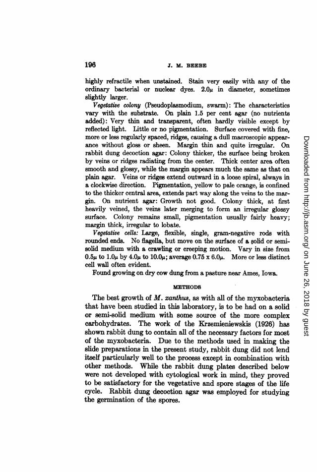
Vegetative colony (Pseudoplasmodium, swarm): The characteristicsvary with the substrate. On plain 1.5 per cent agar (no nutrientsadded): Very thin and transparent, often hardly visible except byreflected light. Little or no pigmentation. Surface covered with fine,more or less regularly spaced, ridges, causing a dull macroscopic appear-ance without gloss or sheen. Margin thin and quite irregular. Onrabbit dung decoction agar: Colony thicker, the surface being brokenby veins or ridges radiating from the center. Thick center area oftensmooth and glossy, while the margin appears much the same as that onplain agar. Veins or ridges extend outward in a loose spiral, always ina clockwise direction. Pigmentation, yellow to pale orange, is confinedto the thicker central area, extends part way along the veins to the mar-gin. On nutrient agar: Growth not good. Colony thick, at firstheavily veined, the veins later merging to form an irregular glossysurface. Colony remains small, pigmentation usually fairly heavy;margin thick, irregular to lobate.
Vegetative cells: Large, flexible, single, gram-negative rods withrounded ends. No flagella, but move on the surface of a solid or semi-solid medium with a crawling or creeping motion. Vary in size fromO.Spu to 1.0ku by 4.Ou to 10.Oj; average 0.75 x 6.Op. More or less distinctcell wall often evident.Found growing on dry cow dung from a pasture near Ames, Iowa.
METHODS
The best growth of M. xcanthus, as with all of the myxobacteriathat have been studied in this laboratory, is to be had on a solidor semi-solid medium with some source of the more complexcarbohydrates. The work of the Krzemieniewskis (1926) hasshown rabbit dung to contain all of the necessary factors for mostof the myxobacteria. Due to the methods used in making theslide preparations in the present study, rabbit dung did not lenditself particularly well to the process except in combination withother methods. While the rabbit dung plates described belowwere not developed with cytological work in mind, they provedto be satisfactory for the vegetative and spore stages of the lifecycle. Rabbit dung decoction agar was employed for studyingthe germination of the spores.
196
on June 26, 2018 by guesthttp://jb.asm
.org/D
ownloaded from

MYXOCOCCUS XANTHUB, N. BP.
Incubation was, for the most part, at room temperature. Thework was carried on largely during the summer months and thelaboratory temperatures of 22 to 270C. proved to be satisfactory.Temperatures above 30'C. were not beneficial. At room tem-perature germination occurred in about 24 hours.
Culture methodRabbit dung plates were prepared by placing two or three pieces
of rabbit dung in each petri dish and sterilizing, with the lids on,for one hour at 15 pounds pressure. When cool the plates wereremoved from the autoclave and allowed to stand over night inorder to dry somewhat. Plain 1.5 per cent agar (no nutrientsadded) was then prepared. This was melted and sterilized inthe autoclave for 20 minutes at 15 pounds pressure. When cooledto about 60'C. enough of the liquid agar was added to each plateto fill it to a depth of about one-half the diameter of the rabbitdung. This required 15 to 20 ml. of agar. Care was taken notto cover the dung with agar. Before the agar had a chance toharden, the pieces of dung were moved to the center of the platewith a flamed needle. When the agar had set the plates wereinverted. Inoculations were made by transferring one or twofruiting bodies from the stock cultures to the dung at the levelof the surface of the agar. Diffusion of the water-soluble partsof the dung into the surrounding agar made growth possible onthe surface of the latter. Microscopic slide preparations weremade from the colonies that developed on the agar around theimbedded pieces of dung.Dung decoction agar was used in studying germination. This
substrate was made by adding one liter of distilled water to 100grams of dry dung. The mixture was heated to boiling and thenallowed to infuse at room temperature for 24 hours. The solidmaterial was then filtered off, and the filtrate made up to one literby the addition of distilled water. Fifteen grams of agar wereadded and the solution sterilized in the autoclave for 30 minutesat 15 pounds pressure. When cooled to 60'C. plates were poured.A fairly heavy suspension of spores was then made by trans-
ferring a few fruiting bodies to a sterile slide, inclosed in a sterilepetri dish, on which a drop or two of sterile water had been placed.
197
on June 26, 2018 by guesthttp://jb.asm
.org/D
ownloaded from

J. M. BEEBE
The fruiting bodies were broken up with a flamed needle. Loop-fuls of this suspension were then transferred to various pointsmarked on the dung decoction agar plates. Incubation was atroom temperature and slides were made at 12-, 24-, 48- and 72-hour intervals.
Methods of slide preparationOriginally an attempt was made to study the cells of M. xanthus
by employing the usual type of smear technique. This was foundto be entirely unsatisfactory. It was impossible to spread thematerial out on the slide, once it had been stripped from the agarsubstrate, due to the membranous nature of the colony. Thosefew cells that had been teased away from the mass were often sobadly bent and distorted that the size and shape were questionable.Also, the fact that no indication of the disposition of the cells onthe colony was to be had by this method made it imperative touse some other means.
Cover slip preparations were finally decided upon as beingmost satisfactory. For this purpose number one cover slips,seven-eighths inch square, were used. These were cleaned welland stored in alcohol. Just before use they were rinsed in 95 percent alcohol and flamed. This was repeated twice. After thefinal flaming the cover slip was placed, by means of sterile forceps,over that section of the colony to be studied, and pressed downcarefully to assure good contact. The only difficulty was withair bubbles, and it was found that with a little care these couldbe pressed out from under the glass. Generally the cover slipswere left in place for about two minutes, though the time factorwas not critical. The cover slips were then removed with sterileforceps, care being taken to raise the glass, not to slide it. Partof the plan was to obtain a picture of cell distribution and anysliding motion would, of course, disturb the location of thebacteria.When dried, the cover slips were placed in 95 per cent ethyl
alcohol for fixation. A fixing time gf three minutes was found P5be satisfactory. Various mixtures of alcohol and xylol were also
198
on June 26, 2018 by guesthttp://jb.asm
.org/D
ownloaded from
MYXOCOCCUS XANTHUS, N. SP.
tried as fixatives but no improvement could be noted over thealcohol alone. After fixation the slides were allowed to dry inthe air.
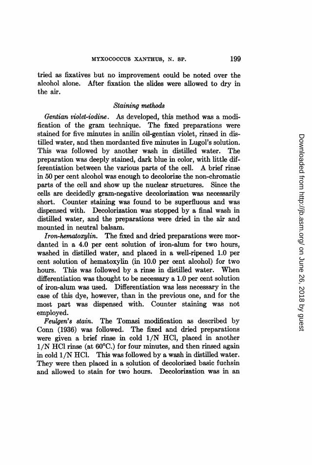
Staining methodsGentian violet-iodine. As developed, this method was a modi-
fication of the gram technique. The fixed preparations werestained for five minutes in anilin oil-gentian violet, rinsed in dis-tilled water, and then mordanted five minutes in Lugol's solution.This was followed by another wash in distilled water. Thepreparation was deeply stained, dark blue in color, with little dif-ferentiation between the various parts of the cell. A brief rinsein 50 per cent alcohol was enough to decolorize the non-chromaticparts of the cell and show up the nuclear structures. Since thecells are decidedly gram-negative decolorization was necessarilyshort. Counter staining was found to be superfluous and wasdispensed with. Decolorization was stopped by a final wash indistilled water, and the preparations were dried in the air andmounted in neutral balsam.
Iron-hematoxylin. The fixed and dried preparations were mor-danted in a 4.0 per cent solution of iron-alum for two hours,washed in distilled water, and placed in a well-ripened 1.0 percent solution of hematoxylin (in 10.0 per cent alcohol) for twohours. This was followed by a rinse in distilled water. Whendifferentiation was thought to be necessary a 1.0 per cent solutionof iron-alum was used. Differentiation was less necessary in thecase of this dye, however, than in the previous one, and for themost part was dispensed with. Counter staining was notemployed.
Feulgen's stain. The Tomasi modification as described byConn (1936) was followed. The fixed and dried preparationswere given a brief rinse in cold 1/N HC0, placed in another1/N H1 rinse (at 60'C.) for four minutes, and then rinsed againin cold 1/N HC1. This was followed by a wash in distilled water.They were then placed in a solution of decolorized basic fuchsinand allowed to stain for two hours. Decolorization was in an
199
on June 26, 2018 by guesthttp://jb.asm
.org/D
ownloaded from

J. M. BEEBE
acidified potassium metabisulphite solution. After rinsing in dis-tilled water and drying, the cover slips were mounted in neutralbalsam. Counter staining was not found to be practical.The quality of the basic fuchsin used in this test is of prime
importance. The first dye lot that was tried gave very poorresults; another lot, certified for the particular purpose, was foundto be satisfactory. Checks were run on slides of onion root tipin order to make sure that the procedure, as well as the dye, wascorrect before attempts were made to stain bacteria.
Other stainsA number of other dyes, for the most part the usual bacterial
dyes, was tried in connection with this study. The majorityof them were found to be unsatisfactory. Spore stains such asthe Ziehl-Neilsen stain, the malachite green stain, etc., gave nega-tive results on both spores and vegetative cells of M. xanthus.Both types of cells decolorized rapidly and completely. How-ever, some of the differences between the spores of this organismand those of the true spore-forming bacteria were emphasized.Gram's stain gave negative results. Loeffler's methylene blue,while satisfactory in part, gave results inferior to those obtainedwith gentian violet and iron-hematoxylin. Safranin and carbolfuchsin were not satisfactory. Aceto-carmine and iron-aceto-carmine were expected to give good reactions but rather poorresults were had with both. Sudan III was tried in an attemptto show that the vacuoles occasionally seen within the cells wereof fatty materials, but without success, and iodine in the form ofLugol's solution and as a dilute alcoholic solution was used asa test for glycogen. It gave negative results in the case of thevegetative cells, doubtfully positive in the case of the spores.
Forty-five millimeter petri dishes were employed for all pro-cedures that required more than a few minutes for mordantingand staining. These were more satisfactory than larger con-tainers since the small preparations could be processed separately
MORPHOLOGY AND CYTOLOGY
The germination of the spores, or resting cells, might be con-sidered as the beginning of the life cycle of M. xanthus, but the
200
on June 26, 2018 by guesthttp://jb.asm
.org/D
ownloaded from

MYXOCOCCUS XANTHUS, N. SP.
vegetative phase appears to be the dominant one; for that reasonthe vegetative phase is considered first. The vegetative cells
A
; -
FIG. 1. RECONSTRUCTION OF THE LIFE CYCLE OF M. XANTHUS
Originally drawn to a scale: 1 cm. = 1 p,. A, mature spore; B, early germinalstage; C-H, germination; I-L, vegetative phases; M, transitional; N-0, pro-phase; P, early sporulation stage; Q-R, sporulation; S, mature spore.
of Myxococcus xanthus are long, flexible, gram-negative rods withrounded ends (Figs. 1 (I-L), 9 to 11). They have no flagella butappear to move by means of a crawling or creeping motion on top
9201
jI
f'.
II&K
?i
on June 26, 2018 by guesthttp://jb.asm
.org/D
ownloaded from

J. M. BEEBE
of the layer of slime with which they pave the substrate. Theexact nature of this motion is not well understood, but it has beensuggested to be the result of an asymmetric excretion of slime bythe cell, producing more pressure on one end than on the other.No wriggling motion such as is usually associated with motilebacteria can be observed, but the cell can be seen to change itsposition, from time to time, in relation to some fixed point.Examinations of living preparations of M. xanthus have shownthe cells to travel at a rate of about 7/i per minute. The cellsalways move away from the center of the colony, but not in astraight line: there is always a curving of the path in a clockwisedirection. This varies in intensity but may usually be noted tosome degree.The distribution of the cells on the colony as shown in figures 9
to 11 is characteristic not only of this particular species but ofthe genus. Very seldom is more than a single layer of cells ob-served on a colony. The cells are usually arranged in small groupsof from two or three to a dozen or more, lined up with their longaxes parallel, and moving in the same direction. The entire groupmoves as a unit, along a "front." While a few of the cells seemto be traveling independently, the large majority always followthe same slightly curved path toward the margin of the colony.Generally they are not packed closely together, but are separatedby about the diameter of one cell.The vegetative cells average 6.0/i in length by about 0.75,u in
diameter. Unstained, the cells are long flexible rods, usuallyrounded on the ends. However, they occasionally appear to beslightly tapered at one or both ends, particularly those cells atthe margin of the colony; very few are spindle-shaped. Moreoften than not the younger unstained cells show several highlyrefractile granular bodies, varying in number from two to eight,while the older vegetative cells show one or two rather largerefractile bodies at or near the center of the cell. The fixing andstaining procedures previously outlined appear to have very littleeffect on cell morphology, the only noticeable change being anextremely slight swelling at the ends of the younger cells. Thisgives them the cylindrical appearance of the older cells.
202
on June 26, 2018 by guesthttp://jb.asm
.org/D
ownloaded from

MYXOCOCCUS XANTHUS, N. SP.
The cells stain fairly well with most of the usual bacterial dyes,in some cases a thin cell wall being visible. The center of thecell is occupied by a deeply-staining body approximately one-third the length of the cell and equal in diameter to the cell; i.e.,about 2.0 by 0.75,j. As far as can be told at the present time thisbody has no limiting wall or membrane, but is rather a mass ofcompact nuclear material. It has a marked affinity for gentianviolet and for iron-hematoxylin, and gives a positive reaction withFeulgen's stain. The remaining portions of the cell take all stainslightly. Occasionally the cell appears to contain vacuoles at oneor both ends but tests for glycogen and for fats have failed toindicate the nature of the vacuolar material. Functionally thecondensed body located at the center of the cell seems to be nu-clear, also. Vegetative reproduction is by means of transversefission. Prior to cell fission a division of the nucleus takes place.Stages in this process may be seen in figures 1 (I-L), 9 to 11. Atfirst the nucleus enlarges longitudinally to almost twice its lengthand then begins to constrict at a point near its center, producinga dumbbell-shaped body. The halves finally pull apart andmigrate toward opposite ends of the cell. After nuclear divisionhas been completed actual cell fission begins. This also is accom-plished by constriction and not by the formation of a transversewall. The steps in the process are shown in figures 1 (I-L) and9 to 11. No case has been observed in which the daughter cellsremain united in pairs after cell division has been completed.Each new cell contains the single, deep-staiing, compact nu-cleus. Sometimes, during periods of rapid growth, a secondnuclear division takes place before fission begins, and in suchcases cells with four nuclei may be noted. These are not to beconfused with a later stage in which four stainable bodies aretypical.The vegetative phase continues for 4 to 10 days, the time de-
pending upon conditions of temperature and environment. Atroom temperature (21 to 250C.) and on rabbit dung plates thefirst signs of fruiting body formation begin to be noticed afterfive to six days. Morphologically, a slight increase in the diame-ter of the cells is the. first indication of the change about to occur
203
on June 26, 2018 by guesthttp://jb.asm
.org/D
ownloaded from

J. M. BEEBE
(fig. 1 (M), 12). Most of the cells measure approximately 1.01.in diameter in this early transitional phase, and this increase indiameter may also be accompanied by a slight decrease in length.At first there is not much change in the internal structure of thecell, but soon the nucleus is noted to have broken up, and par-ticles of deep-staining material are to be seen throughout thelength of the cell (figs. 1 (M), 12 to 16). During this stage thecells begin their migrations toward some predetermined "fruitingcenter" on the colony.The stimulus for the gathering of the cells from within a given
area for the express purpose of sporulation and the formation offruiting bodies is not understood. No apparent change in tem-perature or environment is necessary although a lack of foodmaterial may play a part. More likely would be the productionof metabolic products by the cells, or a slight change in the mois-ture content of the medium. At any rate, the motion of the cellstoward the margin of the colony suddenly ceases, and all cellswithin a given area begin to move toward a central point. Thisis best shown in figure 17. As the bacteria approach this point,the morphological and cytological changes taking place are par-ticularly marked. Observations by several workers on a speciesof Bacillus, and by Krzemieniewski (1928) and Badian (1930) onspecies of myxobacteria, have indicated that a number of changesoccur within the cell prior to sporulation; whether, as in the caseof species of the Bacillaceae, the spore is formed as a structurewithin the cell, or whether, as in the case of Myxococcus xanthusand other myxobacteria, the cell is the spore, rearrangement andredistribution of chromatin material appears to be a preliminarystep in sporulation.As they approach the location where the fruiting body is to
be formed, or is in the process of formation, the cells increase indiameter and become perceptably shorter (figs. 1 (N), 3, 14,15, 16), and rearrangements in the chromatin material becomequite apparent. The chromatin previously distributed through-out the length of the cell collects in four more or less distinctmass (figs. 12 to 16). These bodies are not arranged withinthe cell in any particular fashion in the earlier stages, nor do they
~204
on June 26, 2018 by guesthttp://jb.asm
.org/D
ownloaded from

MYXOCOCCUS XANTHUS, N. SP. 205
1a)
N O 0
FIG. 2. DIAGRAMMATIC OUTLINE OF NUCLEAR DIVISIONS; NOT DRAWN TO SCALEA-F, vegetative phases; G-J, prophase; K, late prophase; L, chromosomal
fusion; M, nuclear fusion; N-0, spore, mononucleate, diploid; P-Q, early ger-minal, diploid; R-S, germination; T, binucleate vegetative; U, post-germinaldivision; V, haploid vegetative cells.
on June 26, 2018 by guesthttp://jb.asm
.org/D
ownloaded from
J. M. BEEBE
t9
__
0*1.
3I/0, .
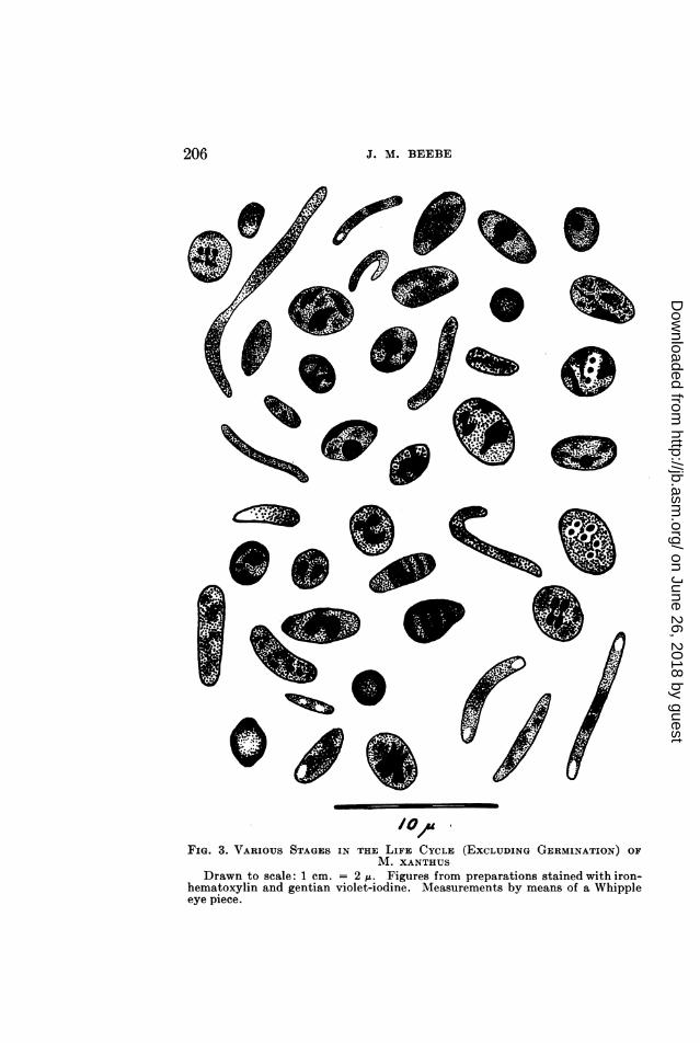
FIG. 3. VARIOUS STAGES IN THE LIFE CYCLE (EXCLUDING GERMINATION) OFM. XANTHUS
Drawn to scale: 1 cm. = 2 1. Figures from preparations stained with iron-hematoxylin and gentian violet-iodine. Measurements by means of a Whippleeye piece.
206
wokk on June 26, 2018 by guesthttp://jb.asm
.org/D
ownloaded from

MIYXOCOCCUS XANTHUS, N. SP.
seem to have any particular shape although their outlines aresharp. In general, they give the cell a banded appearance; twoof them are usually located at the poles, the other two near thecenter of the cell. At this stage the cell is about 1.0 to 1.5pt indiameter by 4.0 to 5.0,u in length. There is a good deal of varia-tion in cell size during these phases, and cells as broad as 2.0/uhave been noted.The next phase is typified by an elongation of the four struc-
tures into rod-shaped bodies or chromosomes. These becomearranged in pairs with their long axes parallel to each other andto the long axis of the cell. One pair tends to locate toward eachend of the cell. These rod-shaped chromosomes then begin to1)reak up into chains of bead-like bodies that are interpreted aschromomeres. The break is not complete, however, for thechromomeres remain in a chain, and under proper lighting canbe seen to be held together by slender threads of chromatinmaterial. There is little doubt that these bodies are chromosomalin nature; they stain readily with the nuclear dyes, particularlyiron-hematoxylin, and seem to function as prophase chromosomesin cells of higher forms. The chromomeres are refractile, andwere so photographed. It will be noted in figure 17 that thosecells nearer the margins of the illustration are in slightly differentfocus and show the chromomeres as deeply stained bodies withinthe cells. The number of chromomeres to each chromosome isdifficult to determine, though total counts, i.e., the total numberof chromomeres per cell, varied up to about 28. This wouldindicate a maximum of seven to each chromosome, if they weredivided evenly between the four structures. In this stage thechromosomes are still typically in pairs, one toward each endof the cell. This stage appears to be of brief duration, havingbeen observed only a few times during the examination of 350to 400 slides, but it is thought to be typical. The illustrations,figures 17 and 18, were made from a slide in which nearly all ofthe cells were in this particular stage and afforded a good oppor-tunity to study the structures. In all other cases only a few cellsin the "prophase" stage were to be noted, and these more or lessisolated from each other. In appearance, however, they were
207
on June 26, 2018 by guesthttp://jb.asm
.org/D
ownloaded from

J. M. BEEBE
all very similar. To this author's knowledge this is the firsttime any structures resembling prophase chromosomes have beenobserved in bacteria of any kind.These long chain-like bodies soon shorten to oval, rod- or
comma-shaped structures (figures 1 (P), 2 (K), 3, 23, 24, 25, 26)and appear to undergo a type of autogamous fusion. The cells,in many instances, are nearly spherical, often 2.5 to 3.0,u in diame-ter or larger, allowing the chromosomes to assume almost anyposition within the cell. In some cells they are seen to be paired,some of these being shown in figure 3, while in other cells only twolarge bodies are to be noted, indicating a fusion has occurred.This corresponds to the phenomenon described by Badian (1930)as taking place in Myxococcus virescens and considered by thatauthor as being autogamous fusion.During the fusion of the pairs of chromosomes, to form a
binucleate cell, the size of the cells may be seen to begin todecrease (figs. 23, 24), and the staining reactions to become moremarked. Often it is hard to differentiate the internal and externalparts of the cell. The cell wall begins to thicken and stain moredeeply (figs. 1 (Q), 20 to 27) and the two chromatic bodies, ornuclei, if not actually joined together, become closely appressedas though about to unite to form a cell with one large nucleus.It is thought that this actually does take place either during sporeformation or while the cell is in the so-called resting stage. Thelatter assumption appears to be possible. Allen, Appleby andWolf (1937) noted marked changes in nuclear structures in thespores of Bacillus sp. while the cell was supposed to be in a restingcondition. Further indication of nuclear activity during thematured, or nearly matured, spore stage of M. xanthus is the factthat germinating cells frequently show a single large nucleus inthe process of division.The ripe spore is a spherical cell almost exactly 2.OM in diameter.
Unstained, it is highly refractile, and has a thick, easily visiblewall. The spore is highly receptive to all- bacterial and nuclearstains that have been tried, giving in most cases a very dense,completely opaque preparation with no internal structures visible.It is definitely positive to Feulgen's stain, the entire cell becoming
208
on June 26, 2018 by guesthttp://jb.asm
.org/D
ownloaded from
MIYXOCOCCUS XANTHUS, N. SP.
pink. This is not true of the previously described stages in whichonly the nuclear bodies gave a positive reaction. Gentian-violet-iodine gives a dark blue, almost black, color, and iron-hematoxylincolors the spore black with no details visible. Cells so stained areshown in figures 16 to 24. Gram's stain shows the spores, likethe vegetative cells, to he markedly gram-negative, but when asomewhat specialized technique was employed, i.e., intense stain-ing and mordanting and careful destaining, combined with dilutecounter stain, one large gram-positive area within the gram-negative cell could often be seen. Whether this was due to incom-plete destaining or not is uncertain. The fact that the gram-positive areas were not always centrally located might indicatesome differences between their composition and that of the restof the cell. If it were entirely a matter of incomplete destainingit would be reasonable to expect that the area would be centrallylocated since the entire cell wall is likely to be of equal perme-ability throughout. This would appear to support the assump-tion that the two nuclei in the immature spore combine to forma single large nucleus. By the time the cell reaches this stage itis imbedded in the slimy material holding the fruiting bodytogether (fig. 5).The differences between the spores of M. xanthus and those of
species of Bacillaceae are notable. Spore stains such as Ziehl-Neilsen and the malachite green stains that show a definite dif-ferentiation between the spore and the sporangium in the case ofthe true bacteria show no difference whatever between the sporesand the vegetative cells of M. xanthus. Decolorization is com-plete in both cases. On the other hand, stains such as Loeffler'smethylene blue, gentian violet and the like, that ordinarily indi-cate the presence of a spore by a complete absence of color (withthe ordinary techniques), produce a deep, opaque colorationin the spores of M. xanthus. Species of Bacillus generally formthe spore within the cell, whereas in M. xanthus the spore isthe cell, the entire structure of which takes part in sporulation.
Germination of the spores seems to occur only when a newenvironment is made available. It would hardly seem possiblethat the growth of a single layer of bacteria over the surface of
209
on June 26, 2018 by guesthttp://jb.asm
.org/D
ownloaded from

J. M. BEEBE
the substrate and the eventual production of fruiting bodieswould exhaust the food supply of that area. It is more likelythat the various metabolic products formed by the cell, or anunfavorable change, or lack of change, in moisture content, mightcause the environment to become unsuitable for germination.Once fruiting has taken place little, if any, more vegetative growthis to be seen in that given area. Attempts to promote germina-
0 s ~~~~~~~~~~~~~~~~~~~e.FIG. 4. STAGES IN THE GERMINATION OF M. XANTHUS
Drawn to scale: 1 cm. = 1 u. A, spore; B, early stage in germination; divisionof nucleus has already occurred; C, first stage in formation of new cell: nucleusdividing; D, later stage; E, germination nearly complete: nucleus dividing; F,newly germinated cell and old spore wall; G, binucleate vegetative cell beforefirst division.
tion on the same substrate on which fruiting took place failed.Transfer of some of the fruiting bodies to a fresh medium pro-duces a vigorous new growth.
Studies on the germination of the spores were carried on bygrowing the cells on a dung decoction agar, rather than on steri-lized rabbit dung imbedded in agar. Slide preparations madeafter 12 hours incubation at room temperature showed no signsof any changes having occurred in the spores. At the end of a
210
on June 26, 2018 by guesthttp://jb.asm
.org/D
ownloaded from

MYXOCOCCUS XANTHUS, N. SP.
.i _
41*'
y4
I *4011
A
a
wGo%_ ,- k,
s; -
.4'P
eV,Ado- S
It%,
/ _b
FIG. 5. Fruiting bodies of A1. xanthus growing on rabbit dung. About 20 X.FIG. 6. Germinating cells in various stages. Gentian violet-iodine. 1860 XK.FIG. 7. Same as No. 6. Iron-hematoxylin. 1160X.FIG. 8. Same as No. 7. Newly formed binucleate cells may be seen.
211
allell".. a
-46
on June 26, 2018 by guesthttp://jb.asm
.org/D
ownloaded from
J. MI. BEEBE
24-hour incubation period at least half of the cells had begun theprocess of germination (fig. 6) while some of them had com-pleted it.The first indication of a change was the lessened affinity of the
cell for dyes (figs. 1 (B), 6 to 8). During the earliest germinalphases, the internal structures of the cells are very difficult toobserve (figs. 1 (A), 6 to 7) but, shortly, one or two stainablebodies are to be noted (figs. 1 (B), 4, 7, 8). There next appears asoftening at some point on the cell wall, which has in generalbecome much thinner, and a slight bulge appears. The stain-able body, nucleus, migrates toward that point (figs. 1 (C), 4,6, 7) and moves into the vegetative cell as the latter forms. Oftenthe nucleus is to be seen as a dumbbell-shaped body during thisstage (fig. 4, C, D, E) and may take a position at the distal endof the newly germinating cell and remain there until germinationis complete (fig. 1, D-G). As a general thing nuclear division iscomplete by the time the cell has germinated, division beingaccomplished by constriction as in the case of the vegetativenucleus. The new binucleate vegetative cell (fig. 8) is com-pletely freed of the spore wall when it has reached a length of 3.0to 4.OA. Until one division of this binucleate cell has occurred itcan hardly be considered a typical vegetative cell. This post-germinal division seems to take place soon after germination,giving rise to two mononucleate vegetative cells. The new cellsacquire the ability to move within a short time, and after 48 hourswill usually have formed a colony large enough to see with theunaided eye.
DISCUSSION
The cells of Myxococcus xanthus go through a comparativelycomplex morphological cycle. In the vegetative (dominant)phase the cells are long, slender, flexible rods with rounded orslightly tapered ends. Multiplication is by transverse fission bymeans of constriction rather than by the formation of a transverseseptum. The cells crawl over a layer of slime which they excreteon the substrate. Motion always follows a clockwise spiral pathaway from the center of the colony. The cells are often arrangedin small groups which move as a unit.
9211
on June 26, 2018 by guesthttp://jb.asm
.org/D
ownloaded from
MYXOCOCCUS XANTHUS, N. SP. 213
Vt~~~~~~~~~~~~~~-J...................an-
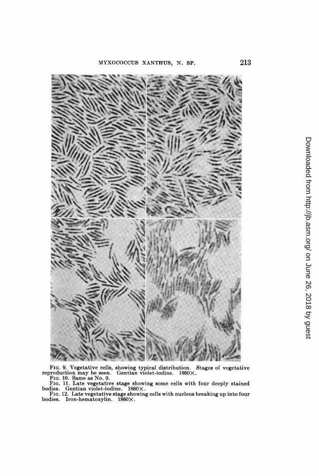
FIG. 9. Vegetative cells, showing typical distribution. Stages of vegetativereproduction may be seen. Gentian violet-iodine. 1860X.
FIG. 10. Same as No. 9.FIG. 11. Late vegetative stage showing some cells with four deeply stained
bodies. Gentian violet-iodine. 1860X.FIG. 12. Late vegetative stage showing cells with nucleus breaking up into four
bodies. Iron-hematoxylin. 1860X.
on June 26, 2018 by guesthttp://jb.asm
.org/D
ownloaded from
J. M. BEEBE
After several days in the vegetative phase the cells begin togather around various fruiting centers. Changes in morphologyare apparent, particularly a shortening of the cells. As theyapproach the point where the fruiting body is in process of forma-tion the cells may become almost spherical. By the time theyare incorporated in the slimy mass of the fruiting body the cellhas become a perfectly spherical, thick-walled, non-motile spore.
Germination occurs when the spores -are placed in a new envi-ronment. The first indication of germination is a thinning of thecell wall. At some point on the wall a slight bulge appears. Thisdevelops, by means of a process somewhat similar to budding,into an elongate rod-shaped cell. When this cell has reached alength of 3.0 to 4.0,u it becomes detached from the old spore wallby constriction at the point of emergence. After a single post-germinal division the cell is typically vegetative and begins thelife cycle again.
Cytologically the life cycle of M. xanthus is correspondinglycomplex. In the vegetative phase a single large nucleus, occupy-ing the central third of the cell, is to be seen. This body has amarked affinity for such nuclear stains as iron-hematoxylin andgentian violet, and gives a positive Feulgen reaction. Thereseems to be no limiting membrane enclosing the nucleus, but it israther composed of a compact condensed mass of nuclear proto-plasm. Its boundaries are quite definite. It seems to be granularin structure and apparently contains the equivalent of one pairof chromosomes (fig. 2 A). There is no evident arrangement inthreads or chains such as might be found in the close spiremephase in the cells of higher plants. Nuclear division is by meansof non-random amitosis. Prior to cell division the nucleus in-creases in size (fig. 2 B), along with the increase in size of the cellitself. Each particle of which the nucleus is composed mustdouble in size and split, the various halves migrating towardopposite poles of the cell. This corresponds to the splitting ofchromosomes before a straight somatic nuclear division: eachnew cell receives its portion of chromatin material carrying heredi-tary genes. After enlarging, the nucleus begins to constrict at apoint near the middle (fig. 2 C), the halves pulling toward opposite
214
on June 26, 2018 by guesthttp://jb.asm
.org/D
ownloaded from

MYXOCOCCUS XANTHUS, N. SP.
4
IWWFIG. 13. Late vegetative and early transitional stages. Cells with two and
four chromosomal bodies. Iron-hematoxylin. 1860X.FIG. 14. Same.FIG. 15. Same. Gentian violet-iodine. 1860X.FIG. 16. Various stages in spore formation. Gentian violet-iodine. 1860X.
215
.among.
on June 26, 2018 by guesthttp://jb.asm
.org/D
ownloaded from
J. M. BEEBE
ends of the cell. Just before division has been accomplished, andbefore cell fission is obviously under way, the nucleus has theappearance of a dumbbell-shaped body (fig. 2 C). This condi-tion has been at times previously described as the normal condi-tion of the nucleus; actually it is a stage of comparatively briefduration. Nuclear division is always completed before cell fis-sion begins (fig. 2 D); in a rapidly growing colony two nucleardivisions may take place before the cell itself has completed onedivision. Such cells appear to have four nuclei. This conditionis not similar to a later stage in which four bodies, chromosomes,are to be seen within the cell. The two should not be confused.Once nuclear division is complete, the cell constricts at the middle(fig. 2 E), producing two new mononucleate daughter cells. Thistype of vegetative reproduction continues for an indefinite periodof time.When the cells begin to converge upon a fruiting center definite
changes in cell structure begin to be seen. The first noticeablechange is an increase, slight in some cases, in the size of the nucleus(fig. 2 F). The cell at this point is quite broad, and the nucleus,at first occupying a central position as in the typical vegetativephase, begins to spread out and become dispersed through thelength of the cell; at this particular stage it might be considereddiffuse (fig. 2 H). Shortly, however, four masses of nuclear mate-rial are to be noted, each corresponding to a chromosome (fig. 2 I).At first these are irregularly shaped masses, occupying variouspositions in the cell, but as the cell progresses toward the fruitingcenter the bodies within become more definite until four distinctstructures may be seen. They elongate to rod-shaped bodies,pair up, and each chromosome breaks up into a chain of chromo-meres (fig. 2 J). In this stage the chromosomes are similar, ifnot identical, to the prophase chromosomes in the cells of higherplants. Chemically and functionally these structures are chromo-somal in nature. This stage is of brief duration, having beenobserved only a few times during the examination of numbers ofslides, but it is thought to be a typical step in the mitotic divisionof the nucleus of M. xanthus.Having become arranged in pairs the prophase chromosomes
216
on June 26, 2018 by guesthttp://jb.asm
.org/D
ownloaded from
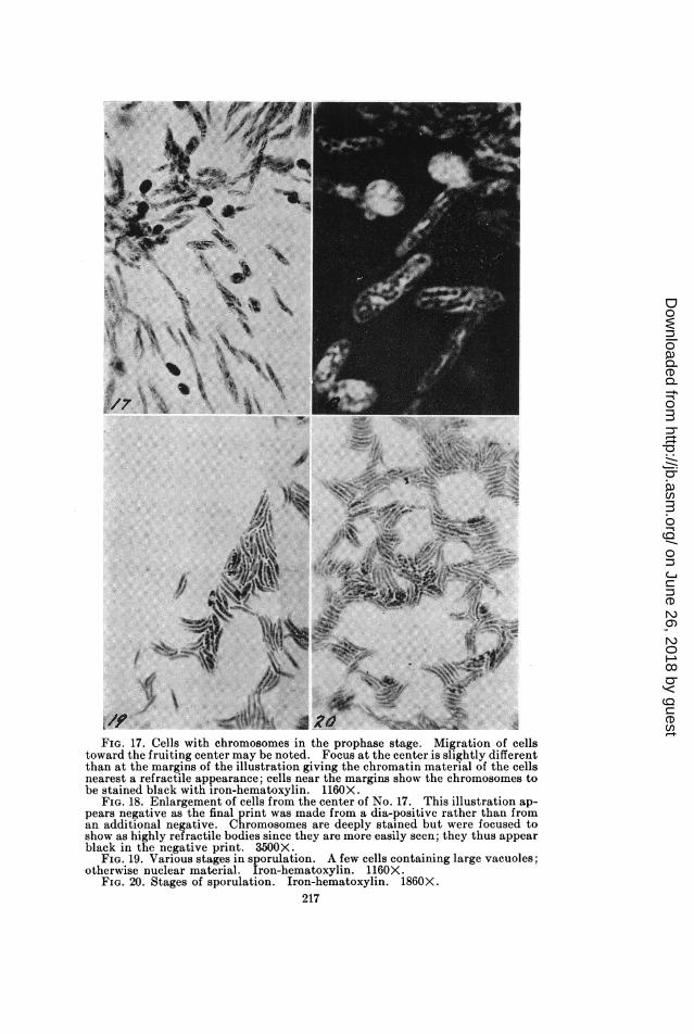
FIG. 17. Cells with chromosomes in the prophase stage. Migration of cellstoward the fruiting center may be noted. Focus at the center is slightly differentthan at the margins of the illustration giving the chromatin material of the cellsnearest a refractive appearance; cells near the margins show the chromosomes tobe stained black with iron-hematoxylin. 1160X.
FIG. 18. Enlargement of cells from the center of No. 17. This illustration ap-pears negative as the final print was made from a dia-positive rather than froman additional negative. Chromosomes are deeply stained but were focused toshow as highly refractive bodies since they are more easily seen; they thus appearblack in the negative print. 3500X.
FIG. 19. Various stages in sporulation. A few cells containing large vacuoles;otherwise nuclear material. Iron-hematoxylin. 1160X.
FIG. 20. Stages of sporulation. Iron-hematoxylin. 1860X.217
on June 26, 2018 by guesthttp://jb.asm
.org/D
ownloaded from
J. M. BEEBE
21 U2
I~~~~~~~~~~Is
4
2*'
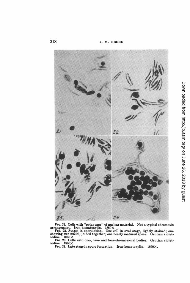
FIG. 21. Cells with "polar caps" of nuclear material. Not a typical chromatinarrangement. Iron-hematoxylin. 1860X.
FIG. 22. Stages in sporulation. One cell in oval stage, lightly stained; oneshowing two nuclei, joined together; one nearly matured spore. Gentian violet-iodine. 1860X.
FIG. 23. Cells with one-, two- and four-chromosomal bodies. Gentian violet-iodine. 1860X.
FIG. 24. Late stage in spore formation. Iron-hematoxylin. 1860X.
t'I'llBy/Iw _r
_-
/"aIf
O.-I
i4,
Aakv
218
on June 26, 2018 by guesthttp://jb.asm
.org/D
ownloaded from

NIYXOCOCCUS XANTHUS, N. SP.
begin to shorten, becoming rod-, L- or comma-shaped (fig. 2, K).The cells are much thickened and shortened by this time, and thearrangement of these pairs of chromosomes varies considerably.No correlation to the long axis of the cell has been noted. Finallya union of the pairs of chromosomes is accomplished, producingthe binucleate cell (fig. 2, L), each nucleus containing the equiva-lent of one pair of chromosomes. As a rule the two nuclei areabout the same size, although in some cases one may appear tobe somewhat larger than the other. Rosca (1937) based histheory of sexual union on the pairing of two bodies unlike in size.It is thought that any difference in the size of these two nucleiin M. xanthus is purely incidental.At this point the cell has become almost spherical, and the cell
wall has begun to thicken. The spore is already imbedded in thefruiting body and must have lost all power of locomotion. Nomotile spores have ever been seen. It is quite probable that asecond nuclear union takes place at this time or after the sporehas been formed. Due to the extreme affinity of the cell fordyes and the thickness of the cell wall it has been found impossibleto observe changes in the nuclear structure during these phases.However, the division of a single large nucleus in the early stagesof germination points toward a previous autogamous nuclearfusion (fig. 2, M-N) and it is thought that the spore goes throughthe resting stage as a large single nucleus, diploid in nature, sur-rounded by a thick cell wall, and containing little, if any, othercytoplasmic material. That the spore is basically nuclear isshown by the dye reactions, particularly the definitely positiveFeulgen reaction. No vacuoles of any sort have been notedwithin the spores, and the test for glycogen was questionablypositive. Apparently little reserve food material is present. Anoccasional pre-spore stage cell may show one or two vacuoles,the remainder of the cell giving positive nuclear reactions. Thesecells are not often seen, however, and attempts to ascertain thenature of the vacuolar material were unsuccessful.
Germination of the spores is first indicated by a lessened affinityfor dyes. At this time the large single nucleus initiates a pre-germinal division (fig. 2, Q) that progresses as the spore germi-
219
on June 26, 2018 by guesthttp://jb.asm
.org/D
ownloaded from

220 Jo. M. BEEBE
* 0e ltWs
\ styr
9. . 9n
08 . *
4s
9'~~~~~T_: *
t#\ ~/ oTits~sFIG. 25. Stages in spore formation. One cell (left) containing four deeply
stained bodies. Iron-hematoxylin. 1160X.FIG. 26. Various stages in sporulation. Iron-hematoxylin. 1160X.FIG. 27. Same. Gentian violet-iodine. 1860X.FIG. 28. Migration of cells toward fruiting center. The lightly stained mate-
rial appearing in "folds" is the slime upon which the cells rest. (Wrinkled duringpreparation). Matured spores on left; cells in vegetative and transitional stagesat center. Note binucleate cell above left of center. Iron-hematoxylin. 1160X.
on June 26, 2018 by guesthttp://jb.asm
.org/D
ownloaded from

MYXOCOCCUS XANTHUS, N. SP.
nates. By the time the newly emerged cell is ready to separatefrom the old spore wall (fig. 2, S) the nucleus has divided, pro-ducing a rod-shaped binucleate cell. Unless a chromosomal unionhas been affected during the spore stage this nuclear split is areduction-division for the purpose of producing vegetative cellswith the typical number of chromosomes. Following the divisionof the nucleus the cell elongates, constricts at the middle, anddivides, producing two typical vegetative cells, each with a singlecompact mass of nuclear material in the center.
If any sexual union were to be noted it would of necessity occurimmediately following the post-germinal nuclear division, result-ing in four haploid cells. The union of a pair of these cells wouldbe necessary for the formation of typical vegetative cells. How-ever, no indication of cell conjugation has been observed, and it isthought that this step, which is typical of some of the fungi, isnot included in the life cycle of M. xanrthus.
BUMMARY
A new species of Myxococcus producing an orange-coloredfruiting body is diagnosed and described, and the name Myxo-coccus xanthus proposed.Methods of growing the bacterium on media, utilizing rabbit
dung as the source of nutrient material, are described.Microscopic slide preparations were made by pressing clean,
sterile cover slips down on the growing colony. The adheringcells are fixed, stained and mounted in neutral balsam.The life cycle of M. xaanthus is relatively complex. In the
vegetative stage the cells are long, flexible, single rods, thatmove over the surface of the substrate by a crawling or creepingmotion. They are grouped on the substrate in small clumps,their long axes parallel, and move in clockwise paths as a unittoward the margin of the colony. As the cells go into the sporestage they shorten and become perfectly spherical by the timethey are imbedded in the slime of the fruiting body. Germina-tion is by a process analogous to budding.
Evidence is presented supporting the theory of a compact orcondensed nucleus. In the vegetative phase the nucleus is a
221
on June 26, 2018 by guesthttp://jb.asm
.org/D
ownloaded from
222 J. M. BEEBE
single compact mass of nuclear protoplasm that divides prior tocell fission. It has a marked affinity for nuclear dyes, and isFeulgen-positive. During the transitional phase the nucleusbreaks up into four chromosomes that are stained by gentianviolet and iron-hematoxylin. In the prophase the chromosomesare shown to be made up of chromomeres. An autogamous fusionof chromatin material occurs before the mature spore has beenformed, and nuclear division, presumably meiotic, takes placeduring germination of the spores.
The author wishes to express his thanks and appreciation toDr. J. E. Sass, of the Department of Botany, Iowa State College,for his most helpful assistance on certain of the technical prob-lems involved, and to Dean R. E. Buchanan, of the Departmentof Bacteriology, Iowa State College, under whose direction thiswork is being carried on, for his kind advice and criticism.
BIBLIOGRAPHYALLEN, L. A., APPLEBY, J. C., AND WOLF, J. 1937 Chromatin arrangements in
spore-forming bacilli. Nature, 139, (3514), 412-413.ALLEN, L. A., APPLEBY, J. C., AND WOLF, J. 1939 Cytological appearances in
a spore-forming bacillus. Zentr. Bakt. Parasitenk., II, 100, 3-16.BADIAN, JEAN. 1930 Z cytologji miksobakteryj (Zur Zytologie der Myxobak-
terien). Acta Soc. Botan. Poloniae, 7, 55-71.BADIAN, JEAN. 1935 Sur la cytologie du Bacillus megatherium. Acta Soc.
Botan. Poloniae, 12, (1), 69-74.BROOKE, JAMES W. 1936 Possible bipolar nuclear distribution in bacteria.
Proc. Soc. Exptl. Biol. Med., 34,185-188.CHANCE, H. L. 1938 Mitosis-like activity in Bacillus sp. J. Bact., 36, 347-349.CONN, H. J. 1936 Biological stains. Commission on Standardization of
Biological Stains, Geneva, N. Y.HOLLANDE, A. -CH. 1932 etude cytologique de quelques microbes pathogenes
a l'homme. Arch. zool., exptl. g6n. Paris, 72, 445-576.HOLLANDE, A. -CH. 1930 Nouvelle technique pour la mise en evidence rapide
et certain du chondriome dans les cellules. Compt. rend. soc. biol.,104, 473-474.
HOLLANDE, A. -CH., AND HOLLANDE, G. 1932 Cytologie des Bacillus mega-therium (de Bary) et Bacillus mycoides (Fltigge). Compt. rend. soc.biol., 109, 803-806.
KNAYSI, GEORGES. 1938 Cytology of bacteria. Botan. Rev., 4, 83-112.KRZEMIENIEWSKI, H., AND KRZEMIENIEWSKI, S. 1926 Miksobakterje Polski
(Die Myxobakterien von Polen). Acta Soc. Botan. Poloniae, 4, 1-54.
on June 26, 2018 by guesthttp://jb.asm
.org/D
ownloaded from
MYXOCOCCUS XANTHUB, N. BP. 223
KRZEMIENIEWSKI, H., AND KRZEMIENIEWSKI, S. 1928 Morfologj komorkimiksobakteryj (Zur Morphologie der Myxobakterienzelle). ActaSoc. Botan. Poloniae, 5, 46-90.
LEWIS, I. M. 1934 Cell inclusions and endospore formation in Bacillus mycoides.J. Bact., 28, 133-143.
LEWIS, I. M. 1940 The genus Spirillum Ehbg. with special reference to cellinclusions and the chromidial theory. J. Bact., 40, 271-284.
LINDEGREN, C. C., AND MELLON, R. R. 1933 Observations on a chromosomalmechanism for the bacterial cell. J. Bact., 25, 47-49.
ROscA, VALENTINE. 1937 Contribution A l'tude de la structure cytologiquedes batteries. Arch. roumaines path. exptl. microbial., 10, 267-279.
STOUGHTON, R. H. 1929 The morphology and cytology of Bacterium malvace-arum. Proc. Roy. Soc. (London) B, 105, 469-484.
on June 26, 2018 by guesthttp://jb.asm
.org/D
ownloaded from