Embed Size (px)
Citation preview

ARTICLE
Received 7 Jun 2016 | Accepted 3 Jan 2017 | Published 17 Feb 2017
Arylmethylamino steroids as antiparasitic agentsReimar Krieg1, Esther Jortzik2, Alice-Anne Goetz3, Stephanie Blandin3, Sergio Wittlin4,5, Mourad Elhabiri6,
Mahsa Rahbari2, Selbi Nuryyeva6,7, Kerstin Voigt8, Hans-Martin Dahse8, Axel Brakhage8, Svenja Beckmann9,
Thomas Quack9, Christoph G. Grevelding9, Anthony B. Pinkerton10,11, Bruno Schonecker12, Jeremy Burrows13,
Elisabeth Davioud-Charvet6, Stefan Rahlfs2 & Katja Becker2
In search of antiparasitic agents, we here identify arylmethylamino steroids as potent
compounds and characterize more than 60 derivatives. The lead compound 1o is fast acting
and highly active against intraerythrocytic stages of chloroquine-sensitive and resistant
Plasmodium falciparum parasites (IC50 1–5 nM) as well as against gametocytes. In P. berghei-
infected mice, oral administration of 1o drastically reduces parasitaemia and cures the
animals. Furthermore, 1o efficiently blocks parasite transmission from mice to mosquitoes.
The steroid compounds show low cytotoxicity in mammalian cells and do not induce acute
toxicity symptoms in mice. Moreover, 1o has a remarkable activity against the blood-feeding
trematode parasite Schistosoma mansoni. The steroid and the hydroxyarylmethylamino
moieties are essential for antimalarial activity supporting a chelate-based quinone methide
mechanism involving metal or haem bioactivation. This study identifies chemical scaffolds
that are rapidly internalized into blood-feeding parasites.
DOI: 10.1038/ncomms14478 OPEN
1 Institute of Anatomy II, University Hospital Jena, Teichgraben 7, 07743 Jena, Germany. 2 Biochemistry and Molecular Biology, Interdisciplinary ResearchCentre, Justus Liebig University Giessen, Heinrich Buff Ring 26-32, 35392 Giessen, Germany. 3 Universite de Strasbourg, CNRS, Inserm, MIR UPR9022/U963,F-67000 Strasbourg, France. 4 Swiss Tropical and Public Health Institute, Socinstrasse 57, PO Box, 4002 Basel, Switzerland. 5 University of Basel, Petersplatz1, 4001 Basel, Switzerland. 6 UMR 7509 Centre National de la Recherche Scientifique and University of Strasbourg, European School of Chemistry, Polymersand Materials (ECPM), 25, rue Becquerel, F-67087 Strasbourg, France. 7 New York University Abu Dhabi, PO Box 129188, Abu Dhabi, UAE. 8 Leibniz Institutefor Natural Product Research and Infection Biology-Hans Knoll Institute (HKI), Adolf-Reichwein-Strasse 23, 07745 Jena, Germany. 9 Institute of Parasitology,Biomedical Research Centre, Justus Liebig University Giessen, Schubertstrasse 81, 35392 Giessen, Germany. 10 Conrad Prebys Center for Chemical Genomics,Sanford-Burnham-Prebys Medical Discovery Institute, 10901 North Torrey Pines Road, La Jolla, California 92037, USA. 11 Conrad Prebys Center for ChemicalGenomics, Sanford-Burnham-Prebys Medical Discovery Institute, 6400 Sanger Road, Orlando, Florida 32827, USA. 12 Institute of Organic andMacromolecular Chemistry, Friedrich Schiller University Jena, Humboldtstrasse 10, 07743 Jena, Germany. 13 Medicines for Malaria Venture, 20 Route de Pre-Bois, 1215 Geneva 15, Switzerland. Correspondence and requests for materials should be addressed to K.B. (email: [email protected]).
NATURE COMMUNICATIONS | 8:14478 | DOI: 10.1038/ncomms14478 | www.nature.com/naturecommunications 1

Malaria caused by the unicellular apicomplexan parasitePlasmodium still threatens about 3.2 billion peopleworldwide. In 2015 there were an estimated 214 million
cases of malaria and 438,000 deaths1. Artemisinin-basedcombination therapies are the currently recommendedtreatment for uncomplicated P. falciparum malaria1, however,parasites exhibiting artemisinin-resistance have now beenreported2,3. Thus, the discovery and development of novelantimalarials and transmission-blocking agents is of globalimportance4,5. Schistosomiasis (bilharzia) ranks second tomalaria as a parasitosis affecting more than 240 million peoplein the tropics and subtropics6. A vaccine against these parasites isnot yet available, and praziquantel (PZQ) is the only commonlyused drug for treatment, justifying the fear of upcomingresistance7.
In connection with our work on steroids as chiral ligands formetal ions8 we became interested in the biological activity ofsteroid compounds. In general, steroids possess a conformationof their lipophilic framework, which allows the placementof substituents in well-defined spatial environments andconformational freedom. Also, there is a great structuralvariability of the tetracyclic framework, including estratrienes,androstanes, and cholestanes. In addition, ring junctions(5a/5b, 14a/14b and so on) can differ considerably. Functionalgroups can be attached at up to 27 positions in the frameworkor on side chains, and advances in synthetic chemistry overthe last several years have made accessing many of thesederivatives possible. In addition to their use in pharmaceuticals,there has also been growing interest in the use of steroidderivatives as rigid homochiral model compounds, for example,biomimetics, or as templates for chemo-, regio- andstereoselective investigation. Thus, one approach to discovernew biologically active compounds is to combine a steroidskeleton with structural elements possessing appropriatebiological activities.
Interestingly, during a screening program for potentialantiprotozoal drugs, the naturally occurring steroid 3b-amino-22,26-epiminocholest-5-ene (sarachine) from the leaves of Saracapunctate, had been isolated as a hit. Despite its simplesubstitution patterns, it was reported to exhibit activity againstthe malaria parasite Plasmodium falciparum9, and a series ofamino steroids having side chains similar to that of sarachinewere prepared from deoxycholic acid as the starting material10.To this end, the most active derivative of this series containeda chloroquinoline moiety in the side chain, which might becontributing to biological activity. The advantage of employinghydrophobic steroid units is their membrane permeability, pavingthe way for biologically active hybrid molecules. On the basis ofthis knowledge o-pyridiniumalkyl ethers of steroidal phenolswere synthesized and indeed the combination of the hydrophobicsteroid moiety with a hydrophilic group (ferrocenylmethylaminogroup, N-alkyl-pyridinium groups)11,12 led to compoundswith antimicrobial activity. In continuation of this work, the3-methoxy-estra-1,3,5(10)-triene unit was combined with theo-hydroxybenzylamino group13 and resulted in compoundswith antimalarial activity which were systematically optimizedusing structure-activity relationship approaches.
Here we report on arylmethylamino steroids with excellentantimalarial activity. The compounds are active in vitro againsthuman and murine Plasmodium asexual blood stages as well asagainst P. falciparum gametocytes. Furthermore they exhibitin vivo activity in Plasmodium berghei-infected mice and blockthe transmission of parasites to mosquitoes. Notably thecompounds also have fatal effects on adult Schistosoma mansoni.The lipophilic steroid carrier of these antiparasitic leadcompounds is likely to facilitate membrane permeation and
bioavailability whereas the essential hydroxyarylmethylaminomoiety points to a chelate-based quinone methide mechanisminvolving metal or haem bioactivation.
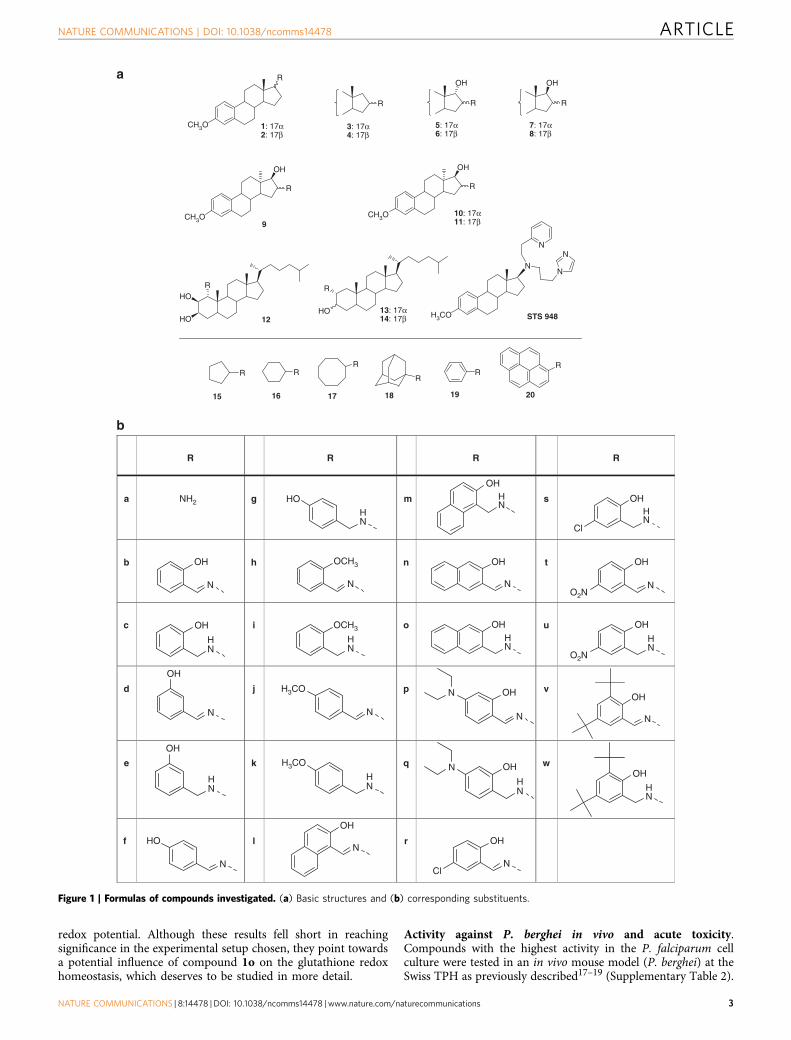
ResultsChemistry. The low molecular weight steroid compoundsdescribed here are based on a substituted steroidal pharmaco-phore and represent novel chemical matter14. All compounds arederived from amino steroids with varying constitutions of thebasic gonane core (series 1–14) and varying functional groupsR (substitution patterns c, e, g, i, k, m, q, s, u and w, Fig. 1).In brief, the compounds were synthesized via condensation ofamino steroids with arylaldehydes, and subsequent NaBH4-reduction of obtained Schiff-bases furnished the desiredcompounds in high yields. The synthesis could also be carriedout as a one-pot procedure without isolation of Schiff-bases. Thisis especially beneficial in case of cis-configured amino alcohols(series 5, 8, 11 and 14) where reaction with aldehydes leads torapidly exchanging tautomeric Schiff-based/oxazoline equilibria.Complete synthetic details can be found in SupplementaryNote 1, 1H and 13C NMR spectra of the most relevantcompounds are provided in Supplementary Figs 1–6.
Activity against Plasmodium falciparum blood stages in vitro.The activity of the steroid compounds was tested against the asexualblood stages of the malaria parasite P. falciparum (strain 3D7) inthe 3H-hypoxanthine incorporation assay. As shown in Table 1, thecompounds are highly active against red blood cell stages of P.falciparum with IC50 values in the nanomolar range. The mostactive compound 1o had an IC50 value of 4.1±1.6 nM on 3D7(n¼ 6), (± indicates s.d. throughout the manuscript, unlessotherwise stated) which is comparable to the in vitro activity ofclinically employed antimalarials such as chloroquine (IC50¼ 8.6nM) and artemisinin (IC50¼ 17.3 nM) using the same methods(72 h 3H-hypoxanthine incorporation assay)15. The second mostactive compound 2o showed an IC50 value of 6.6±2.1 nM (n¼ 4)on 3D7 (for data on all compounds tested, please seeSupplementary Table 1). Notably, the compounds were found tobe more active against chloroquine-resistant parasites as tested forthe P. falciparum strain Dd2: 1o: 1.0±0.9 nM (n¼ 4); 2o:2.0±1.2 nM (n¼ 4); chloroquine: 90.2 nM; artemisinin: 20.4 nM(ref. 15) (Supplementary Fig. 7). A similar phenomenon has beenreported for other antimalarial compounds as diverse as 8-aminoquinolines and amadantine. The underlying mechanism ofthis very promising activity of the steroid compounds deservesfurther attention.
To assess the rapidity of growth inhibition induced by thesteroid compounds, the so-called IC50 speed assay16 wasconducted for compound 1o on P. falciparum NF54 (Fig. 2a,b).In this assay, a comparison of IC50 values, which are obtainedside-by-side after 72, 48, and 24 h of compound incubation, iscarried out (the 72 h assay is the standard IC50 assay otherwiseused in our study). For compound 1o, no significant IC50 shiftswere determined across all time points, indicating that thecompound is fast acting like chloroquine or artesunate and notslow acting like pyrimethamine.
To further study the mechanism of action of the steroidcompounds and evaluate the potential involvement of redoxstress, compound 1o was tested in cell culture on thechloroquine-sensitive P. falciparum 3D7 strain expressing thecytosolic glutathione redox sensor hGrx1-roGFP2(Supplementary Note 2). As indicated in Supplementary Fig. 8,24 h incubation with 20, 50 and 100 nM of compound 1o ledto a dose-dependent increase of the redox ratio of the probe,pointing to increased oxidation and alterations in the intracellular
ARTICLE NATURE COMMUNICATIONS | DOI: 10.1038/ncomms14478
2 NATURE COMMUNICATIONS | 8:14478 | DOI: 10.1038/ncomms14478 | www.nature.com/naturecommunications
redox potential. Although these results fell short in reachingsignificance in the experimental setup chosen, they point towardsa potential influence of compound 1o on the glutathione redoxhomeostasis, which deserves to be studied in more detail.
Activity against P. berghei in vivo and acute toxicity.Compounds with the highest activity in the P. falciparum cellculture were tested in an in vivo mouse model (P. berghei) at theSwiss TPH as previously described17–19 (Supplementary Table 2).
CH3O
a
b
CH3OCH3O
H3CO
R
R R
OH
OH
OH
R
R
OH
R
HO
HO
R
HO
R
N
N
N
N
R RR
RR
R
1: 17α2: 17β
3: 17α4: 17β
5: 17α6: 17β
7: 17α8: 17β
10: 17α11: 17β
13: 17α14: 17β
9
STS 94812
15 16 17 18 19 20
R R R R
a NH2 g m s
b h n t
c i o u
d j p v
e k q w
f l r
OH
HO
OH
OCH3
OCH3
H3CO
H3CO
O2N
O2N
HN
HHN
HN
OH
OH
HN
N
OH
N
N
N
OH
HN
OH
OH
Cl
HN
N
OH
HON
OH
HN
Cl
OHHN
OH
N
OH
NNN
OH
OH
OH
N
HN
HN
N
N N
Figure 1 | Formulas of compounds investigated. (a) Basic structures and (b) corresponding substituents.
NATURE COMMUNICATIONS | DOI: 10.1038/ncomms14478 ARTICLE
NATURE COMMUNICATIONS | 8:14478 | DOI: 10.1038/ncomms14478 | www.nature.com/naturecommunications 3

Initially, the compound 2c was tested, which exhibited an IC50
of 73 nM against P. falciparum 3D7 in vitro. The compound wasactive when applied to the mice subcutaneously (s.c.) or orallyover three consecutive days. Subcutaneous application of3� 10 mg kg� 1 reduced parasitaemia by 47% and increasedthe life span of the animals from 6 days to 10 days(parasitized control mice were killed on day 4 to prevent deathotherwise occurring at day 6); 3� 30 mg kg� 1 s.c. reducedparasitaemia by 87% and increased the life span from 6 to
14 days. After oral application of 3� 100 mg kg� 1 2c,parasitaemia decreased by 99%, and survival time increased to16 days. As shown in Supplementary Table 2 compounds 1c and1o (optimized on the basis of 2c) were studied in more detail.Compound 1c, which had an in vitro activity againstP. falciparum 3D7 of 7.4 nM, was also active after s.c. and oralapplication and showed a similar in vivo activity pattern as 2c.Parasitaemia was drastically decreased, and the life span of theanimals was enhanced.
Before carrying out further P. berghei in vivo experiments,compound 1o, which had the best in vitro activity againstP. falciparum 3D7 (72 h incubation, IC50¼ 4.1 nM) and a strongactivity against P. berghei ex vivo (24 h incubation,IC50¼ 3.1±2.4 nM (n¼ 3) and methylene blue which served asa control resulted in 2.6±1.2 nM (n¼ 3)), were tested in an ‘acutetoxicity’ in vivo model. The compound was applied at anaccumulative dose of 100 mg kg� 1 (per os (p.o.). as well asintraperitoneally (i.p.)). The first application was 5 mg kg� 1, 2 hlater 15 mg kg� 1, 2 h later 30 mg kg� 1 and again 2 h later50 mg kg� 1. No acute toxicity symptoms were observed in thatexperiment. Moreover, we performed pharmacokinetics studies inmice, dosing i.v., i.p. and p.o. (Supplementary Table 5).1o displayed moderate clearance (29.6 ml min� 1 kg� 1) witha long half-life (48 h) after i.v. dosing. The compound showedmodest oral bioavailability (%Fo5) but sustained plasma levels ofB100 nM after oral dosing at 100 mg kg� 1. In contrast,high exposure and sustained levels of B2mM were obtainedafter i.p. dosing at 100 mg kg� 1. On the basis of these data, wedecided to perform the following P. berghei in vivo experimentsat doses of 4� 100 mg kg� 1. After i.p. administration of4� 100 mg kg� 1 1o, all mice treated were cured; after oraladministration two thirds of the animals were cured, which isconsistent with the improved exposure after i.p. dosing. Notably,a single dose (1� 100 mg kg� 1) p.o. also reduced parasitaemiaby 98.46% and increased the life span from 6 to 14 days.
Effects on transmission and gametocytes. We further tested theactivity of compound 1o on parasite transmission from mice toAnopheles gambiae mosquitoes. For this, P. berghei-infected mice
Table 1 | Antiplasmodial effects as well as antiproliferative and cytotoxic activity of selected steroid compounds on mammaliancells.
Compound Antiplasmodial activity Antiproliferative activity Cytotoxicity
IC50 [nM] GI50[lM] CC50[lM]
Pf (3D7) HUVEC GI50 K-562 GI50 HeLa CC50
1c 7.4 17.1 5.9 41281o 4.1 108.5 5.0 41132c 73 10.0 5.1 42.92o 6.6 13.8 2.5 41133c 42 4.1 3.8 20.74c 63 3.1 2.6 12.05c 160 2.2 2.0 11.36c 676 4.4 2.5 11.37c 120 1.2 2.0 10.18c 224 2.5 0.5 5.99c 999 11.2 8.4 48.810c 84 2.9 2.5 11.511c 141 22.0 0.1 6.412c 4,360 12.4 16.5 38.013c 465 23.7 17.7 85.314c 208 6.7 8.2 21.8STS 948 551 7.2 6.6 18.3
Strong antiproliferative effects are judged to occur at a GI50: r2 mM. All values are mean values of at least three independent determinations that differed by less than 20%. SDs for antiplasmodialactivity are provided.
IC50 48 h
(nM)
IC50 24 h
(nM)
IC50 24 h / IC50 72 h 20
a
b
Fol
d ch
ange
in IC
50w
ith in
crea
sing
ass
ay d
urat
ion
10
3
2
1
0
IC50 48 h / IC50 72 h
IC50 72 h / IC50 72 h
IC50 72 h
(nM)
1o 3.4 ± 0.2 2.7 ± 0.4
Chloroquine 7.5 ± 0.9 7.4 ± 1.1
Artesunate 5.5 ± 0.8 5.7 ± 0.5 4.7 ± 0.3
Pyrimethamine
1o Chloroquine Artesunate Pyrimethamine
220 ± 61
2.9 ± 0.2
8.3 ± 2.1
30.4 ± 7.6 15.2 ± 1.6
Figure 2 | IC50 values of 1o and established antimalarial drugs as
determined in the IC50 speed assay on P. falciparum NF54 in vitro18.
(a) Provided are mean values±s.d. from three biological replicates. (b) The
fold change in IC50 over time is indicated, showing that only pyrimethamine
is a slow-acting compound.
ARTICLE NATURE COMMUNICATIONS | DOI: 10.1038/ncomms14478
4 NATURE COMMUNICATIONS | 8:14478 | DOI: 10.1038/ncomms14478 | www.nature.com/naturecommunications

received a daily injection of compound 1o for 3 days starting24 h post passage and were exposed to mosquito bites 24 h aftereach injection. Tween 80 (Tw80) and methylene blue (MB) servedas negative and positive controls, respectively. As with MB,compound 1o limited parasite multiplication in mice, albeit toa lesser extent (Fig. 3a). Importantly, three doses were sufficientto fully block parasite transmission to mosquitoes (Fig. 3b,c,4 days post passage). A reduction of parasite transmission tomosquitoes was already visible 24 h after the first treatment(Fig. 3b,c, 2 days post-passage), with reduced infection prevalenceand parasite loads in mosquitoes fed on compound 1o-treatedmice compared with control mice.
Parasite gametocytes are the only stage infectious to mosqui-toes. To determine whether compound 1o has any activity onthese sexual stages, we exposed P. falciparum gametocytes to drugtreatment at different times during their maturation. A strongreduction of gametocyte viability was observed for all stages afterexposure to low concentrations of compound 1o (relative survivalbelow 50% for concentrations45 nM), although we neverdetected complete killing of gametocytes, even at concentra-tions41 mM (Fig. 3d). The fact that early stage gametocytessurvived better at 100 mM than at 1 mM suggests that at highconcentrations, the compound could have a gametostatic activity.Taken together, these data and the activity of compound1o against P. berghei asexual parasites ex vivo suggest thatthe transmission blocking effect observed above is explainedby a combination of the activity of 1o on gametocytes anda reduced gametocytemia following the impairment of asexual
stages, and/or a decrease in gametocyte commitment ontreatment.
Antimicrobial profile and effects on Schistosoma mansoni. Tolearn more about specificity and potential application ofthe steroid compounds, the two best compounds 1o and 2o weretested for their antimicrobial spectrum at the Hans KnollInstitute, Jena (Supplementary Tables 3 and 4). The compoundsshowed moderate activity against selected bacteria andweak activity against fungi. Antimicrobial activity was detectablein 4 out of 11 cases (Escherichia coli, Mycobacterium vaccae,Sporobolomyces salmonicolor and Candida albicans); however,micromolar concentrations of the compounds were required.No biologically relevant inhibition was determined for Bacillus,Pseudomonas, Staphylococcus, Enterococcus or Penicillium(Supplementary Table 3). The antifungal profile against 6 asco-mycetes and 2 zygomycetes indicated weak activity only(Supplementary Table 4).
Interestingly, however, the steroid compounds were found tohave remarkable physiological and morphological effects onadult Schistosoma mansoni, leading to the death of thistrematode parasite in vitro. To investigate the effects of thecompounds 1o, 2o and 1c in more detail, we made use of anestablished in vitro culture for adult S. mansoni20. The threecompounds were tested in concentrations of 1–100 mM each,and their effects were first checked via bright-field microscopywith respect to physiological parameters such as pairing stability,
1 2 3 40
2
4
6
8
10
12
Days post passage
Par
asite
mia
(%
)
0
20
40
60
80
100 *** *** ****** *** ***
1
10
100
1,000 *** *** ****** *** ***
Tw80
MB
1o
******
Pre
vale
nce
(%
)N
o.
pa
rasi
tes
pe
r m
osq
uit
o
a b
c
108 125 116 63 96 101 80 89 100n=
32 4Days post passage
–5 0 50
50
100 EarlyMidLate
log10 (concentration, nM)
Pro
port
ion
of v
iabl
e ga
met
ocyt
esre
lativ
e to
con
trol
(%
)
d
1o
–3 3
Figure 3 | Effect of compound 1o on P. berghei multiplication in mice and on parasite transmission to mosquitoes. (a) Naive mice were injected i.v. with
infected blood and treated daily for 3 days starting 24 h post passage with compound 1o (100 mg kg� 1), with methylene blue (MB, 15 mg/kg), or with
vehicle (Tw80) as positive and negative controls, respectively. Parasitemia was monitored daily and the mean± s.d. of three independent experiments are
plotted. Groups of mosquitoes were fed on mice 24 h after each treatment. (b) Prevalence (percentage of infected mosquitoes) and (c) infection intensity
(mean number of parasites per infected midgut) were determined 7 days post infectious blood feeding. The results of four independent experiments were
pooled, and the mean±s.e. of the mean is shown (b,c). Significance for differences between treatments was calculated using generalized linear models
with treatment, time, and repeats as variables, ***P value o0.001 (a–c). n, number of mosquitoes. (d) Effects of compound 1o on early, mid, and late
stage P. falciparum gametocytes. Representative data sets are given (4.5% gametocytaemia) out of three independent experiments. Survival of treated
gametocytes was calculated for each concentration relative to parasites exposed to vehicle.
NATURE COMMUNICATIONS | DOI: 10.1038/ncomms14478 ARTICLE
NATURE COMMUNICATIONS | 8:14478 | DOI: 10.1038/ncomms14478 | www.nature.com/naturecommunications 5

egg production, and survival. Starting with 1 mM (1c, 2o) and5 mM (1o), respectively, all compounds showed a negativeinfluence on worm physiology. Using 10 mM and more, themost remarkable effects on all three parameters were observedwith 2o, followed by 1c and 1o. Using 2o, reduced motilityand viability were observed starting at 1 mM, while 10 mM ofthis compound caused the death of the parasites within a week(Supplementary Fig. 9). This was accompanied by tegumentalinvaginations and oedema-like swellings of the body. CLSMindicated an enlarged gut lumen and degradation ofthe gastrodermis, which led to the accumulation of degradedtissue and aggregate formation. Furthermore, we observeda disorganization of oocytes within the ovary and reduced sizesof testicular lobes in males (see Fig. 4 and detailed explanations inthe legend).
All tested compounds affected physiology and morphology inS. mansoni that included changes in the female ovary as wellas in the gut of both genders, finally leading to the death of theblood fluke. Interesting was the tissue dilatation of the gutassociated with the degradation of the gastrodermis and theaccumulation of particle aggregates of remarkable size. In thisintensity, such a phenotype has not yet been observed withother substances applied to adult schistosomes in vitro20,although it resembled a phenotype previously observed foradults treated with imatinib or its derivatives dasatinib ornilotinib to some extent. However, in none of the latter caseswere tegumental invaginations or a tissue dilatation of thisintensity observed, nor such a strong accumulation of aggregates.It should further be noted that in comparison with a unicellularorganism such as Plasmodium, it usually takes much higher drugconcentrations (accompanied by lower selectivity indices) totarget multicellular organisms such as Schistosoma. In the case ofthe arylmethylamino steroids, lower micromolar concentrationswere required to see clear effects on schistosomes. This isexactly in the range of the gold standard praziquantel, which istoxic for schistosomes at 5–10 mM but no longer at 1 mMin vitro21.
Cytotoxicity and pharmacokinetic parameters. Selected steroidcompounds were tested for their antiproliferative activity inHUVEC cells and K-562 cells with GI50-values between 0.1 and108.5 mM. Cytotoxicity tests were carried out on HeLa
Control
D E F
G H I
J K L
M N O
P Q R
S T U
B CTu
Te
TM
Sv
Hs
Vs
Te
Te
T
T
F
Te
Te
FF
mO O
Sv
Sv
Vs
Vs
Te
M
M
M
M
MM
M
Mu
G
Oo
F
E
F
iOOG
mO
M
M
Ga
Ga
G
G
M
M
M
O
Oo
F
M
M
mOiO Gy
FGy
Gy
Gy
T
TG
G
Ga
Te
Te
FO
GOe
M
M
F
F
Sv
T
M
G
Ga
Ga
Ga
G
G
Gy
iO
mO
mO
O
GG
M
T
F
F
F
Tu
GO
iO
G
Gy
Te
Ga Te
M
M
F
Tu
GaGO
OmO
M
M
F
Oo
Gy
1o
2o
1c
a b c
d e f
g h i
j k l
m n o
J K Lp q r
s t u
Figure 4 | Morphological effects on adult S. mansoni in vitro. Compounds
1o, 2o, and 1c (5 mM each) were administered over 6–13 days before effects
were investigated via confocal laser scanning microscopy. (a–c) Untreated
schistosome couples exhibiting a smooth tegumental surface with
tubercles. (a) The testes are composed of lobes containing spermatogonia
and differentiated spermatozoa accumulating in the sperm vesicle. (b) The
gut lumen is surrounded by the gastrodermis. (c) The ovary of the female
exhibits a bulb-like structure with mature oocytes at the broader, posterior
part and immature oocytes at the narrow, anterior part. (d,e) The ootype is
the egg-forming organ. (d–i) 1o (9-day treatment); swellings and
invaginations occurred at the tegument. Arrows mark aggregates of
degradation products of the gastrodermis within the gut lumen. (f) The
number of mature oocytes was reduced, some occurred within the anterior
part of the ovary, which normally only contains immature oocytes.
(g,h) After 13- day treatment, swellings, invaginations and the size of the
aggregates increased. (i) No more immature oocytes occurred.
(j–o) 2o (5 mM; 6-day treatment); (j) the diameter of the testicular lobes
was reduced, the ovary was disorganized. (k,l; arrows) Oedema-like
swellings, gastrodermis degradation, and aggregate formation were visible.
(m) After 9-day treatment, diameter of the testicular lobes and number of
spermatozoa within the seminal vesicle were reduced. (n) The morphology
of the ovary was disturbed, (o) and eggs were deformed. (p–u) 1c (5 mM;
9-day treatment); (p) diameter of the testicular lobes and ovary appeared
normal, number of mature oocytes was smaller. (p,q) Aggregate-like
degradation products of the gastrodermis occurred within the gut lumen;
size of aggregates was larger in males (p,q) than in females (r). Gut
swelling and tegument invaginations were not obvious. (s) After 13-day
treatment, the diameter of the testicular lobes was reduced. (t,u) Gut
swelling and tegument invagination appeared. Morphology of ovary was
disturbed and number of mature oocytes reduced. (s–u) Degradation
aggregates appeared within gut lumen and oesophagus area. After 9 days,
precipitates were larger in males (s,t) compared with females (u). E, egg;
F, female; G, gut; Ga, gastrodermis; Gy, gynecophoral canal; Hs, head
sucker; M, male; T, testis; Te, tegument; Tu, tubercle: O, ovary; Oe,
oesophagus; mO, mature oocytes; iO, immature oocytes; Oo, ootype;
Sv, seminal vesicle; Vs, ventral sucker. Scale bars, 100mm, except
d (400mm) and o (50mm).
ARTICLE NATURE COMMUNICATIONS | DOI: 10.1038/ncomms14478
6 NATURE COMMUNICATIONS | 8:14478 | DOI: 10.1038/ncomms14478 | www.nature.com/naturecommunications
cells, resulting in CC50-values between 5.9 and 4128 mM,which is 2–4 orders of magnitude higher than the IC50 valuesdetermined against malaria parasites (Table 1). In addition,cytotoxicity tests with compound 1o were carried out onimmortalized human hepatocytes, Fa2N-4 cells, resulting in anLC50450mM. The best compound 1o has furthermorebeen tested in an acute toxicity in vivo model at the SwissTropical Institute, Basel. The compound was applied at anaccumulative dose of 100 mg kg� 1 (p.o. as well as i.p.), but noacute toxicity symptoms were observed.
First pharmacokinetic data for the best compound 1o werecollected at the Sanford Burnham Prebys Medical DiscoveryInstitute (Supplementary Table 6). Aqueous solubility, plasmastability, plasma protein binding, hepatic microsome stability,and membrane permeability using the parallel artificialmembrane permeability assay (PAMPA) were determined.Although compound 1o was found to have rather poor aqueoussolubility (o1 mg ml� 1 across a range of pH), it displayedgood stability in plasma (62 and 58% remaining, respectively,after a 3 h incubation with human and mouse plasma) andmoderate microsomal stability (B38% remaining after a 1 hincubation with human or mouse liver microsomes).
Influence of metal or haem binding and catalysis. To gain moreinsight into the mechanism of action of the steroid compounds,the binding capacities of the most potent compounds 1o and2o towards Cu(II) and Fe(III) were first evaluated underquasi-physiological experimental conditions (pH 7.5). Bothcompounds were shown to lead to 1:1 stoichiometric complexeswith stability constants log KLM (L¼ 1o or 2o, M¼Cu(II)or Fe(III)) ranging from¼ 4.1 to 4.7 (Supplementary Note 3,Supplementary Fig. 17, and Supplementary Methods).Compounds 1o and 2o share the same bidentate N,O bindingsite suitable for speciation of soft divalent or hard trivalentmetal ions.
Interestingly, the most potent antimalarial compounds(1o and analogues found in the present work) are the ortho-substituted derivatives, suggesting that generation of radicals isthe most probable explanation for the increased efficacy of thechemical series. Similar ortho-substituted ligands of transientmetal complexes, such as the reduced benzoylmenadionemetabolite of the antimalarial plasmodione, which presents anoxygen-rich bidentate site suitable for Fe(III) chelation22, werereported to become redox-inactive when their effects wereantagonized with the known iron(III) chelator desferoxamine(DFO). In this study a most potent antagonistic effect(sum FIC50¼ 2.4) induced by DFO was observed whencombined with plasmodione in cell culture assays using RBCsparasitized by P. falciparum 3D7.
In parallel, the ability of a broader range of compounds(1o, 2o, 1a and 1h, used as a negative control, see Fig. 1) tointeract with haem was evaluated (Supplementary Note 3,Supplementary Table 7, Supplementary Figs 10–16 andSupplementary Methods). Regardless of the nature of thecompound, strong interactions (KD in the mM range) with thehaem dimer were measured (that is, haematin predominatesas a p-p dimer in solution at pH 7.5) and compare wellwith chloroquine CQ, a well-known antiparasitic drug that actsas a haemozoin inhibitor. p-p-interactions and hydrophobicinteractions are likely the driving forces accounting for suchstrong interactions. It therefore appears that 1o and 2o displayingan N,O bidentate site are capable of interacting with electroactivemetal ions such as Cu(II) or Fe(III). With respect to haemligation, the steroid unit was demonstrated to be of greatimportance. The capacities of these systems to prevent haemozoin
formation were then investigated using the ESI-MS collision-induced dissociation method. Interestingly, 1o (DV50¼ 289 V,bottom plateau of 20% at 400 V) and 2o (DV50¼ 280 V,bottom plateau of 21% at 400 V) were found to be the mostpotent compounds to prevent haemozoin crystallization and arebetter than the well-known antiparasitic drugs chloroquine(DV50¼ 201 V, bottom plateau of 0% at 400 V) or amodiaquine(DV50¼ 212 V, bottom plateau of 7% at 400 V). The significantresidual amount of haem 1o or haem 2o adducts at a highfragmentor voltage of 400 V is indicative of covalent binding,likely through quinone methide formation23 and cross-linking tohaem. The 2-hydroxyarylmethylamino moiety was furtherdemonstrated to be of crucial importance since modelcompounds 1a and 2a, which are lacking such an electroactiveunit, are less efficient. The covalent character of the haem adductsobserved with 1o and 2o was further substantiated byco-incubation of the compounds and haem at pHo1 followedby ESI-MS.
In conclusion, 1o and 2o bearing a 2-hydroxyarylmethylaminomoiety are potent metal (copper or iron) chelators andlikely covalently associate with haematin via quinone methideformation and subsequent cross-linking. No significant differencehas been found between 1o and 2o, showing that steroidconformation is not of crucial importance. However, the presenceof the steroid moiety has been clearly demonstrated to favourthe physiochemical properties towards inhibition of haemozoinformation, leading to increased antimalarial activities of bothlead agents. On the basis of these data and taking into accountthe fact that parasitized red blood cells contain high amountsof haem and free iron, it can be estimated that morethan 80% of compound 1o is present in complexed form withinthe infected RBC (Supplementary Note 4 and SupplementaryFig. 18).
DiscussionWe identified and characterized arylmethylamino steroidsas a class of compounds with remarkable activity against malariaparasites and schistosomes. Transmission-blocking activity, oralavailability, and low cytotoxicity further support their potential.
The presently synthesized and available compound poolcomprises about 60 steroid derivatives and a series ofnon-steroidal analogues that can be employed for SAR studiesand considerations concerning a lead structure for furtheroptimization (Fig. 5). Present SAR data indicate that thehydrophobic steroid component and a hydroxyarylmethylaminogroup are essential for the antimalarial action of the compounds.This gives rise to some key considerations (Fig. 5): the deliveryof small molecules to living cells is typically mediated bypolymers, lipids, sophisticated nano-sized drug deliverysystems24, or by altering membrane permeability, for example,via covalent conjugation of target molecules with cholanederivatives25. In our context, the lipophilic steroid moiety isconsidered to mediate cellular uptake and intracellular transportprocesses. Steroid conjugates often readily enter living cells,either via membrane association and/or endocytotic pathways asknown from cholane derivatives26 or specifically via steroidbinding sites as known from steroid hormones27. In our study,the highest biological activity was found among estratrienederivatives (series 1–11), while cholane derivatives (series 12–14)were less efficient. De facto, none of the non-steroid-derivedanalogs (series 15–20) exhibited antimalarial activity comparableto the steroid compounds. The IC50 values on P. falciparum3D7 asexual blood stages were about 2 mM for 17c, 4 mM for 18c,5.6 mM for 20c,46 mM for 15c and 16c, and 415 mM for 19c.Notably, 18c is derived from the lipophilic 1-adamantylamine,
NATURE COMMUNICATIONS | DOI: 10.1038/ncomms14478 ARTICLE
NATURE COMMUNICATIONS | 8:14478 | DOI: 10.1038/ncomms14478 | www.nature.com/naturecommunications 7

which is closely related to the neuroprotective agent memantine(3,5-dimethyl-1-adamantylamine). On the basis of these data,the steroid part is essential for the biological activity of thecompounds. This interesting new chemical entity seems tovectorize the aminocresols to parasitized red blood cells andis potentially recognized by transporters. The present studymight therefore initiate new investigations based on the useof steroids to target not only drugs, but also fluorophores orother chemical probes, to cells of interest. On the basis ofthe properties of our compounds, the sex hormone-bindingglobulin (SHBG), the principal carrier of oestrogensand androgens in the blood of almost all vertebrate species28,may be involved in drug transport in the in vivo experiments.Furthermore, in our approach, a lipophilic moiety joinedto a hydrophilic one could result in an inhibitor that mightdestabilize cell membranes as known from phosphatidylcholineinhibitors29,30.
Notably, the 2-hydroxyarylmethylamino-group is present inall of our best-acting derivatives. Such compounds are wellknown for forming ortho-quinone methides of high inherentreactivity29,31,32. There is some evidence that quinone methideintermediates, which may alter or inhibit the action ofbiomolecules via covalent cross-linking, play a key role (Fig. 5).In our approach, methylation of the phenolic group (R¼ i and k)or movement of the OH function to the meta-position (R¼ e)prevents the formation of quinone methides. Even thesecompounds exhibit a clearly decreased antimalarial activity.Movement of the phenolic group to the para-position recoversbiological activity. Introduction of bulky tert-butyl groupsinto ortho- and para-positions prevents cross-linking capability(6w). All these findings make a quinone methide mechanism
permitted by R very likely. Furthermore, ortho phenols are mostefficient, while their methyl ethers as well as meta-substitutedphenols are less active (Fig. 5). This fact supports theparticipation of a chelation mechanism, including activationprocesses and ongoing redox processes mediated by metals. Therecovery of antimalarial activity from meta- to para-substitutedphenols, however, suggests that additional mechanisms arelikely to support activity, as the latter cannot act as bidentateligands. Given the lipophilicity of our steroid compounds aswell as their basic nature, it is furthermore most likely that theyaccess the food vacuole of the parasite. At the pH of the organelle(5.0), these molecules will be protonated and concentrated.Protonation of an aminocresol will increase the potential fora site-specific formation of a reactive species, limiting any safetyrisk. Furthermore, the SAR demonstrates that compounds nothaving an aminocresol retain potency (albeit reduced) suggestingthat the structures retain other pharmacology unrelated to thiscovalent binding; this is an innovation—delivering a compoundhaving polypharmacology—with the benefits such as resistanceselection apparent.
When proposing steroid-based compounds as anti-infectiveagents, a potential intrinsic biological activity of the steroidmoiety determined by its steric and electronic fit with steroidbinding sites should be considered. In oestrogens, hormoneaction is mainly determined by the specific interaction of the3- and 17b-OH functions. Both binding sites are impairedin series 1–8 and 10–11 by 3-OH methylation and inseries 1–6 additionally by the lack of the 17b-OH function.Therefore, potential side effects based on steroid hormoneactivity are unlikely to occur, particularly taking the short-termcourse of an antimalarial therapy into account. Furthermore, in
Steroid moietya
b
c
X
Impossible Impossible1e (434)2e(741)
1g (116)
1k (1,010)
1i (872)
1c (7.4), 2c(73)1o (4.1), 2o (6.6)
Impossible
Possible
Unlikely
Possible Plausible
Unlikely
Novel leads
Unlikely
Influence of steroid moiety (IC50 (nM) values for R = c)
1 (7.4) > 3 (42) ≈≈ 4 (63) ≈ 2 (73) ≈ 10 (84) ≈ 7 (120) ≈ 11 (141) ≈ 5 (160) > 8 (224) ≈ 14 (208) > 13 (465) > 6 (676) > 9 (999) >> 12 (4,360)
HOX
NH
OX
NH
X O
NH
HN
HO
1cH3CO H3CO
HN
HO
1o
H
CH3
H
CH3
Quinone methideformation
Metalchelation
Example(IC50 (nM))
R
Figure 5 | Working hypothesis for the mode of action and optimization of R. (a) Ortho-hydroxylated arylmethyl amines exhibit the highest antimalarial
activity. A chelate involving quinone methide mechanism is substantiated via structure/activity considerations. Both the redox pair phenol/quinone and the
highly reactive quinone methide itself may be causative for biological activity. (b,c) The steroid core also plays a crucial role. The highest activity is found
for estratriene derivatives.
ARTICLE NATURE COMMUNICATIONS | DOI: 10.1038/ncomms14478
8 NATURE COMMUNICATIONS | 8:14478 | DOI: 10.1038/ncomms14478 | www.nature.com/naturecommunications

our experiments no initial liabilities were identified at highmM concentrations in vitro and at 100 mg kg� 1 in vivo inP. berghei-infected mice. However, more extensive and thoroughtesting will be necessary during further preclinical development ofone of the two best compounds or optimized analogues.
Finally, phenoxide binding to haem iron33 might contributeto the antimalarial effects of our compounds where the2-hydroxyarylamino moiety may chelate or associate withmetals. For derivatives with this configuration, an increasedantimalarial activity was found compared with the 4-isomer,which may also furnish quinone methide intermediates but doesnot chelate metals. Utilizing metal catalytic effects as a drivingforce and their dependence on substitution patterns of quinonemethide precursors have been described in other contexts34
and were studied in detail for our antimalarial compounds(see above).
The most potent steroid compounds discovered here actuallyrepresent a subclass of aminocresols, a group of antimalarialsthat have shown up in various antimalarial studies since the1940s and include amodiaquine. However, none of thesecompounds has so far had a steroid attached. A prominentexample of aminocresols is WR194965 (ref. 35). This compoundhas strong antimalarial activity against P. falciparum andP. vivax and was even taken into human trials. However, sincemefloquine was discovered at the same time, WR194965 wasnot followed up in detail. This is also the reason why onlylimited toxicity data are available. A second example is MK4815(from Merck), which was also identified via high-throughputscreening and was found to be orally active in a P. berghei mousemodel, even when applied as a single dose36. However, theIC50 against P. falciparum 3D7 was about one order of magnitudehigher than that of our steroid-based compounds. Indeed thereare additional theoretical concerns that exist for aminocresols ingeneral. First, there is the potential for unwanted toxicityassociated to quinone methide formation. Second, dependingon the lipophilicity and the basicity of the compounds, there ispotential for cardiovascular and CNS safety issues. All of thesewould need to be carefully monitored in preclinical safety studiesbefore starting human studies. However, even in reasonablydetailed toxicology studies, the respective compounds, includingour steroid derivatives, have hardly shown any toxicity.Furthermore, as indicated by initial studies in cell culture usinga genetically encoded redox probe (Supplementary Fig. 8),the redox stress induced by the steroid compounds mightcontribute to their mechanism of action but is unlikely torepresent a major risk for patients with glucose-6-phosphatedehydrogenase deficiency. Interestingly, Jacobus Pharmaceuticalshas recently delivered a second generation WR194965 namedJPC3210 with reported excellent efficacy and half-life37. Much hasalready been said about pan-assay interference compounds(PAINs) that cannot be developed into selective covalentlybinding drugs38,39. However, not all compounds with PAINcharacteristics should be discarded a priori as antimicrobialagents because the compounds are expected to be administeredfor rather short-term treatment compared with drugs acting inother fields such as the treatment of cancer or cardiovasculardiseases. Although the authors of course fully agree that thepotential safety issues of PAINs need to be taken into accountthoroughly, it is a big challenge to fight dogmas and taboos in thefield of anti-infective drug discovery. Furthermore, evenwith PAINS characteristics, compounds with excellent potenciesshould be considered for chemical probe discovery40 to helpand improve our understanding of drug targets/transportersand pathways.
The next steps for further optimizing our steroid compoundsinclude in-depth analyses of pharmacokinetic and toxicological
properties, further improvement of lipophilicity and ADMEproperties including oral bioavailability and enhancement ofpotency in vivo. One starting point could be the furtheroptimization of R, for example, by introducing suitable leavinggroups. Furthermore, the potential of combining the steroidcompounds with known antimalarial agents should be system-atically evaluated. To reduce potential hormone activity a basicnitrogen function has already been introduced into thecompounds. Further approaches could be taken includingthe preparation of analogues with altered substitution patternsand altered stereochemistry at the gonane core and theintroduction of a second (or third) hydroxyarylmethylaminogroup. A more detailed discussion of lead optimization is given inSupplementary Note 1. On the basis of our studies andthe corresponding considerations we propose here a novelapproach to drug development for fighting malaria, schistoso-miasis and potentially other parasitic diseases.
MethodsCultivation of Plasmodium falciparum. The chloroquine-sensitive P. falciparumstrain 3D7 Netherlands and the chloroquine-resistant strain Dd2 (kindly providedby Michael Lanzer, Heidelberg in 2000) were grown in continuous culture asdescribed by Trager and Jensen41 with slight modifications42. Unless otherwisestated, parasites were maintained at 1 to 10% parasitaemia and 3.3% haematocritin an RPMI 1640 culture medium supplemented with Aþ erythrocytes(purchased from the blood bank of the University Hospital Giessen andMarburg, UKGM), 0.5% lipid-rich bovine serum albumin (Albumax), 9 mM(0.16%) glucose, 0.2 mM hypoxanthine, 2.1 mM L-glutamine, and 22mg ml� 1
gentamicin. All incubations were carried out at 37 �C in 3% O2, 3% CO2, and94% N2 unless otherwise stated. Synchronization of parasites in culture to ringstages as a starting population was carried out via treatment with 5% (w/v) sorbitol.The parasites were used for the experiments delineated below.
IC50 values on P. falciparum in vitro and drug combinations. Isotopic drugsensitivity assays were employed as described41 to investigate the susceptibilityof P. falciparum to the steroid compounds. The procedure depends on theincorporation of radioactive 3H-hypoxanthine, which is taken up by the parasite asa precursor of purine deoxynucleotides for DNA synthesis43. In 96-well microtitreplates (NuncR), a twofold serial dilution of the starting concentration of eachpharmacologically active compound to be tested was carried out. Parasites wereincubated at a parasitaemia of 0.25% (470% ring forms) and 1.25% haematocrit inhypoxanthine-free medium. After 48 h, 0.5 mCi 3H-hypoxanthine was added toeach well, and the plates were incubated for another 24 h. The cells of each wellwere then collected on a glass fibre filter (Perkin-Elmer, Rodgau-Jugesheim,Germany), washed, and dried. Their radioactivity in counts per minute wasconsidered to be proportional to the number of parasites in the well. All IC50 valueswere determined at least in quadruple. IC50 values (drug concentrations thatproduce 50% reduction in the uptake of 3H-hypoxanthine) were calculated.
IC50 values on P. falciparum gametocytes in vitro. Sensitivity of P falciparumgametocytes to lead compound 1o was determined by measuring the activity ofluciferase in luciferase-expressing gametocytes after an exposure of 72 h to differentconcentrations of compound 1o.
The pfs16-GFP-Luc (NF54 background) parasite strain44 was maintainedat 1 to 10% parasitaemia in an RPMI 1640 culture medium supplementedwith Aþ erythrocytes (2% haematocrit), 10% decomplemented human serum,9 mM (0.16%) glucose, 0.2 mM hypoxanthine, 2.1 mM L-glutamine, and20 mg ml� 1 gentamicin. Parasite cultures were incubated at 37 �C in 5% O2,5% CO2, and 90% N2. Asexual blood stage parasites were first synchronizedby 5% D-sorbitol (wt vol� 1) treatment for 10 min at 37 �C for two or moresuccessive growth cycles. Synchronous parasites at 3% haematocrit were cultured in10 cm Petri dishes to 10% parasitaemia (ring stage), at which pointgametocytogenesis was induced by feeding cultures with a mixture of conditionedand fresh media (1:1). The next day, trophozoite cultures were diluted fourfold.N-acetyl glucosamine (NAG; Sigma-Aldrich) was added to a final concentration of50 mM after all schizonts had ruptured. NAG treatment was maintained for thenext two to three cycles of reinvasion to eliminate all asexual forms from theculture. Immature gametocyte stages (I and II) were purified on a Percollgradient45 and incubated directly in 96-well plates at 9%, 12%, or 45%gametocytaemia depending on the experiment (150 ml well� 1). Gametocytecultures were exposed for 72 h to dilutions of compound 1o, methylene blue (MB)(Sigma M9140) or DMSO (0.5%) starting at day 3, 8, or 11 after gametogenesisinduction. For this, compound 1o (10 mM) and MB (10 mM) were dissolved in100% DMSO and H2O, respectively, diluted 1:100 in RPMI, and further diluted inRPMI to the proper concentrations. 150 ml of these solutions were added to the
NATURE COMMUNICATIONS | DOI: 10.1038/ncomms14478 ARTICLE
NATURE COMMUNICATIONS | 8:14478 | DOI: 10.1038/ncomms14478 | www.nature.com/naturecommunications 9
gametocyte culture in triplicate. Following drug exposure, parasites were cultivatedfor two additional days in normal medium before centrifugation and freezing at� 80 �C, and luciferase activity was measured.
Luciferase assays in P. berghei and P. falciparum in vitro. Plates frozen at� 80 �C were thawed at 37 �C. The cells were lysed at room temperature in10ml 1x luciferin lysis buffer (Luciferase Cell Culture Lysis 5� reagent, Promega),and 100ml luciferase assay substrate was added to each well (Luciferase AssaySystem, Promega). Luciferase activity was measured on a luminescence plate reader(Mithras LB940 luminometer, Berthold Technologies). The mean of the luciferaseactivity was calculated for each technical triplicate and normalized as follows:(Lucx� LucRBC)/(Lucsolvent� LucRBC) with Lucx and Lucsolvent¼mean luciferaseactivity for parasites incubated with compound x and with solvent (DMSO for1o and PBS for MB), respectively, and LucRBC¼mean luciferase activity ofnon-infected red blood cells (background). IC50 values were calculated usingPrism (GraphPad, log(inhibitor) versus normalized response—variable slope) inthree independent experiments for the P. falciparum gametocyte assay and five forthe P. berghei ex vivo drug assay.
In vivo testing in the P. berghei mouse model. The animal experimentsdescribed here were performed at the Swiss Tropical and Public Health Institute(Swiss TPH, Basel, Switzerland), they were approved by the Swiss CantonalAuthorities (‘Kantonales Veterinaramt Basel Stadt’) adhering to local and nationalregulations of laboratory animal welfare in Switzerland. The best compoundswere tested in the murine P. berghei model essentially as described17–19. Theinfection was initiated at day 0 with the P. berghei GFP ANKA malaria strain(donation from AP Waters and CJ Janse, Leiden University). From donor mice(female NMRI mice, 18–20 grams (about 3 weeks) from Charles River Germany)with B30% parasitaemia, heparinized blood was taken and diluted in physiologicalsaline to 108 parasitized erythrocytes/ml. An aliquot (0.2 ml) of this suspensionwas injected intravenously into experimental and control groups of mice(female NMRI mice, 18–20 grams (about 3 weeks) from Charles River Germany).Usually, in untreated control mice parasitaemia rose regularly to B30% byday 3 post-infection. Control mice died between days 6 and 7 post infection.In the experiments described here, however, control animals (n¼ 5) were killed onday 4 post infection for ethical reasons.
The steroid compounds were prepared at an appropriate concentration asa solution/suspension in Tween 80/alcohol (7:3, Tween 80 and absolute ethanol,respectively), followed by a 10x dilution in water. They were administered togroups of three mice in three doses (24, 48 and 72 h post infection) or four doses(4, 24, 48 and 72 h post infection). The route of administration was i.p., p.o., s.c. ina volume of 10 ml kg� 1.
The degree of infection (parasitaemia expressed in % of infected erythrocytes)was determined by FACS analysis on day 4 (96 h post infection). Thedifference of the mean infection rate of the control group (¼ 100%) to thetest group was calculated and expressed as a percent reduction. For example,activity determination with a mean of 2% parasitaemia in treated mice anda mean of 40% parasitaemia in the control animals is calculated as follows:(40� 2%)/40%� 100¼ 95% activity. The survival time in days was recordedup to 30 days after infection. A compound was considered curative if theanimal survived to day 30 after infection with no detectable parasites (confirmed bylight microscopy).
In vivo maintenance of P. berghei parasites. The animal experiments describedin this part were performed at the Institut de Biologie Moleculaire et Cellulaire(Strasbourg, France), using facilities and protocols adhering to national regulationsof laboratory animal welfare in France. Facilities and protocols have been certifiedby the regional veterinary services (authorization N� F67-482-2) and by thenational ethics committee in animal experimentation (authorization N� 04480.02),respectively. 8- to 12-week-old male Hsd:ICR mice (bred in-house from couplespurchased from Envigo) were infected with the P. berghei ANKA malaria strainconstitutively expressing GFP or GFP luciferase46,47. For this, blood was taken byheart puncture from a donor mouse with a parasitaemia of 3–5%, diluted inPBS to 108 parasitized erythrocytes per ml. 0.2 ml of this suspension wasinjected intravenously into mice. Parasitaemia was monitored via FACS analysis(FACSCalibur, BD Bioscience). The P. berghei parasites maintained in vivo werethen used for the ex vivo drug assay and transmission-blocking assays.
Determination of IC50 values on P. berghei parasites ex vivo. Sensitivity ofP. berghei asexual stages to lead compound 1o was determined by measuring theactivity of luciferase in a parasite expressing constitutively GFP-Luciferase afteran exposure of 24 h to different concentrations of compound 1o in ex vivoconditions46. Briefly, infected blood was collected by heart-puncture froma mouse with parasitaemia between 1 and 3%. Blood was washed twice in RPMI1640 culture medium supplemented with fetal calf serum (FCS) at 25% andresuspended in the same medium. Infected blood samples were exposed for 24 h todilutions of compound 1o and methylene blue (Sigma) (0, 1, 3, 10, 30, 100, and300 nM) or DMSO (0.2%). For this, compound 1o (10 mM) and MB (10 mM) weredissolved in 100% DMSO and H2O, respectively, diluted 1:100 in RPMI, and
further diluted in RPMI to the proper concentrations. 50 ml of these solutionswere added to each well containing 50 ml of infected blood (final haematocrit 2%)in triplicate. The plate was incubated for 24 h at 37 �C in 5% O2, 5% CO2 and90% N2; supernatant was removed before freezing at � 80 �C; and luciferaseactivity was measured.
In vivo transmission-blocking assay. Anopheles gambiae mosquitoes(G3 strain, donation from the Kafatos laboratory at EMBL in 2002) were bredfollowing standard procedures48.
Compound 1o was prepared in solution/suspension in Tween 80/absolutealcohol (7:3), followed by a 10x dilution in water to reach the final concentration of10 mg ml� 1. ProveBlue (Inresa Pharma), a heavy metal-free solution of MB(5 mg ml� 1) was used as a positive control. Both solutions were injected i.p. ininfected mice as daily doses (24, 48 and 72 h post passage) (100 mg kg� 1 for1o and 15 mg kg� 1 for MB). Negative control mice were i.p. injected with anequivalent quantity of solvent.
Parasitaemia was determined daily via FACS analysis (FACSCalibur, BDBioscience). In parallel, groups of 40 Anopheles gambiae mosquitoes (4–7 days old)were allowed to take a blood meal on treated and control infected mice(see paragraph ‘In vivo maintenance of P. berghei parasites for ex vivo drugassay and transmission-blocking assay’) at 48, 72, 96 and 120 h post infection(one group of 40 mosquitoes per mouse per day). For this, mice were anaesthetizedvia an i.p. injection of 5% Rompun (Xylazine) and 10% Imalgene (Ketamine)diluted in saline solution (100 ml per 10 g) and exposed to mosquito bites for15 min. Fully engorged mosquitoes were sorted, and their midgut was dissected in1� PBS at 7 days post infection. Each midgut was imaged using a Nikonfluorescence microscope AZ100, and fluorescent parasites (oocyst stage) werecounted using the Image J watershed program. The prevalence (percentage ofinfected mosquitoes) and infection level (average number of parasites per midgutin mosquitoes carrying at least one parasite) were plotted using Prism. Threeindependent experiments were performed, and significance for differences betweentreatments was calculated using generalized linear models with treatment, timeand repeats as variables using the JMP software (SAS). Data were modelled withnormal distributions and an identity link for parasitemia, a logit link forprevalence, and a log link for the level of infection.
Inhibition tests on bacteria and yeast. The following organisms were tested atthe Hans Knoll Institute in Jena, for their susceptibility to the compounds 1o and2o: Bacillus subtilis JMRC:STI:10880, Staphylococcus aureus JMRC:ST:10760,Escherichia coli JMRC:ST:33699, Pseudomonas aeruginosa JMRC:ST:33772,Pseudomonas aeruginosa JMRC:ST:337721, Staphylococcus aureus JMRC:ST:33793(multi-resistant), Enterococcus faecalis JMRC:ST:33700 (Vancomycin-resistant),Mycobacterium vaccae JMRC:STI:10670, Sporobolomyces salmonicolor(Basidiomycetes yeast) JMRC:ST:35974, Candida albicans (Ascomycetes yeast)JMRC:STI:25000, Penicillium notatum JMRC:STI:50164.
The bacteria were propagated in Mueller Hinton Broth (Oxoid)49. Yeast waspropagated in YPD (yeast extract peptone dextrose): 1% yeast extract, 10 g;2% peptone, 20 g; 2% agar, 20 g; 2% dextrose (glucose), 20 g; Sigma-Y1375. Allbacteria and yeast suspensions were freshly made. The cultures were incubated at37 �C for 16 h. Afterwards, the bacteria concentration from each culture wasdetermined, and 34 ml of nutrient agar was loaded with 107 cells. These cultureswere stored at 6-8 �C and could be used for seven days for the preparation of testplates. 34 ml of nutrient agar was liquefied and inoculated with the calculatedamount of inoculum bacteria suspension at 48–50 �C, a temperature suitable forvegetative forms of bacteria. The inoculated nutrient media (34 ml each) wereimmediately poured into the prepared test plates (3 mm layer). With a punchingdevice, 12 holes per test plate were punched out. For inhibiting bacterial growth,ciprofloxacin was used as a reference substance. The test substances were dissolvedin distilled water, and 5 mg ml� 1 and 50ml was added to each bacterial culture.For inhibition of fungal growth, amphotericin B served as the reference substance.The test substances were diluted to 10 mg ml� 1 in DMSO/methanol; 50 ml wasadded to each culture.
Bacterial growth inhibition assay. Configuring the test plate; volumes/punchedhole: 50ml; test solutions/test plate: 1� 50 ml reference substance. Test plates werecultivated for 18 h at 37 �C. Analysis: Reading the inhibition zones (IZ): As IZ, thezone in which no growth can be determined with the naked eye is measured; thetiniest colonies on the edge of the inhibition zone were therefore not taken intoconsideration. The diameter (f) of the IZ is measured in mm. The analysis ofIZ f is carried out internationally with the help of statistic procedures, especiallyregression analysis. With each test approach, the reference substance was broughtalong as a control; if there were deviations from allowed IZ f, the test wasrepeated. An analysis at 50 mg test substance/diffusion zone showed the following:� 1¼ attempt not measurable; 0¼ no effect; IZ f 15–20 mm¼ 1 moderate effect;21–25 mm¼ 2 good effect;425 mm¼ 3 very good effect.
The inhibition of bacterial growth was determined using bacterial culturesgrown in Muller-Hinton Bouillon, which was configured at barely visible opacitywith the McFarland Standard Nr. 0.5, which indicates a bacterial density of
ARTICLE NATURE COMMUNICATIONS | DOI: 10.1038/ncomms14478
10 NATURE COMMUNICATIONS | 8:14478 | DOI: 10.1038/ncomms14478 | www.nature.com/naturecommunications
108 ml� 1. This bacterial suspension was diluted with Muller-Hinton Bouillonusing 2-fold dilution series in the agar diffusion test.
Analysis: The growth in the inhibition zones around the holes containingactive substances is thereby compared with the growth in the growth control(no active substance). Bacterial growth was determined according to fourmeasurements: p¼ presence of colonies in the inhibition zone, P¼ presence ofmany colonies in the inhibition zone, A¼ indication of inhibition, F¼ facilitation.
Spore inhibition test. The following test organisms were used for their suscept-ibility to compounds 1o and 2o: Alternaria alternata JMRC:SF:09317, Arthrodermabenhamiae JMRC:ST:35888, Aspergillus fumigatus JMRC:Afum:00073¼ATCC46645, Aspergillus terreus JMRC:SF:06307, Candida albicans JMRC:ST:35864,Lichtheimia corymbifera JMRC:SF:09682, Penicillium chrysogenumJMRC:SF:10137, Rhizopus arrhizus (syn. R. oryzae) JMRC:SF:05857¼ATCC56536, CBS 112.07.
Fungi were grown on 3% malt extract solidified medium for 1 to 2 weeks atroom temperature. Then the plates were washed with 7 ml sterile distilled water,and the spore suspension was filtered through 4 layers of sterile Miracloth(Kalbiochem, USA) to remove the hyphae. The concentration of the sporesuspension was set to 2� 104 conidia ml� 1 for the in vitro assays.
The spore inhibition assay took place in 96-well microplates (flat base). 50 ml ofthe prepared spore suspension was seeded and then mixed with 50 ml of therespective inhibitor substance. Tests were started with end concentrations of1 mg ml� 1 and were then reduced in a minimum of 10 halving steps. Thusa 2 mg ml� 1 stock solution diluted in half ten times with solvent (distilled water,2% DMSO in distilled water, or 2% DMF in distilled water) and each 50 ml wastransferred to 50ml spore suspension. For each test substance, 12 holes on themicrotiter plate were filled. After 24 h incubation (25 �C), the germination of thespores was macroscopically and microscopically examined directly in the microtiterplate via measurement of the optical density and light microscopy using an inversestereomicroscope with subsequent photodocumentation, respectively. If necessary,the plates were further incubated for another 24 or 48 h, depending on the extentof fungal germination. The germination was completely inhibited if no myceliumdevelopment occurred.
Schistosome in vitro culture including inhibitor treatment and subsequentmorphological analysis. To maintain the life-cycle of S. mansoni (in-houseLiberian strain)50, Biomphalaria glabrata was used as an intermediate snail hostand the Syrian gold hamster as the final host (Mesocricetus auratus; males andfemales; 8 weeks old at the time of infection; purchased from Janvier, France).Adult worms were recovered from hamsters via hepatoportal perfusion 49 dayspost infection using M199 medium (Gibco) as described previously20,50.Experiments with hamsters were done in accordance with the EuropeanConvention for the Protection of Vertebrate Animals Used for Experimental andOther Scientific Purposes (ETS No 123; revised Appendix A) and had beenapproved by the Regional Council (Regierungsprasidium) Giessen (V54-19 c 20/15c GI 18/10).
Following perfusion, worm couples were collected using featherweight tweezersand washed with M199 medium before keeping in culture in M199 supplementedwith FCS (Gibco; 10%), HEPES (Sigma; 1M, 1%), and antibiotic/antimycoticmixture (Sigma; 1%) at 37 �C and 5% CO2 (5). Inhibitor treatment was performedup to 14 days with 1–100 mM of each of the compounds 1c or 1o. Control coupleswere kept in culture for the same time periods without adding the compound, butotherwise they were treated using the same conditions. During the treatmentperiods, pairing stability, egg production, and vitality were monitored daily. Pairingstability of schistosome pairs was approved when males kept their female partnerswithin the gynecophoral canal while being sucked with their ventral suckers to thePetri dish. Egg production was determined via bright-field microscopy, countingeggs each day. Separation of couples and detaching from the Petri dish and/or ifworms were lying on their sides were evaluated as signs of decreasing vitality.Absence of motility and/or gut peristalsis were judged as signs of death.
Selected samples were morphologically analysed via confocal laser scanningmicroscopy (CLSM) using a Leica TSC SP2 microscope, a 488 nm He/Ne laser,and a 470 nm long-pass filter in reflection mode as described earlier (3, 5). In short,worm couples were prepared via fixation for at least 24 h in AFA (ethanol 95%,formaldehyde 3%, and glacial acetic acid 2%), stained for 30 min with 2.5%hydrochloric carmine (Certistain, Merck), and destained in acidic 70% ethanol.After dehydration for 5 minutes in 70, 90 and 100% ethanol, worms were preservedas whole-mounts in Canada balsam (Merck) on glass slides.
Cytotoxicity and antiproliferative effects on mammalian cells. Compoundswere tested for their antiproliferative activity on HUVEC and K-562 cells and forcytotoxicity on HeLa cells using standard protocols. K-562 (DSM ACC 10),HUVEC (ATCC CRL-1730), and HeLa (DSM ACC 57) cells were cultured asdescribed previously51. To assay the antiproliferative and cytotoxic effect,procedures were carried out as described before12. In brief, for each experimentwith K-562 and HeLa B10,000 cells were seeded per well of the 96-wellmicroplates. For the cytotoxicity assay, the HeLa cells were preincubated for48 h without the test substances. Cells were incubated for 72 h at 37 �C in
a humidified atmosphere and 5% CO2. Suspension cultures of K-562 in microplateswere analysed by an electronic cell analyzer system CASY 1 (SCHARFE,Reutlingen, Germany). The monolayer of the HUVEC and HeLa cells were fixed byglutaraldehyde (MERCK) and stained with a 0.05% solution of methylene blue(SERVA). After gently washing, the stain was eluted by 0.2 ml of 0.33 N HCl in thewells. The optical densities were measured at 660 nm in a microplate reader. Thecalculations of the different values of GI50 and CC50 were performed with thesoftware Magellan (TECAN).
Furthermore, cytotoxicity tests were carried out on immortalized humanhepatocytes, Fa2N-4 cells (XenoTech, cat.no. IFH15), at the Sanford-BurnhamMedical Research Institute. Cells were incubated with a range of concentrations(0.01–50mM) of compound 1o for 24 h. Cell viability was determined withcellular ATP levels by using the Luminescence ATP Detection Assay System(ATPlite 1 step).
Pharmacokinetics. Pharmacokinetic data were obtained for compound 1o at theSanford-Burnham Medical Research Institute according to established procedures.Compound solubility in an aqueous solution was measured using an automatedkinetic solubility method. The concentration of the compound in a saturatedpH-buffered aqueous solution was determined via UV absorbance (250–498 nm)and compared with the spectra of a precipitation-free reference solution. Theplasma stability of the compound was determined at a single concentration usingspecies-specific plasma. The percentage of parent compound remaining after 3 hwas determined via LC-MS/MS. The percentage of compound 1o bound to plasmaproteins was determined through rapid equilibrium dialysis and quantified viaLC-MS/MS. Metabolic stability of the compound was determined at a singleconcentration using species-specific liver microsomes that contained cytochromeP450s, flavin-monooxygenases, carboxyl-esterases, epoxide hydrolases, and otherdrug-metabolizing enzymes. The quantification was carried out via LC-MS/MS.Membrane permeability of compound 1o was measured using an in vitro modelfor the passive transport from the gastrointestinal tract into the blood system.A double-sink parallel artificial membrane permeability assay PAMPA, ina 96-well format was employed.
Data availability. All relevant data are available from the authors on request.
References1. WHO. World Malaria Report. 2015.2. Wells, T. N., van Huijsduijnen, R. H. & Van Voorhis, W. C. Malaria medicines:
a glass half full? Nat. Rev. Drug Discov. 14, 424–442 (2015).3. Sinha, S., Medhi, B. & Sehgal, R. Challenges of drug-resistant malaria. Parasite
21, 61 (2014).4. Gamo, F. J. et al. Thousands of chemical starting points for antimalarial lead
identification. Nature 465, 305–310 (2010).5. Guiguemde, W. A. et al. Chemical genetics of Plasmodium falciparum. Nature
465, 311–315 (2010).6. World Health Organization. Schistosomiasis. Fact sheet N� 115 (2015)
Available at http://www.who.int/mediacentre/factsheets/fs115/en/.7. Cioli, D., Pica-Mattoccia, L., Basso, A. & Guidi, A. Schistosomiasis control:
praziquantel forever? Mol. Biochem. Parasitol. 195, 23–29 (2014).8. Schonecker, B. & Lange, C. Steroids as chiral model compounds for selective
reactions with metals (review). J. Organomet. Chem. 691, 2107–2124 (2006).9. Moretti, C. et al. A novel antiprotozoal aminosteroid from Saracha punctata.
J. Nat. Prod. 61, 1390–1393 (1998).10. Sharma, U., Srivastava, K., Puri, S. K. & Singh, C. Amino steroids as
antimalarial agents. Med. Chem. Res. 17, 326–334 (2008).11. Krieg, R., Wyrwa, R., Mollmann, U., Gorls, H. & Schonecker, B. Novel
(N-ferrocenylmethyl)amines and (N-ferrocenylmethylen)imines derived fromvicinal steroid amino alcohols and amines: Synthesis, molecular structure andbiological activity. Steroids 63, 531–541 (1998).
12. Lange, C. et al. Omega-pyridiniumalkylethers of steroidal phenols: newcompounds with potent antibacterial and antiproliferative activities. Bioorg.Med. Chem. 12, 3357–3362 (2004).
13. Dubs, M., Krieg, R., Gorls, H. & Schonecker, B. Reactions of the fourdiastereomeric 16-amino-17-hydroxy-3-methoxyestra-1,3,5(10)-trienes witharomatic ortho-hydroxy and heteroaromatic aldehydes and with 1,3-dicarbonylcompounds - molecular structures of condensation products and of copper(II)complexes. Steroids 65, 305–318 (2000).
14. Becker, K., Krieg, R. & Schonecker, B. DE 10 2010 047714.1 (Derivate vonSteroidbenzylaminen mit antiparasitarer, antibakterieller, antimykotischerund/oder antiviraler Wirkung; 06.10.2010). An international patent has beenfiled (European Patent Application No. 11819158.4-1451).
15. Kasozi, D., Mohring, F., Rahlfs, S., Meyer, A. J. & Becker, K. Real-time imagingof the intracellular glutathione redox potential in the malaria parasitePlasmodium falciparum. PLoS Pathog. 9, e1003782 (2013).
16. Le Manach, C. et al. Fast in vitro methods to determine the speed of action andthe stage-specificity of antimalarials in Plasmodium falciparum. Malar. J. 12,424 (2013).
NATURE COMMUNICATIONS | DOI: 10.1038/ncomms14478 ARTICLE
NATURE COMMUNICATIONS | 8:14478 | DOI: 10.1038/ncomms14478 | www.nature.com/naturecommunications 11

17. Vennerstrom, J. L. et al. Identification of an antimalarial synthetic trioxolanedrug development candidate. Nature 430, 900–904 (2004).
18. Ridley, R. G. et al. Antimalarial activity of the bisquinoline trans-N1,N2-bis-(7-chloroquinolin-4-yl)cyclohexane-1,2-diamine: Comparison of twostereoisomers and detailed evaluation of the S,S enantiomer, Ro 47-7737.Antimicrob. Agents Chemother. 41, 677–686 (1997).
19. Peters, W. Chemotherapy and Drug Resistance in Malaria Vol. 1(Academic Press, 1987).
20. Beckmann, S. & Grevelding, C. G. Imatinib makes a fatal impact onmorphology, pairing stability, and survival of adult S. mansoni in vitro. Int. J.Parasitol. 40, 521–526 (2010).
21. Magalhaes, L. G. et al. In vitro schistosomicidal activity of curcuminagainst Schistosoma mansoni adult worms. Parasitol. Res. 104, 1197–1201(2009).
22. Bielitza, M. et al. Antimalarial NADPH-consuming redox-cyclers as superiorglucose-6-phosphate dehydrogenase deficiency copycats. Antioxid. RedoxSignal. 22, 1337–1351 (2015).
23. Modica, E., Zanaletti, R., Freccero, M. & Mella, M. Alkylation of amino acidsand glutathione in water by o-quinone methide. Reactivity and selectivity. J.Org. Chem. 66, 41–52 (2001).
24. Zhang, Y., Chan, H. F. & Leong, K. W. Advanced materials and processing fordrug delivery: The past and the future. Adv. Drug. Deliv. Rev. 65, 104–120 (2013).
25. Hussey, S. L., He, E. & Peterson, B. R. Synthesis of chimeric 7a-substitutedestradiol derivatives linked to cholesterol and cholesterylamine. Org. Lett. 4,415–418 (2002).
26. Sugandhia, E. W., Slebodnicka, C., Falkinham, J. O. & Gandour, R. D. Synthesisand antimicrobial evaluation of water-soluble, dendritic derivatives of epimeric5a-cholestan-3-amines and 5a-cholestan-3-yl aminoethanoates. Steroids 72,615–626 (2007).
27. Hussey, S. L., Muddana, S. S. & Peterson, B. R. Synthesis of a b-estradiol-biotinchimera that potently heterodimerizes estrogen receptor and streptavidinproteins in a yeast three-hybrid system. J. Am. Chem. Soc. 125, 3692–3693 (2003).
28. Avvakumov, G. V., Cherkasov, A., Muller, Y. A. & Hammond, G. L. Structuralanalysis of sex-hormone-binding globuline reveal novel ligands and function.Mol. Cell. Endocrinol. 316, 13–23 (2010).
29. Dechamps, S. et al. Rodent and nonrodent malaria parasites differ in theirphospholipid metabolic pathways. J. Lipid. Res. 51, 81–96 (2010).
30. Richier, E. et al. Potent antihematozoan activity of novel bisthiazolium drugT16: evidence for inhibition of phosphatidylcholine metabolism in erythrocytesinfected with Babesia and Plasmodium spp. Antimicrob. Agents Chemother. 50,3381–3388 (2006).
31. Willis, N. J. & Bray, C. D. ortho-Quinone Methides in natural productsynthesis. Chem. Eur. J 18, 9160–9173 (2012).
32. Thompson, D. C., Thompson, J. A., Sugumaran, M. & Moldeus, P. Biologicaland toxicological consequences of quinone methide formation. Chem. Biol.Interact. 86, 129–162 (1992).
33. de Villiers, K. A., Marques, H. M. & Egan, T. J. The crystal structure ofhalofantrine-ferriprotoporphyrin IX and the mechanism of action ofarylmethanol antimalarials. J. Inorg. Biochem. 102, 1660–1667 (2008).
34. Krieg, R., Oehring, H. & Halbhuber, K.-J. Towards versatile, chromogenic, andmetal associating substrates for the determination of peroxidatic activity/hydrogen peroxide by chemical designing of Schiff base derivatives. Cell. Mol.Biol. 47, OL209–OL234 (2001).
35. Schmidt, L. H. & Crosby, R. Antimalarial activities of WR-194,965, an alpha-amino-o-cresol derivative. Antimicrob. Agents. Chemother. 14, 672–679 (1978).
36. Powles, M. A. et al. MK-4815, a potential new oral agent for treatment ofmalaria. Antimicrob. Agents Chemother. 56, 2414–2419 (2012).
37. Chavchich, M. et al. Lead selection of a new aminomethylphenol, JPC-3210, formalaria treatment and prevention. Antimicrob. Agents Chemother. 60,3115–3118 (2016).
38. Dahlin, J. L., Inglese, J. & Walters, M. A. Mitigating risk in academic preclinicaldrug discovery. Nat. Rev. Drug Discov. 14, 279–294 (2015).
39. Baell, J. B. & Holloway, G. A. New substructure filters for removal of pan assayinterference compounds (PAINS) from screening libraries and for theirexclusion in bioassays. J. Med. Chem. 53, 2719–2740 (2010).
40. Arrowsmith, C. H. et al. The promise and peril of chemical probes. Nat. Chem.Biol. 11, 536–541 (2015) Corrigendum in: Nat. Chem. Biol 11, 541 (2015).
41. Trager, W. & Jensen, J. B. Human malaria parasites in continuous culture.Science 193, 673–675 (1976).
42. Akoachere, M. et al. In vitro assessment of methylene blue on chloroquine-sensitive and -resistant Plasmodium falciparum strains reveals synergistic actionwith artemisinins. Antimicrob. Agents Chemother. 49, 4592–4597 (2005).
43. Desjardins, R. E., Canfield, C. J., Haynes, J. D. & Chulay, J. D. Quantitativeassessment of antimalarial activity in vitro by a semiautomated microdilutiontechnique. Antimicrob. Agents Chemother. 16, 710–718 (1979).
44. Adjalley, S. H. et al. Quantitative assessment of Plasmodium falciparum sexualdevelopment reveals potent transmission-blocking activity by methylene blue.Proc. Natl Acad. Sci. USA 108, E1214–E1223 (2011).
45. Fernandez, V. in Methods in Malaria Research (eds Moll, K., Perlmann, H.,Scherf, A. & Wahlgren, M.) 26–27 (American Type Culture Collection, 2008).
46. Franke-Fayard, B. et al. A Plasmodium berghei reference line that constitutivelyexpresses GFP at a high level throughout the complete life cycle. Mol. Biochem.Parasitol. 137, 23–33 (2004).
47. Franke-Fayard, B. et al. Simple and sensitive antimalarial drug screeningin vitro and in vivo using transgenic luciferase expressing Plasmodium bergheiparasites. Int. J. Parasitol. 38, 1651–1662 (2008).
48. Benedict, M. Q. Methods in Anopheles Research (MR4 Vector Activity, CDCAtlanta, 2007).
49. Mueller, H. J. & Hinton, J. Ein proteinfreies Medium zur Isolation vonGonococcus und Meningococcus. Proc. Soc. Exp. Biol. Med. 48, 330–333(1941).
50. Grevelding, C. G. The female-specific W1 sequence of the Puerto Rican strainof Schistosoma mansoni occurs in both genders of a Liberian strain. Mol.Biochem. Parasitol. 71, 269–272 (1995).
51. Ziehl, M., He, J., Dahse, H. M. & Hertweck, C. Mutasynthesis of aureonitrile: anaureothin derivative with significantly improved cytostatic effect. Angew. Chem.Int. Ed. Engl. 44, 1202–1205 (2005).
AcknowledgementsThe work was supported by the Deutsche Forschungsgemeinschaft (BE 1540/23-1 withinSPP 1710 to K.B.), by the Laboratoire d’Excellence (LabEx) ParaFrap (grant LabExParaFrap ANR-11-LABX-0024 to E.D.-C. and S.A.B.), by the Equipement d’Excellence(EquipEx) I2MC (grant ANR-11-EQPX-0022 to S.A.B.), by the ERC Starting Grant No260918 to S.A.B., and by funding from CNRS (E.D.-C. and S.A.B), Inserm (S.A.B), andthe University of Strasbourg (E.D.-C. and S.A.B). M.E. and E.D.-C. are grateful to theNew York University Abu Dhabi Undergraduate Research Program for a researchsummer funding to S.N. We also acknowledge Dr Layton Smith and the pharmacologycore at Sanford Burnham Prebys Medical Discovery Institute for obtaining the in vitroADME data.
Author contributionsR.K. and B.S. synthesized, purified, and characterized compounds of series 1-20. E.J. andS.R. contributed to the characterization of the effects of the compounds on Plasmodiumblood stage parasites and to writing the manuscript and preparing the figures. S.W.contributed the experiments in the P. berghei mouse model and the speed assay. A.-A.Gand S.A.B. characterized the activity of compound 1o on P. berghei ex vivo, onP. falciparum gametocytes, and on parasite transmission to mosquitoes. E.D.-C., M.E.and S.N. studied haem interaction and the effects of metals on the activation of thearylmethylamino steroids and analysed the physiochemical data with respect to anti-malarial activities. M.R. characterized the influence of compound 1o on cellular redoxpotential. K.V. and A.B. contributed the tests on the antimicrobial profile of thecompounds. H.-M.D. determined cytotoxicity and antiproliferative activity onmammalian cells. S.B., T.Q. and C.G.G. characterized the effects of the arylmethylaminosteroids on S. mansoni. A.B.P. contributed the data on solubility and plasma/microsomalstability in mice and humans. J.B. contributed valuable discussions and comments on themanuscript. K.B. designed and coordinated the study, supervised the experiments onblood-stage parasites, contributed to SAR analyses, and wrote most parts of the manu-script. All authors contributed to figure production and commented on the manuscript.
Additional informationSupplementary Information accompanies this paper at http://www.nature.com/naturecommunications
Competing financial interests: The authors declare no competing financialinterests.
Reprints and permission information is available online at http://npg.nature.com/reprintsandpermissions/
How to cite this article: Krieg, R. et al. Arylmethylamino steroids as antiparasitic agents.Nat. Commun. 8, 14478 doi: 10.1038/ncomms14478 (2017).
Publisher’s note: Springer Nature remains neutral with regard to jurisdictional claims inpublished maps and institutional affiliations.
This work is licensed under a Creative Commons Attribution 4.0International License. The images or other third party material in this
article are included in the article’s Creative Commons license, unless indicated otherwisein the credit line; if the material is not included under the Creative Commons license,users will need to obtain permission from the license holder to reproduce the material.To view a copy of this license, visit http://creativecommons.org/licenses/by/4.0/
r The Author(s) 2017
ARTICLE NATURE COMMUNICATIONS | DOI: 10.1038/ncomms14478
12 NATURE COMMUNICATIONS | 8:14478 | DOI: 10.1038/ncomms14478 | www.nature.com/naturecommunications